Multiple Crown Size Variables of the Upper Incisors in Patients with
Supernumerary Teeth Compared with Controls
K. Khalaf a , , R. N. Smith b*, C. Elcock c, A. H. Brook b
a
Department of Child Dental Health, School of Dental Sciences, University of Newcastle, Framlington Place, Newcastle upon Tyne NE2 4BW, UK
b
International Collaborating Centre in Oro-facial Genetics and Development, University of Liverpool, Edwards Building, Daulby Street, Liverpool L69 3GN, UK c
Department of Oral Health and Development, University of Sheffield, School of Clinical Dentistry, Claremont Crescent, Sheffield, S10 2TA, UK
* Corresponding author. Dr RN Smith. 0151 7065118 E-mail address: r.n.smith@liv.ac.uk
Keywords: Size variables Upper incisors Supernumerary
Image Analysis System
Abstract
Objectives: As part of ongoing studies of the aetiology of dental anomalies the aims of this study were to identify multiple components of tooth size of the upper
permanent incisors in 34 patients with supernumerary teeth and to compare them with those in a control group to determine whether the presence of a supernumerary tooth has a local effect on the size of the surrounding dentition.
Method: The labial and occlusal aspects of the clinical crowns of the upper permanent central and lateral incisors on the study models of 74 subjects were digitally imaged and measured using an image analysis system and automated macro (34 patients with supernumerary in the upper incisor region: 17 males and 17 females and 40 controls: 20 males and 20 females). The macro defined seventeen variables from each view. From the labial view these were: the mesio-distal and occluso-gingival length and additional measurements along 25 and 75% of the mesio-distal line and at 25, 50 and 75% along the occluso-gingival line such that all these sub-divisions extended to the periphery of the tooth. From the occlusal view these were: the mesio-distal and labio-lingual lengths, and additional variables that sub-divided the mesio-distal again at 25 and 75% along the length and at 25, 50 and 75% along the labio-lingual dimension. Principal Component Analysis (PCA) was used to identify the key factors with the most random variability. Comparisons were then carried out between the
supernumerary cases and control group using 2-way ANOVA.
Conclusions: A number of factors of tooth size were identified and found to be larger in the supernumerary group compared to the control (7 for upper central and 8 for upper lateral incisors); the majority reached the 0.05 significance level. Tooth crown size of the upper central incisor was affected more than that of the upper lateral incisor, supporting a local field effect.
Introduction
Odontometric studies of tooth crown dimensions have mainly used the mesio-distal and/or labio-lingual dimensions 1-5. More comprehensive measurements would permit the investigation of major components and determination of tooth crown size. Based on a clinical assessment of anomalies of tooth number and size, Brook 6 has suggested an association between hypodontia and microdontia, and between supernumerary teeth in any location and megadontia. Subsequently he developed a model explaining the aetiology of anomalies of tooth number and size 7. Hypodontia and microdontia are more common in females and supernumerary teeth and megadontia are more common in males 6, 7. According to this model the aetiology of anomalies of tooth number and size is multifactorial, with both genetic and environmental factors playing a role. Using odontometric measurements of the clinical crowns of patients with hypodontia and their first-degree relatives, and patients with supernumerary teeth in the upper incisor region and a control group, Brook et al 8 have supported the clinical implications of this mathematical model. An association has been found between the presence of any supernumerary teeth and large tooth size of the whole dentition 9. Further, it was reported that in patients with any supernumerary teeth, the mesio-distal and the labio-lingual crown dimensions are influenced differently than in patients with
size in greater detail, in seeking to understand the factors involved in normal development and dental anomalies of number and size.
Therefore the aims of this study were to investigate multiple descriptive components of tooth size of the upper incisors in patients with supernumerary teeth and to
compare them with those in a control group, to determine whether the presence of a supernumerary tooth has a local size effect on the size of the surrounding teeth.
Sample
The study investigated 74 patients consisting of 34 patients with supernumerary teeth and 40 control patients. There were 17 males and 17 females in each group. Their age ranged from 10.5 to 14.0 years and the mean age of the supernumerary group was 12.2 years, compared with a mean of 12.8 years for the controls with 90% being 12 years of age. The supernumeraries were all within the upper incisor region.
Method
To detect a difference of 1.5 mm or more between the group means of the mesio-distal or labio-lingual dimensions with 0.05 Alpha and 80% power, 15 subjects would be required for each group. In case of any exclusion of some of the upper central and lateral incisors due to their unsuitability for conducting the measurements, 17 patients were included in the supernumerary group.
Alginate impressions (Alginoplast, Bayer) were taken of the subjects dentition and then cast in dental plaster (Kaffir D, British Gypsum), following the manufacturers‟ instructions. The study models were numbered and a description sheet was kept to record the details of each study model in terms of case type, gender, age, type, number and location of the supernumerary teeth. Each study model was imaged and measured blind.
The upper central and lateral incisors on each study model were imaged and measured from both labial and occlusal views using an established image analysis technique 8, 9. Teeth that were not fully erupted, or had cavities, restorations, excessive tooth wear or severe crowding were excluded from both the supernumerary and control groups. The tooth surface to be imaged was positioned horizontally so that it was parallel to the lens of the camera and a linear scale was placed alongside the study model for calibration.
Seventeen variables from each view were measured using an automated macro. From the labial view these were: the mesio-distal and occluso-gingival (termed MD and OG2, Figure 1). MD is the maximum distance between the mesial and distal proximal areas of the tooth at the contact areas with the adjacent teeth, derived by drawing a line between the contact points, and OG2 is a line produced automatically
The perimeter on both image views is a line drawn around the outer periphery of the tooth as visible from that view. There area was defined as the surface area within the periphery of the tooth from both views. Each measurement was divided into two portions (one of which was measured) bounded by the mesio-distal, labio-lingual or occluso-gingival dimensions (Figures 3 and 4 and Tables 1 and 2). The total area was divided into four partial areas (two of which were measured) using the mesio-distal and labio-lingual or occluso-gingival dimensions (described in Tables 1 and 2). Measurement repeatability was assessed from 20 models which were re-imaged and the measurements from each study model set compared using limits of agreement 11. Intra-class correlation coefficients were used to compare the left/right variance within patients (no significant differences were found between sides) to the variation between patients, subsequently each pair of corresponding measurements from the left and right upper incisors were averaged 12.
To calculate the principal component analysis (PCA) a factor analysis was performed. The mean values were subtracted from each variable measure and a
study and remaining factors detail the key information regarding the variable
relationships. These factors were then compared between groups and genders using 2-way ANOVA.
Results
The limits of agreement (not shown for brevity) displayed substantial or excellent levels of intra-operator repeatability.
1. Results of Principal Component Analysis
Upper central incisors
The first seven factors were extracted in the supernumerary group and together these accounted for 94.79% of the total variance in the upper central incisor measurements (Figure 5 and Table 3). The scree plot displays factor variance before rotation. Variable loadings were examined to interpret the factors with sets of highly loaded variables on a factor indicating a common underlying trait. A factor score for each case was calculated as a single or group of variables representing the identified factor. The seven factors were as follows:
Factor 1: The large component of tooth length (LCOTL). Factor score = average (MD.B and MD.O).
Factor 2: Tooth size from the occlusal view (TSFOV). Factor score = A.O. Factor 3: Tooth size from the labial view (TSFBV). Factor score = A.B.
Factor 4: The occlusal component of tooth size from the labial view (OCOTSFBV). Factor score = AO.B.
Factor 6: The small component of tooth length from the occlusal view (SCOTLFOV). Factor score = MD3.O.
Factor 7: The small component of tooth length from labial view (SCOTLFBV). Factor score = MD3.B.
Upper lateral incisors
The first eight factors were extracted in the supernumerary group and these together accounted for 94.36% of the total variance in the upper lateral incisor measurements (Figure 6 and Table 4). Again the scree plot displays factor variance before rotation. The eight factors were as follows:
Factor 1: The large component of tooth length (LCOTL). Factor score = average (MD.B, MD.O).
Factor 2: Tooth size from the occlusal view (TSFOV). Factor score = A.O.
Factor 3: The occlusal component of tooth size from the labial view (OCOTSFBV). Factor score = AO.B.
Factor 4: The buccal component of tooth size from the occlusal view (BCOTSFOV). Factor score = AB.O.
Factor 5: Tooth size of the mesial half from the labial view (TSOMHFBV). Factor score = AM.B.
Factor 6: The small component of tooth length from the occlusal view (SCOTLFOV). Factor score = MD3.O.
Factor 7: The small component of tooth length from the labial view (SCOTLFBV). Factor score = MD3.B.
The factor scores (Tables 3 and 4) portray the effect each has on the actual shape/size of a tooth with factor 1 having the greatest effect and factor 8 the least (Table 4. For example factor 1 is responsible for 30.60% of the variability in shape and is derived from the effect of the mesio-distal dimension from both views).
2. Results of comparisons between supernumerary and control groups
Tables 5 and 6 show the results of the comparison study of size variables between the supernumerary and control groups for the upper central and lateral incisors respectively.
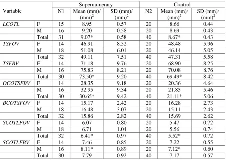
The range of the mean difference between the supernumerary group and control for the central incisors was 0.40-0.99 mm and 0.17- 9.54 mm2 for lineal and tooth surface area measurements respectively and for the lateral incisors 0.26-0.96 mm and 0.60- 3.07 mm2 respectively. Not all the differences were significant at the 0.05 level. Variables which were found to have significant differences between groups for the upper central incisors were: LCOTL, TSFBV, OCOTSFBV, SCOTLFOV and SCOTLFBV in males only. Whereas for the upper lateral incisors these were BCOTSFOV, TSOMHFBV, SCOTLFOV in males only and SCOTLFBV.
Discussion
The upper incisor teeth were chosen in the present study to compare crown size variables between the supernumerary and control groups due to their proximity to the site of the supernumerary teeth which in the present study were located in the
of any supernumerary tooth, on adjacent tooth size, being larger than controls and related directly proportionally to the proximity to the supernumerary tooth 9.
Teeth are irregular objects and therefore defining descriptive variables of their shape and size has previously been largely un-investigated. The present method used various linear, perimeter and tooth surface area measurements covering the labial and occlusal aspects of the upper incisors. These measurements together form an enhanced analysis of crown size. Some of the measurements are interrelated and convey the same
meaning as representatives of tooth size. Thus PCA was used to examine the structure of tooth size parameters and identify key components. As the supernumerary tooth patients were the study group of interest and may have variation in tooth size which may not be present in the normal population, PCA was applied to the supernumerary group only and the calculated key component variables were then compared with the variables measured in the control group. Seven factor variables were extracted as discriminative components of tooth size of the upper central incisors accounting for 95% of the total variance, indicating only a small loss of information. Similarly eight factor variables were extracted as discriminative components of tooth size of the upper lateral incisors, accounting for 94% of the total variance.
and DCOTH) were found. It appears that TSOMHFBV and DCOTH variables in the upper lateral incisor have replaced TSFBV in the upper central incisor.
Most previous studies of crown dimensions have mainly used the mesio-distal and labio-lingual parameters, and these have been made manually, thus offering limited information and a high degree of subjectivity. The method of the present study has identified and measured the main components of tooth size by image analysis,
offering detailed and more accurate description of tooth size with a high level of intra-operator repeatability.
When there was no significant interaction between genders and groups, the genders were combined for group comparison and groups were combined for gender
comparison. In the presence of a significant interaction comparisons between groups were performed with genders separate.
Males had larger tooth size variables than females across both supernumerary and control groups. However none of the differences reached the 0.05 significance level, except for one variable i.e. SCOTLFOV for the upper lateral incisors in the
supernumerary group. This agrees with the findings of Garn et al., on sexual dimorphism for the mesio-distal and labio-lingual measurements in the human dentition, which were least for the incisor group 18-21.
Most tooth size variables for the upper incisors were found to be larger in the supernumerary group than in the control. However not all the differences were
significant at the 0.05 level. These findings agree with a previous study by the authors
9
The mean differences provide more insight in the patterning of crown size variation in the supernumerary group and help to investigate the control of development of teeth and the aetiology of supernumerary teeth. The significant differences found in the cervical, occlusal, lingual, and labial measurements across the clinical crowns of the upper central and lateral incisors suggest that the factors which have contributed to the aetiology of the supernumerary teeth have also influenced crown formation of the upper incisors throughout development, with periodic activities more noticeable at the start and end of tooth crown formation.
Although crown diameters in both upper central and lateral incisors were larger in the supernumerary group, the variation was more noticeable in the upper central incisor crown, supporting the local field effect suggested by the authors in a previous study 9. Future research will analyse the relationship of the findings with the local field effect and then investigate tooth shape comparisons utilising 3D methodology, providing new and actual „on-surface‟ variables.
Conclusions
By PCA seven key components for the upper central incisor and eight components for the upper lateral incisor were identified for crown size. These components were found to be larger in the supernumerary patients than in the control group, with the majority reaching the 0.05 significance level. Tooth crown size of the upper central incisor was more affected than that of the upper lateral incisor, supporting a local field effect theory.
References
1). Moorrees CFA. The Aleut dentition. A correlative study of dental characteristics in an Eskimo people, Harvard University Press, Cambridge, Massachusetts. 1957.
2). Harris EF, Nweeia MT. Tooth size of Ticuna Indians, Colombia, with phenetic comparisons to other Amerindians. Am J Phys Anthropol 1980;53: 81-91.
3). Bishara SE, Garcia AF, Jakobsen JR, Fahl JA. Mesiodistal crown dimensions in Mexico and the United States. Angle Orthod 1986; 65:315-323.
4). Chaushu S, Sharabi S, Becker A. Tooth size in dentitions with buccal canine ectopia. Eur J Orthod 2003;25:485-91.
5). Ling JY, Wong RW. Tooth dimensions of Southern Chinese. Homo 2007;58:67-73.
6). Brook AH. Dental anomalies of number, form and size: Their prevalence in British schoolchildren. J Int Assoc Dent Child 1974;5:37-53.
8). Brook AH, Elcock C, Al-Sharood MH, McKeown HF, Khalaf K, Smith RN. Further studies of a model for the aetiology of anomalies of tooth number and size in humans. Conn Tiss Res 2002; 43:289-95.
9). Khalaf K, Robinson DL, Elcock C, Smith RN, Brook AH. Tooth size in patients with supernumerary teeth and a control group measured by image analysis system. Arch oral Biol 2005;50:243-8.
10). Brook AH, Griffin RC, Smith RN, Townsend GC, Kaur G, Davis GR and Fearne J (2008) Tooth Size patterns in Patients with Hypodontia and Supernumerary teeth. Arc oral Biol In press.2008
11). Bland JM, Altman DG. (1986). Statistical methods for assessing agreement
between two methods of clinical measurement. Lancet, 1(8476):307-310.
12). Fliess JL. The design and analysis of clinical experiments, Wiley, New York; Chichester, 1986; Chapter 1:1-32.
13). Stafne EC. Supernumerary teeth. Dental Cosmos 1932;74:653-659.
14). Luten JR. The prevalence of supernumerary teeth in primary and mixed dentitions. J Int Assoc Dent Child 1967;34:346-353.
16). Nasif MM, Ruffalo RC, Zullo T. Impacted supernumerary teeth: a survey of 50 cases. Journal of the American Dental Association 1983;106:201– 204.
17). Rajab LD, Hamdan MA. Supernumerary teeth: review of the literature and a survey of 152 cases. Int J Paediatr Dent 2002;12:244-54.
18). Garn SM, Lewis AB, Kerewsky RS. Sex differences in tooth size. J Dent Res 1964;43:306.
19). Garn SM, Lewis AB, Kerewsky RS. Sexual dimorphism in the buccolingual tooth diameter, J Dent Res 1966a;45:1819.
20). Garn SM, Swindler DR, Kerewsky RS. A canine “field” in sexual dimorphism in tooth size. Nature 1966b;212:1501-1502.
Figure Legends
Figure 1. Buccal view of an upper right central incisor, measurements include mesio-distal (MD) length, additional mesio-mesio-distal lengths, at 25, 50 and 75% along a line perpendicular to the MD at its midpoint (MD1.B, MD2.B, MD3.B), and occluso-gingival lengths, at 25, 50 and 75% along the MD line (OG1, OG2, OG3), and tooth surface perimeter (P).
Figure 2. Occlusal view of an upper right central incisor measurements, including bucco-lingual lengths, at 25, 50 and 75% along the MD line (BL1, BL2, BL3) similar in principle to the occluso-gingival lengths from the buccal view. MD1.O, MD2.O and MD3.O are tooth widths taken from 25, 50 and 75% along the length of a perpendicular bisector of the MD at its midpoint.
Figure 3. Buccal view of an upper right central incisor tooth showing partial measurements
a) mesio-distal measurements bounded by perimeter trace and OG2: MD1M.B, MD2M.B and MD3M.B at 25, 50 and 75% respectively.
b) occluso-gingival measurements bounded by perimeter trace and MD: OG1, OG2 and OG3 at 25, 50 and 75% respectively.
Figure 4. Occlusal view of an upper right central incisor tooth showing partial measurements
b) bucco-lingual measurements bounded by perimeter trace and MD: BL1, BL2 and BL3 at 25, 50 and 75% respectively.
Figure 5. Scree plot, before rotation, for factor extraction of upper central incisor size variables in the supernumerary group.
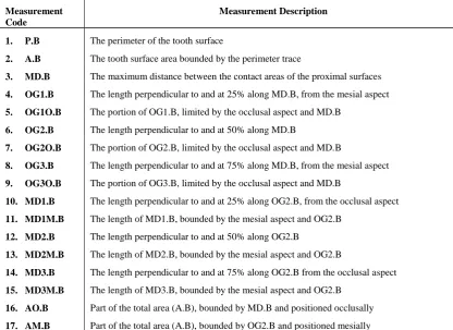
Table 1 Variables measured from the buccal view of each tooth.
Measurement Code
Measurement Description
1. P.B The perimeter of the tooth surface
2. A.B The tooth surface area bounded by the perimeter trace
3. MD.B The maximum distance between the contact areas of the proximal surfaces
4. OG1.B The length perpendicular to and at 25% along MD.B, from the mesial aspect
5. OG1O.B The portion of OG1.B, limited by the occlusal aspect and MD.B
6. OG2.B The length perpendicular to and at 50% along MD.B
7. OG2O.B The portion of OG2.B, limited by the occlusal aspect and MD.B
8. OG3.B The length perpendicular to and at 75% along MD.B, from the mesial aspect
9. OG3O.B The portion of OG3.B, limited by the occlusal aspect and MD.B
10. MD1.B The length perpendicular to and at 25% along OG2.B, from the occlusal aspect
11. MD1M.B The length of MD1.B, bounded by the mesial aspect and OG2.B
12. MD2.B The length perpendicular to and at 50% along OG2.B
13. MD2M.B The length of MD2.B, bounded by the mesial aspect and OG2.B
14. MD3.B The length perpendicular to and at 75% along OG2.B from the occlusal aspect
15. MD3M.B The length of MD3.B, bounded by the mesial aspect and OG2.B
16. AO.B Part of the total area (A.B), bounded by MD.B and positioned occlusally
Table 2 Variables measured from the occlusal view of each tooth.
Measurement Code
Measurement Description
1. P.O The perimeter of the tooth surface
2. A.O The tooth surface area bounded by the perimeter trace
3. MD.O The maximum distance between the contact areas of the proximal surfaces
4. BL1.O The length perpendicular to and at 25% along MD.O, from the mesial aspect
5. BL1B.O The portion of BL1.O, limited by the buccal aspect and MD.O
6. BL2.O The length perpendicular to and at 50% along MD.O
7. BL2B.O The portion of BL2.O, limited by the buccal aspect and MD.O
8. BL3.O The length perpendicular to and at 75% along MD.O, from the mesial aspect
9. BL3B.O The portion of BL3.O, limited by the buccal aspect and MD.O
10. MD1.O The length perpendicular to and at 25% along BL2.O, from the buccal aspect
11. MD1M.O The length of MD1.O, bounded by the mesial aspect and BL2.O
12. MD2.O The length perpendicular to and at 50% along BL2.O
13. MD2M.O The length of MD2.O, bounded by the mesial aspect and BL2.O
14. MD3.O The length perpendicular to and at 75% along BL2.O from the buccal aspect
15. MD3M.O The length of MD3.O, bounded by the mesial aspect and BL2.O
16. AB.O Part of the total area (A.O), bounded by MD.O and positioned buccally
Table 3 Total and percent variance of the first seven factors for upper central incisor size variables in the supernumerary group. Note: the “Rotaion Sums of Squared Loadings” shows factor variance after rotation (Varimax) which was used to transform the initial matrix into one that is easier to interpret.
Table 4 Total and percent variance of the first eight factors for upper lateral incisor size variables in the supernumerary group. Note: the “Rotation Sums of Squared Loadings” shows factor variance after rotation (Varimax) which was used to transform the initial matrix into one that is easier to interpret.
Thus six of the factors are common between the central and lateral incisors.
Factor Rotation Sums of Squared Loadings
Total % of Variance Cumulative %
1 10.40 30.60 30.60
2 5.22 15.36 45.96
3 4.08 12.00 57.95
4 3.75 11.04 68.99
5 3.65 10.73 79.73
6 2.24 6.59 86.31
7 1.56 4.58 90.89
8 1.18 3.47 94.36
Factor Rotation Sums of Squared Loadings
Total % of Variance Cumulative %
1 11.17 32.87 32.87
2 5.04 14.82 47.69
3 4.57 13.43 61.12
4 4.29 12.61 73.73
5 4.08 12.00 85.73
6 2.22 6.53 92.26
Table 5 Variables of upper central incisors of supernumerary and control groups.
Variable
Supernumerary Control
N1 Mean (mm)/ (mm)2
SD (mm)/ (mm)2
N2 Mean (mm)/ (mm)2
SD (mm)/ (mm)2
LCOTL F 15 8.95 0.57 20 8.66 0.44
M 16 9.20 0.58 20 8.69 0.43
Total 31 9.07* 0.58 40 8.67* 0.43
TSFOV F 14 46.91 8.52 20 48.48 5.96
M 18 51.08 6.01 20 46.14 5.05
Total 32 49.11 7.51 40 47.31 5.58
TSFBV F 14 71.18 9.76 20 68.90 8.25
M 16 75.83 8.21 20 70.08 8.76
Total 30 73.50* 9.20 40 69.49* 8.42
OCOTSFBV F 14 28.35 9.18 20 20.36 4.64
M 16 32.95 9.34 20 21.85 5.46
Total 30 30.65* 9.42 40 21.11* 5.06
BCOTSFOV F 14 15.17 2.42 20 16.28 2.73
M 18 16.48 3.07 20 15.11 2.43
Total 32 15.86 2.82 40 15.69 2.62
SCOTLFOV F 14 6.07 0.80 20 5.47 0.72
M 18 6.71 1.04 20 5.56 0.74
Total 32 6.41* 0.97 40 5.52* 0.72
SCOTLFBV F 14 7.46 0.85 20 7.22 0.55
M 16 8.11* 0.89 20 7.12* 0.60
Total 30 7.79 0.92 40 7.17 0.57
Key: N1= number of teeth measured from individuals with supernumeraries; N2= number of teeth measured from control subjects; M= males; F= females. SD= standard deviation; * = P < 0.05
Table 6 Variables of upper lateral incisors of supernumerary and control groups.
Variable
Supernumerary Control
N1 Mean (mm)/ (mm)2
SD (mm)/ (mm)2
N2 Mean (mm)/ (mm)2
SD (mm)/ (mm)2
LCOTL F 17 6.95 0.58 19 6.92 0.45
M 17 7.26 0.38 18 6.75 0.56
Total 34 7.10 0.51 37 6.84 0.51
TSFOV F 15 36.40 5.52 18 36.39 4.75
M 17 36.72 4.93 16 33.68 6.22
Total 32 36.57 5.13 34 35.15 5.56
OCOTSFBV F 17 17.25 5.27 17 14.50 4.43
M 15 20.21 4.13 16 16.75 4.21
Total 32 18.63 4.93 33 15.56 4.41
BCOTSFOV F 15 10.70 1.41 18 11.05 1.87
M 17 12.26* 2.19 16 9.95* 1.88 Total 32 11.53 2.00 34 10.55 1.93
TSOMHFBV F 17 21.22 3.10 17 20.66 2.54
M 15 21.70 2.09 16 21.06 3.33
Total 32 21.45* 2.65 33 20.85* 2.90
SCOTLFOV F 15 4.96* 0.50 18 5.21 0.68
M 17 5.85* 0.68 16 4.89* 0.79
Total 32 5.43 0.75 34 5.06 0.74
SCOTLFBV F 17 5.48 0.82 17 5.23 0.84
M 15 5.84 0.79 16 5.26 0.50
Total 32 5.65* 0.81 33 5.24* 0.69
DCOTH F 17 6.36 0.79 17 6.51 0.66
M 15 6.56 0.61 16 6.67 0.77
Total 32 6.46 0.71 33 6.58 0.71