JOURNAL OFVIROLOGY, July 2011, p. 6867–6881 Vol. 85, No. 14 0022-538X/11/$12.00 doi:10.1128/JVI.00229-11
Copyright © 2011, American Society for Microbiology. All Rights Reserved.
HIV-1 Nef Disrupts Intracellular Trafficking of Major Histocompatibility
Complex Class I, CD4, CD8, and CD28 by Distinct Pathways That
Share Common Elements
䌤
Jolie A. Leonard,
1Tracy Filzen, Christoph C. Carter,
1,2Malinda Schaefer,
3† and Kathleen L. Collins
1,4*
Graduate Program in Cellular and Molecular Biology,1Medical Scientist Training Program,2Department of
Immunology,3and Internal Medicine,4University of Michigan, Ann Arbor, Michigan
Received 1 February 2011/Accepted 26 April 2011
The Nef protein is an important HIV virulence factor that promotes the degradation of host proteins to augment virus production and facilitate immune evasion. The best-characterized targets of Nef are major histocompatibility complex class I (MHC-I) and CD4, but Nef also has been reported to target several other proteins, including CD8, CD28, CD80, CD86, and CD1d. To compare and contrast the effects of Nef on each protein, we constructed a panel of chimeric proteins in which the extracellular and transmembrane regions of the MHC-I allele HLA-A2 were fused to the cytoplasmic tails of CD4, CD28, CD8, CD80, CD86, and CD1d. We found that Nef coprecipitated with and disrupted the expression of molecules with cytoplasmic tails from MHC-I HLA-A2, CD4, CD8, and CD28, but Nef did not bind to or alter the expression of molecules with cytoplasmic tails from CD80, CD86, and CD1d. In addition, we used short interfering RNA (siRNA) knockdown and coprecipitation experiments to implicate AP-1 as a cellular cofactor for Nef in the downmodulation of both CD28 and CD8. The interaction with AP-1 required for CD28 and CD8differed from the AP-1 interaction required for MHC-I downmodulation in that it was mediated through the dileucine motif within Nef (LL164,165AA) and did not require the tyrosine binding pocket of the AP-1subunit. In addition, we
demon-strate a requirement for-COP as a cellular cofactor for Nef that was necessary for the degradation of targeted molecules HLA-A2, CD4, and CD8. These studies provide important new information on the similarities and differences with which Nef affects intracellular trafficking and help focus future research on the best potential pharmaceutical targets.
Nef is an important virulence factor for human immunode-ficiency virus type 1 (HIV-1) pathogenesis that functions to increase viral spread and promote disease progression. The significance of Nef expression on disease progression is high-lighted by the delayed progression to AIDS observed in a cohort infected with HIV with a deletion of the nef open reading frame and long terminal repeat (LTR) (8, 25, 27, 54, 55). Similarly, rhesus macaques infected with SIV⌬Nef dem-onstrated low viral loads and delayed disease progression (48). Nef is a 25- to 34-kDa myristoylated early viral gene product that acts as a multifunctional adaptor protein, containing a number of protein-protein interaction domains. Additionally, Nef has several disordered regions that confer flexibility to facilitate the exposure of different interaction domains in response to the local environment (33; also reviewed in reference 3).
Nef has been implicated in signal transduction, intracellular trafficking, and viral infectivity. One of its best-studied activi-ties is its role in promoting immune evasion from anti-HIV cytotoxic T lymphocytes (CTL) (22, 56, 78, 85, 93). Nef also downmodulates CD4, the HIV receptor, from the plasma
membrane (32) to prevent superinfection (5) and allow the more efficient release of budding virus (51, 74). In addition, Nef has been reported to downmodulate a number of other surface molecules, including CD8 (mediated through its sub-unit), CD28, CD80, CD86, and CD1d (12–14, 18, 19, 80, 84). Relative to major histocompatibility complex class I (MHC-I) and CD4, little is known about the mechanisms and the func-tional significance of the downmodulation of these molecules by Nef.
We have demonstrated previously that Nef disrupts MHC-I trafficking by binding to the cytoplasmic tail of MHC-I in the endoplasmic reticulum or early Golgi, and this complex sub-sequently recruits adaptor protein 1 (AP-1) in thetrans-Golgi network (62, 72, 79, 89–91, 93a). AP-1 then directs MHC-I into an endolysosomal pathway as opposed to the cell surface (77, 89–91). In contrast, CD4 anterograde traffic to the plasma membrane is not inhibited in the presence of Nef. Instead, Nef promotes CD4 internalization at the plasma membrane (11, 23, 58, 68, 77). CD4 downmodulation has been shown to re-quire AP-2, and it has been proposed that Nef-dependent AP-2 recruitment to the cytoplasmic domain of CD4 at the plasma membrane promotes clathrin-dependent endocytosis (16, 44, 80). Our laboratory and others also have demonstrated that Nef utilizes a -COP-dependent pathway to ultimately target MHC-I and internalized CD4 to lysosomes for degra-dation (4, 29, 68, 77, 93a).
The other putative targets of Nef, CD28, CD8, CD80, CD86, and CD1d, are expressed in a variety of immune cells,
* Corresponding author. Mailing address: 1150 W. Medical Center Dr., 3514 MSRB I, Ann Arbor, MI 48109. Phone: (734) 615-1320. Fax: (734) 615-5252. E-mail: klcollin@umich.edu.
† Present address: Emory Vaccine Center, Emory University, At-lanta, GA.
䌤Published ahead of print on 4 May 2011.
6867
on November 7, 2019 by guest
http://jvi.asm.org/
significant downmodulation of CD80, CD86, or CD1d by HIV-1 Nef in a variety of cell lines or primary antigen-presenting cells (APCs). The downmodulation of CD8and CD28 was partially dependent on AP-1 expression, and we detected a Nef-dependent interaction of AP-1 with the CD8and CD28 cytoplasmic tails that relied on the dileu-cine sorting signal within Nef. Additionally, we found that Nef promoted the degradation of CD8and that this func-tion of Nef required -COP expression. These new data help define the most relevant cellular targets of HIV-1 Nef and provide insights into the mechanism by which HIV-1 promotes disease.
MATERIALS AND METHODS
Cell culture.Bosc and 293T viral packaging cells were cultured in high-glucose Dulbecco’s modified essential medium (DMEM) supplemented with 10% fetal bovine serum (FBS) and 2 mM penicillin, streptomycin, and glutamine (PSG). CEM-SS and SupT1 cells lines were maintained in RPMI 1640 supplemented with 10% FBS and 2 mM PSG.
Preparation of T-cell lines expressing HLA-A2 chimeric molecules.Stable cell lines were made as previously described (3). Briefly, each construct was intro-duced into CEM-SS or SupT1 cells using the murine stem cell virus (MSCV) retroviral vector pseudotyped with vesicular stomatitis virus G protein (VSV-G). A uniform population was selected by culturing the cells in neomycin.
Primary T-cell preparation.Leukopaks were obtained from the New York Blood Center. Peripheral blood mononuclear cells (PBMCs) were purified using
Ficoll gradients. Nonadherent cells were stimulated with 10g/ml
phytohemag-glutinin (PHA; Sigma-Aldrich) overnight, and the next day 50 U/ml interleukin-2 (IL-2) was added.
Preparation of macrophages and DCs.Primary monocytes were obtained from whole PBMCs by positive selection using a magnetic sorting kit (EasySep human
CD14 selection kit; StemCell Technologies). The CD14⫹cells were induced to
mature into macrophages by culturing in RPMI plus 10 ng/ml granulocyte-macrophage colony-stimulating factor (GM-CSF) (R&D Systems) for 5 days.
The CD14⫹cells were induced to mature into dendritic cells (DC) by culture in
RPMI plus 800 U/ml GM-CSF and 500 U/ml IL-4, with the addition of 100 U/ml
tumor necrosis factor alpha (TNF-␣) on day 3 (all from R&D Systems). DCs
were stimulated 9 days in total before infection.
DNA constructs.The CD80, CD1d, and CD8cytoplasmic tails were fused to the extracellular and transmembrane regions of hemagglutinin (HA)-tagged HLA-A2 using PCR cloning methods. HA–HLA-A2 (1) was used as the PCR template. The following oligonucleotide primers were used: HA–
HLA-A2/CD8(forward primer, 5⬘-CGGGATCCACCATGCGGGTCACG
GCG-3⬘; reverse primer, 5⬘-GTGGTCGCTGCTGTGATGTGGTGCTGCC
GGCGGAGGAGAGCCCGGCTTCGTTTCATGAAACAATTTTACAAAT
AACTCGAGCGG-3⬘), HA–HLA-A2/CD80 (forward primer, 5⬘-CGGGATC
CACCATGCGGGTCACGGCG-3⬘; reverse primer, 5⬘-AGCTGTGGTCGC
TGCTGTGATGTGGAGATGCAGAGAGAGAAGGAGGAATGAGAGA
TTGAGAAGGGAAAGTGTACGCCCTGTATAACTCGAGCGG-3⬘), and
HA–HLA-A2/CD1d (forward primer, 5⬘-CGGGATCCACCATGCGGGTCA
CGGCG-3⬘; reverse primer, 5⬘-GCTGCTGTGATGTGGTCCCGGTTTAAG
AGGCAAACTTCCTATCAGGGCGTCCTGTGACCGCTCGAGCGG-3⬘).
The CD28 tail was fused to HA–HLA-A2 in a two-step cloning method. First
a 3⬘fragment was created using two overlapping oligonucleotides that span
the entire CD28 cytoplasmic tail sequence along with a forward primer that appended a 15-bp sequence, which overlapped with the transmembrane
re-gion of HLA-A2, and a reverse primer to introduce a 3⬘XhoI site. The 5⬘
CD86 cytoplasmic tail fragment with 15 bp 5⬘of the CD86 tail that
over-lapped the transmembrane region of HLA-A2 using full-length CD86 as the
template, forward primer 5⬘-GTGGTCGCTGCTGTGATGTGGAAATGGAA
GAAGAAGAAGC-3⬘, and reverse primer 5⬘-AAGTGATACATGTTTTTAACT
CGAGCGG-3⬘. A second PCR was performed to create HA–HLA-A2 with a 15-bp
3⬘region that overlapped with the 5⬘sequence of the CD86 tail. HA–HLA-A2 was
the template, and the primer sequences were 5⬘-CGGGATCCACCATGCGGGT
CACGGCG-3⬘(forward primer) and 5⬘-GTGGTCGCTGCTGTGATGTGGAAA
TGGAAGAAGAAGAAGC-3⬘(reverse primer). A third round of PCR, using the
products of the first and second PCRs, resulted in the full-length HA–HLA-A2/
CD86 chimera. The following primers were used: forward primer, 5⬘-CGGGATC
CACCATGCGGGTCACGGCG-3⬘; reverse primer, 5⬘-AAGTGATACATGTTTT
TAACTCGAGCGG-3⬘. The PCR products were digested with BamHI and XhoI
and then ligated into the BglII and XhoI sites of MSCV2.2 (2). The resulting constructs were sequenced to ensure that no mutations were introduced during cloning.
Cloning ofnefalleles into pMIG.nefalleles were cloned into the BglII and EcoRI sites of the pMIG vector obtained from Luk Van Parijs (MIT) (27). Nef
alleles containing a 5⬘ BamHI and 3⬘ EcoRI site were PCR amplified from
cloned HIV genomes obtained through the AIDS Research Reference Reagent
Program, Division of AIDS, NIAID, NIH (7, 8, 12). The following 5⬘
oligonu-cleotides for oligonucleotide primers were used: 5⬘Nef-1,5⬘-CGGGATCCACC
ATGGGTGGCAAGTGGTCAAAA-3⬘ (used with all except 92UG037,
93BR029, and 92RW009); 5⬘Nef-2, 5⬘-CGGGATCCACCATGGGTAACAAG
TGGTCAAAG-3⬘(used with 92UG037); and 5⬘Nef-4, 5⬘-CGGGATCCACCA
TGGGTAGCAAGTGGTCAAAA-3⬘(used with 93BR029 and 92RW009). The
following 3⬘oligonucleotides were used: 89.6, 5⬘-CGGAATTCTCAGTTCTTG
AAGTACTCCGGATG-3⬘; 92UG037, 5⬘-CGGAATTCTCAGCAGTCTTTGT
AAAACTCCGG-3⬘; 93BR020, 5⬘-CGGAATTCTCAGTCTTGGTAGTACTC
CGGATG-3⬘; NL4-3/84ZR, 5⬘-CGGAATTCTCAGCAGTTCTTGAAGTACT
CCGG-3⬘; PEYYKDC, 5⬘-CGGAATTCTCAGCAGTCTTTGTAGTACTCCG
G-3⬘(93BR029, 92RW009, and 94UG114); and YU-2, 5⬘-CGGAATTCTCAG
TTCTTGTAGTACTCCGGATG-3⬘. All alleles were sequenced to ensure that
no mutations were introduced during cloning.
shRNA constructs.FG12 short hairpin RNA (shRNA) lentiviral vectors were constructed as previously described (71, 77). shNC and a construct targeting
-COP were previously described (77). The target sequence for shAP1␥began at
position 1201: GATCCCCGCAGACTGTGCATCTGGAATTCAAGAGATTC
CAGATGCACAGTCTGCTTTTTGGAAA. The AP2␣ target sequence was
GATCCCCGTCAGAGTTCCGACAGAACTTCAAGAGAGTTCTGTCGGA
ACTCTGACTTTTTGGAAA. The target sequence for shAP3␦began at
posi-tion 4180: GATCCCCGAGAGGTTGCAGTCAGAACTTCAAGAGAGTTCT GACTGCAACCTCTCTTTTTGGAAA.
Viral transductions.Adenovirus was prepared by the University of Michi-gan Gene Vector Core, and the infection of CEM-SS cells was performed as previously described (72, 89). Murine retroviral vector preparation and trans-duction were performed as described previously (66, 88, 91). shRNA-con-taining lentivirus preparations and transductions also have been described (71, 77). The infection of PBMCs with HIV constructs was performed as described in reference 22. Primary APCs were infected with adenovirus at multiplicities of infection (MOIs) titrated to generate similar Nef expression levels compared to what we typically obtain in CEM cells. In these experi-ments, CEM cells were infected at an MOI of 50, primary macrophages were infected at an MOI of 250, and primary DCs were infected at an MOI of 500.
Western blot analyses and immunoprecipitations.Cells were lysed in phos-phate-buffered saline (PBS), 0.3% 3-[(3-cholamidopropyl)-dimethylammonio]-1-propanesulfonate (CHAPS), 0.1% SDS, pH 8, 1 mM phenylmethylsulfonyl fluoride (PMSF) and normalized for total protein and separated by SDS-poly-acrylamide gel electrophoresis. Endoglycosidase H (endo H) (NEB) digestion was performed according to the manufacturer’s protocol. Immunoprecipitations
on November 7, 2019 by guest
http://jvi.asm.org/
were performed as previously described (91). Briefly, cells were lysed in 1% digitonin (Wako) and normalized for total protein. Lysates were precleared overnight with A/G beads (Pierce) and isotype control antibody (BD Biosci-ences). BB7.2 cross-linked A/G beads were used for immunoprecipitation and were washed five times with 0.1% digitonin buffer prior to elution with 150 mM dithiothreitol (DTT).
Samples were separated by SDS-PAGE. HA–HLA-A2 and the chimeric pro-teins were detected using the anti-HA antibody HA.11 (1:5,000; Covance). Nef
was detected with AG11 (1:5,000) and -COP with M3A5 (2), which were
purified as described previously (45). Adaptor protein subunits were detected
with anti-AP-2␣, anti-AP1␥, and anti-AP3␦(all 1:500; BD Bioscience) or Ry/1
anti-AP1(1:5,000) (86). The secondary antibody for anti-Nef,-COP, HA,
AP-2␣, and anti-AP3␦was horseradish peroxidase (HRP)-rat anti-mouse IgG1
(Zymed), for anti-AP1␥was HRP-rat anti-mouse (nonspecific) (Zymed), and for
anti-1 was HRP-goat anti-rabbit (Zymed).
Flow cytometry.Intact cells were stained in fluorescent-activated cell sorter buffer
(2% FBS, 1% human serum, 1% HEPES, and 0.025% NaN3in PBS). Primary cells
were preincubated with 10% Fc-receptor blocker (Accurate Chemical and Scientific Corps) prior to primary antibody incubation. Chimeric molecules were detected with HA.11 (1:50; Covance) or BB7.2 (1:500). Endogenous proteins were detected with BB7.2 (1:500), OKT4 (1:500), and OKT8 (1:500), all prepared by the University of
Michigan Hybridoma Core and purified as previously described (45), or anti-CD8
[image:3.585.42.536.68.507.2]5F2 (1:100; Santa Cruz), CD28.2 (1:100; Biolegend), CD80 (1:100; BD Pharmin-gen), CD86 (1:100; BD PharminPharmin-gen), and CD1d (1:100; eBioscience). Isotype-specific secondary antibodies conjugated to phycoerythrin (PE), Alexa fluor 488, or Alexa fluor 647 were used at 1:250 (Invitrogen).
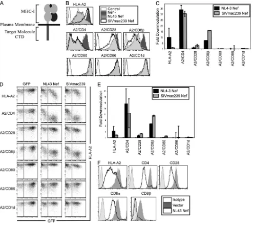
FIG. 1. Nef downmodulates HLA-A2, A2/CD4, A2/CD8, and A2/CD28 in CEM T cells. (A) Diagram of chimeric molecules. (B) Flow-cytometric analysis of CEM-SS T cells expressing the indicated chimeric proteins and transduced with the indicated adenoviral vector. Cells were analyzed at 3 days postinfection (dpi). (C) Quantitation of flow cytometry experiments shown in panel B. The mean fold downmodulation⫾ standard deviations is shown.n⫽3. (D) Flow-cytometric analysis of CEM-SS T cells transduced with a retroviral vector expressing GFP alone or NL43 Nef and the GFP reporter. (E) Quantitation of flow cytometry experiments shown in panel D. The mean fold downmodulation of GFP⫹ cells⫾standard deviations is shown.n⫽3. (F) Flow-cytometric analysis of SupT1 cells transduced with the indicated adenoviral vector. Cells were analyzed at 3 dpi. Sup T1 cells express endogenous CD4, CD28, and CD8.
VOL. 85, 2011 HIV-1 Nef DOWNMODULATION OF MHC-I, CD4, CD8, AND CD28 6869
on November 7, 2019 by guest
http://jvi.asm.org/
RESULTS
Nef targets HLA-A2, CD4, CD8, and CD28 through their cytoplasmic tail domains. Nef has been reported to down-modulate a large number of host proteins, including MHC-I, CD4, CD28, CD8, CD80, CD86, and CD1d. To assess the relative effects of Nef on these targets, we created a panel of chimeric proteins in which the extracellular and transmem-brane regions of hemagglutinin epitope (HA)-tagged HLA-A2 were fused to the cytoplasmic tail domain of each protein of interest (Fig. 1A). This allowed us to use the same antibodies and establish assays to directly compare the effect of Nef on each cytoplasmic tail domain. We then measured the impact of HIV-1 and simian immunodeficiency virus (SIV) Nef on the cell surface expression of each chimeric molecule. We found that HLA-A2, the HLA-A2 chimera with the CD4 cytoplasmic
tail (A2/CD4), and the HLA-A2 chimera with the CD8 cyto-plasmic tail (A2/CD8) were downmodulated by both HIV and SIV Nef expressed using an adenoviral vector system in a CD4⫹ T-cell line, CEM-SS (3.7-, 8.5-, and 4.0-fold, respec-tively) (Fig. 1B and C). HIV-1 Nef had a lesser (1.8-fold) effect on the HLA-A2 chimera with the CD28 cytoplasmic tail (A2/ CD28), whereas SIV Nef downmodulated A2/CD28 to a greater extent (3-fold) (Fig. 1B and C), as previously demon-strated (84). The chimeric molecules with cytoplasmic tails from CD80, CD86, and CD1d (A2/CD80, A2/CD86, and A2/ CD1d, respectively) were not sufficient for downmodulation by either Nef protein (1.1-, 1.1-, and 1.3-fold downmodulation, respectively) (Fig. 1B and C).
[image:4.585.42.541.67.449.2]Similar results were obtained when Nef was expressed using a retroviral vector that also expresses green fluorescent protein
FIG. 2. NL43 Nef does not downmodulate CD80, CD86, or CD1d. (A) Flow-cytometric analysis of U937 cells and THP-1 cells transduced with the indicated retroviral vector and treated as indicated with LPS, PMA, or DMSO (Undiff.). The cells were analyzed 5 days postinfection (dpi). (B) Quantitation of flow cytometry experiments shown in panel A. The mean fold downmodulation⫾standard deviations is shown.n⫽3. (C) Flow-cytometric analysis of THP-1 cells treated as described for panel A. The surface expression levels of endogenous HLA-A2, CD4, and CD1d expression were measured at 5 dpi. (D) Quantitation of flow cytometry experiments shown in panel C. The mean fold downmodulation⫾ standard deviation is shown.n⫽3. (E) Flow-cytometric analysis of primary antigen-presenting cells that were transduced with the indicated adenoviral vector. Endogenous HLA-A2, CD4, CD80, and CD86 expression levels were assessed at 3 dpi. Primary macrophages were GM-CSF treated prior to transduction; dendritic cells were GM-CSF, IL-4, and TNF-␣treated prior to transduction. (F) Quantitation of flow cytometry experiments shown in panel E. The mean fold downmodulation⫾standard deviations is shown.n⫽3.
on November 7, 2019 by guest
http://jvi.asm.org/
(GFP) from an internal ribosomal entry site (38). Again we observed the strong downmodulation of HLA-A2, A2/CD4, and A2/CD8 and the weak downmodulation of A2/CD28 (Fig. 1D and E). In addition, we found that HIV-1 Nef did not significantly affect A2/CD80, A2/CD86, or A2/CD1d (Fig. 1D and E). Similar results were achieved using another T-cell line (SupT1) that expressed wild-type, endogenous CD4, CD8 (both␣/␣and␣/dimers), and CD28 plus exogenous HLA-A2 (Fig. 1F).
Endogenous CD80 and CD86 are not downmodulated by Nef in primary APCs.Because CD80, CD86, and CD1d are nor-mally expressed in antigen-presenting cells (APCs), we em-ployed the human monocytic cell lines THP-1 and U937 for further experiments with these molecules. We were unable to
[image:5.585.43.539.68.473.2]detect significant cell surface expression of endogenous CD80 or CD86 in either cell line (data not shown). Therefore, we created stable U937 and THP-1 lines expressing the chimeric molecules. Nef was introduced into undifferentiated cells using a retroviral vector, and then the cells were stimulated with lipopolysaccharide (LPS) and phorbol myristate acetate (PMA) to induce macrophage differentiation. Overall, we ob-served that Nef was less active in the APC lines than in T cells, downmodulating HLA-A2 2- to 3-fold in APC lines and 17-fold in CEM cells (Fig. 2B). Even so, Nef had minimal effect on the CD80 and CD86 chimeras in all three cell lines (0.8 to 2.0-fold) (Fig. 2A and B), indicating that the cytoplasmic tails of CD80 and CD86 were not sufficient for downmodulation by HIV-1 Nef even in macrophage cell lines. In contrast, we
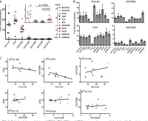
FIG. 3. Nef-induced downmodulation of MHC-I, CD4, CD8, and CD28 is conserved across multiple clades. (A) Quantitation of downmodulation of the indicated molecule in CEM-SS cells following transduction with bicistronic murine retroviral vectors expressing the indicated Nef. The mean fold downmodulation of each molecule in GFP-positive cells as determined by flow cytometry is shown.n⫽3. The inset displays the data for CD28, CD1d, CD80, and CD86, with an expandedyaxis to highlight differences among these molecules that are of a small magnitude. The statistical significance was determined by two-way analysis of variance. (B) Quantitation of Nef-dependent downmodulation of the indicated molecule by each Nef variant. The mean fold downmodulation in the GFP-positive cells⫾ standard deviations is shown.n⫽3. (C) Relative ability of Nef variants to downmodulate each target protein, plotted as fold downmodulation. YU-2 was removed as an outlier.
VOL. 85, 2011 HIV-1 Nef DOWNMODULATION OF MHC-I, CD4, CD8, AND CD28 6871
on November 7, 2019 by guest
http://jvi.asm.org/
observed a decrease in MHC-I HLA-A2 surface expression under all conditions. Because PMA also reduces CD4 surface expression (7), we were unable to assess the effect of Nef on CD4 in the PMA-treated cells.
We used the same experimental approach to examine the ability of HIV-1 Nef to downmodulate full-length endogenous CD1d in the THP-1 cell line, which also expresses endogenous HLA-A2. We observed minimal downmodulation of CD1d by Nef in either undifferentiated or differentiated THP-1 cells (Fig. 2C and D).
To examine the ability of Nef to downmodulate full-length CD80 and CD86, we obtained primary antigen-presenting cells from human peripheral blood mononuclear cells. These cells were stimulated with GM-CSF alone or GM-CSF, IL-4, and TNF-␣to induce macrophage- or monocyte-derived dendritic cell phenotypes, respectively (76, 94). We observed no signif-icant effect of Nef on the surface expression of endogenous CD80 and CD86 in either cell type (Fig. 2E and F). As con-trols, we demonstrated the downmodulation of endogenous MHC-I HLA-A2 and CD4 by Nef, albeit to a lesser extent than that in T cells (Fig. 2E and F).
Multiple HIV-1 Nef variants downmodulate HLA-A2, CD4, CD8, and CD28.All experiments thus far utilized Nef from the NL4-3 molecular clone of HIV (1). While many of the known functional domains of Nef are conserved, significant sequence variation does occur. Therefore, we examined whether the activity of Nef varied among HIV isolates. Al-though we observed variation in the magnitude of MHC-I HLA-A2 downmodulation, all Nef variants tested (except YU-2) reduced HLA-A2 surface expression dramatically (6- to 35-fold) (Fig. 3A and B). Similarly, all of the Nef proteins downmodulated endogenous CD4 as well as A2/CD8 and A2/CD28. CD4 was downmodulated to the greatest extent, followed by HLA-A2 and then A2/CD8. A2/CD28 was only moderately affected by all of the Nef variants tested. In con-trast, none of the Nef proteins reduced A2/CD80 or A2/CD86 surface expression more than 1.5-fold (Fig. 3A, inset).
Interestingly, some of the Nef variants downmodulated A2/ CD1d more than 1.5-fold (Fig. 3A, inset). 93BR020 Nef, a clade F isolate (31), downmodulated A2/CD1d to the greatest extent. Thus, certain HIV subtypes may evolve the ability to significantly affect CD1d trafficking, although the effect was comparatively small.
Notably, Nef proteins that demonstrated higher
downmodu-lation activity on one target protein did not necessarily dem-onstrate similarly high activity for all other targets examined (Fig. 3B). When relative Nef activities were plotted for each target molecule, a number of correlative observations were made (Fig. 3C). HLA-A2 and A2/CD8were negatively cor-related, indicating that the ability of Nef to downmodulate these proteins did not coevolve. There is a weak positive cor-relation between HLA-A2 and A2/CD28, indicating that Nef sequences that contribute to the downmodulation of these targets are partially shared. Because CD4 and CD8 are anal-ogous molecules, one can hypothesize that CD8
downmodu-FIG. 4. CD4 and CD8 are downmodulated by Nef in HIV-1-infected PBMCs. Flow-cytometric analysis of PBMCs infected with HXB ePLAP HIV with or without Nef, pseudotyped with the HXB envelope, is shown. Surface marker and intracellular Gag stains were performed at 3 days postinfection (dpi).
FIG. 5. Nef physically associates with the cytoplasmic domains of HLA-A2, CD4, CD8, and CD28 and recruits AP-1 to HLA-A2, CD8, and CD28. Immunoprecipitation (IP) and Western blot anal-ysis of the indicated chimeric molecule expressed in CEM T cells transduced with the indicated adenoviral vector are shown. The cells were harvested at 3 days postinfection (dpi) after an overnight incu-bation in ammonium chloride to inhibit degradation. Lysates were immunoprecipitated with an antibody directed against HLA-A2 (BB7.2), and the presence of HA–HLA-A2, Nef (A), or AP-1 (B) was detected by Western blot analysis.
on November 7, 2019 by guest
http://jvi.asm.org/
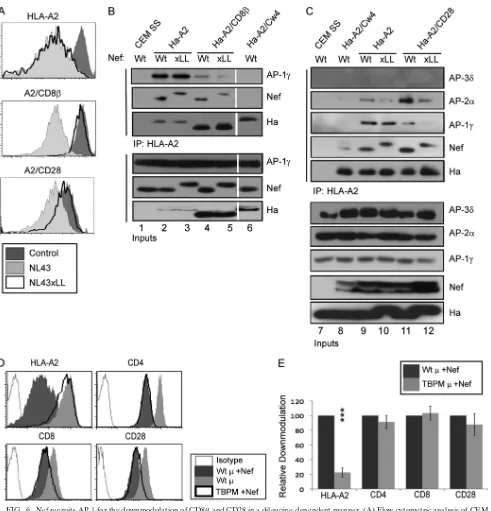
[image:6.585.310.528.394.632.2]FIG. 6. Nef recruits AP-1 for the downmodulation of CD8and CD28 in a dileucine-dependent manner. (A) Flow-cytometric analysis of CEM SS T cells expressing the indicated chimeric protein and transduced with the indicated adenoviral vector. Nef LL164,165AA is indicated as xLL.
(B) Immunoprecipitation and Western blot analysis of the indicated chimeric molecule expressed in CEM T cells transduced with the indicated adenoviral vector. The cells were harvested at 3 days postinfection (dpi). Lysates were immunoprecipitated with an antibody directed against HLA-A2 (BB7.2), and the presence of Nef, AP-1 subunits, and MHC-I (HA) were detected by Western blot analysis. Intervening lanes were removed, as indicated by the white gap. The data are representative of four independent experiments. (C) Immunoprecipitation and Western blot analysis of the indicated chimeric molecule expressed in CEM T cells transduced with the indicated adenoviral vector and treated as described for panel B. AP-2 coprecipitation is representative of two of four experiments. (D) Flow-cytometric analysis of SupT1 T cells transduced with an adenoviral vector expressing Nef and a retroviral vector expressing the indicatedsubunit of AP-1. TBPM, tyrosine binding pocket mutant subunit of AP-1; Wt, wild-typesubunit of AP-1. The cells were analyzed by flow cytometry at 3 dpi. (E) Quantitation of flow data shown in panel D. The relative fold downmodulation, where the fold downmodulation of each molecule in the presence of Wt AP-1 is set to 100%, is shown. Error bars represent standard deviations;***,P⬍0.00001;n⫽3.
VOL. 85, 2011 HIV-1 Nef DOWNMODULATION OF MHC-I, CD4, CD8, AND CD28 6873
on November 7, 2019 by guest
http://jvi.asm.org/
lation is an evolutionary by-product of CD4 downmodulation. Significantly, however, there was no correlation between CD4 and A2/CD8downmodulation (Fig. 3C).
HIV-1 Nef downmodulates CD4 and CD8 in primary T lym-phocytes.CD8 can be expressed as a CD8␣/␣homodimer or as a CD8␣/heterodimer. Because Nef targets CD8, only the CD8␣/heterodimer can be affected by Nef (80). T lympho-cytes, DCs, and NK cells all are CD8⫹, but the CD8␣/ het-erodimer is expressed exclusively in T cells. Typically, T lym-phocytes are either CD4⫹or CD8⫹, and only the CD4⫹subset can be infected by HIV. Thus, it is unclear how Nef might come in contact with the CD8molecule. Significantly, a small population of CD4⫹CD8⫹T lymphocytes circulates in periph-eral blood (9, 30, 64, 65, 97), and the frequency of these double-positive cells increases in response to infection with a number of viruses, including HIV (42, 52, 59). Additionally,
these cells have been reported to be susceptible to infection by HIV (6, 20, 40, 41, 50, 92, 96). While the role of CD4⫹CD8⫹ double-positive cells is not entirely clear, they are known to exhibit cytolytic activity as well as the antigen-dependent se-cretion of gamma interferon (IFN-␥) and IL-2. Moreover, they are enriched for HIV-specific responses in chronically infected patients (39, 64, 82, 95). Importantly, these cells are reported to express the CD8␣/heterodimer (64, 81). Thus, this popu-lation represents a potential physiologic target of HIV in which Nef would encounter CD8.
[image:8.585.81.499.68.434.2]To investigate the ability of HIV to infect CD4⫹CD8⫹cells, we infected PBMCs with HIV HXB ePLAP (17) pseudotyped with an HIV envelope (HXB) that lacked Nef expression (HIV nef mutant) so that the expression of cell surface markers would be maintained. As expected, we observed similar rates of infection in CD4⫹CD8⫹and CD4⫹CD8⫺cells (16 and
FIG. 7. Nef requires AP-1 and-COP for maximal downmodulation of CD8 and CD28. (A) Flow-cytometric analysis of CEM SS cells expressing the indicated chimeric protein and transduced with lentivirus expressing shRNA and GFP and subsequently transduced with the indicated adenoviral vector at 3 days after lentivirus infection. Flow-cytometric analysis was performed at 3 days after adenoviral transduction. Histograms represent GFP-expressing cells. (B) Quantitation of the flow data shown in panel A. The fold downmodulation of the indicated molecule in the presence of shRNA is shown. (C) Western blot analysis confirming the specific knockdown of AP-1 subunits in CEM SS cells. (D) Western blot analysis confirming adaptor protein knockdown in SupT1 cells. (E) Relative downmodulation of the indicated molecule in SupT1 T cells transduced with the indicated lentivirus expressing shRNA and GFP plus a bicistronic retrovirus expressing Nef and a PLAP marker gene. shNC, negative control. The cells were analyzed by flow cytometry at 3 days after retroviral transduction. The fold downmodulation normalized to the negative control in cells positive for both GFP and PLAP is shown.*,P⬍0.05;**,P⬍0.01;***,P⬍0.001.
on November 7, 2019 by guest
http://jvi.asm.org/
14%, respectively) (Fig. 4, upper). In contrast, when these cells were infected with an HIV HXB ePLAP that expressed Nef, most of the infected cells became negative for both CD4 and CD8 (Fig. 4, lower). Therefore, CD4⫹CD8⫹cells are a phys-iologically relevant target of HIV-1, and Nef actively down-modulates both CD4 and CD8 in these cells.
Nef recruits AP-1 to the cytoplasmic tail of CD8and CD28.
Prior research has shown that Nef disrupts MHC-I and CD4 host protein trafficking by binding to sequences in their cyto-plasmic tail domains (77, 89). Here, we confirmed that Nef coprecipitated with HLA-A2 and A2/CD4. Moreover, we ob-served that Nef coprecipitated with A2/CD8 and A2/CD28 but not A2/Cw4, A2/CD80, A2/CD86, or A2/CD1d (Fig. 5A). A2/Cw4 is a negative control in these experiments, as Nef does not bind to or downmodulate HLA-C (or HLA-E or HLA-F) MHC-I allotypes (21, 35, 90). Thus, NL43 Nef physically asso-ciates with each of the proteins it potently downmodulates in T cells.
In Nef-expressing cells, HLA-A2 also coprecipitates with the clathrin adaptor protein AP-1 (when degradation is inhibited by ammonium chloride), and this interaction is required for Nef to disrupt MHC-I trafficking (72, 91). Similarly, we ob-served AP-1 coprecipitation with A2/CD28 and A2/CD8(Fig. 5B), although the interaction with A2/CD8 seemed weaker than that observed with HLA-A2 and A2/CD28. In contrast, we did not observe AP-1 coprecipitating with A2/CD4, A2/ Cw4, A2/CD80, A2/CD86, or A2/CD1d.
We also asked whether the other adaptor proteins, AP-2 and AP-3, coprecipitated with the chimeras. In two of five experi-ments, we observed AP-2␣recruitment to A2/CD28 (Fig. 6C), which would be consistent with previous reports of AP-2 in-volvement in CD28 downmodulation (80, 84). However, we were unable to detect the coprecipitation of any other chime-ras with AP-2 or AP-3 (data not shown).
Nef recruits AP-1 to CD8and CD28 through its dileucine motif.The AP proteins can utilize either Yxxor (E/D)xxxLL motifs in cargo molecules (x stands for any amino acid, and indicates a hydrophobic residue). While Nef recruits AP-1 to the tyrosine residue in the MHC-I cytoplasmic tail, such a sequence is not present in the cytoplasmic tail domains of CD28 or CD8. ⌱nterestingly, Nef also contains a conserved canonical dileucine motif (ExxxLL165) that has been reported
to mediate binding to AP-1, AP-2, and AP-3 (11, 24, 28, 34, 42, 43, 53, 67). Furthermore, Nef LL164,165AA is defective at
downmodulating CD8 or CD28 yet retains the ability to downmodulate HLA-A2 (Fig. 6A) (80). Thus, we hypothesized that the recruitment of AP-1 to CD8or CD28 occurs through this dileucine motif. Consistently with our hypothesis, Nef LL164,165AA did not support the coprecipitation of AP-1 with A2/CD8and A2/CD28 (Fig. 6B, lanes 4 and 5, and C, lanes 11 and 12) but did promote the coprecipitation of AP-1 with HLA-A2 (Fig. 6B and C). Therefore, Nef can recruit AP-1 to cytoplasmic tail domains using two different signals, depending upon which cytoplasmic tail domain Nef is bound to.
In experiments where we observed AP-2 coprecipitation with the CD28 cytoplasmic tail, we were able to determine that the AP-2 recruitment also depended upon the dileucine motif in Nef (Fig. 6C).
The subunit of heterotetrameric adaptor protein com-plexes recognizes tyrosine-based motifs (10, 63), whereas an
interface between theand large subunit (␣,␦, or␥) recog-nizes dileucine motifs (15, 26, 47). For MHC-I downmodula-tion, Nef promotes an interaction between the tyrosine binding motif in the1 subunit of AP-1 and a conserved tyrosine in the cytoplasmic tail of MHC-I. An AP-1 mutant with an inactive tyrosine binding pocket acts as a dominant-negative inhibitor of MHC-I downmodulation (72, 91). To confirm that the downmodulation of CD8and CD28 did not utilize the AP-1 binding pocket, we tested the impact of the tyrosine-binding pocket mutant (TBPM) on these pathways. In contrast to its effect on MHC-I, TBPM did not significantly inhibit CD8 or CD28 downmodulation by Nef (Fig. 6D and E). Similarly, the knockdown of the AP-11 subunit had no sig-nificant effect on CD8or CD28 downmodulation by Nef (Fig. 7A and B). Importantly, the depletion of the AP-11 subunit did not significantly reduce AP-1␥expression, and AP-1␥ de-pletion had a partially destabilizing effect on AP-11 (Fig. 7C). Therefore, AP-1 involvement in CD8and CD28 downmodu-lation by Nef is independent of the AP-1 tyrosine binding activity but still could depend on AP-1 dileucine motif binding. Based on the requirement for the Nef dileucine motif for CD8 and CD28 downmodulation as well as the lack of an effect of knocking down the AP-1subunit, we next examined the effect of knocking down the large subunits of the clathrin-associated adaptor proteins, which contain in part the dileu-cine motif recognition site (15, 26, 47). Knockdown was achieved using lentiviral vectors expressing shRNAs directed against the large subunits of AP-1␥, AP-2␣, AP-3␦, and
[image:9.585.322.518.69.232.2]-COP. Western blot analysis confirmed that we efficiently and specifically reduced the expression of each target (Fig. 7D). We found that knocking down AP-1␥and-COP significantly reduced the downmodulation of both CD8and CD28 by Nef,
FIG. 8. CD28 is recycled rapidly in Nef-expressing cells. The mea-surement of recycling in CEM T cells expressing A2 or HLA-A2/CD28 and transduced with the indicated adenovirus is shown. Cells were harvested at 3 days postinfection (dpi), incubated with 150 mg/ml cycloheximide for 2 h to inhibit protein synthesis, and stripped of stainable HLA-A2 by removing the 2-microglobulin with an acid wash (50 mM glycine, 100 mM NaCl, pH 3.4). The cells then were incubated at 37°C in culture medium plus cycloheximide. Triplicate samples were removed to ice at the indicated time points, stained for surface HLA-A2 expression (BB7.2), and analyzed by flow cytometry. Recycling is plotted as a percentage of steady-state levels, where the mean fluorescence of each time point was divided by the mean fluo-rescence of cells that were not acid stripped. n⫽2.
VOL. 85, 2011 HIV-1 Nef DOWNMODULATION OF MHC-I, CD4, CD8, AND CD28 6875
on November 7, 2019 by guest
http://jvi.asm.org/
6876
but the effect was only partial (Fig. 7E). The partial effects we observed may be due to incomplete knockdown or Nef utili-zation of multiple adaptor proteins in redundant pathways to achieve the downmodulation of CD8 and CD28. Additionally, AP-2␣knockdown had a small but significant effect on CD28 downmodulation, which is consistent with previous reports and the interaction we observed between CD28 and AP-2 (Fig. 7E) (80, 84). Surprisingly, the knockdown of AP-2 had no signifi-cant impact on CD4 downmodulation by Nef, despite a body of evidence tying CD4 downmodulation to this adaptor in other cell systems (16, 44, 80). It is possible CD4 downmodulation by Nef can utilize multiple adaptor proteins, and the degree to which it does so might vary in different cell systems. However, this possibility remains ambiguous because when we attempted to knock down more than one adaptor protein large subunit, the toxicity was too great to assess Nef function (data not shown).
Nef increases CD28 recycling.We were surprised to observe the robust recruitment of adaptor proteins to A2/CD28 in the presence of Nef because the effect of Nef on the steady-state surface expression of A2/CD28 is relatively small. Interest-ingly, CD28 is internalized at a rate similar to, or even greater than, that of CD4 in the presence of Nef (80 and data not shown). To address this discrepancy, we performed a recycling assay in which we observed a significant increase in CD28 recycling in the presence of Nef (Fig. 8). Thus, Nef may ini-tially induce a potent downmodulation of CD28 through the recruitment of AP-1 and/or AP-2 in parallel or redundant pathways, and then a subset of CD28 is rapidly recycled to the plasma membrane. Thus, the effect of Nef on CD28 may vary depending on the recycling rates in different cell types.
Nef utilizes -COP in CD8 and CD28 downmodulation.
We also observed that the knockdown of-COP significantly reduced the effect of Nef on CD8and CD28 surface expres-sion (Fig. 7E and 9A). This is interesting because-COP is required for the Nef-dependent trafficking of HLA-A2 and CD4 to lysosomes for degradation (77).-COP knockdown has different effects on the cell surface expression of MHC-I and CD4 in Nef-expressing cells. Whereas-COP knockdown increases cell surface levels of MHC-I in Nef-expressing cells,
-COP knockdown does not always alter CD4 surface expres-sion in Nef-expressing cells (Fig. 7E) (77). Instead, CD4 accu-mulates in intracellular vesicles and is unable to recycle to the cell surface in some cell types (68, 77).
Nef contains two-COP binding sites, RR17,19and EE155,156 (68, 77). Nef RR17,19AA is defective at targeting MHC-I for
degradation while Nef EE155,156AA is defective at targeting CD4 for degradation, and both have a partially reduced ability to coprecipitate with -COP (77). When both domains are mutated (R/E), Nef is unable to efficiently bind-COP and is
defective at promoting the degradation of either MHC-1 or CD4 (77). We assessed the relative contributions of these Nef domains in CD8and CD28 downmodulation and found that the R/E double mutant was defective at downmodulating CD8and CD28, while each of the single-mutant Nef proteins demonstrated intermediate defects (Fig. 9B and C). Similar results were observed whether we assayed the chimeric panel (Fig. 9B) or the full-length target molecules (Fig. 9C).
Combined effects of AP-1 and-COP.To further investigate the contribution of AP-1 and-COP to A2/CD8degradation, we again utilized the shRNA lentivirus system to knock down AP-1␥ and -COP both singly and in combination. We ob-served that the knockdown of either AP-1 or-COP reduced the ability of Nef to downmodulate HLA-A2 and A2/CD8. However, knocking down both adaptors in combination did not have a significant additional inhibitory effect on down-modulation (Fig. 9A). Thus, the effect of Nef on CD8results from effects of at least two cellular trafficking factors and possibly others (80).
To examine whether HIV-1 Nef promoted the degradation of CD8, we performed Western blot analysis focusing on the mature, endoglycosidase H (endo H)-resistant form of each molecule. As reported previously, Nef expression reduces the amount of endo H-resistant HLA-A2, and this degradation is reversed by knocking down AP-1 or-COP (Fig. 9D, left, and E). As previously observed, knocking down -COP also af-fected the migration of MHC-I (Fig. 9D), probably by disrupt-ing the traffickdisrupt-ing of enzymes required for proper glycosyla-tion. -COP knockdown does not inhibit the trafficking of HLA-A2 through the Golgi membrane, and it does not disrupt gross Golgi structure (77).
In addition, we found that the knockdown of-COP dra-matically reduced the degradation of A2/CD8(Fig. 9D, right, and E). A small but statistically significant effect of knocking down AP-1 on A2/CD8degradation also was noted (Fig. 9E).
DISCUSSION
In summary, we have found that HIV-1 Nef has three major cellular targets: MHC-I, CD4, and CD8. The ability of Nef to downmodulate endogenous CD8 was confirmed in HIV-in-fected primary CD4⫹CD8⫹T cells. We also report here that the effect of Nef on MHC-I and CD8 depended at least in part on the expression of AP-1 and-COP. In addition, we found that the AP-1 binding motif utilized by Nef to recruit AP-1 to cytoplasmic tails varied depending on which cytoplasmic tail domain Nef was bound to. Finally, we found that the mutation of both-COP binding sites in Nef reduced the activity of Nef against CD8. Thus, one explanation for the capacity of Nef to
FIG. 9.-COP is required for Nef-mediated degradation of HLA-A2/CD8. (A) Quantitation of relative downmodulation (normalized to the control) of the indicated molecule expressed in CEM-SS cells transduced with the indicated lentivirus expressing shRNA and a retroviral vector expressing Nef. The cells were harvested at 3 days after retroviral transduction.n⫽3. (B and C) Quantitation of the fold downmodulation of the indicated molecule expressed in CEM-SS cells (B) or Sup T1 cells (C) transduced with a retroviral vector expressing Nef or the indicated Nef mutants. Cells with equal GFP expression, to control for Nef levels, were analyzed at 3 days postinfection (dpi). The mean fold downmodulation⫾ standard deviations is shown.n⫽3.*,P⬍0.05;**,P⬍0.01;***,P⬍0.001. (D) Western blot analysis of endonuclease H (endo H)-treated lysates from the cells described in panel A. (E) Quantitation of endo H-resistant bands. The endo H-resistant bands (indicated by “R” in the figure) were quantified and normalized to the loading control, and shNC was set to 100% remaining.*,P⬍0.05;**,P⬍0.01;***,P⬍0.001;n⫽3.
VOL. 85, 2011 HIV-1 Nef DOWNMODULATION OF MHC-I, CD4, CD8, AND CD28 6877
on November 7, 2019 by guest
http://jvi.asm.org/
FIG. 10. (A and B) Schematic representation of the two AP-1 binding sites that Nef utilizes. (A) When bound to the MHC-I cytoplasmic domain, Nef recruits AP-1 through the tyrosine recognition site in the AP-1subunit (91). In this model, the three-way complex requires amino acid residues in both MHC-I, especially Y320, as well as in Nef, M20in particular (although assays using purified proteins and a Nef–MHC-I
cytoplasmic tail fusion protein did not demonstrate a requirement for M20) (62, 72, 79, 91). When bound to the CD8 or CD28 cytoplasmic tail,
Nef recruits AP-1 through the dileucine recognition site at the interface between the␥and subunits of AP-1 (47). (C to E) Schematic representation of the Nef domains involved in-COP recruitment. (C) Nef residues R17and R19are required for-COP involvement in MHC-I
degradation (77). (D) A diacidic motif in Nef, EE155,156, is required for-COP involvement in CD4 degradation (29, 68, 77). (E) Either or both
Nef domains likely participate in-COP recruitment for the degradation of CD8 by Nef. (F) Model of the trafficking pathways Nef uses to achieve the downmodulation and degradation of target proteins. Nef blocks anterograde transport in an AP-1-dependent pathway (46, 72, 83). Nef also induces the internalization of target molecules at the plasma membrane through an association with AP-1 or AP-2 (11, 16, 24, 44, 59, 77, 80, 84). These pathways likely converge at a-COP-dependent step (77). Because AP-1 knockdown did not block the-COP-dependent degradation of A2/CD8, Nef may be able to direct some targets into endolysosomal compartments by a pathway in which only-COP recruitment is necessary and clathrin-associated adaptor proteins are dispensable. PM stands for plasma membrane, LY indicates lysosome, and LE/MVB indicates late endosome or multivesicular body compartments.
6878
affect multiple targets is that it has evolved redundant mech-anisms to recruit the same adaptor proteins.
The downmodulation of CD8, which participates in the rec-ognition of antigen in association with MHC-1, provides a second Nef-dependent mechanism by which HIV-infected cells could avoid CTL lysis. While CD8⫹T cells generally are not thought to be targeted by HIV, there is mounting evidence that a subset of CD8⫹T cells upregulate CD4 in response to acti-vation stimuli and that these cells demonstrate antiviral activ-ity, thus creating an infectible target cell, the inhibition of which would be beneficial to HIV (30, 49, 50, 81, 92). Nef might also access CD8⫹T cells through cell-cell contacts, as was described to occur between infected and bystander T cells (61). Cell-cell conduits also have been described between Nef-expressing APCs and bystander B cells. Moreover, these nano-tubular conduits have been shown to transmit Nef (70). If Nef were transferred when CTLs contacted an infected target, the transmitted Nef could downmodulate CD8 and reduce the efficacy of the anti-HIV-specific CTL attack. Finally, there are reports of CD8-tropic HIV strains providing another potential scenario in which Nef could encounter CD8 (36, 37, 57, 69, 75, 87).
This study is the first to assess the contribution of AP-1 and
-COP to the downmodulation of CD28 and CD8. Another group has reported a partial requirement for AP-2 in the downmodulation of CD4 and CD8 that we did not observe here (80). However, the literature is conflicting on this point; some groups report a defect in CD4 downmodulation when AP-2 is knocked down, while other groups observe no effect (15, 73, 80). Additionally, the codepletion of Eps15 and AP-2 may be required to observe an inhibition of Nef activity against CD4 (44). The knockdown of adaptor protein subunits is tech-nically difficult, as the highly efficient depletion of these pro-teins can be lethal to cells. For these reasons, we may have underestimated the contribution of AP-2 in CD28 and CD8 downmodulation by Nef in our system. AP-1- and AP-2-de-pendent mechanisms may represent parallel or redundant pathways that Nef can utilize depending upon cellular local-ization and the availability of cellular adaptors.
Here, we have presented direct evidence that Nef can recruit AP-1 by utilizing either the tyrosine motif recognition site or the dileucine recognition site of AP-1, and that this is deter-mined by which cytoplasmic tail domain Nef is bound to (Fig. 10A and B). Previous studies found that the dileucine motif in Nef is required for the glutathioneS-transferase (GST)-Nef pulldown of AP-1 complexes from mammalian cell lysates (11, 43) and for interactions detected in yeast two- and three-hybrid analyses (11, 24, 28, 34, 42, 43, 53, 67), but the dileucine motif is dispensable for MHC-I downmodulation even though AP-1 is required (60). CD4 downmodulation by Nef requires the dileucine motif but does not involve AP-1 (34). Thus, the functional significance of the interaction between AP-1 and the dileucine motif has remained unexplained. Here, we provide evidence that this interaction is utilized to target CD8 (and CD28).
We also have identified-COP as a common factor in the downregulation and degradation of all three major targets of Nef: MHC-I, CD4, and CD8. The evidence presented here is consistent with a model in which Nef utilizes AP-1 or AP-2 to direct cellular targets into endosomal compartments where
Nef subsequently recruits-COP to route targets to the lyso-somes for degradation as previously proposed for MHC-I and CD4 (Fig. 10F). However, the fact that the degradation of CD8 was far more dependent on-COP expression than on AP-1 expression indicates that-COP also directly targets intracel-lular CD8 for degradation in Nef-expressing cells without the need for additional adaptor proteins (Fig. 10F). The require-ment for -COP in multiple Nef pathways may represent a good target for the therapeutic inhibition of Nef.
CD28 appears to be a unique target of Nef, in that it is downmodulated consistently by a panel of Nef variants, but the effect is small relative to that of the other targets of Nef. The small effect of Nef on steady-state surface CD28 seems at odds with the observation that CD28 is internalized at a rate similar to that of CD4 in the presence of Nef (80 and data not shown). This discrepancy may be explained by the observation that CD28 recycling is significantly increased in Nef-expressing cells. SIV Nef downmodulates CD28 more potently than HIV-1 Nef, therefore CD28 downmodulation may be more important for SIV pathogenesis. It also is possible that CD28 recycling is regulated in some cell systems, and that a physio-logically relevant set of conditions exists that would result in less recycling of CD28 and more downmodulation of CD28 by HIV-1 Nef.
We were unable to reproduce data reported by another group that CD80 and CD86 were downmodulated by Nef. There were some differences between the cell lines used in our study and those in the previous report. The previous studies used endogenously expressed CD80 and CD86 in the U937 monocytic cell line. However, even when replicating the pub-lished culture conditions, we were able to observe only low levels of CD80 on the cell surface and no CD86 at all, leading us to believe that our clone of the U937 line is inherently different from that used to report the downmodulation of CD80 and CD86. This may be the cause for the discrepancy between our results and others and is the reason that we utilized primary macrophages and DCs as a highly relevant system for investigating Nef activity against these potential targets. We were unable to detect an effect of Nef on CD80 or CD86 under established stimulation and culture conditions for primary macrophages and DCs, indicating that the Nef target-ing of these proteins must occur only under specialized cir-cumstances that we were unable to replicate. CD1d also ap-pears to be a special case, in that not all Nef isolates retain the ability to reduce the surface expression of this molecule, yet one or more Nef variants may be capable of doing so to a limited extent. This may be rare, evolutionarily driven by some infrequently occurring host factor, or a feature of certain HIV-1 clades.
In summary, we have identified three major targets of HIV-1 Nef, MHC-I, CD4, and CD8, and these results were general-izable to multiple Nef variants. We also demonstrated that the downmodulation of CD8 and CD28 required AP-1 recruitment and was dependent upon the Nef dileucine motif. This obser-vation indicates that Nef can interact with AP-1 through either the tyrosine-binding pocket or dileucine recognition domain of AP-1, depending upon which cytoplasmic tail Nef is associated with. Finally, we discovered a role for-COP in the downregu-lation and degradation of CD8 as well as MHC-I and CD4,
VOL. 85, 2011 HIV-1 Nef DOWNMODULATION OF MHC-I, CD4, CD8, AND CD28 6879
on November 7, 2019 by guest
http://jvi.asm.org/
We also thank Linton Traub (University of Pittsburgh) for his gift of antibody to AP-1; Alice Telesnitsky (University of Michigan) for her gift of SupT1 cells; and Beatrice Hahn (University of Pennsylvania) for her gift of HIV-1 molecular clones (31).
REFERENCES
1.Adachi, A., et al.1986. Production of acquired immunodeficiency syndrome-associated retrovirus in human and nonhuman cells transfected with an
infectious molecular clone. J. Virol.59:284–291.
2.Allan, V. J., and T. E. Kreis.1986. A microtubule-binding protein associated
with membranes of the Golgi apparatus. J. Cell Biol.103:2229–2239.
3.Arold, S. T., and A. S. Baur.2001. Dynamic Nef and Nef dynamics: how structure could explain the complex activities of this small HIV protein.
Trends Biochem. Sci.26:356–363.
4.Benichou, S., et al.1994. Physical interaction of the HIV-1 Nef protein wih
-cop, a component of non-clathrin coated vesicles essential for membrane
traffic. J. Biol. Chem.269:30073–30076.
5.Benson, R. E., A. Sanfridson, J. S. Ottinger, C. Doyle, and B. R. Cullen.1993. Downregulation of cell-surface CD4 expression by simian immunodeficiency
virus Nef prevents viral super infection. J. Exp. Med.177:1561–1566.
6.Betts, M. R., et al.2004. The functional profile of primary human antiviral
CD8⫹T-cell effector activity is dictated by cognate peptide concentration.
J. Immunol.172:6407–6417.
7.Bigby, M., P. Wang, J. F. Fierro, and M. S. Sy.1990. Phorbol myristate acetate-induced down-modulation of CD4 is dependent on calmodulin and
intracellular calcium. J. Immunol.144:3111–3116.
8.Birch, M. R., et al.2001. An examination of signs of disease progression in
survivors of the Sydney Blood Bank Cohort (SBBC). J. Clin. Virol.22:263–
270.
9.Blue, M. L., J. F. Daley, H. Levine, and S. F. Schlossman.1985. Coexpression of T4 and T8 on peripheral blood T cells demonstrated by two-color
fluo-rescence flow cytometry. J. Immunol.134:2281–2286.
10.Boll, W., et al.1996. Sequence requirements for the recognition of
tyrosine-based endocytic signals by clathrin AP-2 complexes. EMBO J.15:5789–5795.
11.Bresnahan, P. A., et al.1998. A dileucine motif in HIV-1 Nef acts as an internalization signal for CD4 downregulation and binds the AP-1 clathrin
adaptor. Curr. Biol.8:1235–1238.
12.Chaudhry, A., et al.2005. The Nef protein of HIV-1 induces loss of cell surface costimulatory molecules CD80 and CD86 in APCs. J. Immunol.
175:4566–4574.
13.Chaudhry, A., et al.2008. HIV-1 Nef induces a Rab11-dependent routing of endocytosed immune costimulatory proteins CD80 and CD86 to the Golgi.
Traffic9:1925–1935.
14.Chaudhry, A., et al.2007. A two-pronged mechanism for HIV-1 Nef-medi-ated endocytosis of immune costimulatory molecules CD80 and CD86. Cell
Host Microbe1:37–49.
15.Chaudhuri, R., O. W. Lindwasser, W. J. Smith, J. H. Hurley, and J. S. Bonifacino.2007. Downregulation of CD4 by human immunodeficiency virus type 1 Nef is dependent on clathrin and involves direct interaction of Nef
with the AP2 clathrin adaptor. J. Virol.81:3877–3890.
16.Chaudhuri, R., R. Mattera, O. W. Lindwasser, M. S. Robinson, and J. S. Bonifacino.2009. A basic patch on alpha-adaptin is required for binding of human immunodeficiency virus type 1 Nef and cooperative assembly of a
CD4-Nef-AP-2 complex. J. Virol.83:2518–2530.
17.Chen, B. K., R. T. Gandhi, and D. Baltimore.1996. CD4 down-modulation during infection of human T cells with human immunodeficiency virus type
1 involves independent activities of vpu, env, and nef. J. Virol.70:6044–6053.
18.Chen, N., et al.2006. HIV-1 down-regulates the expression of CD1d via Nef.
Eur. J. Immunol.36:278–286.
19.Cho, S., et al.2005. Impaired cell surface expression of human CD1d by the
formation of an HIV-1 Nef/CD1d complex. Virology337:242–252.
20.Cochrane, A., et al.2004. High levels of human immunodeficiency virus
infection of CD8 lymphocytes expressing CD4 in vivo. J. Virol.78:9862–
9871.
21.Cohen, G. B., et al.1999. The selective downregulation of class I major histocompatibility complex proteins by HIV-1 protects HIV-infected cells
from NK cells. Immunity10:661–671.
sigma1 and alpha/sigma2 hemicomplexes of clathrin adaptors AP-1 and AP-2
harbor the dileucine recognition site. Mol. Biol. Cell18:1887–1896.
27.Dyer, W. B., et al.1997. Lymphoproliferative immune function in the Sydney Blood Bank Cohort, infected with natural nef/long terminal repeat mutants, and in other long-term survivors of transfusion-acquired HIV-1 infection.
AIDS11:1565–1574.
28.Erdtmann, L., et al.2000. Two independent regions of HIV-1 Nef are required for connection with the endocytic pathway through binding to the
mu 1 chain of AP1 complex. Traffic1:871–883.
29.Faure´, J., et al.2004. ARF1 regulates Nef-induced CD4 degradation. Curr.
Biol.14:1056–1064.
30.Flamand, L., et al.1998. Activation of CD8⫹T lymphocytes through the T-cell receptor turns on CD4 gene expression: implications for HIV
patho-genesis. Proc. Natl. Acad. Sci. U. S. A.95:3111–3116.
31.Gao, F., et al.1998. A comprehensive panel of near-full-length clones and reference sequences of non-subtype B isolates of human immunodeficiency
virus type 1. J. Virol.72:5680–5698.
32.Garcia, J. V., and A. D. Miller.1991. Serine phosphorylation-independent
downregulation of cell-surface CD4 by nef. Nature350:508–511.
33.Geyer, M., et al.2002. Subunit H of the V-ATPase binds to the medium chain of adaptor protein complex 2 and connects Nef to the endocytic
machinery. J. Biol. Chem.277:28521–28529.
34.Greenberg, M., L. DeTulleo, I. Rapoport, J. Skowronski, and T. Kirch-hausen.1998. A dileucine motif in HIV-1 Nef is essential for sorting into
clathrin-coated pits and for downregulation of CD4. Curr. Biol.8:1239–1242.
35.Greenberg, M. E., et al.1997. Co-localization of HIV-1 Nef with the AP-2 adaptor protein complex correlates with Nef-induced CD4 down-regulation.
EMBO J.16:6964–6976.
36.Gulzar, N., S. Balasubramanian, G. Harris, J. Sanchez-Dardon, and K. F. Copeland.2008. Infection of CD8⫹CD45RO⫹memory T-cells by HIV-1
and their proliferative response. Open AIDS J.2:43–57.
37.Gulzar, N., et al.2008. Proportion of HIV-1 infected CD8⫹CD4- T
lym-phocytes in vivo. Curr. HIV Res.6:585–596.
38.Hahn, W. C., et al.2002. Enumeration of the simian virus 40 early region
elements necessary for human cell transformation. Mol. Cell. Biol.22:2111–
2123.
39.Howe, R., et al.2009. Phenotypic and functional characterization of
HIV-1-specific CD4⫹CD8⫹double-positive T cells in early and chronic HIV-1
infection. J. Acquir. Immune Defic. Syndr.50:444–456.
40.Hughes, G. J., et al.2008. HIV-1-infected CD8⫹CD4⫹T cells decay in vivo
at a similar rate to infected CD4 T cells during HAART. AIDS22:57–65.
41.Imlach, S., et al.2001. Activated peripheral CD8 lymphocytes express CD4 in vivo and are targets for infection by human immunodeficiency virus type
1. J. Virol.75:11555–11564.
42.Janvier, K., et al.2003. HIV-1 Nef stabilizes the association of adaptor
protein complexes with membranes. J. Biol. Chem.278:8725–8732.
43.Janvier, K., et al.2003. Recognition of dileucine-based sorting signals from HIV-1 Nef and LIMP-II by the AP-1 gamma-sigma1 and AP-3 delta-sigma3
hemicomplexes. J. Cell Biol.163:1281–1290.
44.Jin, Y. J., et al.2005. HIV Nef-mediated CD4 down-regulation is adaptor
protein complex 2 dependent. J. Immunol.175:3157–3164.
45.Kasper, M. R., and K. L. Collins.2003. Nef-mediated disruption of HLA-A2
transport to the cell surface in T cells. J. Virol.77:3041–3049.
46.Kasper, M. R., et al.2005. HIV-1 Nef disrupts antigen presentation early in
the secretory pathway. J. Biol. Chem.280:12840–12848.
47.Kelly, B. T., et al.2008. A structural explanation for the binding of endocytic
dileucine motifs by the AP2 complex. Nature456:976–979.
48.Kestler, H. W., III, et al.1991. Importance of the nef gene for maintenance
of high virus loads and for development of AIDS. Cell65:651–662.
49.Kitchen, S. G., et al.2004. CD4 on CD8(⫹) T cells directly enhances effector function and is a target for HIV infection. Proc. Natl. Acad. Sci. U. S. A.
101:8727–8732.
50.Kitchen, S. G., Y. D. Korin, M. D. Roth, A. Landay, and J. A. Zack.1998.
Costimulation of naive CD8(⫹) lymphocytes induces CD4 expression and
allows human immunodeficiency virus type 1 infection. J. Virol.72:9054–
9060.
51.Lama, J., A. Mangasarian, and D. Trono.1999. Cell-surface expression of
on November 7, 2019 by guest
http://jvi.asm.org/
CD4 reduces HIV-1 infectivity by blocking Env incorporation in a Nef- and
Vpu-inhibitable manner. Curr. Biol.9:622–631.
52.Laux, I., et al.2000. Response differences between human CD4(⫹) and
CD8(⫹) T-cells during CD28 costimulation: implications for immune
cell-based therapies and studies related to the expansion of double-positive
T-cells during aging. Clin. Immunol.96:187–197.
53.Le Gall, S., et al.1998. Nef interacts with mu subunit of clathrin adaptor complexes and reveals a cryptic sorting signal in MHC I molecules. Immunity
8:483–495.
54.Learmont, J., et al.1992. Long-term symptomless HIV-1 infection in
recip-ients of blood products from a single donor. Lancet340:863–867.
55.Learmont, J. C., et al.1999. Immunologic and virologic status after 14 to 18 years of infection with an attenuated strain of HIV-1. A report from the
Sydney Blood Bank Cohort. N. Engl. J. Med.340:1715–1722.
56.Lewinsohn, D. A., et al.2002. HIV-1 Vpr does not inhibit CTL-mediated
apoptosis of HIV-1 infected cells. Virology294:13–21.
57.Livingstone, W. J., M. Moore, D. Innes, J. E. Bell, and P. Simmonds.1996. Frequent infection of peripheral blood CD8-positive T-lymphocytes with
HIV-1. Edinburgh Heterosexual Transmission Study Group. Lancet348:
649–654.
58.Macchi, B., G. Graziani, J. Zhang, and A. Mastino.1993. Emergence of double-positive CD4/CD8 cells from adult peripheral blood mononuclear cells infected with human T-cell leukemia virus type I (HTLV-I). Cell
Im-munol.149:376–389.
59.Mangasarian, A., et al.1997. The HIV-1 Nef protein acts as a connector with
sorting pathways in the Golgi and at the plasma membrane. Immunity6:67–
77.
60.Mangasarian, A., V. Piguet, J. K. Wang, Y. L. Chen, and D. Trono.1999. Nef-induced CD4 and major histocompatibility complex class I (MHC-I) down-regulation are governed by distinct determinants: N-terminal alpha helix and proline repeat of Nef selectively regulate MHC-I trafficking. J.
Vi-rol.73:1964–1973.
61.Muratori, C., et al.2009. Massive secretion by T cells is caused by HIV Nef in infected cells and by Nef transfer to bystander cells. Cell Host Microbe
6:218–230.
62.Noviello, C. M., S. Benichou, and J. C. Guatelli.2008. Cooperative binding of the class I major histocompatibility complex cytoplasmic domain and human immunodeficiency virus type 1 Nef to the endosomal AP-1 complex
via its mu subunit. J. Virol.82:1249–1258.
63.Ohno, H., et al.1995. Interaction of tyrosine-based sorting signals with
clathrin-associated proteins. Science269:1872–1875.
64.Ortolani, C., E. Forti, E. Radin, R. Cibin, and A. Cossarizza.1993.
Cyto-fluorimetric identification of two populations of double positive
(CD4⫹,CD8⫹) T lymphocytes in human peripheral blood. Biochem.
Bio-phys. Res. Commun.191:601–609.
65.Parel, Y., and C. Chizzolini.2004. CD4⫹CD8⫹double positive (DP) T cells
in health and disease. Autoimmun. Rev.3:215–220.
66.Pear, W. S., G. P. Nolan, M. L. Scott, and D. Baltimore.1993. Production of high-titer helper-free retroviruses by transient transfection. Proc. Natl. Acad.
Sci. U. S. A.90:8392–8396.
67.Piguet, V., et al.1998. Mechanism of Nef-induced CD4 endocytosis: Nef
connects CD4 with the mu chain of adaptor complexes. EMBO J.17:2472–
2481.
68.Piguet, V., et al.1999. Nef-induced CD4 degradation: a diacidic-based motif in Nef functions as a lysosomal targeting signal through the binding of
beta-COP in endosomes. Cell97:63–73.
69.Potter, S. J., D. E. Dwyer, and N. K. Saksena.2003. Differential cellular
distribution of HIV-1 drug resistance in vivo: evidence for infection of CD8⫹
T cells during HAART. Virology305:339–352.
70.Qiao, X., et al. 2006. Human immunodeficiency virus 1 Nef suppresses CD40-dependent immunoglobulin class switching in bystander B cells. Nat.
Immunol.7:302–310.
71.Qin, X. F., D. S. An, I. S. Chen, and D. Baltimore.2003. Inhibiting HIV-1 infection in human T cells by lentiviral-mediated delivery of small interfering
RNA against CCR5. Proc. Natl. Acad. Sci. U. S. A.100:183–188.
72.Roeth, J. F., M. R. Kasper, M. Williams, T. M. Filzen, and K. L. Collins.
2004. HIV-1 Nef disrupts MHC-I trafficking by recruiting AP-1 to the
MHC-I cytoplasmic tail. J. Cell Biol.167:903–913.
73.Rose, J. J., et al.2005. CD4 down-regulation by HIV-1 and simian immu-nodeficiency virus (SIV) Nef proteins involves both internalization and
in-tracellular retention mechanisms. J. Biol. Chem.280:7413–7426.
74.Ross, T. M., A. E. Oran, and B. R. Cullen.1999. Inhibition of HIV-1 progeny
virion release by cell-surface CD4 is relieved by expression of the viral Nef
protein. Curr. Biol.9:613–621.
75.Saha, K., et al.2001. Isolation of primary HIV-1 that target CD8⫹T
lymphocytes using CD8 as a receptor. Nat. Med.7:65–72.
76.Schaefer, M. R., et al.2008. A novel trafficking signal within the HLA-C cytoplasmic tail allows regulated expression upon differentiation of
macro-phages. J. Immunol.180:7804–7817.
77.Schaefer, M. R., E. R. Wonderlich, J. F. Roeth, J. A. Leonard, and K. L. Collins.2008. HIV-1 Nef targets MHC-I and CD4 for degradation via a final
common beta-COP-dependent pathway in T cells. PLoS Pathog.4:e1000131.
78.Schwartz, O., V. Marechal, S. Le Gall, F. Lemonnier, and J. Heard.1996. Endocytosis of major histocompatibility complex class I molecules is induced
by the HIV-1 Nef protein. Nat. Med.2:338–342.
79.Singh, R. K., D. Lau, C. M. Noviello, P. Ghosh, and J. C. Guatelli.2009. An MHC-I cytoplasmic domain/HIV-1 Nef fusion protein binds directly to the
mu subunit of the AP-1 endosomal coat complex. PLoS One4:e8364.
80.Stove, V., et al.2005. Human immunodeficiency virus Nef induces rapid
inter-nalization of the T-cell coreceptor CD8alphabeta. J. Virol.79:11422–11433.
81.Sullivan, Y. B., A. L. Landay, J. A. Zack, S. G. Kitchen, and L. Al-Harthi.2001.
Upregulation of CD4 on CD8⫹T cells: CD4dimCD8bright T cells constitute an
activated phenotype of CD8⫹T cells. Immunology103:270–280.
82.Suni, M. A., et al.2001. CD4(⫹)CD8(dim) T lymphocytes exhibit enhanced cytokine expression, proliferation and cytotoxic activity in response to
HCMV and HIV-1 antigens. Eur. J. Immunol.31:2512–2520.
83.Swann, S. A., et al. 2001. HIV-1 Nef blocks transport of MHC class I molecules to the cell surface via a PI 3-kinase-dependent pathway. Virology
282:267–277.
84.Swigut, T., N. Shohdy, and J. Skowronski.2001. Mechanism for
down-regulation of CD28 by Nef. EMBO J.20:1593–1604.
85.Tomiyama, H., H. Akari, A. Adachi, and M. Takiguchi.2002. Different effects of Nef-mediated HLA class I down-regulation on human
immunode-ficiency virus type 1-specific CD8(⫹) T-cell cytolytic activity and cytokine
production. J. Virol.76:7535–7543.
86.Traub, L. M., S. Kornfeld, and E. Ungewickell.1995. Different domains of the AP-1 adaptor complex are required for Golgi membrane binding and
clathrin recruitment. J. Biol. Chem.270:4933–4942.
87.Tsubota, H., et al.1989. CD8⫹CD4- lymphocyte lines can harbor the AIDS
virus in vitro. J. Immunol.143:858–863.
88.Van Parijs, L., et al.1999. Uncoupling IL-2 signals that regulate T-cell proliferation, survival, and Fas-mediated activation-induced cell death.
Im-munity11:281–288.
89.Williams, M., J. F. Roeth, M. R. Kasper, T. Filzen, and K. L. Collins.
2005. Human immunodeficiency virus type 1 Nef domains required for disruption of major histocompatibility complex class I trafficking are also
necessary for coprecipitation of Nef with HLA-A2. J. Virol.79:632–636.
90.Williams, M., et al.2002. Direct binding of human immunodeficiency virus type 1 Nef to the major histocompatibility complex class I (MHC-I)
cytoplasmic tail disrupts MHC-I trafficking. J. Virol.76:12173–12184.
91.Wonderlich, E. R., M. Williams, and K. L. Collins. 2008. The tyrosine binding pocket in the adaptor protein 1 (AP-1) mu1 subunit is necessary for Nef to recruit AP-1 to the major histocompatibility complex class I
cytoplas-mic tail. J. Biol. Chem.283:3011–3022.
92.Yang, L. P., et al.1998. Productive infection of neonatal CD8⫹T
lympho-cytes by HIV-1. J. Exp. Med.187:1139–1144.
93.Yang, O. O., et al.2002. Nef-mediated resistance of human
immunodefi-ciency virus type 1 to antiviral cytotoxic T lymphocytes. J. Virol.76:1626–
1631.
93a.Yi, L., et al.2010. HIV-1 Nef binds a subpopulation of MHC-I throughout its trafficking intinerary and down-regulates MHC-I by perturbing both
antero-grade and retroantero-grade trafficking. J. Biol. Chem.285:30884–30895.
94.Zhou, L. J., and T. F. Tedder.1996. CD14⫹blood monocytes can
differen-tiate into functionally mature CD83⫹dendritic cells. Proc. Natl. Acad. Sci.
U. S. A.93:2588–2592.
95.Zloza, A., et al.2009. Potent HIV-specific responses are enriched in a unique
subset of CD8⫹T cells that coexpresses CD4 on its surface. Blood114:
3841–3853.
96.Zloza, A., Y. B. Sullivan, E. Connick, A. L. Landay, and L. Al-Harthi.
2003. CD8⫹ T cells that express CD4 on their surface
(CD4dimCD8bright T cells) recognize an antigen-specific target, are de-tected in vivo, and can be productively infected by T-tropic HIV. Blood
102:2156–2164.
97.Zuckermann, F. A.1999. Extrathymic CD4/CD8 double positive T cells. Vet.
Immunol. Immunopathol.72:55–66.
VOL. 85, 2011 HIV-1 Nef DOWNMODULATION OF MHC-I, CD4, CD8, AND CD28 6881