The C-Terminal Repeat Domains of nsP3 from the Old World
Alphaviruses Bind Directly to G3BP
Marc D. Panas,aTero Ahola,bGerald M. McInerneya
Department of Microbiology, Tumor and Cell Biology, Karolinska Institutet, Stockholm, Swedena; Department of Food and Environmental Sciences, University of Helsinki, Helsinki, Finlandb
The Old World alphaviruses block stress granule assembly by sequestration of RasGAP SH3-domain binding protein (G3BP). Here, we show that the proline-rich sequences in the hypervariable domain of nonstructural protein 3 (nsP3) of both Semliki Forest virus and Chikungunya virus were dispensable for binding to G3BP. nsP3 variants with or without this domain colocal-ized with G3BP. Furthermore, we show that the C-terminal repeat motifs of nsP3 were sufficient for G3BP binding.
S
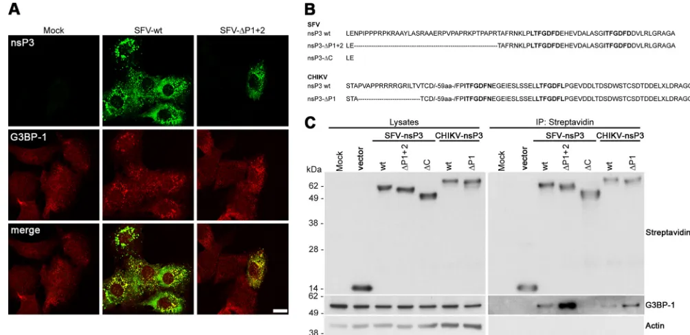
tress granules (SGs) are foci of accumulation of translationally silent mRNP complexes, which are induced during cellular stress (1). Their assembly is dependent on the proteins TIA-1/R (2) and G3BP (3). Many diverse virus infections employ mecha-nisms to restrict the formation of SGs (4,5), which are induced by early events in the replication of many viruses, including the al-phaviruses (6,7). We recently showed that in cells infected with the alphavirus Semliki Forest virus (SFV), nonstructural protein 3 (nsP3) binds RasGAP SH3-domain binding proteins 1 and 2 (G3BP-1 and G3BP-2, respectively), recruits them to foci contain-ing other viral proteins and often double-stranded RNA (dsRNA), and, in doing so, inhibits SG assembly (8). Fros and colleagues (9) also showed that the expression of the closely related Chikungu-nya virus (CHIKV) nsP3 blocks SG assembly by recruitment of G3BP-1 into cytoplasmic foci. These reports therefore describe a function for Old World alphavirus nsP3 in inhibition of the stress response. A surprising difference in the findings of the two groups was that the work of Fros and colleagues suggested that the CHIKV nsP3 sequence binding the SH3 domain of amphiphysins (398-PVAPPRRRR-406 [10]) was essential for colocalization of nsP3 and G3BP (9). In contrast to that work, our study showed that the SFV nsP3 C-terminal L/ITFGDFD repeat motifs at posi-tions 449 to 455 and 466 to 472, both well conserved in the Old World alphaviruses (11), were necessary and sufficient for forma-tion of the nsP3/G3BP complex in infected cells (8). Both these regions, although situated within the hypervariable domain (HVD) of nsP3 (12–15), are highly conserved between both CHIKV and SFV, and it was therefore surprising that the viruses would differ in the region used to bind and recruit G3BP.Recently, a variant of SFV (SFV-⌬P1⫹2) was described, lack-ing the proline-rich SH3-domain bindlack-ing sequences of nsP3 (10). It was shown that the deletion impairs the recruitment of amphiphysin proteins to foci of nsP3 accumulation and delays replication complex formation (10). To determine if this domain of SFV nsP3 was important for the recruitment of G3BP-1, we infected mouse embryonic fibroblasts (MEFs) maintained as de-scribed previously (16) with SFV wild type (wt) or SFV-⌬P1⫹2 at a multiplicity of infection (MOI) of 1, fixed the cells at 8 h postin-fection (hpi), and stained them for nsP3 and G3BP-1. Single-plane images were captured using a supercontinuum confocal TCS SP5 X microscope (Leica, Wetzlar, Germany) with a pulsed white light laser. No deficiency in G3BP-1 recruitment to nsP3-positive foci was detected, relative to SFV wt (Fig. 1A). We did not observe the
persistence of TIA-1-positive SGs in SFV-⌬P1⫹2-infected cells (data not shown) as we had done previously in cells infected with SFV-⌬789, which does not recruit G3BP to viral replication com-plexes or similar structures (8). These data show that the proline-rich region of SFV was not important for the interaction between nsP3 and G3BP-1 in MEFs. G3BP-1 also strongly colocalized with nsP3 in both SFV wt- and SFV-⌬P1⫹2-infected baby hamster kidney (BHK) cells (data not shown).
To test in direct comparison whether the SH3-domain binding domain of either SFV or CHIKV was important for the formation of a complex with G3BP-1 in the absence of other viral sequences, we transfected BHK cells with constructs encoding either wild-type nsP3 or nsP3 lacking the SH3-domain binding domains from both viruses (SFV-wt, SFV-⌬P1⫹2, wt, and
CHIKV-⌬P1), N terminally tagged with the biotin acceptor peptide (BAP), originally described in reference 10. Transfections were per-formed as described previously (8). The boundaries of each of the deletions are presented inFig. 1B. For SFV, we also included a C-terminally truncated construct lacking both the SH3-domain binding domain and downstream sequences, including the repeat motifs (SFV-⌬C). When lysates were precipitated with streptavi-din-coated beads, we detected G3BP-1 in complex with wt nsP3 and with the constructs lacking the SH3-domain binding domains from both viruses (Fig. 1C). Interestingly, for both SFV and CHIKV nsP3, the⌬P1⫹2 and⌬P1 constructs, respectively, bound more G3BP-1 than did wt nsP3, suggesting that the deleted se-quences were in fact antagonistic to G3BP-1 binding. Densitomet-ric analysis of data from three separate experiments revealed that SFV-nsP3-⌬P1⫹2 bound 3.7 times (standard deviation, 2.1) and CHIKV-nsP3-⌬P1 bound 9.3 times (standard deviation, 5.0) more G3BP-1 than did their respective wt constructs. We did not detect G3BP-1 in complex with SFV-⌬C, consistent with our pre-vious observation that the L/ITFGDFD repeat motifs represent the G3BP-binding site in this virus (8). These data suggest that the
Received14 February 2014 Accepted3 March 2014
Published ahead of print12 March 2014
Editor:M. S. Diamond
Address correspondence to Gerald M. McInerney, [email protected]. Copyright © 2014, American Society for Microbiology. All Rights Reserved.
doi:10.1128/JVI.00439-14
on November 7, 2019 by guest
http://jvi.asm.org/
formation of a complex containing nsP3 and G3BP-1 does not require the proline-rich, SH3-domain binding domain of SFV or CHIKV nsP3 protein.
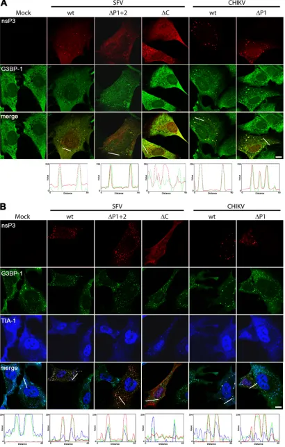
The subcellular localization of the proteins was also analyzed after transfection into MEFs (Fig. 2A). We have used these cells in the past for analysis of viral protein localization and SG formation (6,8). Cells were fixed 22 h after transfection and stained for nsP3, G3BP-1, and TIA-1. All three SFV nsP3 constructs exhibited gen-erally diffuse localization with occasional punctate staining. These puncta are foci of accumulation of the viral protein with its cellu-lar interacting partners that have been observed in other studies on alphavirus nsP3 function (8–11,17,18). SFV wt nsP3 appeared in foci in about 50% of transfected MEFs, while foci in cells trans-fected with nsP3-⌬P1⫹2 and nsP3-⌬C were much more rare. Since, in this experiment, we were primarily interested in the in-teraction and localization of G3BP-1 with nsP3, we chose to re-strict our analyses to cells displaying these foci. Colocalization studies are more meaningful when the proteins are not displaying completely diffuse cytoplasmic staining. The nsP3 puncta were, in the case of SFV wt nsP3 and nsP3-⌬P1⫹2, also G3BP-1 positive (Fig. 2A) but not TIA-1 positive (data not shown). This can be more easily appreciated in the RGD profile analyses shown under the merged images. Foci of nsP3-⌬C staining did not contain G3BP-1, as expected from the lack of interaction in the coimmu-noprecipitation experiment (Fig. 1C). Punctate localization was more obvious for CHIKV nsP3 wt and⌬P1, where almost all transfected cells displayed such localization. Both CHIKV nsP3 variants colocalized very well with G3BP-1 but not with TIA-1.
In our previous work (8), we showed that nsP3, when ex-pressed in the absence of other viral sequences in an inducible cell
line, also colocalized with G3BP-1 but not with TIA-1. When those cells were treated for 1 h with sodium arsenite, however, wt nsP3 was found in foci that were G3BP-1 and also TIA-1 positive, representing SGs, suggesting that nsP3 binding alone does not completely block the function of G3BP-1 in SG formation (8). To test whether MEFs transfected with the BAP-nsP3 fusion con-structs were capable of mounting a stress response, we treated them with 0.5 mM sodium arsenite to induce the formation of SGs. We found that while mock-transfected cells efficiently formed G3BP-1- and TIA-1-positive SGs, all nsP3 variants which were capable of G3BP-1 binding (SFV wt, SFV-⌬P1⫹2, CHIKV wt, and CHIKV-⌬P1) remained colocalized with G3BP-1 but also displayed some enrichment of TIA-1 in those foci (Fig. 2B). In cells transfected with these constructs, the localization of TIA-1 with G3BP-1 was weaker than that in nearby nontransfected cells or in mock-transfected cells but readily detectable. This was con-sistent with our previous work and suggested that nsP3–G3BP-1 bindingper seis not enough to fully inhibit SG formation in re-sponse to sodium arsenite. SFV nsP3-⌬C, which does not bind G3BP-1, did not inhibit the formation of SGs and did not colocal-ize with G3BP-1 or TIA-1, as expected. Together, the results inFig. 2show that both SFV and CHIKV nsP3 colocalized with G3BP-1 under normal and stress conditions and that this colocalization was not dependent on the proline-rich domain but rather on se-quences downstream.
In contrast to these results, Fros and colleagues suggested that the sequence between amino acids (aa) 398 and 406 of CHIKV nsP3, including the SH3-domain binding domain, was essential for the formation of nsP3/G3BP-1 foci in Vero cells (9). Our CHIKV-⌬P1 construct contained a larger deletion (residues 398 FIG 1SFV and CHIKV nsP3 sequences binding the SH3 domain of amphiphysins are not required for G3BP-1 binding. (A) MEFs were infected with SFV wt or SFV-⌬P1⫹2 at an MOI of 1. At 8 hpi, cells were fixed and stained for nsP3 and G3BP-1. Results are representative of two independent experiments. Images of single focal planes were processed using Adobe Photoshop. Bar, 10m. (B) Extreme C-terminal sequences of SFV wt nsP3, nsP3-⌬P1⫹2, nsP3-⌬C, and CHIKV wt nsP3 and nsP3-⌬P1. The C-terminal repeat sequences are in bold type. (C) BHK cells were mock transfected or transfected with the indicated constructs. Cell lysates were prepared 16 h after transfection, precipitated with streptavidin-coated beads, and separated by SDS-PAGE. Lysates and precipitates were probed with streptavidin and with G3BP-1 and actin antisera. Results are representative of three independent experiments. IP, immunoprecipitation.
Old World Alphavirus nsP3 C-Terminal Repeats Bind G3BP
on November 7, 2019 by guest
http://jvi.asm.org/
[image:2.585.37.539.66.310.2]FIG 2SFV and CHIKV nsP3 sequences binding the SH3 domain of amphiphysins are not required for localization to G3BP-1-positive foci. MEFs were mock transfected or transfected with pEBB/PP-SFVnsP3, nsP3-⌬P1⫹2, or nsP3-⌬C or pEBB/PP-CHIKVnsP3 or nsP3-⌬P1. After 22 h, transfected cells were mock treated (A) or treated with sodium arsenite (B) for 1 h, fixed and stained with streptavidin (for nsP3) and with G3BP-1 and TIA-1 antisera, and analyzed by confocal microscopy. Images of single focal planes were processed using Adobe Photoshop. Bars, 10m. Profiles were calculated using the RGB profiler tool in ImageJ.
on November 7, 2019 by guest
http://jvi.asm.org/
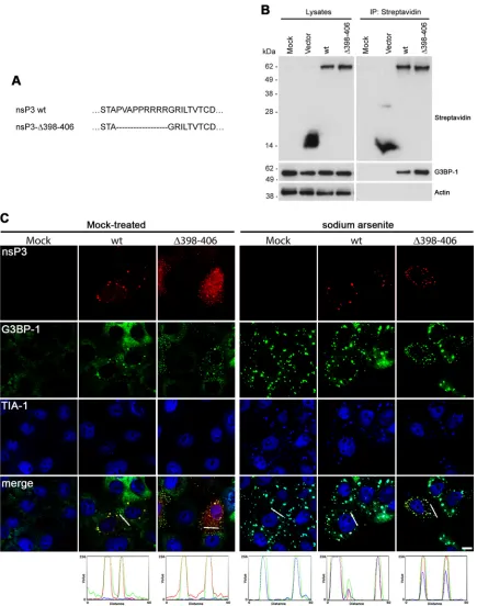
[image:3.585.88.498.37.681.2]FIG 3CHIKV nsP3 sequences binding the SH3 domain of amphiphysins are not required for localization to G3BP-1-positive foci in Vero cells. (A) Amino acid sequence of⌬398 – 406 mutation. (B) Vero cells were mock transfected or transfected with pEBB/PP vector or with CHIKVnsP3 or nsP3-⌬398 – 406. Cell lysates were prepared 22 h after transfection, precipitated with streptavidin-coated beads, and separated by SDS-PAGE. Lysates and precipitates were probed with streptavidin and with G3BP-1 and actin antisera. Results are representative of three independent experiments. (C) Vero cells were mock transfected or transfected with pEBB/PP-CHIKVnsP3 or nsP3-⌬398 – 406. At 22 h, transfected cells were mock treated (left panels) or treated with sodium arsenite (right panels) for 1 h, fixed and stained with Cy3-streptavidin (for nsP3) and with G3BP-1 and TIA-1 antisera, and analyzed by confocal microscopy. Images of single focal planes were processed using Adobe Photoshop. Bar, 10m. Profiles were calculated using the RGB profiler tool in ImageJ.
Old World Alphavirus nsP3 C-Terminal Repeats Bind G3BP
on November 7, 2019 by guest
http://jvi.asm.org/
[image:4.585.76.511.64.616.2]to 412), but we nevertheless demonstrated strong binding of that mutant protein to G3BP-1 in BHK cells. To address the discrep-ancy directly, we constructed a CHIK-nsP3-⌬398 – 406 construct (Fig. 3A) and analyzed its ability to bind G3BP-1 in Vero cells. Cells were transfected with constructs encoding BAP tag alone or BAP-tagged CHIKV-nsP3-wt or CHIKV-nsP3-⌬398 – 406, and lysates were precipitated with streptavidin-coated beads and ana-lyzed by immunoblotting as done before. Again, we detected somewhat stronger binding of the CHIKV nsP3 construct lacking the proline-rich domain to G3BP-1, relative to the wt protein (Fig. 3B), indicating that deletion of the proline-rich domain did not inhibit nsP3’s ability to form a complex with G3BP-1 in Vero cells. We also analyzed the localization of the nsP3 variants in Vero cells (Fig. 3C). Similarly to their localization in transfected MEFs, both CHIKV nsP3 wt and⌬398 – 406 localized to predominantly punc-tate structures in Vero cells with some diffuse staining visible, more obviously so for the mutant (Fig. 3C, left panels). Puncta formed by both wt and mutant proteins contained G3BP-1, con-firming the observation that both nsP3 variants formed a complex with the cellular protein. These puncta were likely not SGs, since TIA-1 was never found in wt nsP3- or nsP3⌬398-406- and G3BP-1-positive foci under these conditions. When similarly transfected cells were treated with sodium arsenite, however, both variants of nsP3 were found in foci that were G3BP-1 and TIA-1 positive, which we therefore identified as SGs (Fig. 3C, right panels). The diffuse staining for the viral proteins was no longer observed, in-dicating that the majority of the protein, whether or not diffuse under normal conditions, was recruited to SGs upon arsenite stress. In conclusion to the data shown inFig. 3, despite repeating very closely the experiment of Fros and colleagues, we were not able to repeat their observations and conclude that the formation of a complex containing nsP3 and G3BP-1 in Vero cells does not require the proline-rich, SH3-domain binding domain of Old World alphavirus nsP3 protein.
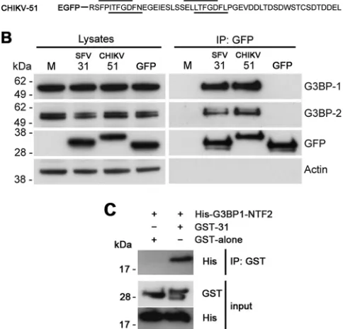
We have previously shown that the C-terminal L/ITFGDFD repeat domain of SFV nsP3 is necessary and sufficient for forma-tion of a complex with G3BP-1 (8). To determine whether the C-terminal repeat domain of CHIKV nsP3 was also sufficient for G3BP-1 complex formation in the absence of the proline-rich SH3-domain binding domain or other viral sequences, we gener-ated an enhanced green fluorescent protein (EGFP) construct fused to 51 amino acids from the C terminus of CHIKV nsP3 (CHIKV-51), similar to the SFV-31 construct previously de-scribed (8) (Fig. 3A). After transfection of these constructs into BHK cells, immunoprecipitation with GFP antisera revealed that both constructs efficiently formed complexes with G3BP-1 and also with G3BP-2 (Fig. 3B).
Several studies have previously demonstrated an interaction between nsP3 from the Old World alphaviruses and G3BP-1 and -2 (8,9,19–21). These studies examined the interaction in infected cells or in cells transfected with mutant constructs, and in no case can it be excluded that the binding is indirect and that another protein or RNA may be involved. In order to determine if the nsP3-G3BP interaction involves direct binding, we constructed vectors for expression inEscherichia coliof the nuclear transport factor 2 (NTF2)-like domain of G3BP-1 (residues 1 to 139; shown to be necessary for the nsP3 interaction [19]) fused to the His tag (His-G3BP1-NTF2) and 31 residues of the SFV nsP3 C terminus fused to the C terminus of glutathioneS-transferase (GST-31). The fusion proteins were separately expressed inE. coliBL21 T7
Express and purified using an ÄKTAprime Plus unit with HisTrap or GST GraviTrap columns (GE Healthcare). His-G3BP1-NTF2 (2 mg/ml) was mixed with GST (2 mg/ml) or GST-31 (2 mg/ml) in 20 mM Tris HCl, 300 mM NaCl, 30 mM imidazole, 5 mM MgCl2,
10% glycerol, pH 8, and incubated for 90 min with GST antisera (Abcam) at room temperature. When immunoprecipitates were analyzed by SDS-PAGE, we detected His-G3BP1-NTF2 in the sample containing GST-31 but not in that containing GST alone (Fig. 4C). These data show that the C-terminal 31 residues of SFV nsP3 were capable of binding to the NTF2-like domain of G3BP-1 in the absence of other cellular or viral factors and suggest, there-fore, that in Old World alphavirus-infected cells, the binding of nsP3 to G3BP is direct.
In this work, we have shown that the C-terminal L/ITFGDFD repeat regions of both SFV and CHIKV nsP3 are necessary and sufficient for G3BP binding. Supporting our findings, recently published work with chimeric Sindbis virus and Venezuelan equine encephalitis virus (VEEV) revealed that the HVD of VEEV, which contains a proline-rich sequence potentially binding SH3 domains but lacks repeat motifs with homology to the SFV L/ITF GDFD repeats, does not form a complex with G3BP (17,18). We conclude, therefore, that for the Old World but not the New World alphaviruses, the G3BP binding site resides in the C-termi-nal repeat motifs of nsP3. This contributes to an emerging picture FIG 4The C-terminal repeat motifs of SFV and CHIKV nsP3 are sufficient for G3BP binding. (A) Amino acid sequences of the C termini of the SFV-31 and CHIKV-51 constructs used in this study. The repeat sequences are underlined. (B) BHK cells were mock transfected or transfected with pEGFP-SFV-31, pEGFP-CHIKV-51, or pEGFP-C1. Cell lysates were prepared 16 h after trans-fection, immunoprecipitated with GFP antisera, and separated by SDS-PAGE. Lysates and precipitates were probed for G3BP-1, G3BP-2, GFP, and actin antisera. Results are representative of three independent experiments. (C) Pu-rified GST, GST-nsP3-31, or His-G3BP-1-NTF2 protein was mixed, immuno-precipitated with GST antisera, and separated by SDS-PAGE. Lysates and pre-cipitates were probed with GST or His antisera. Data are representative of three independent experiments.
Panas et al.
on November 7, 2019 by guest
http://jvi.asm.org/
[image:5.585.300.542.87.319.2]of distinct sets of host interactions for the New and Old World alphaviruses (17,22).
ACKNOWLEDGMENT
This work was supported by a grant from the Swedish Cancer Foundation to G.M.M.
REFERENCES
1.Anderson P, Kedersha N.2009. RNA granules: post-transcriptional and epigenetic modulators of gene expression. Nat. Rev. Mol. Cell Biol.10: 430 – 436.http://dx.doi.org/10.1038/nrm2694.
2.Kedersha NL, Gupta M, Li W, Miller I, Anderson P.1999. RNA-binding proteins TIA-1 and TIAR link the phosphorylation of eIF-2 alpha to the assembly of mammalian stress granules. J. Cell Biol.147:1431–1442.http: //dx.doi.org/10.1083/jcb.147.7.1431.
3.Tourriere H, Chebli K, Zekri L, Courselaud B, Blanchard JM, Bertrand E, Tazi J.2003. The RasGAP-associated endoribonuclease G3BP assem-bles stress granules. J. Cell Biol.160:823– 831.http://dx.doi.org/10.1083 /jcb.200212128.
4.Beckham CJ, Parker R.2008. P bodies, stress granules, and viral life cycles. Cell Host Microbe3:206 –212.http://dx.doi.org/10.1016/j.chom .2008.03.004.
5.Reineke LC, Lloyd RE.2013. Diversion of stress granules and P-bodies during viral infection. Virology436:255–267.http://dx.doi.org/10.1016/j .virol.2012.11.017.
6.McInerney GM, Kedersha NL, Kaufman RJ, Anderson P, Liljeström P. 2005. Importance of eIF2␣phosphorylation and stress granule assembly in alphavirus translation regulation. Mol. Biol. Cell16:3753–3763.http: //dx.doi.org/10.1091/mbc.E05-02-0124.
7.Venticinque L, Meruelo D.2010. Sindbis viral vector induced apoptosis requires translational inhibition and signaling through Mcl-1 and Bak. Mol. Cancer9:37.http://dx.doi.org/10.1186/1476-4598-9-37.
8.Panas MD, Varjak M, Lulla A, Eng KE, Merits A, Karlsson Hedestam GB, McInerney GM.2012. Sequestration of G3BP coupled with efficient translation inhibits stress granules in Semliki Forest virus infection. Mol. Biol. Cell23:4701– 4712.http://dx.doi.org/10.1091/mbc.E12-08-0619. 9.Fros JJ, Domeradzka NE, Baggen J, Geertsema C, Flipse J, Vlak JM,
Pijlman GP.2012. Chikungunya virus nsP3 blocks stress granule assem-bly by recruitment of G3BP into cytoplasmic foci. J. Virol.86:10873– 10879.http://dx.doi.org/10.1128/JVI.01506-12.
10. Neuvonen M, Kazlauskas A, Martikainen M, Hinkkanen A, Ahola T, Saksela K. 2011. SH3 domain-mediated recruitment of host cell amphiphysins by alphavirus nsP3 promotes viral RNA replication. PLoS Pathog.7:e1002383.http://dx.doi.org/10.1371/journal.ppat.1002383.
11. Varjak M, Zusinaite E, Merits A.2010. Novel functions of the alphavirus nonstructural protein nsP3 C-terminal region. J. Virol.84:2352–2364.
http://dx.doi.org/10.1128/JVI.01540-09.
12. Galbraith SE, Sheahan BJ, Atkins GJ.2006. Deletions in the hypervari-able domain of the nsP3 gene attenuate Semliki Forest virus virulence. J. Gen. Virol.87:937–947.http://dx.doi.org/10.1099/vir.0.81406-0. 13. Oberste MS, Parker MD, Smith JF.1996. Complete sequence of
Vene-zuelan equine encephalitis virus subtype IE reveals conserved and hyper-variable domains within the C terminus of nsP3. Virology219:314 –320.
http://dx.doi.org/10.1006/viro.1996.0254.
14. Strauss EG, Levinson R, Rice CM, Dalrymple J, Strauss JH. 1988. Nonstructural proteins nsP3 and nsP4 of Ross River and O’Nyong-nyong viruses: sequence and comparison with those of other alphaviruses. Virol-ogy164:265–274.http://dx.doi.org/10.1016/0042-6822(88)90644-7. 15. Vihinen H, Ahola T, Tuittila M, Merits A, Kaariainen L. 2001.
Elimination of phosphorylation sites of Semliki Forest virus replicase protein nsP3. J. Biol. Chem.276:5745–5752.http://dx.doi.org/10.1074 /jbc.M006077200.
16. Breakwell L, Dosenovic P, Karlsson Hedestam GB, D’Amato M, Liljeström P, Fazakerley J, McInerney GM.2007. Semliki Forest virus nonstructural protein 2 is involved in suppression of the type I interferon response. J. Virol. 81:8677– 8684.http://dx.doi.org/10.1128/JVI.02411-06.
17. Foy NJ, Akhrymuk M, Akhrymuk I, Atasheva S, Bopda-Waffo A, Frolov I, Frolova EI.2013. Hypervariable domains of nsP3 proteins of New World and Old World alphaviruses mediate formation of distinct, virus-specific protein complexes. J. Virol.87:1997–2010.http://dx.doi.org/10 .1128/JVI.02853-12.
18. Foy NJ, Akhrymuk M, Shustov AV, Frolova EI, Frolov I.2013. Hyper-variable domain of nonstructural protein nsP3 of Venezuelan equine en-cephalitis virus determines cell-specific mode of virus replication. J. Virol. 87:7569 –7584.http://dx.doi.org/10.1128/JVI.00720-13.
19. Cristea IM, Carroll JW, Rout MP, Rice CM, Chait BT, MacDonald MR. 2006. Tracking and elucidating alphavirus-host protein interactions. J. Biol. Chem.281:30269 –30278.http://dx.doi.org/10.1074/jbc.M603980200. 20. Frolova E, Gorchakov R, Garmashova N, Atasheva S, Vergara LA,
Frolov I. 2006. Formation of nsP3-specific protein complexes during Sindbis virus replication. J. Virol. 80:4122– 4134. http://dx.doi.org/10 .1128/JVI.80.8.4122-4134.2006.
21. Gorchakov R, Garmashova N, Frolova E, Frolov I.2008. Different types of nsP3-containing protein complexes in Sindbis virus-infected cells. J. Virol.82:10088 –10101.http://dx.doi.org/10.1128/JVI.01011-08. 22. Garmashova N, Gorchakov R, Volkova E, Paessler S, Frolova E, Frolov
I.2007. The Old World and New World alphaviruses use different virus-specific proteins for induction of transcriptional shutoff. J. Virol.81: 2472–2484.http://dx.doi.org/10.1128/JVI.02073-06.
Old World Alphavirus nsP3 C-Terminal Repeats Bind G3BP