CONTENTS
S.NO CONTENT PAGE. NO
1. INTRODUCTION 1-4
2. AIM AND OBJECTIVE 5
3. REVIEW OF LITERATURE 6-17
4 MATERIALS AND METHODS 18-32
5. RESULTS 33-39
6. DISCUSSION 40-49
7. SUMMARY AND CONCLUSION 50-51
1
INTRODUCTION
Dental caries is one of the major causes for tooth loss in all age groups and
affects both primary and permanent teeth. Dental caries is a dynamic demineralization
and remineralisation process. (1) Cavitations occur when demineralization process is
ahead of remineralisation process with initial carious lesion appearing as a white spot
lesion. (2)
Saliva and gingival crevicular fluid mainly contain calcium, phosphate, and
fluoride ions. The presence of these minerals at neutral pH helps in maintaining
equilibrium condition between the mineral content of tooth and oral fluids. However
when there is a drop in the oral fluid pH (below 5.5), dissolution of hydroxyapatite
(HA) crystals and release of calcium and phosphate ions from tooth surface into oral
fluids happen which is referred as demineralization. (3)
The demineralization process can be stopped by creating an environment
conductive for remineralisation by various remineralising agents.(4) The process of
restoring lost mineral ions in the tooth structure and strengthening the lattice work is
known as remineralisation.(5)
The remineralised enamel crystallites are generally more resistant to
decalcification and also have the same orientation as the original enamel crystallites
(6)
. The early enamel lesions have a potential for remineralisation with an increased
resistance to further acid challenge, particularly with the use of enhanced
remineralisation treatments (7). Thus invasive treatments of pre cavitated lesions are
not required. Various remineralising agents like fluoride (8), Casein phosphopeptide,
2
Enamelon, CPP stabilized amorphous calcium phosphate with fluoride (CPP-ACPF
Recaldent) (11,12), has been studied in both in-vitro (13-15) & in-vivo studies (16,17) .
Remineralising agents are available in various forms such as restorative
materials, pit & fissure sealants, chewing gums, mouth rinses and dentifrices.(18,19)
One of the most effective remineralising agents in caries prevention is fluoride.
Nevertheless, some concerns have been expressed about fluorosis and total fluoride
intake.(18,19) In recent years, fluoride alternatives have been proposed, which include
CPP-ACP and nano-hydroxyapatite (NHA) because of their anticariogenic
characteristics.(20,21)
The modern dental practice is focused on prevention and minimal
intervention; replacement of lost tooth substance with a bio mimetic material is
considered as one of the fundamentals of minimal intervention dentistry. Several
methods were introduced to remineralise an early tooth structure loss. (22, 23)
Nano hydroxyapatite (n-HAp) is one of the most biocompatible and bioactive
materials with marked affinity to the enamel surface. These nano-sized particles are
similar to the apatite crystals of tooth enamel in morphology and crystal structure. [24]
Having better affinity for hydroxyapatite crystals of enamel.
NHA toothpastes were first investigated in Japan in the 1980s. Studies have
reported more or comparable remineralising effects for NHA toothpastes in
comparison to other toothpastes containing amino fluoride and fluoride. (25,26) Daily
tooth brushing with NHA toothpaste can provide adequate amounts of HA and enrich
3
In current practice, Nano hydroxyapatite has been widely used as an effective
anti caries agent mainly because of its unique potential to bring about
remineralisation. (28) The size of the calcium phosphate crystals also plays an
important role in the formation of hard tissue and also has a significant impact on its
intrinsic properties, solubility & biocompatibility. (29)
Calcium sodium phosphate silicate bioactive glass (BAG) (Novamin) is
another material introduced to aid in remineralisation. Calcium sodium phospho
silicate disintegrates and gives off sodium that gets exchanged with hydrogen cations
(H+ or H3O+) when it comes in contact with saliva, which results in the release of
calcium (Ca²+) and phosphate (PO4²) ions from the particle structure. (30-32)
The result of transient increase in pH that brings about the precipitation of
calcium and phosphate ions from the saliva, to form a calcium phosphate layer on the
tooth surfaces. Ca-P complexes crystallize to form a hydroxy carbonate apatite that is
chemically and structurally similar to biological apatite. (30, 33)
Bioactive glass when contacts with saliva, rapidly releases sodium, calcium
and phosphorous ions into saliva that are available for remineralisation of the tooth
surface. These ions are released form hydroxycarbonate apatite (HCA) directly. They
also attach to the tooth surface and continue to release ions and remineralise the tooth
surface after the initial application. These particles have been shown to release ions
and transform into HCA for up to 2 weeks. Ultimately, these particles will completely
transform into HCA. (34)
Novamin adheres to exposed dentin surface and forms a mineralized layer that
4
Tri Calcium Phosphate (TCP) is a new hybrid material created with a milling
technique that fuses beta tricalcium phosphate (ß-TCP) and sodium lauryl sulfate or
fumaric acid. This blending results in a “functionalized” calcium and a “free”
phosphate ions, designed to increase the efficacy of fluoride remineralisation (36, 37).
ß-TCP is similar to apatite crystals and possesses unique calcium environments capable
of reacting with fluoride and enamel. As the phosphate floats freely, the exposed
calcium environments are protected by preventing the calcium from prematurely
interacting with fluoride. TCP provides catalytic amounts of calcium to boost fluoride
efficacy and may be well designed to coexist with fluoride in a mouth rinse or
dentifrice because it will not react before reaching the tooth surface (38). When TCP
finally comes in contact with the tooth surface and is moistened by saliva, the
protective barrier breaks down, making the calcium, phosphate and fluoride ions
which are present in the teeth. The fluoride and calcium then reacts with weakened
enamel to provide a seed for enhanced mineral growth relative to fluoride alone.
Micro hardness tests are commonly used to study the physical properties of
materials and they are widely used to measure the hardness of teeth (39-41). This
5
AIM AND OBJECTIVE
AIM
To evaluate the remineralisation efficacy of three different novel
remineralising agents namely - Bioactive glass, Tri-calcium Phosphate,
Nanohydroxyapetite.
OBJECTIVE
To compare the efficacy of three different novel remineralising agents namely-
Bioactive glass, Tri-calcium Phosphate, Nanohydroxyapetite on artificially created
6
REVIEW OF LITERATURE
King et al (2006) (42), prepared enamel blocks of extracted human third molars
in an in vitro study to compare the efficacy of 10% NHA toothpaste, 900 ppm sodium
fluoride and 900 ppm sodium monofluorophosphate (MPF) and the results showed
that all the three agents remineralised enamel lesions and no significant difference
was noted between their efficacy. Differences in the results might be explained by the
use of polarized light microscopy and microradiography and the differences in the
materials used.
EC Reynolds (2008) (43), The NovaMin TM technology is based on calcium
sodium phosphosilicate bioactive glass which is claimed to release calcium and
phosphate ions intra-orally to help the self-repair process of teeth. No published
studies could be found supporting the remineralisation of enamel subsurface lesions in
vitro or in situ. Furthermore, no published studies could be found showing an
anticariogenic efficacy of NovaMin TM in animal models or other caries model
systems or randomized, controlled caries clinical trials. This technology appears to be
at a very early stage of development.
Huang et al (2009) (42), prepared 129 bovine enamel blocks to compare the
efficacy of 1%, 5%, 10% and 15% NHA and sodium fluoride with using of Vickers
microhardness tester and scanning electron microscopy and they found that optimal
concentration (10%) of NHA resulted in remineralisation of initial enamel lesions.
HA, which is a bioactive and biocompatible material, is one of the primary
components of tooth mineral content. HA is expected to significantly enhance
7 higher efficacy than HA for this purpose due to its nano-size particles (11). NHA has
hydrophilic and wetting characteristics and is capable of producing a thin but tightly
bound layer on the tooth surface, resulting in higher surface hardness and
remineralisation. HA is capable of obstructing the dentinal tubules and thus, relieves
tooth hypersensitivity. (13)
Chanya Chuenarrom et al (2009) (44), conducted a study to evaluate the
effect of indentation loads and times on Knoop and Vickers microhardness tests for
human enamel and dentin and they concluded that the difference of indentation times
was not influential on KHN and VHN values of enamel and dentin for the same
indentation loads but the KHN values of enamel and the VHN values of dentin were
affected by variation of indentation loads.
Huang sB et al (2009) (45), conducted to determine the effect of
nano-hydroxyapatite-containing toothpastes on initial enamel lesions under dynamic
pH-cycling conditions and studies concluded that nano-HA has the potential to
remineralise initial enamel lesions. A concentration of 10% nano-HA may be optimal
for remineralisation of early enamel caries.
Huang in (2009) and Swarup in (2012) (46), used fluoride in combination
with nanohydroxyapatite and concluded that nanohydroxyapatite produced a new
surface layer over the demineralised enamel, with morphology similar to that of
biologic enamel. They also stated that 10% nanohydroxyapatite is optimal for
remineralisation of early carious lesions.
Braurer D et al (2009) (47), performed a study to understand the effect of
8 increased in Sio2-CaO-P2O5-NaO5 system while network connectivity was kept
constant. It was observed that incoporation of fluoride in bioactive glass, decreased it
Tg which means that the glass has reduced hardness and is more bioactive. Also the
onset of crystallization and peak temperatures were decreased when CaF2 was
increased.
S.B. Huang et al 2009, Shengbin et al 2010 and S. Huang et al 2011 (48),
reviewed the surface chemical properties and morphological structure of
hydroxyapatite has been claimed to play the most important part in re-mineralization
of early caries lesions. Three studies showed significant improvement in surface
microhardness post treatment with nano-hydroxyapatite toothpaste similar to a review
by Kim et al 2007.
Itthagarun et al 2010, Peter et al 2011 (49), has shown that lesions can be
re-hardened by deposition of hydroxyapatite that is initially deposited near the surface
layer of the enamel and this was found to be significant. Future studies are required
for further assessment of the potential of nano-hydroxyapatite toothpaste on
enamelin-vivo.
Peter et al (2011) (50),evaluated the effects of nano-hydroxy apatite
(n-HAp)tooth pastes on remineralization of bovine and dentine subsurface lesions and
they concluded that tooth pastes containing nano-hydroxy apatite revealed higher
remineralising effects compared to amine fluoride toothpastes with bovine dentine,
and comparable trend were obtained for enamel.
Arathi rao et al(2011)(51), reviewed a concentration of 10%
9 hydroxyapatite has been used in toothpastes (as fillers) and pit-and-fissure sealants.
and also the crystals of hydroxyapapite can effectively penetrate the dentin tubules
and obturate them and can cause closure of the tubular openings of the dentin with
plugs within 10 minutes as well as a regeneration of a surface mineral layer.
Tschoppe et al (2011) (52), prepared 85 dentin blocks of bovine teeth to
compare the efficacy of 7%, 20% and 24% NHA toothpastes and 0.14% amine
fluoride by using microradiography and they found that NHA toothpastes had greater
efficacy for remineralisation of initial lesions compared to amine fluoride.
Najibfard et al (2011) (53), compared 10% and 5% NHA and a combination of
10% NHA + 1100 ppm NAF in an in vivo study based on microradiographs, the
results showed comparable remineralising effect of the toothpastes evaluated. This
finding is in contrast to our findings, which might be attributed to differences in
concentration of NHA and study designs. We used Vickers microhardness tester but
Najibfard et al evaluated the remineralising effect with microradiography. Differences
in the results might be explained by the use of polarized light microscopy and
microradiography and the differences in the materials used.
Tschoppe P et al (2011) (54), conducted a study on Enamel and dentine
remineralisation by nano-hydroxyapatite toothpastes concluded that toothpastes
containing nano-hydroxyapatite revealed higher remineralising effects when
compared to amine fluoride toothpastes.
Yuan et al (2012) (55), used 48 dentin specimens to compare the efficacy of
10 (EDS), they concluded that NHA was highly capable of obstructing the tubules and
remineralising the tooth structure, which is in accord with our study results.
J.Shanti Swarup, Arathi Rao (2012) (56),conducted a study about to explore
the effects of synthetically processed nanosized biomimetic HA particles in causing
remineralisation of the early enamel lesion in comparison with 2%sodium fluoride &
they concluded that 10% biomimetic nanohydroxyapatite of the particle size 20nm
has the potential to remineralise initial enamel caries under in vitro conditions when
compared with 2% sodium fluoride. This documented biomimetic apatite coating on
the demineralised enamel suffices the need for a synthetic enamel biocompatible
material able to repair early enamel lesions. nano hydroxyapatite would therefore be
beneficial in promoting remineralisation with regular daily usage.
Goswami M, Saha S1, Chaitra TR (2012) (57), done a study to determine the
effect of nano-hydroxyapatite concentrations on initial enamel lesions under dynamic
pH-cycling conditions. It was concluded that nano-hydroxyapatite had the potential to
remineralise initial enamel lesions. A concentration of 10% nano-hydroxyapatite may
be optimal for remineralisation of early enamel caries.
Somkamol Vanichvatana and Prim Auychai (2013) (58), compared the
efficacy of CPP–ACPF and fTCP (calcium phosphate pastes) with that of
conventional 0.1%fluoride toothpaste in remineralising enamel on artificial caries
lesions and they concluded that all three groups remineralised the enamel slab lesions,
indicating model sensitivity to fluoride. Given the differences in usage amounts and
treated regimens, Clinpro Tooth Creme provided similar benefits to the 0.1% fluoride
toothpaste, however, no additional benefit of Tooth Mousse Plus was observed when
11
Tabari M et al. (2013)(59), prepared nano-hydroxyapatite preparation by
in-situ hybridization method and found that application of nano-hydroxyapatite
preparation increased microhardness of tooth whether applied before or after iron
drop exposure, but results were found better after the application of iron drop
exposure. Application of nanohydroxyapatite preparation in our study also showed
increase in microhardness of enamel after iron drop exposure.
Balakrishnan A et al (2013) (60), evaluated the remineralisation potential of
various dentifrices over a period of 30 days and concluded that the extent of
remineralisation achieved was dose dependant and increased with increasing the time
of exposure and duration of the study .
Namrata Patil et al (2013) (61), compared the remineralising potential of three
agents (CPP-ACP, CPP-ACP & fluoride and tricalcium phosphate & fluoride) on
artificial enamel carious lesion and they concluded All the three remineralising agents
in the study could effectively remineralise artificial enamel caries and they reported
that TCP & fluoride-based products performed better than CPP-ACP-based products
in remineralising artificial enamel caries.
Sai Sathya Narayana et al (2014) (62), Remineralisation efficiency of
bioactive glass on artificially induced carious lesion – in vitro and concluded that
bioactive glass is an effecive remineralising agent. NovaMin® : It is the trade name
for a calcium sodium phosphosilicate bioactive glass, which is originally developed
for the treatment of hypersensitivity by the physical occlusion of dentinal tubules. (7)
Haghgoo et al (2014)(63), found no differences between NHA and NaF
12 remineralisation significantly increased, and they used the remineralising agent in the
form of a mouthwash.
Su-Yeon Jo et al (2014) (64), examined the effects of fluoridated, casein
phosphopeptide–amorphous calcium phosphate complex (CPP-ACP)-containing, and
functionalized β-tricalcium phosphate (fTCP) containing toothpastes on
remineralisation of white spot lesions (WSLs) by using Quantitative light-induced
fluorescence (QLF-D) Biluminator TM and they concluded that fTCP- and CPP-ACP
containing toothpastes seem to be more effective in reducing WSLs than 1,000ppm
fluoride-containing toothpastes.
De Carvalho et al(2014)(65) , reported that nano-HAP group showed
significantly higher difference in surface hardness recovery after using the same pH
cycling method as this study than fluoride varnish. This may be attributed to the
number of applications of the nano-P paste, where it was applied for a total of seven
times, while the fluoride varnish was applied only once. Taking into consideration the
difference in the assessment method as they used KNH, it can be deduced that
increasing the times of application of the nano-P paste could enhance the effect of
remineralisation.
Sai Sathya Narayana et al (2014)(66), Investigated the efficacy of bioactive
glass containing product on remineralisation of artificial induced carious enamel
lesion and to compare its efficiency with other remineralisation products using an
in-vitro pH cycling method and they concluded that Each test group when compared
with the control group showed a significant difference existing for element Ca and P.
13 considered as an effective remineralising agent and F increase was seen in Amflor
followed by CPP-ACPF as both contained fluoride.
Mehta et al (2014) (67),showed that bioactive glass (Novamin) and casein
phosphopeptide-amorphous calcium phosphate successfully remineralised early
enamel caries. However Novamin remineralised the carious lesion more effectively
and CPP-ACP had an amorphous nature and couldn't properly adhere to the enamel
surface. This also led to lower hardness value for CPP-ACP while Novamin showed
higher values of hardness because it attached to the surface more compactly.
Adit Bharat Mehta et al (2014) (68),evaluate and compare the
remineralisation potential of bioactive-Glass (BAG) (Novamin®/
Calcium-sodium-phosphosilicate) and casein phosphopeptide-amorphous calcium phosphate
(CPP-ACP) containing dentifrice and they concluded that both BAG and CPP-ACP are
effective in remineralising early enamel caries. Application of BAG more effectively
remineralised the carious lesion when compared with CPP-ACP.
Nithin m. g & joseph john (2015) (69),they has been done a systematic
review was to evaluate how effective is the remineralisation potential of
nanohydroxyapatite toothpaste on enamel and they concluded that the re-mineralizing
potential of nano-hydroxyapatite toothpaste on enamel, the effectiveness of this
toothpaste seems to be obvious in improving the initial enamel carious lesions.
Samuel B. Low et al (2015) (70),evaluated effectiveness of a commercially
available toothpaste containing potassium nitrate, sodium monoflurophosphate, and
14 reducing dental hypersensitivity in adults and they concluded that a toothpaste
containing potassium nitrate, sodium monoflurophosphate and nano-hydroxyapatite
plus antioxidants phloretin, ferulic acid and silymarin applied daily significantly
decreased tooth pain of dentin hypersensitivity within a two-day and two-week time
period.
Vyavhare et al(2015) and Swarup and Rao (2012) (71), who studied the
effect of adding synthetically processed nanoHAP to toothpaste in remineralising
initial enamel lesion.
MohammadBagherRezvani et al (2015) (74), evaluate the effect of
nano-tricalciumphosphate (n-TCP) and nanohydroxyapatite (n-HAP) on prevention of
restaining of enamel after dental bleaching and they concluded that 10% n-TCP
couldsignificantlymaintaintheresultantcolourandreconstructtheenamelstructureafterble
aching.
Amaechi et al (2015) (72), reported that effectiveness of n-HAp containing
toothpaste (Apagard) to physically occlude dentin tubules as a surrogate measure of
its ability to clinically relieve dentin hypersensitivity.
Udaya Kumar Palaniswamy et al (2016) (73), evaluate remineralising
potential of bioactive glasses (BAGs) and amorphous calcium phosphatecasein
phosphopeptide (ACP‑CPP) on early enamel lesion and concluded that Both the remineralising agents tested in this study can be considered effective in repair and
prevention of demineralization. BAG showed better results initially, but eventually
15
Rithesh kulal et al (2016) (74),they conducted a study about to evaluate and
compare the effects of three different desensitizing agents (15% nano hydroxyapatite
crystals; 5% novamin and 8% proargin) on dentinal permeability and tubule occlusion
in-vitro and they concluded that all the three desensitising agents were effective in
the dentine tubule occlusion. In addition efficacy of nano-hydroxyapatite toothpaste
was greater compared to the other desensitising agents.
Asghar Ebadifar et al (2017) (75),they have done a study to assess the effect
of nano-hydroxyapatite (NHA) on microhardness of artificially created carious lesions
and they concluded that toothpaste containing NHA was more effective than the
toothpaste without NHA for the purpose of remineralisation.
Abhishek Singh et al (2017) (76),compare the relative efficiency of
nanohydroxyapatite (n-Hap) Aclaim, n-HAp Apagard, Clinpro Tooth Crème and
Colgate Total in remineralisation and they concluded that Aclaim and Apagard on
daily application will provide maximum protection against enamel demineralization
in orthodontic patients.
A Arvindkumar et al (2017) (77), compared and evaluated the caries
preventive efficacy of aresin infiltrant, casein phosphopeptideamorphous (CPP-ACP)
and nanohydroxyapatite(nano-HA) on white spot enamel lesion and concluded that
the resin infiltrant showed higher caries inhibition potential and superior acid
resistance than CPP-ACP and nano-HA.
Nilesh Rathi et al (2017) (78), evaluate and compare the microhardness of
deciduous teeth treated with nano-hydroxyapatite and calcium sucrose phosphate after
16 calcium sucrose phosphate have remineralising effect over teeth affected by acid
challenge of iron drops, nanohydroxyapatite preparation showing better results than
calcium sucrose phosphate.
Rakesh Mittal et al (2017) (79), reviewed NovaMin is the brand name of a
particulate bioactive glass. NovaMin is technically described as an inorganic
amorphous calcium sodium phosphosilicate (CSPS) material that was designed based
on a class of materials known as bioactive glasses. It comprises SiO2 (45%), Na2O
(24.5%), CaO (24.5%) and P2O5 (6%) [26]. NovaMin is claimed to release calcium
and phosphate ions intraorally to help the self-repair process of enamel.
A silica-rich surface layer forms through polycondensation of hydrated silica
groups on which precipitation of ions happens which crystallizes over time to form a
hydroxyl-carbonate apatite. Although it is used extensively as a desensitizing agent
reports also claim that the chemical reactions that promote apatite formation may
enhance the remineralisation.
A novel 5,000 ppm fluoride dentifrice, Clinpro 5000, was recently introduced
by 3 M ESPE. This 1.1% NaFsilica-containing paste containing an innovative
functionalized tricalcium phosphate (fTCP) ingredient that, when evaluated in
development formulations, has been shown to boost remineralisation performance
relative to fluoride-only systems.
Umang Jagga et al (2018) (80), compared the remineralising efficacy of
novamin, tricalcium phosphate and they concluded that both novamin and tricalcium
17
Issa Daas et al (2018) (81), compared the effectiveness of nano-hydroxyapatite
(nano-HAP) paste and fluoride varnish in remineralising initial enamel lesion in
young permanent teeth and their ability to resist secondary caries under dynamic pH
cycling quantitatively & qualitatively and they concluded that Nano-HAP paste
showed promising long-term protective effect in terms of surface depositions and
maintaining a smooth surface compared with fluoride varnish.
Niwut Juntavee et al (2018) (82), investigated the effects of
nano-hydroxyapatite (NHA) gel and Clinpro (CP) on remineralisation potential of enamel
and cementum at the cavosurface area of computeraided design and computer-aided
manufacturing ceramic restoration and they concluded that NHA gel and CP were
capable of remineralisation of the enamel and cementum.NHA was more capable in
the remineralisation process than CP. NHA was extremely capable in the
remineralisation process for enamel and cementum surrounding the margin of the
18
MATERIALS & METHODS
SOURCE OF SAMPLE
Sixty freshly extracted sound human permanent maxillary first premolars were
collected from the Department of Oral and Maxillofacial Surgery, Vivekanandha
Dental College for Women, Tiruchengode.
MATERIALS USED
Self cure acrylic.
Deionised water.
Demineralising solution.
Tri-Calcium Phosphate (Clin pro tooth paste)
Nano-hydroxyapetite (Aclaim tooth paste)
Bioactive glass (Novamin)
Artificial Saliva.
Armamentarium:
Universal incubator.
Contra angle hand piece (NSK).
Polishing cup.
19
Method of collection of samples:
Sixty maxillary first premolars were collected from the Department of Oral
and Maxillofacial surgery, Vivekanandha Dental College for Women, Tiruchengode,
which were indicated for extraction for therapeutic purpose.
Infection Control protocol for the teeth collected for this study:
Collection, storage, sterilization and handling of extracted teeth were followed
according to the guidelines and recommendations given by:
S.NO Reminerali
sing agents Manufacturer COMPOSITION
1. Tri calcium
phosphate
3M ESPE
Water, Sorbitol, Hydrated silica, Glycerin,
Polyethylene-polypropylene Glycol. Flavor,
Polyethylene Glycol, Sodium lauryl sulfate,
Titanium dioxide, carboxymethyl cellulose,
sodium saccharin, sodium fluoride, tricalcium
phosphate 2. Nano-hydroxy apatite Group Pharmaceuticals Ltd
Sorbital, Glycerin, Silica, Purified water,
Hydroxyapatite, cocamidopropyl betaine,
Hydroxyethyl cellulose, Titanium dioxide,
Flavour, sodium saccharin.
3. Bioactive
glass GlaxoSmithKline
Sodium fluoride, Glycerin,hydrated silica,
calcium sodium phosphosilicate (NOVAMIN),
cocamidopropyl betaine, sodium methyl cocoyl
taurate, aroma, titanium dioxide,
20
Occupational Safety and Health Administration (OSHA) and Centre for Disease
Control & Prevention (CDC):
1. Handling of teeth was always done using gloves, mask and protective eyewear.
2. Teeth were cleaned of any visible blood and gross debris.
3. Distilled water was used in wide mouth plastic jars for initial collection.
4. Teeth were immersed in 10% formalin for 7 days, following which the liquid was
discarded and the teeth were transferred into separate jars containing distilled water.
5. The initial collection jars, lids and the gloves employed were discarded into
biohazard waste receptacles.
6. As and when the teeth were required, they were removed from the jars with cotton
pliers and rinsed in tap water.
Exclusion Criteria:
Teeth with any visible caries, hypoplastic or white spot lesions or any fracture
were excluded from the study.
Inclusion Criteria: Caries-free teeth, extracted for therapeutic purpose were
included.
PROCEDURE
Removal of external residual tissues:
The selected teeth were stored in 10% formalin following extraction and
calculus was mechanically removed using hand scalers.
Preparation of the samples:
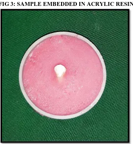
Teeth were embedded in selfcure acrylic with exposure of buccal surface of
enamel following which the samples were then stored in deionised water for one
21
Demineralization solution & artificial saliva Solution preparation:
Both Demineralization solution & Artificial saliva were prepared in the
Department of Biochemistry, Vivekanandha Dental College For Women,
Tiruchengode.
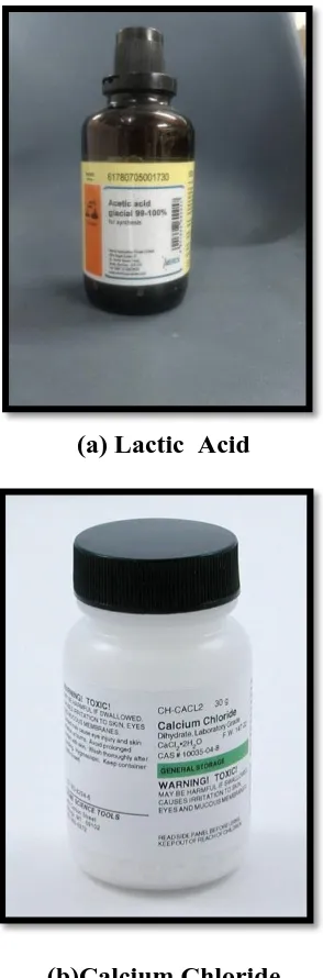
Composition of Demineralising solution:
2.2mM calcium chloride (CaCl2.2H2O)
2.2mM monosodium phosphate (NaH2PO4.7H2O)
0.05M lactic acid.
The final pH was adjusted to 4.5 with 50% sodium hydroxide(NaOH).
Composition of Artificial saliva
2.200g/L gastric mucin
0.381 g/L sodium chloride (NaCl)
0.213 g/L calcium chloride (CaCl2.2H2O)
0.738 g/L potassium hydrogen phosphate (K2HPO4.3H20)
1.114 g/L potassium chloride (KCl)
The final pH was adjusted to 7.00 at 37ᵒc with 85% lactic acid.
Demineralising solution was used to demineralise the surface of the samples
by immersing them in a glass containers containing 50mL of the solution for a period
of 48 hours at 37ᵒC using an incubator. This procedure was aimed at producing a
consistent subsurface lesion, following which demineralization process for 48 hours.
After demineralisation process the samples were washed in deionised water
22
Topical application
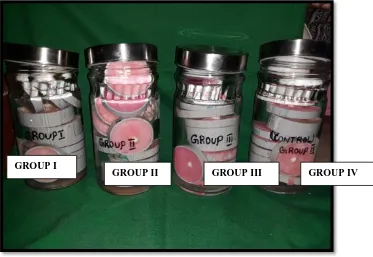
Samples were randomly divided into four groups consisting of 15 samples
each as follows:
Group 1 - Tri-Calcium Phosphate (n=15).
Group 2 - Nanohydroxyapetite (n=15).
Group 3 - Bioactive glass (n=15).
Group 4 - Artificial Saliva (Control) (n=15).
The samples from each group were treated with the respective remineralising
agent (except for the control group) for 4 min , at every 24th hours for a period of 7
days with help of polishing cup attached to a contra-angle hand piece. The samples of
control group was only placed in artificial saliva. The samples of experimental groups
were rubbed with the respective remineralising agent for 4 minutes and placed in
artificial saliva after washing with deionised water.
All samples were incubated in a universal incubator at 37ᵒC between each
remineralising cycle. After 7 cycles of remineralisation, surface micro hardness of the
specimens was determined using a Vicker's micro hardness testing machine (Anna
University, chennai).
The micro hardness values were tabulated & compared to determine the
23
FIG 1: PRE OPERATIVE SAMPLES
[image:36.595.125.505.514.705.2]24
FIG 3: SAMPLE EMBEDDED IN ACRYLIC RESIN
[image:37.595.128.499.450.737.2]25
FIG 5: VICKER' S HARDNESS MACHINE
[image:38.595.204.425.138.727.2]26
FIG 7: SAMPLES IMMERSED IN DEMINERALISING SOLUTION
FIG 8: APPLICATION OF REMINERALISING AGENTS GROUP I
[image:39.595.147.479.475.731.2]27
FIG 9:
[image:40.595.201.428.402.697.2]28
FIG 11: DEMINERALISING SOLUTION
[image:41.595.159.470.478.704.2]29
[image:42.595.151.482.118.362.2]FIG 13: GROUP II
30
31
FIG 16: DEMINERALISING SOLUTION REAGENTS
(a) Lactic Acid
(b)Calcium Chloride
32
FIG 17: ARTIFICIAL SALIVA REAGENTS
(a)Calcium Cloride (b) Potassium Chloride
33
RESULTS
GROUP I (TRICALCIUM PHOSPHATE)
CONSTANT LOAD (P) (N)
AVG. DIA.
LENGTH (D)
(MM)
HV (MPA)
1.854 1.96133 0.158 145.662
1.854 1.96133 0.161 140.2842
1.854 1.96133 0.16 142.0432
1.854 1.96133 0.163 136.8627
1.854 1.96133 0.157 147.5235
1.854 1.96133 0.158 145.662
1.854 1.96133 0.158 145.662
1.854 1.96133 0.161 140.2842
1.854 1.96133 0.159 143.8344
1.854 1.96133 0.158 145.662
1.854 1.96133 0.157 147.5235
1.854 1.96133 0.162 138.5576
1.854 1.96133 0.159 143.8355
1.854 1.96133 0.161 140.2842
34
GROUP II (NANOHYDROXY APATITE)
CONSTANT LOAD (P) (N)
AVG. DIA. LENGTH (D)
(MM)
HV (MPA)
1.854 1.96133 0.143 177.8232
1.854 1.96133 0.142 180.3365
1.854 1.96133 0.141 182.9036
1.854 1.96133 0.140 185.5258
1.854 1.96133 0.141 182.9036
1.854 1.96133 0.144 175.362
1.854 1.96133 0.142 180.3365
1.854 1.96133 0.141 182.9036
1.854 1.96133 0.143 177.8232
1.854 1.96133 0.142 180.3365
1.854 1.96133 0.141 182.9036
1.854 1.96133 0.141 182.9036
1.854 1.96133 0.140 185.5258
1.854 1.96133 0.143 177.8232
35
GROUP III (BIOACTIVE GLASS)
CONSTANT LOAD (P) (N)
AVG. DIA.
LENGTH (D)
(MM)
HV (MPA)
1.854 1.96133 0.202 89.11641
1.854 1.96133 0.199 91.82359
1.854 1.96133 0.198 92.75344
1.854 1.96133 0.196 94.65602
1.854 1.96133 0.199 91.82359
1.854 1.96133 0.200 90.90765
1.854 1.96133 0.201 90.00534
1.854 1.96133 0.195 95.62934
1.854 1.96133 0.197 93.69749
1.854 1.96133 0.198 92.75344
1.854 1.96133 0.198 92.75344
1.854 1.96133 0.196 94.65602
1.854 1.96133 0.197 93.69749
1.854 1.96133 0.202 89.11641
36
GROUP IV(CONTROL)
CONSTANT LOAD (P) (N)
AVG. DIA.
LENGTH (D)
(MM)
HV
(MPA)
1.854 1.96133 0.262 52.9734
1.854 1.96133 0.260 53.79151
1.854 1.96133 0.265 51.78079
1.854 1.96133 0.260 53.79151
1.854 1.96133 0.250 58.18089
1.854 1.96133 0.255 55.92166
1.854 1.96133 0.262 52.9734
1.854 1.96133 0.253 56.80929
1.854 1.96133 0.260 53.79151
1.854 1.96133 0.265 51.78079
1.854 1.96133 0.270 49.88074
1.854 1.96133 0.250 58.18089
1.854 1.96133 0.265 51.78079
1.854 1.96133 0.265 51.78079
37
STATISTICAL ANALYSIS
To analyse the data SPSS (IBM SPSS Statistics for Windows, Version 23.0,
Armonk, NY: IBM Corp. Released 2015) is used. Significance level is fixed as 5% (α
[image:51.595.116.518.399.590.2]= 0.05).
TABLE 1
The Normality tests Kolmogorov-Smirnov and Shapiro-Wilks tests results
reveal that the variable (HV) follows Normal distribution. Therefore, to analyse the
data Parametric method is applied.
One-Way ANOVA To Compare Mean HV Values Between Groups:
Group N MeanHV
(MPa) Std. Dev
Std. Error of Mean
95% CI for Mean
p-value
LB UB
Group -1 15 143.05 3.295 0.8507 141.22 144.87
<0.001
Group -2 15 181.05 2.963 0.7649 179.41 182.69
Group -3 15 92.23 2.067 0.5337 91.08 93.37
Group -4 15 54.02 2.582 0.6667 52.59 55.45
Total 60 117.58 48.857 6.3074 104.96 130.21
The statistical parameters such as mean and SD of micro hardness of samples
were obtained for each group as shown in Table 1. The mean for control was
the lowest 54.02 ± 2.582 MPa. In case of materials used, NHA indicated
highest mean 181.05 ± 2.963MPa, followed by TCP with a mean value of
143.05 ± 3.295 MPa, and bioactive glass with the least mean value of 92.23 ±
38
TABLE 2
ANOVA
Sum of Squares df Mean Square F-value p-value
Between Groups 140405.234 3 46801.745 6123.315 <0.001
Within Groups 428.019 56 7.643
Total 140833.253 59
One-way ANOVA resulted into the F-statistic of 6123.315 with a
corresponding P < 0.001.The test indicated statistically significant difference
of mean microhardness across the groups. Accordingly, pair-wise comparison
of microhardness was performed between groups to determine which groups
differed significantly from other, with results shown in Table 2.
TABLE III
To compare the mean HV values between groups one way ANOVA is applied
followed by Tukey’s HSD post hoc tests for multiple pairwise comparisons.
Tukey Hsd Post Hoc Tests For Multiple Pair Wise Comparisons
Group Mean
Difference p-value
Group -1
Group -2 -38.00 <0.001
Group -3 50.82 <0.001
Group -4 89.03 <0.001
Group -2
Group -3 88.82 <0.001
Group -4 127.03 <0.001
39
CHART
The mean microhardness of Nano hydroxy apatite was higher than other two
experimental groups; however, the difference was statistically significant as
indicated by P < 0.001 while mean values for the three experimental groups
were significantly different from each other.
143.05 181.05 92.23 54.02 0 50 100 150 200
Group -1 Group -2 Group -3 Group -4
40
DISCUSSION
Dental caries is a infectious microbiological disease of teeth that results in
localized dissolution and destruction of calcified tissue(According to sturdevent) and
WHO stated that dental caries is a post eruptive pathological process of external
origin involving softening of the hard tooth tissue & proceeding to the formation of
cavity. It is one of the most commonly occurring oral disease.
White spot lesions are the earliest macroscopic evidence of incipient caries
where typically the enamel surface layer stays intact during subsurface
demineralization (83). At this stage without any intervention, it will eventually
collapse into a full cavity (84).Near neutral pH of saliva has natural buffering capacity
hence demineralisation of the tooth enamel is reversed by saliva in early stages. The
components of saliva which includes calcium ions , phosphate ions, as buffering
agents, fluoride ions and other substances are responsible for its buffering
capacity(85).
Caries is a pH dependent process; as the salivary pH drops below 5.5 (critical
pH) dissolution of enamel begins which is the first step in demineralisation process.
This marks the beginning of early enamel caries(86). In demineralization, the
subsurface layer gets demineralised whereas the surface layer stays consistently
unmutilated (87). The process of demineralisation can be opposed by neutralising the
oral pH which can be achieved by increasing the concentration of salivary calcium
and phosphate ions. This process is termed as remineralisation which involves
41
The strategy of aided remineralisation include direct delivery of ions to the
site where and when they are needed the most [88] .Enamel is the most highly
mineralized tissue in the body. It consists of microscopic crystals of hydroxapatite
arranged in structural layers or rods, also known as prisms and surrounded by
water.[89] Water and protein components in the tooth are important as they form
channels for acids travel into the tooth and the minerals travel resulting in destruction
of the tooth structure will occur .
The principal proteins involved in the hierarchical construction of enamel
apatite crystals includes amelogenin, ameloblastins and proteniases . However; the
proteins that induce or control the process of apatite crystallisation gets partially or
completely degraded or removed during enamel maturation . Enamel, as a nonliving
tissue is mainly composed of inorganic apatite(97 weight%), so even after substantial
mineral loss also It can self repaired.
Prerequisites for Natural Remineralisation:
1. Calcium and Phosphate ions in saliva .
2. Blood rich in minerals, trace elements and vitamins.
3. Dentinal fluid.
4. Salivary pH .
5. Salivary flow .
6. Salivary proteins (90) .
Ideal requirements of remineralising agent includes:
• Should deliver calcium and phosphate ions into the subsurface.
• Should not deliver any excess of calcium.
42 • Should work at an acidic pH so as to stop demineralization during a carious
attack.
• Should work in xerostomic patients also, as saliva cannot effectively stop the
carious process.
• Should be able to boost the remineralising properties of saliva.
• The novel materials should be able to show some benefits over
fluoride.(100,101)
The goal of managing dental caries is to arrest the progression of
demineralising lesion . Various materials have been used in clinical studies to prevent
the dental caries. This study mainly focuses on evaluating the remineralisation
efficacy of three newer novel materials namely- Bioactive glass ,Tricalcium
phosphate, Nanohydroxyapatite.
Few decades ago, bioactive glass modified the functional capabilities of
biomaterial from bio-inactive to bioactive by stimulation of strong response after
implantation in the human body. (Example-osteoprotectivity).
A material to be classified as bioactive, it should have biological response that
results in the formation of a strong chemical bond between the implanted material and
soft / hard tissue. Certain composition of silicate-based glasses with calcium and
phosphorous are in identical proportion to promote the growth of natural bone which
can form such a strong bond without an investing fibrous layer.
Bioactive glass belongs to a group of biomaterial that are now widely used in
the field of dentistry and orthopaedics. With the use of fluoride from conventional
formulation the concomitant cariostatic mechanism can be explained by increasing the
43 the incidence of dental caries have been experienced in the most industrialised
countries which can be attributed largely to the preventive effective of fluoride.
BAG is an extensively studied biomaterial in the field of tissue engineering,
bone regeneration and dentin remineralisation due to the remarkable capability of
forming Hydroxycarbonate Apatite (HCA) (91,92) . Bioactive glass 45S5 (BAG) has
been incorporated into dentifrices, desensitizing pastes and glass ionomer cements
(experimentally). Although, it has been successfully proven that materials based on
bioactive substance have the potential to promote remineralisation, only a limited
number of studies have quantitatively monitored the remineralisation process.
The preventive role of fluoride is mainly due to the formation of calcium
fluoride like precipitate the hampering of demineralisation, whilst fluoride level
needed for remineralisation are assumed to be higher than those required to prevent
lesion formation.
Another material is Tricalcium phosphate - When Tricalcium phosphate
contacts with the tooth structure that is moistened with saliva, the protective barrier
will break down, thereby making calcium, phosphate and fluoride ions available to
induce remineralisation of demineralised surface. (99) Fluoride and calcium ions then
react with weakend demineralised enamel to provide a substrate wherein enhanced
mineral growth can be achieved.
Tricalcium phosphate (β-TCP), sodium laurylsulfate and fumaric acid are
various form of tricalcium phosphate. Blending of these components result in the
functionalised and free phosphate design to increase the fluoride remineralisation
44 enamel thereby possessing the unique calcium ions into enamel capable of reacting
with fluoride ions. When the phosphate ions flow freely, the exposed calcium
environment are protected by preventing the calcium from prematurely interacting
with fluoride.
Tricalcium phosphate effectively provide catalytic amount of calcium that
boost the efficacy of fluoride and may be well designed to coexist with fluoride in a
mouth rinse or dentifrice. Nanohydroxyapatite (nHAp) has gained widespread
acceptance in the field of medicine and dentistry which is attributed to its
biocompability and bioactivity when placed in contact with body tissue. Comparing
the morphology and crystals structure the nano sized particle have similarity to the
apatite crystals of tooth enamel.
Hydroxyapatite Ca10(PO4)6(OH)6 building block of enamel, are the one of the
main structure of dental tissue representing the enamel and dentin about 95% - 97%
wt and responsible for mechanical behaviour of dental tissues. Hydroxyapatite (HA)
is the most stable form of calcium & phosphate ions. Enamel prismatic HA crystals
consist of a weaving of prisms ranging from 3 to 5 μm in diameter. A single prism
reveals a highly organized array of fastened needle like HA crystallites
(approximately 30 nm thick, 60 nm wide, and several millimetres long). Unlike bone,
in enamel and dentine when HA is dissolved or abraded, it cannot spontaneously
remineralise because enamel is deprived of regenerative cells and contrarily dentine
apposition occurs only towards the pulp tissues.(98)
In this study Group II showed the highest hardness value of 181.05 MPa for
Nanohydroxy apatite followed by Group I (Tricalcium phosphate)- 143.05MPa Group
45 Group IV showed the least hardness values compared to other groups (54.02
MPa).Group IV is control which is not treated with any remineralising agent. A goal
of modern dentistry is to manage non cavitated caries lesions non-invasively through
remineralisation in an attempt to prevent disease progression and improve aesthetics,
strength, and function.
The main goal of this present study is comparing the ability of three novel
remineralising agents which will help us to select a suitable material for remineralise
the tooth. The present study shows that ability of nanohydroxy apatite tooth paste
shows the superior remineralising efficacy to enamel and dentin lesion and also
Tricalcium phosphate and bioactive glass also have the ability to remineralise the
enamel and dentin but comparatively lower than the nanohydroxy apatite.
Carbonate hydroxyapatite nanocrystals having size, morphology, chemical
composition and crystallinity comparable to that of dentine are said to remineralise
the enamel.26 A concentration of 10% nanohydroxyapatite is optimal for
remineralisation of early enamel caries.(102-105) Hydroxyapatite has been used in
toothpastes and pit-and-fissure sealants. Hydroxyapapite crystals can effectively
penetrate the dentin tubules and obturate them and can cause closure of the tubular
openings of the dentin with plugs within 10 minutes as well as a regeneration of a
surface mineral layer.(2011,arathi rao).
The results showed significantly higher microhardness following application
of all the three remineralising toothpastes. But Nanohydroxy apatite containing
toothpaste exhibited a higher remineralising effect than fluoridated toothpaste.(93)
46 dental tissues, as it is a relatively simple, rapid, and non destructive method and has
been previously used.[95,96]
Result shows that marked decrease in microhardness values after
demineralization . Similar results were obtained by Diniz in 2009[97] who reported that
the microhardness of bulk enamel was 286.77 VHN which reduced to 38.48 VHN
after demineralization.
After remineralisation , there was a gradual increase in microhardness values
in Enamel to 251.08 VHN at the 28th day which was similar to the control values
(268.38 VHN). (Sullivan, 1995) reported on the ability of fTCP to react with fluoride
at the enamel surface as well as penetrate into subsurface enamel lesions (Karlinsey et
al., 2009a; Karlinsey et al., 2009b; Karlinsey et al., 2009c; Karlinsey et al., 2009d).
Similar to our results, Haghgoo et al (5) observed that nano-HA and novamin were
effective for remineralisation on primary teeth. Vahid Golpayegani et al(4) found that
novamin dentifrice has greater remineralisation effect than fluoride-containing
dentifrices on carious like lesions.
The surface chemical properties and morphological structure of
hydroxyapatite has been claimed to play the most important part in remineralisation of
early caries lesions. Three studies showed significant improvement in surface micro
hardness post treatment with nano-hydroxyapatite toothpaste (S.B. Huang et al 2009,
Shengbin et al 2010 and S. Huang et al 2011) similar to a review by Kim et al 2007.19
This resembles our present study results.
Evidences from the studies (Itthagarun et al 2010, Peter et al 2011) has shown
47 deposited near the surface layer of the enamel and this was found to be significant.
Future studies are required for further assessment of the potential of
nano-hydroxyapatite toothpaste on enamel in-vivo.
Hydroxyapatite, a compound of calcium and phosphate, is a natural substance
that makes up about 75 percent of the weight of dentin. It has excellent biological
properties including non-toxic and non-inflammatory; and it has bio resorption
properties under physiological conditions.
Most of the experimentation on hydroxyapatite has been with studies on
remineralising enamel and on a lesser scale in dentin. Nano-hydroxyapatite crystals
small enough to mimic the size of natural dentinal hydroxyapatite (20 nm) have been
used to repair micrometer-sized tooth surface defects in vitro. The nano-crystals have
been used in tooth pastes and mouth rinses to promote the repair of demineralised
enamel or dentine surfaces.[19]. Some prophylactic products have been shown in vitro
to fill micro defects at the etched enamel surface in as little as a 10 min application
over the enamel and dentine surfaces.[18].
HA, which is a bioactive and biocompatible material, is one of the primary
components of tooth mineral content. HA is expected to significantly enhance
remineralisation of initial enamel and dentin caries, and NHA is believed to have a
higher efficacy than HA for this purpose due to its nano-size particles.(115) NHA has
hydrophilic and wetting characteristics and is capable of producing a thin but tightly
bound layer on the tooth surface, resulting in higher surface hardness and
remineralisation. HA is capable of obstructing the dentinal tubules and thus, relieves
48 In 2016 according to Rithesh kulal reported that the novel biomaterials
nano-hydroxyapatite, novamin and proargin have different modes of action and produce
varying degrees of obliteration of tubules on application and hence, vary in the
amount of blockage of tubules. Nano-hydroxyapatite toothpaste was found to be the
most effective in achieving dentinal tubule occlusion.
Resembling our present study, Pedreira de Freitaset al. compared the effect of
2% neutral NaF and nano-HAP after bleaching treatment and reported that the surface
gloss increased only in the nano-HAP group.
Vickers microhardness test has been used in our study for assessing the
microhardness of teeth on application of various remineralising agents. This test gives
accurate readings in both soft and hard materials that means it gives an identical
hardness numbers on similar materials at different loads(106) .The decalcified teeth lose
the inorganic components leading to decreased hardness, but the accurate readings of
this test made it our choice of analysis for microhardness.
There is no standard condition for enamel and dentine microhardness testing;
therefore, selection of testing conditions depended on the researcher’s decision.
Numerous previous microhardness studies reported results of both KHN and VHN at
different indentation loads and times (107-114). There are many reasons to perform tests
at different conditions. A high load is chosen for the reason that it produces a large
impression, and it is thus easy to measure the indentation diagonal. However, a high
load applied on a soft surface causes an oversize indentation, where the diagonals are
longer than the micrometer scale fitted to the eyepiece of the tester. Therefore in a
49 small load for a comparison between the baseline surface and the eroded surface for
the same indentation load.
Another possible reason for least values obtained may be due to the short
treatment duration. Therefore, it is necessary to have a longer period of application to
be able to detect deposition of calcium and phosphate ions in the demineralised lesion.
Seven-day remineralisation limited to remineralise artificial enamel caries
completely. It is one of the drawbacks observed in the study. Hence, the period of
application for complete remineralisation cannot be determined for all the
remineralising agents used. Although surface remineralisation was confirmed, enamel
subsurface remineralisation was not evaluated.(94)
Within the limitations of this in vitro study, one can infer that remineralisation
takes place with the use of nano-HAP, tricalcium phosphate and bioactive glass.
However, complete remineralisation did not occur within the time span of 7 days.
Our results shows that remineralising efficacy more in NHA toothpaste when
compared to Bioactive glass and tricalcium phosphate toothpaste.
However, when using Vickers microhardness tester for the assessment of
remineralising effect of agents, the researchers should be well aware of the limitations
of this method and generalization of in vitro results to the clinical setting.
This method cannot completely simulate the oral conditions. Furthermore, this
study evaluated the efficacy of a domestically made toothpaste containing NHA,
50
SUMMARY AND CONCLUSION
Summary
The objective of this study was to compare the efficacy of three different
novel remineralising agents namely- Bioactive glass, Tri-calcium Phosphate, Nano
hydroxyapetite on artificially created carious lesion using vicker's microhardness
testing machine.
Sixty extracted human maxillary first premolars were collected and teeth were
embedded in self cure acrylic with the enamel surface exposed and the samples were
then stored in deionised water for one month. Demineralising solution was used to
demineralise the surface of the samples by immersing them in a glass container
containing 50mL of the solution, for a period of 48 hours at 37ᵒC using an incubator.
This procedure was aimed at producing a consistent subsurface lesion. After
48 hours of demineralisation, Samples were then randomly divided into four groups
of 15 samples each according to the material used for remineralisation. In Group 1 -
Tri-Calcium Phosphate (n=15),Group 2 -Nano hydroxyapetite (n=15),Group 3 -
Bioactive glass (n=15), Group 4 - Artificial Saliva (Control) (n=15).The samples from
each group were treated with respective remineralising agent (except for the control
group) using with help of polishing cup attached to a contra-angle hand piece for 4
min, at every 24th hours for a period of 7 days. The control group samples were
51 The samples of experimental groups were rubbed with the respective
remineralising agent for 4 minutes and placed in artificial saliva after washing with
deionised water. All samples were placed in an universal incubator at 37ᵒC between
each remineralising cycle. After 7 cycles of remineralisation, surface micro hardness
of the specimens were determined using Vicker's micro hardness testing
machine(Anna university, chennai). The micro hardness values of each group were
compared to determine the micro hardness of the remineralising agent used.
Conclusion
All the three remineralising agents showed improved surface remineralisation.
Nano-hydroxyapatite (group II) comparatively performed better for
remineralisation followed by Tricalcium phosphate (group I) and Bioactive
glass (group III).
52
BIBLIOGRAPHY
1. Mehta AB, Kumari V, Jose R, Izadikhah V. Remineralisation potential of
bioactive glass and casein phosphopeptide amorphous calcium phosphate on
initial carious lesion: an in-vitro pH-cycling study. J Conserv Dent 2014
Jan;17(1):3-7.
2. Patil N, Choudhari S, Kulkarni S, Joshi SR. Comparative evaluation of
remineralising potential of three agents on artificially demineralized human
enamel: an in vitro study. J Conserv Dent 2013 Mar;16(2):116-120.
3. Palaniswamy UK, Prashar N, Kaushik M, Lakkam SR, Arya S, Pebbeti S. A
comparative evaluation of remineralising ability of bioactive glass and
amorphous calcium phosphate casein phosphopeptide on early enamel lesion.
Dent Res J (Isfahan) 2016 Jul-Aug;13(4):297-302.
4. Pradeep K, rao PK. remineralizing agents in the non invasive treatment of
early carious lesions. Int J Dent case rep 2011;2:73-84.
5. Sai Sathya Narayana, Vinoth Kumar Deepa1, Shafie Ahamed, Emmanuel
Solomon Sathish, Meyappan R, Satheesh Kumar KS. Remineralisation
efficiency of bioactive glass on artificially induced carious lesion an in-vitro
study. Journal of Indian Society of Pedodontics and Preventive Dentistry |
Jan-Mar 2014 | Vol 32| Issue 1.
6. Tanaka t, Yagi N, Ohta t, Matsuo Y, terada H, Kamasaka K,et al. Evaluation
of the distribution and orientation of remineralised enamel crystallites in
subsurface lesions by X-ray diffraction.caries res 2010;44:253-9.
7. Hicks J, Garcia-Godoy F, Flaitz c. biological factors in dental caries: role of
remineralisation and fluoride in the dynamic process of demineralisation and
53
8. Arnold WH, Dorow A, Langenhorst S, Gintner Z, bánóczy J, Gaengler P.
Effect of fluoride toothpastes on enamel demineralization. Mc Oral Health
2006;6:8.
9. Oshiro M,Yamaguchi K, takamizawat, InageH, Watanabe t,Irokawa A, et al.
Effect of cPP-AcP paste on tooth mineralization: An FE-SEM study. J Oral
Sci 2007;49:115-20.
10.Azarpazhooh A, Limeback H. clinical efficacy of casein derivatives: A
systematic review of the literature. J Am Dent Assoc 2008;139:915-24.
11.Reynolds Ec. Remineralisation of enamel subsurface lesions by casein
phosphopeptide-stabilized calcium phosphate solutions. J Dent res
1997;76:1587-95.
12.Reynolds Ec. calcium phosphate-based remineralisation systems: Scientific
evidence Aust Dent J 2008;53:268-73.
13.Kumar VL, Itthagarun A, King NM. The effect of casein
phosphopeptide-amorphous calcium phosphate on remineralisation of artificial caries-like
lesions: An in vitro study. Aust Dent J 2008;53:34-40.
14.Al-Mullahi AM, toumba KJ. Effect of slow-release fluoride devices and
casein phosphopeptide / amorphous calcium phosphate nanocomplexes on
enamel remineralisation in vitro.caries res 2010;44:364-71.
15.Jayarajan J, Janardhanam P, Jayakumar P, Deepika. Efficacy of cPP-AcP
and cPP-AcPF on enamel remineralisation -An in vitro study using
scanning electron microscope and diagnodent. Indian J Dent res
54
16.Reynolds Ec, cai F, cochrane NJ, Shen P,Walker GD, Morgan MV, et
al. Fluoride and case in phosphopeptide amorphous calcium phosphate. J
Dent res 2008;87:344-8.
17.Malekafzali B, Ekrami M, Mirfasihi A, Abdolazimi Z. Remineralising Effect
of Child Formula Dentifrices on Artificial Enamel Caries Using a pH Cycling
Model. J Dent (Tehran). 2015 Jan;12(1):11-7.
18.Li X, Wang J, Joiner A, Chang J. The remineralisation of enamel: a review of
the literature. J Dent. 2014 Jun;42Suppl 1:S12-20.
19.Vyavhare S, Sharma DS, Kulkarni VK. Effect of three different pastes on
remineralization of initial enamel lesion: an in vitro study.J ClinPediatr Dent.
2015 Winter;39(2):149-60.
20.Kalra DD, Kalra RD, Kini PV, Prabhu CA. Nonfluoride remineralisation: An
evidence-based review of contemporary technologies. Journal of Dental and
Allied Sciences. 2014 Jan 1;3(1):24.
21.Abbas HM, Hamza HM, Ahmed HM. Minimal intervention approaches in
remineralizing early carious lesions. J Am Sci 2012;8(3):709-717.
22.Acharya G, Agrawal P, Patri G. Recent biomimetic advances in rebuilding lost
enamel structure. J Int Oral Health 2016 Feb;8(4):527-535.
23.Tschoppe P, Zandim DL, Martus P, Kielbassa AM. Enamel and dentine
remineralization by nano-hydroxyapatite toothpastes. J Dent 2011;39:430-7.
24.Li L, Pan H, Tao J, Xu X, Mao C, Gu X, et al. Repair of enamel by using
hydroxyapatite nanoparticles as the building blocks. Journal of Materials