Isolate-Specific Differences in the Conformational Dynamics and
Antigenicity of HIV-1 gp120
Thaddeus M. Davenport,aMiklos Guttman,bWenjin Guo,cBrad Cleveland,cMaria Kahn,c* Shiu-Lok Hu,c,dKelly K. Leea,b Department of Global Health,aDepartment of Medicinal Chemistry,band Department of Pharmaceutics,cUniversity of Washington, Seattle, Washington, USAc;
Washington National Primate Research Center, Seattle, Washington, USAd
The HIV-1 envelope glycoprotein (Env) mediates viral entry into host cells and is the sole target of neutralizing antibodies. Much of the sequence diversity in the HIV-1 genome is concentrated within Env, particularly within its gp120 surface subunit. While dramatic functional diversity exists among HIV-1 Env isolates— observable even in the context of monomeric gp120 proteins as differences in antigenicity and immunogenicity—we have little understanding of the structural features that distinguish Env isolates and lead to isolate-specific functional differences, as crystal structures of truncated gp120 “core” proteins from diverse isolates reveal a high level of structural conservation. Because gp120 proteins are used as prospective vaccine immunogens, it is critical to understand the structural factors that influence their reactivity with antibodies. Here, we studied four full-length, glycosylated gp120 monomers from diverse HIV-1 isolates by using small-angle X-ray scattering (SAXS) to probe the overall subunit morphology and hydrogen/deuterium-exchange with mass spectrometry (HDX-MS) to characterize the local structural order of each gp120. We observed that while the overall subunit architecture was similar among isolates by SAXS, dramatic iso-late-specific differences in the conformational stability of gp120 were evident by HDX-MS. These differences persisted even with the CD4 receptor bound. Furthermore, surface plasmon resonance (SPR) and enzyme-linked immunosorbance assays (ELISAs) showed that disorder was associated with poorer recognition by antibodies targeting conserved conformational epitopes. These data provide additional insight into the structural determinants of gp120 antigenicity and suggest that conformational dynamics should be considered in the selection and design of optimized Env immunogens.
T
he HIV-1 envelope glycoprotein (Env) facilitates viral entryinto host cells through a series of receptor-mediated confor-mational changes that lead to fusion of the viral and host mem-branes. Env is a heavily glycosylated trimer of the gp120 surface subunit and gp41 transmembrane subunit heterodimers. As the primary target of the humoral immune response against HIV-1 (1–3), Env is the focus of intensive vaccine design efforts (4). HIV-1 escape from neutralizing antibodies generates exceptional diversity within the Env gene, which is particularly concentrated within the variable loops of gp120 (V1 to V5) (5–8). It is widely believed that an effective antibody-based HIV-1 vaccine would need to elicit antibodies capable of recognizing diverse Env iso-lates, ideally including “broadly” neutralizing antibodies (NAbs) as well as nonneutralizing antibodies with antibody-dependent cellular cytotoxicity (ADCC) effector functions, which appeared to correlate with protection in the RV144 HIV-1 vaccine trial (9). Indeed, the hopeful results of the RV144 trial, which provided evidence that vaccine-induced protection against HIV-1 may be possible (10), suggested that monomeric gp120 is a relevant HIV-1 vaccine immunogen and highlighted the importance of understanding the structural features that distinguish gp120 pro-teins and influence gp120 reactivity with neutralizing and ADCC-active antibodies (11–13).
Although the sequence and functional diversity of HIV-1 Env
have been well-described (8,14,15), the degree of structural
vari-ability among global Env isolates, which must be overcome by broadly cross-reactive neutralizing and ADCC-active antibodies, is poorly understood. Similarly, it is unclear what structural fea-tures are associated with improved antibody recognition of Env immunogens and how these features vary among immunogens derived from distinct HIV-1 isolates. Cryo-electron microscopy studies have provided evidence that trimeric Env from distinct
isolates can adopt different quaternary conformations on the virus
surface (16,17), but the detailed structural differences underlying
these large-scale morphological rearrangements have not been re-solved. Crystal structures of the HIV-1 gp120 core, with variable loops and glycosylation largely removed, have been determined for a number of Env isolates from multiple clades (18–26). The available structures indicate that the gp120 core is organized into a conserved inner domain composed of three layers (22), a heavily glycosylated outer domain, and a bridging sheet subdomain that forms upon CD4 binding (18). Furthermore, these truncated gp120 structures reveal a striking degree of structural
conserva-tion in the gp120 core across clades (26,27). This conservation
contrasts with the significant functional variability among diverse Env isolates, including differences in sensitivity to neutralizing
antibodies (14), coreceptor usage (28,29), CD4 reactivity (30),
dependence upon CD4 (31,32), antigenicity (15), and
immuno-genicity (33). This discrepancy between the functional diversity of Env and the apparent structural conservation of gp120 has often been attributed to quaternary structural differences in trimeric
Env, which may not be apparent in monomeric gp120 (16,21).
However, differences in the antigenicity (15) and immunogenicity
Received6 June 2013Accepted25 July 2013 Published ahead of print31 July 2013
Address correspondence to Kelly K. Lee, [email protected]. * Present address: Maria Kahn, PATH, Seattle, Washington, USA.
Supplemental material for this article may be found athttp://dx.doi.org/10.1128 /JVI.01535-13.
Copyright © 2013, American Society for Microbiology. All Rights Reserved. doi:10.1128/JVI.01535-13
on November 7, 2019 by guest
http://jvi.asm.org/
(33) of gp120 proteins from diverse isolates suggest that there is substantial conformational heterogeneity among isolates, even in the context of monomeric gp120.
The apparent homogeneity of the available gp120 core structures is rooted in the fact that the most divergent features of gp120, the variable loops and glycans (34), are truncated in order to facilitate crystallization. This is problematic for un-derstanding the relationship between gp120 structure and an-tigenicity, because these elements are primary regulators of antibody binding to Env (35–37). Variable loop truncation has also been reported to allow the core to adopt the receptor-bound state, even in the absence of receptor, potentially favor-ing uniformity among the core structures (26). Additionally, most structures feature gp120 core in complex with a stabiliz-ing ligand, such as soluble CD4 (sCD4) or antigen bindstabiliz-ing antibody fragments (Fabs), which may influence the observed core structure (21). The available crystal structures of trun-cated core constructs thus provide a detailed picture of how antibodies and receptors recognize the conserved elements of gp120, but the structural basis for isolate-specific antigenic dif-ferences among gp120 proteins remains poorly characterized.
To better understand the structure-function relationship that governs gp120 Env antigenicity, it is necessary to study constructs with intact variable loops (V1 to V5) and glycosyla-tion under native condiglycosyla-tions. The critical importance of char-acterizing gp120 in solution was supported by previous work that suggested that the global disorder of gp120 Env may dis-favor antibody and receptor binding (38). Conformational flexibility is difficult to assess by X-ray crystallography, how-ever, and requires complementary approaches, such as hydro-gen-deuterium exchange with mass spectrometry (HDX-MS), which can elucidate the dynamic characters of proteins in so-lution. HDX-MS was used in two recent studies to examine disorder and structural flexibility in gp120 and to demonstrate that regions throughout the subunit, including the CD4 and coreceptor binding sites, are disordered in unliganded minimal
core and full-length gp120 in solution (39,40). Additionally, a
recent study used molecular dynamics modeling to identify isolate-specific differences in residue communication net-works within gp120 that contribute to differences in coupled motions among isolates. The study highlighted the role that molecular movements may play in generating conformational diversity among isolates, even within the gp120 core (41).
Here, we investigated the extent of isolate-specific structural differences in a panel of full-length gp120s in their unliganded and CD4-bound forms, and we examined whether site-specific differ-ences in dynamics relate to differdiffer-ences in association with sCD4 and antibodies directed against a variety of linear and conforma-tional epitopes. Four Env isolates with widely divergent pheno-types were compared (Table 1): SF162 (42), HXB2 (43), 1084i (44), and 1157ip (45). These isolates are derived from clades B and C, exhibit differences in coreceptor usage, vary in their sensitivity to neutralization by the CD4 binding site-directed antibody IgG1-b12, and exhibit a range of variable loop lengths and number of potential N-linked glycosylation sites (PNGS) (Table 1). Of these, HXB2 and 1084i are the most divergent isolates, with a sequence identity of 71.4%, based on 486 overlapping amino acids. Given the functional diversity of these Env isolates and the variability of their gp120 amino acid sequences, we hypothesized that there are measurable isolate-specific differences in solution structure and stability among the gp120s and that differences such as these might contribute to the antigenic variability of gp120 proteins.
We used small-angle X-ray scattering (SAXS) and HDX-MS to characterize the full-length, glycosylated gp120 monomers in so-lution. SAXS provides a low-resolution structure for proteins in solution (46–48) and can identify the location of the large V1/V2 loops relative to the conserved core (40). HDX-MS provides com-plementary information on solvent accessibility and local struc-tural dynamics by measuring the rate of deuterium incorporation into the amide groups of the protein backbone (49). Amides that are buried deep within a protein’s hydrophobic core or that par-ticipate in stable hydrogen bond networks, such as those in pro-tein secondary structures, take on deuterium slowly, while those in solvent-accessible, flexible, or dynamic regions of the native protein take on deuterium rapidly (50).
[image:2.585.43.549.78.152.2]We observed that while the overall architecture of full-length, glycosylated gp120 is similar among diverse isolates, there are clear isolate-specific differences in the conforma-tional dynamics of both unliganded and CD4-bound gp120, particularly within the gp120 inner domain. Our results pro-vide insight into the structural factors that contribute to natu-rally occurring antigenic differences among full-length, glyco-sylated gp120 proteins and suggest that conformational dynamics should be considered in the selection and design of enhanced HIV-1 Env immunogens.
TABLE 1Characteristics of the four Env isolates
Isolate Source Clade
Coreceptor usage
b12 IC50a
(g/ml)
Loop length (no. of aa) No. of PNGS
V1 V2 V3 V4 V5 C1 V1/V2 C2 V3 C3 V4 C4 V5 C5 Total 1157ip Plasma C CCR5b 0.7c 21 43 35 28 13 1 6 8 1 3 4 2 2 0 27
1084i Plasma C CCR5d 20e 16 44 35 21 10 1 6 6 1 3 4 2 1 0 24
HXB2 TCLAf B CXCR4g 0.01h 26 40 36 34 11 1 5 8 1 3 4 1 1 0 24
SF162 CSFi B CCR5g ⬍0.01h 25 40 35 28 12 1 3 6 1 3 5 1 1 0 21
aThe 50% inhibitory concentration of IgG-b12.
b
Data were obtained from reference25. cData were obtained from reference102.
d
Data were obtained from reference44. eData were obtained from reference103.
f
TCLA, T cell line adapted cells. gData were obtained from reference101.
h
Data were obtained from reference104. iCSF, cerebrospinal fluid.
on November 7, 2019 by guest
http://jvi.asm.org/
MATERIALS AND METHODS
Antibodies and CD4 constructs.Antibodies IgG1-b12 (51–54), 17b (18,
35,55–57), VRC01 (58), A32 (35,59), and soluble two-domain CD4 (60) and CD4-IgG2 were obtained from the NIH AIDS Reagents Program. The antibodies N5i5, B18, and C4 were generously provided by Yongjun Guan and George Lewis (University of Maryland) (61,62). Antibody M90 was purchased from Advanced BioScience Laboratories, Inc. (Rockville, MD) (63). Antibody CA13 (ARP3119) was obtained through the Programme EVA Center for AIDS Reagents of the National Institute for Biological Standards and Control (NIBSC; Hertfordshire, United Kingdom).
Expression and purification of gp120 glycoproteins.Full-length gp120 glycoproteins HXB2, SF162, 1084i, and 1157ip K197N (referred to simply as 1157ip throughout the text) were expressed in African green monkey kidney cells (BSC40) infected with vaccinia virus encoding the corresponding gp120 sequence within the thymidine kinase locus as de-scribed previously (64). Plasmids carrying genes for Env sequences for HXB2 and SF162 were kindly provided by Leo Stamatatos, while those for 1157ip and 1084i were kindly provided by Ruth Ruprecht. All gp120 con-structs possessed the same signal sequence, derived from HXB2. Briefly, cells were maintained at 37°C and 5% CO2in Dulbecco’s modified Eagle’s
medium (DMEM) supplemented with 10% fetal bovine serum (FBS) and 1% penicillin-streptomycin (Pen/Strep). Infection of BSC40 cells was car-ried out using a multiplicity of infection of 3, after which the cells were cultured in DMEM with 5% FBS and 1% Pen-Strep for 48 h. The culture supernatant was clarified by centrifugation, and Empigen BB was added to a final concentration of 0.25% to inactivate virus particles. After clarifica-tion, gp120 was purified byGalanthus nivalislectin affinity, DEAE ion exchange, and size exclusion chromatography on a HighLoad 26/600 Su-perdex 200 column (GE Healthcare). Purified monomeric gp120s were homogenous, as determined by denaturing and reducing as well as blue native polyacrylamide gel electrophoresis (PAGE) (see Fig. S1 in the sup-plemental material).
Hydrogen-deuterium exchange with mass spectrometry.HDX-MS was carried out as described previously (40) with a few modifications. Specifically, full-length, glycosylated gp120 proteins were diluted into a D2O-based phosphate buffer (10 mM phosphate, 144 mM sodium chlo-ride, 85% D2O; pH 7.4) to a final gp120 concentration of 100g/ml. The sCD4 complexes of 1084i, 1157ip, and HXB2 were formed by incubating gp120 with a 3-fold molar excess of sCD4 for 1084i and HXB2 or both 3-fold and 5-fold molar excesses of sCD4 for 1157ip at room temperature for 1 h and at 4°C overnight. Soluble CD4-bound SF162 expressed in CHO cells was previously studied by using HDX-MS (40). CD4-bound gp120 samples were diluted to a final gp120 concentration of 100g/ml and were deuterated under conditions that were identical to those for the unliganded gp120 samples. Exchange reactions were quenched at 12 s, 1 min, 5 min, 30 min, and 4 h by adding an equal volume of ice-cold 1 M tris(2-carboxyethyl)phosphine (TCEP) in 0.2% formic acid titrated with sodium hydroxide such that the pH of the final solution was 2.5. The quenched exchange reaction was digested with pepsin (2:1 mass ratio) on ice for 5 min prior to flash-freezing in liquid nitrogen. In addition to these experimental samples, three control samples were prepared in parallel as previously described (40): an undeuterated sample, a “zero” exchange sample to correct for H-to-D exchange during pepsin digestion, and a fully deuterated sample to correct for D-to-H back-exchange during di-gestion and chromatography. Quenched and digested HDX samples were frozen in liquid nitrogen and stored at⫺80°C.
Deuterium uptake was monitored by liquid chromatography-MS (LC-MS) using a Waters Acquity ultraperformance LC (UPLC) system connected to a Waters Synapt high-definition MS quadrupole time of flight instrument. HDX samples were thawed on ice rapidly and loaded onto a 100-l injection loop within a closed, insulated box with all lines and columns kept on melting ice to minimize back exchange. Peptides were trapped on an Acquity UPLC bridged ethylsiloxane/silica hybrid shield reverse-phase C18guard column (2.1 by 5 mm; 1.7-m particle size;
Waters) in a running buffer of 0.1% trifluoroacetic acid (TFA). Peptides
were eluted from the guard column and further resolved on a Hypersil gold reverse-phase C18UPLC column (50 by 1 mm; 1.9-m particle size;
Thermo Scientific) by using a 15-min linear gradient from 5% solvent B to 100% solvent B (solvent A, 5% acetonitrile [ACN], 0.05% TFA; solvent B, 80% ACN, 0.05% TFA). To minimize sample carryover between runs, three “sawtooth gradients” from 5% to 80% acetonitrile over the course of 1 min were used to wash the main column, while the trap column was washed with successive injections of 10% formic acid, 80% methanol, 2:1 isopropanol-acetonitrile, and 80% acetonitrile between samples (65). Mass shifts were analyzed using HX Express (66), and the percent deu-teration was calculated as shown in equation 1 (wheremis the peptide mass centroid at a given time point,m0%is the zero time point peptide
mass centroid, andm100%is the fully deuterated peptide mass centroid):
%D⫽100⫻ (m⫺m0%) (m100%⫺m0%)
(1) The estimated standard deviations for the percent deuteration values were calculated from independently prepared duplicate deuteration ex-periments using equation 2 as implemented in Microsoft Excel (wherenis the number of replicates,xiis the measured value, andxis the average
value):
s⫽
冑
1(n⫺1)i
兺
⫽1n
(xi⫺x)2 (2)
Peptic fragments were identified using a combination of tandem MS (MS/MS) data and the exact mass with the aid of the Protein Prospector program (http://prospector.ucsf.edu) as previously described (40). While glycan moieties on glycopeptides can retain deuterium, potentially affect-ing the observed rate of deuterium uptake (67), we observed that variabil-ity in site-specific glycosylation patterns (changes in the number of man-nose, galactose, and fucose groups) had only a minor impact on the rate of deuterium exchange. Therefore, glycopeptide deuteration plots reflect the average deuterium uptake values for all available glycoforms. The heat map figures (for example, those inFig. 2and5, below) were generated using Pymol (68).
Surface plasmon resonance.All surface plasmon resonance (SPR) experiments were carried out on a Biacore T100 instrument (GE Health-care) at 25°C in HBS-EP⫹buffer (10 mM HEPES, 150 mM NaCl, 3 mM EDTA, and 0.05% P-20 at pH 7.4). Interactions between each gp120 and sCD4 were measured by immobilizing sCD4 (1 to 2g/ml in 10 mM sodium acetate [pH 5.5]) to densities of 85 response units (RU) and 185 RU on a CM5 chip by standard amine coupling with ethyl(dimethyl-aminopropyl) carbodiimide/N-hydroxysuccinimide (EDC/NHS). Flow cell 1 was activated with EDC/NHS and capped with ethanolamine as the reference surface. After testing a variety of analyte flow rates to minimize potential mass transfer effects, gp120 injections were performed at 30
l/min with a 4-min association phase and an 11-min dissociation phase. Duplicate gp120 samples were prepared independently with a 2-fold di-lution series ranging from 1M to 15 nM. Regeneration of the sCD4 surface was performed with a 30-s injection of 10 mM glycine (pH 1.5) at 10l/min.
Interactions between gp120s and gp120-specific antibodies were mea-sured using an antibody capture method. The four flow cells of a CM5 chip were immobilized with either goat anti-human or goat anti-mouse Fc-specific antibodies (Jackson ImmunoResearch Laboratories, Inc.) at 30g/ml in 10 mM sodium acetate buffer (pH 5.5) to a final density of
⬃15,000 RU via amine coupling. Flow cell 1 was used as a reference sur-face, and flow cells 2, 3, and 4 were used to capture gp120-specific anti-bodies by injecting antianti-bodies (diluted to 0.1 to 2g/ml in HBS-EP) at a flow rate of 10l/min with empirically determined contact times to achieve the desired surface density (typically around 60 RU). Indepen-dently prepared duplicate samples of gp120 were then injected over the captured antibody surface. The specific conditions for capture and gp120 injection, including flow rates, contact, and dissociation times and analyte concentrations are summarized in Table S1 in the supplemental material.
Structural Dynamics and gp120 Antigenicity
on November 7, 2019 by guest
http://jvi.asm.org/
Injections of sCD4-bound gp120 were performed using gp120 concentra-tions from 125 to 1.9 nM, all in the presence of 625 nM sCD4 (a 5-fold molar excess at a minimum). Blank injections contained the same con-centration of sCD4. The goat anti-human or goat anti-mouse surface was regenerated with a 30- to 40-s injection of 10 mM glycine (pH 1.5) at 10
l/min. All data were double reference subtracted, and curves were fit using a 1:1 Langmuir binding model within the BIAcore Evaluation soft-ware. The dissociation rates of gp120 from 17b and N5i5 were too slow to obtain reliable fits of the off-rates, even after more than an hour of disso-ciation. However, by fixing the dissociation rate to 0 in a custom binding model within the Biacore Evaluation software, it was possible to obtain estimates of the association rates. Fits of the experimental data using the 1:1 binding model and the fixed off-rate model produced comparable results, as judged by similar chi-square values (see Table S2 in the supple-mental material). Estimated errors for on- and off-rates and dissociation constants were calculated from independently prepared duplicate con-centration series for each analyte-ligand pair by using equation 2.
ELISAs.ELISAs were performed by immobilizing 50 ng of gp120 per well of an Immulon 2HB plate (Thermo Scientific) in phosphate-buffered saline (PBS) at 4°C overnight. The following day, plates were blocked in a solution of PBS with 2% bovine serum albumin (BSA) and 0.01% Tween 20 for 2 h at room temperature and washed with Tris-buffered saline with 0.02% Tween 20. Primary antibodies were added at a starting concentra-tion of 10g/ml for human antibodies or 5g/ml for mouse antibodies, diluted with a 4-fold dilution series into antibody dilution buffer (PBS with 1% BSA and 0.01% Tween 20), and allowed to incubate at room temperature for 1 h. Plates were washed, and secondary horseradish per-oxidase (HRP)-conjugated goat anti-human or goat anti-mouse second-ary antibodies (Jackson ImmunoResearch) were added to wells at 1:5,000 in antibody dilution buffer for 1 h at room temperature. After a final set of washes, the plates were developed with SureBlue Reserve trimethylbenzi-dine (KPL, Inc.) and stopped with 1 M HCl, and absorbance at 450 nm was measured on a Tecan Infinite M200 plate reader. Methods used in the peptide competition ELISA are described in the supplemental material (see Fig. S19).
Small angle X-ray scattering.SAXS measurements were conducted on beam line 4-2 at the Stanford Synchrotron Radiation Laboratory (69). In-line size exclusion with SAXS was used to generate high-quality data sets for each gp120 protein. In these experiments, 100l of each sample at 3 to 5 mg/ml was injected onto a high-resolution Sepharose 200 column (GE Healthcare) with a flow rate of 50l/min in PBS. The focused 11-keV X-ray beam irradiated the eluted sample flowing through a thin-wall quartz capillary cell, placed at 1.7 m upstream of an MX 225HE detector (Rayonix, Evanston, IL). One-second exposures were collected every 5 s through the course of each run. The detector pixel numbers were con-verted to the momentum transfer (q⫽4 ⫻sin/, where 2⫻ is the scattering angle andis the X-ray wavelength of 1.127 Å, calibrated with silver behenate powder at the capillary position. Protein scattering data were processed by using MarParse, scaled for the transmitted beam inten-sity integrated for each exposure, and azimuthally averaged (69). A buffer blank was obtained from the average of the first 100 data points (before the void volume) and subtracted from the subsequent protein scattering data. The radii of gyration (Rg), the root-mean squared distance from the molecules’ center of mass, was calculated from the slope of the Guinier plot and from the P(r) pairwise distance distribution histogram (77), Rg and I(0) for each frame were batch analyzed using the autoRg program (70), and frames with stable Rg values were merged using Primus (71). The one-dimensional (1D) SAXS curves were processed using GNOM to determine the Rg andDmaxvalues from the pairwise distance distribution functions (72–74). Low-resolution shape reconstructions were per-formed following the procedure described previously (40). In brief, DAMMIN (75) was used to generate a set of bead models, which were then aligned with each other. Positions of low occupancy were filtered using DAMAVER and DAMFILT to generate the final model (76).
RESULTS
Full-length, glycosylated gp120s exhibit similar morphologies based on SAXS.Small-angle X-ray scattering was used to study large-scale structural differences among the four isolates. Previous studies used this technique to observe changes in the position of V1/V2 loops upon sCD4 binding (40). If any of the isolates exhib-ited a distinct difference with regard to variable loop position, we would expect to observe this by SAXS. Because minor populations of dimeric contaminant can lead to large changes in the scattering profile, scattering data were collected with on-line size exclusion chromatography to ensure that only the homogenous monomeric population was sampled. The scattering patterns for the four
gp120s are shown inFig. 1Aand are essentially superimposable.
The inset forFig. 1Ashows that the Guinier plots for each gp120
sample were linear, confirming that the SAXS data from size ex-clusion chromatography-eluted samples were free of artifacts due to aggregation or interparticle effects and hence suitable for fur-ther analysis (77). Rg values for the four gp120s were in excellent agreement (Fig. 1B). The SAXS data suggested that only slight differences in the radii of gyration and maximal dimensions exist among the four gp120s examined, with general dimensional trends mirroring the expected increases in size based upon V1/V2
size and degree of glycosylation (Tables 1and2). Anab initio
shape reconstruction was also performed for the gp120 mono-mers in their unliganded state. A comparison of reconstructions
for 1157ip and 1084i gp120 is shown inFig. 1C; all four gp120
reconstructions are compared in Fig. S2A in the supplemental material. Overall, the raw SAXS data and reconstructions indi-cated that the gp120 monomers possess similar morphologies. Additionally, the subunit organization observed for these gp120s matched previous observations with SF162 gp120 from Chinese hamster ovary (CHO) and 293E cell expression systems, with a prominent bulge positioned atop the gp120 core proximal to the likely V3 loop position. We inferred that this bulge corresponds to the position of V1/V2 in unliganded gp120 (40).
The Porod-Debye region of the scattering curve along with Kratky plots were also compared (see Fig. S2B and C), as these can show differences in the degree of globular character and
compact-ness (77, 78). These plots were nearly superimposable for the
gp120s from the four isolates, indicating that they exhibited sim-ilar degrees of globular structure. Therefore, it appears that on a global scale, the gp120s were similar in their overall architecture, and the differences in deuterium uptake observed by HDX-MS (see below) largely reflect local differences in structural order and dynamics.
Isolate-specific differences in unliganded gp120 conforma-tional dynamics measured by HDX-MS.For the set of gp120s studied here, observable peptides provided deuteration informa-tion for 62 to 69% of the residues in the mature gp120 sequences: 62% for 1157ip, 67% for HXB2, and 69% coverage for both 1084i and SF162 (see Fig. S3 to S6 in the supplemental material). There was excellent peptide coverage of the gp120 inner domain
se-quence (⬃98%, on average). In contrast, coverage of the outer
domain and variable loops was lower and differed substantially among the isolates, due to greater sequence variability and glyco-sylation in these regions (see Fig. S7 in the supplemental material). For example, an L369P mutation between the clade C and B iso-lates likely disrupted a potential pepsin cleavage site (79) in the clade B isolates and yielded differential coverage of the CD4
on November 7, 2019 by guest
http://jvi.asm.org/
ing loop across clades. Similarly, peptides spanning V1 and V4, the most densely glycosylated regions of gp120, were not observed for any isolates. Overall, differences in peptide coverage limited, but did not eliminate, our ability to characterize and compare the stability of certain regions of gp120 across isolates (see Fig. S8 in the supplemental material).
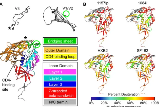
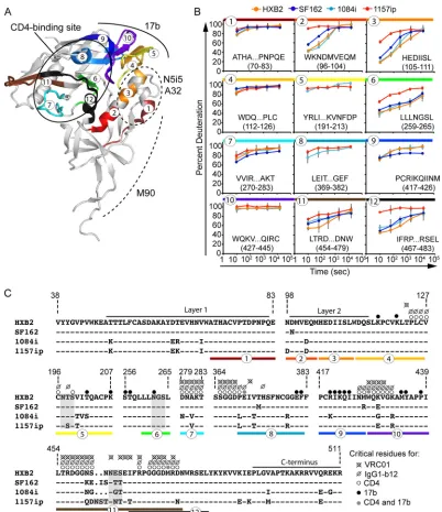
In order to measure the local dynamics of each full-length, glycosylated gp120, we quantified deuterium incorporation for every observable peptide in each isolate as a function of deutera-tion time (see Fig. S9 to S12 in the supplemental material). These data were summarized in the form of heat maps (Fig. 2; see also Fig. S13 in the supplemental material), in which the percent deu-terium uptake for each peptide is indicated by the color on the gp120 core crystal structure with N- and C-terminal extensions (22). These heat maps make it possible to identify regions of order and disorder within full-length gp120 in solution. Notably, a number of peptides from layer 1 and the bridging sheet, which
map to ordered␣-helices and-sheets in core crystal structures
(Fig. 2A), were found to be rapidly deuterated based on HDX-MS analysis of unliganded, full-length gp120s in solution (Fig. 2B). Such discrepancies illustrate that secondary structures within the
core regions of gp120 may be quite dynamic in the context of full-length gp120 in solution.
Heat maps enable qualitative comparison of differences in
stability among the four gp120s.Figure 2Bshows that there
were many similarities among the isolates; for example, the CD4 binding loop, bridging sheets, and portions of the inner domain exhibited a relatively high degree of structural dynam-ics in all cases. Additionally, similar regions of gp120 were protected in all four instances (e.g., portions of the outer
do-main and the seven-stranded-sandwich) (Fig. 2, 3, and4).
While there were general similarities, there were also abundant differences that could be observed among the four gp120s. Of the gp120s from the four isolates, 1157ip’s stood out as the most dynamic; however, additional apparent differences in deuterium uptake over time (see Fig. S13 in the supplemental material) among the other gp120s suggest that each may have a unique stability profile.
Comparisons of homologous peptides revealed significant variability, especially within the inner domains.A more focused, direct comparison of individual peptides that were similar or
identical among the isolates is shown inFig. 3and4. Excluding
glycoform variants, we identified 60, 49, 51, and 53 unique pep-tides for 1084i, 1157ip, HXB2, and SF162, respectively. Of these peptides, 29 were present in all four isolates, 9 were common to three isolates, and 10 were common to two isolates. Altogether, these peptides allowed us to compare deuterium uptake for 91% of residues within the inner domain and 42% of residues within the outer domain, bridging sheet, or variable loops across at least two isolates. However, it was only possible to compare deuterium uptake among four isolates for 81% of the inner domain residues
[image:5.585.137.450.64.291.2]FIG 1Similar architecture of gp120 across isolates observed by SAXS. (A and B) SAXS curves (A) and pairwise distance distribution [P(r)] plots (B) show that the four full-length gp120s are similar in shape and size. Guinier plots (A, inset) indicate that the gp120 specimens did not exhibit aggregation or interparticle effects, which would have complicated the analysis. a.u., arbitrary units. (C) DAMMIN shape reconstructions of unliganded 1084i (left) and 1157ip (right) full-length gp120 (gray envelopes), with the gp120 extended core from 3JWD.pdb and containing the V3 loop from 2B4C.pdb (blue spheres) docked into the SAXS shape envelope. The V1/V2 stem in the CD4-bound conformation does not fit into the unliganded SAXS density. Instead, a lobe of mass atop the gp120 core is attributed to V1/V2 proximal to the likely V3 position in unliganded full-length gp120 (40). Two-domain sCD4 was not present in the SAXS experiment; however, it is shown as a red ribbon to aid in the interpretation of the SAXS envelopes.
TABLE 2SAXS-derived gp120 dimensions
gp120
Rg (Å) determined via:
Dmax(Å)
Guinier GNOM
1157ip 37.4⫾0.1 37.7⫾0.1 132 1084i 36.6⫾0.1 36.8⫾0.1 130 HXB2 37.7⫾0.1 37.9⫾0.1 130 SF162 36.6⫾0.1 367.⫾0.1 130
Structural Dynamics and gp120 Antigenicity
on November 7, 2019 by guest
http://jvi.asm.org/
[image:5.585.40.285.650.724.2]and 17% of the residues in the outer domain, bridging sheet, or variable loops (see Fig. S8 in the supplemental material).
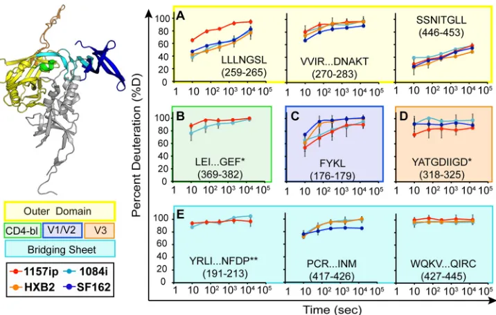
The stability of the gp120 inner domain varied substantially among the four isolates (Fig. 3). Layer 1 peptides were saturated with deuterium at the earliest time point in every isolate (Fig. 3A), indicating that this region is poorly ordered in the solution struc-ture of full-length gp120. Because the deuterium uptake was so fast for these peptides, we could not detect differences among the isolates in this region. In contrast, layer 2 peptides (amino acids [aa] 96 to 104, 105 to 111, and 211 to 222) showed various degrees of protection from deuterium uptake at early time points in 1084i, HXB2, and SF162, but the corresponding peptides from 1157ip gp120 were nearly fully exchanged at matched time points (Fig. 3B; see also Fig. S14 in the supplemental material). Layer 3 pep-tides (aa 227 to 258 and aa 467 to 483) (Fig. 3C) and peppep-tides from
the 7-stranded-sandwich (aa 84 to 95, 223 to 225, and 484 to
501) (Fig. 3D) showed a similar trend, in which 1157ip peptides were rapidly deuterated relative to those derived from other iso-lates. Therefore, it appears that inner domain secondary struc-tures, which exhibit similar, though not identical, stabilization in the other three isolates, are substantially more dynamic in the context of 1157ip gp120.
Although few outer domain peptides could be directly com-pared across all four isolates, much of the outer domain sequence was comparable between at least two isolates (Fig. 4; see also S15 in the supplemental material). Most peptides from the outer domain were similarly stabilized among those isolates for which compara-ble peptides were availacompara-ble (e.g., aa 270 to 283 and aa 446 to 453). However, the LLLNGSL peptide (aa 259 to 265), which is directly linked to the inner domain, was deuterated more rapidly in 1157p
than for the other isolates (Fig. 4A). The CD4 binding loop pep-tide (aa 369 to 382) was only slightly more dynamic in 1157ip than 1084i (Fig. 4B). Interestingly, the CD4 binding site-proximal pep-tide (aa 350 to 361) was significantly more stable in SF162 than in HXB2 (see Fig. S15 in the supplemental matrial). Overall, these results revealed conformational variations of the outer domain across isolates, although the extent of this variation appears lim-ited relative to that observed for the inner domain.
Peptides from the variable loops and bridging sheet that were common to multiple isolates revealed few isolate-specific differ-ences in deuterium uptake, partially due to the fact that these peptides were typically saturated with deuterium at the earliest
time point of deuteration (Fig. 4CtoE). The peptides FYKL (Fig.
4C) and YATGDIIGD (Fig. 4D) from V2 and V3, respectively, showed subtle differences in protection across isolates, but more complete coverage of these regions is needed to better characterize the extent of structural differences in the variable loops. Peptides
within bridging-sheet beta strands (-strands)2, -3, and -21 (aa
112 to 127, 191 to 213, and 427 to 445, respectively) exhibited rapid deuterium uptake rates that were similar among all available isolates (Fig. 4E; see also Fig. S15). The PCRIKQIINM peptide (aa
417 to 426), which partly covers-strands 19 and 20 of the
bridg-ing sheet, showed slight protection at early time points in all avail-able isolates (Fig. 4E), perhaps due to hydrogen bonding with
-sheet 17, downstream of the CD4 binding loop, as observed in
the core crystal structure (22).
In summary, significant isolate-specific differences in stability were observed throughout gp120. Most notably, peptides map-ping to the gp120 inner domain were consistently more disor-dered in 1157ip than in matched peptides from other isolates.
FIG 2Summary of isolate-specific differences in unliganded gp120 conformational dynamics. (A) The organization of monomeric gp120 into inner, outer, and bridging sheet domains is indicated by colors on the gp120 core with N- and C-terminal extensions (PDB ID 3JWD). The inner domain is further divided into mobile layers 1, 2, and 3 (magenta, cyan, and blue, respectively) that protrude from the 7-stranded-sandwich (red) (22). Variable loops V1/V2 and V3 (from PDB 3U4E and PDB 2B4C, respectively) are included for reference. The positions of these elements in the context of the gp120 core are indicated by an asterisk for V3 and a green circle for V1/V2. The approximate location of the CD4 binding site is indicated with a black oval. The structures shown here and throughout the text reflect that of gp120 in a receptor-bound conformation, because no structure is currently available for full-length, unliganded gp120. These structures may not necessarily reflect the unliganded structure of full-length gp120 in solution. (B) Heat maps of HDX-MS data for unliganded gp120 reveal qualitative isolate-specific differences in structural dynamics. Colors mapped onto the gp120 core (PDB ID 3JWD) indicate the percent deuteration of peptides after 1 min of incubation in a deuterated buffer; warm colors correspond to high levels of deuterium uptake (dynamic regions), and cool colors correspond to low levels of deuteration (ordered or “protected” regions). Regions where peptide information is missing are indicated in gray.
on November 7, 2019 by guest
http://jvi.asm.org/
[image:6.585.137.447.65.274.2]Many (but not all) peptides from the outer domain were similarly protected among the isolates that could be directly compared. Peptides mapping to the variable loops and bridging sheets were typically disordered, but poor peptide coverage and rapid satura-tion with deuterium made it difficult to discern differences in dynamics in these regions.
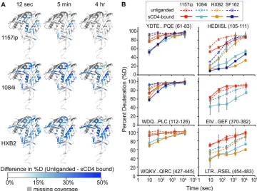
Isolate-specific differences persist in CD4-bound gp120.
Be-cause CD4 binding has been shown to stabilize gp120 (38,80) and
induce ordering of the conserved CD4 and coreceptor binding
sites (39,40), we hypothesized that the CD4-bound forms of the
gp120s are more structurally similar among isolates than unligan-ded gp120. To test this, we performed HDX-MS on sCD4-gp120 complexes for each isolate (Fig. 5). Overall, sCD4-bound gp120s were stabilized relative to their unliganded forms, taking on less deuterium than unliganded gp120s at matched time points (see Fig. S9 to S11 in the supplemental material). This was likely a result of multiple factors, including solvent occlusion, hydrogen bonding between sCD4 and the gp120 backbone, and a conforma-tional change within gp120 that led to the formation of a more stable secondary structure. For regions of gp120 outside the CD4 binding site, for example, the bridging sheet and layers 1 and 2 of the inner domain, CD4-induced HDX protection is likely due to stabilization of secondary structures. The impact of sCD4 binding on deuterium uptake by gp120 is shown in the difference maps of Fig. 5A, in which blue areas indicate protection relative to unli-ganded gp120. Similar patterns of protection were observed among the four isolates, most notably in the CD4 binding loop, the bridging sheet, and layers 1, 2, and 3 of the inner domain (Fig. 5A; see also Fig. S16 in the supplemental material). However, the
degree of protection as a result of sCD4 binding—the downward shift in deuterium uptake between unliganded and sCD4-bound gp120 —varied among the isolates (Fig. 5B). Deuterium uptake plots for matched peptides from the sCD4-bound gp120s revealed that in contrast to 1084i and HXB2, 1157ip was poorly stabilized in the presence of sCD4 (Fig. 5B; see also Fig. S17 and S18). For example, 1084i and HXB2 peptides from the inner domain (e.g., aa 61 to 83, 105 to 111, and 454 to 483), bridging sheet (e.g., aa 112 to 126 and aa 427 to 445), and the CD4 binding loop for 1084i (aa 369 to 382) were dramatically stabilized in the presence of sCD4 relative to 1157ip. Notably, the sCD4-induced stabilization of 1084i and HXB2 was comparable to that observed for CHO cell-expressed SF162 gp120 (Fig. 5B, dark blue lines) demonstrated in previous work (40). Increasing sCD4 in the 1157ip-sCD4 mixture, from a 3-fold to a 5-fold molar ratio, did not increase the apparent protection of sCD4-stabilized peptides (data not shown), which suggests that sCD4 was present at saturating concentrations and all available gp120 was bound by sCD4. Thus, it appears that while similar regions of gp120 are stabilized as a result of sCD4 binding to diverse isolates, the sCD4-bound gp120s maintain different lev-els of structural dynamics and, as in the unliganded state, 1157ip gp120 remains relatively dynamic even with sCD4 bound.
Epitope stability and antigenicity: linear epitope-specific an-tibodies.In order to determine whether differences in structural dynamics observed by HDX-MS affected ligand association with gp120, we used SPR to quantify the kinetics of sCD4 and antibody binding to the four gp120s and ELISAs to measure the levels of antibody stably bound to gp120. The determinants of antigenicity are complex and include, as minimal requirements, that (i) the
FIG 3Deuterium exchange profiles of homologous inner domain peptides. Each graph shows the deuterium uptake (percent deuteration) over time for peptides throughout the gp120 inner domain, which are either identical or homologous among the four gp120s. Each line in the graph reflects the deuterium uptake for that peptide in the context of a different isolate, as indicated in the figure legend. The peptide sequence and amino acid position (HXB2 numbering) are indicated for each graph. (A to C) Peptides from layers 1, 2, and 3 are color coded in the ribbon diagram, and deuteration uptake plots are grouped within the magenta, cyan, and blue boxes, respectively. (D) Peptides from the 7-stranded-sandwich are bounded by a red box. A comparison of the deuterium uptake curves for a given peptide from multiple isolates revealed differences in stability, for example, the WKNDMVEQM peptide is more dynamic in 1157ip than in the other isolates. **, deuteration data obtained by subtraction of overlapping peptides. Error bars reflect standard deviations, calculated as described in Materials and Methods.
Structural Dynamics and gp120 Antigenicity
on November 7, 2019 by guest
http://jvi.asm.org/
[image:7.585.97.491.66.306.2]residues that make up the antibody epitope be present in the pri-mary amino acid sequence, and (ii) these residues, which form the antibody epitope, must be exposed or accessible for antibody rec-ognition (81). Beyond these basic requirements, we hypothesized that the local conformational stability of an antigen might also impact the kinetics and/or the affinity of an antibody’s interaction with its epitope, as has been previously proposed (81).
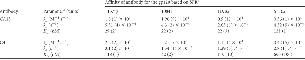
The antibodies B18, C4, and CA13 have been reported to
rec-ognize linear epitopes within helix 1 of gp120 (62,82), and we
confirmed their specificity in a peptide competition ELISA (see Fig. S19 in the supplemental material). The core epitopes of the three antibodies were absolutely conserved in all four isolates (Fig. 6B), yet exhibited significant differences in conformational dy-namics based on HDX-MS (Fig. 3B). There was no detectable difference in linear epitope-specific antibody binding to the four gp120s by ELISA (Fig. 6A). We also investigated isolate-specific differences in the kinetics of gp120 binding to captured B18, CA13, and C4 by SPR (see Fig. S20 in the supplemental material). Although 1157ip stood out among the four isolates, with a more dynamic helix 1 based on HDX-MS, it was not distinguished by substantially different rates of association with or dissociation from these linear epitope-specific antibodies (Table 3). Further-more, the affinities of C4 and CA13 for 1157ip were comparable to those observed for 1084i and HXB2, both of which featured a
more ordered helix 1(Table 3andFig. 6C). In contrast, SF162,
which was similar to 1084i and HXB2 in helix 1 stability, had affinities for CA13 and C4 that were at least 4-fold weaker than those of the other isolates. These findings suggest that the weaker recognition of SF162 by this panel of helix 1-specific antibodies may be due to factors other than the conformational stability of helix 1 (e.g., tertiary structural differences).
Dynamics and conformational epitope antigenicity: CD4 binding site. Because the majority of broadly cross-reactive HIV-1 NAbs and ADCC-active antibodies target discontinuous epitopes within gp120, we sought to determine whether differ-ences in the stability and structural dynamics of gp120 correlated with differences in the antigenicity of conformational epitopes such as the CD4 binding site. A summary of the differences in the primary sequence and dynamics of peptide segments involved in
conformational antibody epitopes is shown inFig. 7. Many of the
peptides that are directly involved in CD4 binding site-ligand in-teractions (Fig. 7A) are either saturated with deuterium at the
earliest time point (peptides 4 and 10 inFig. 7B) or are similarly
disordered in the isolates for which data are available (e.g.,
pep-tides 7 and 8 inFig. 7B). However, peptide 12, which contains a
number of CD4 contact residues and helix 5, is significantly more dynamic in 1157ip (Fig. 7B). In an ELISA, the CD4 binding site ligands CD4-IgG2 and IgG1-b12 bound strongly to the two clade B isolates, SF162 and HXB2, and relatively weakly to the clade C isolates (Fig. 8). This pattern of differential binding across clades was particularly evident for IgG1-b12 and suggested that the ob-served differences in CD4 binding site ligand recognition of gp120 might be partly due to clade-specific primary sequence differences within the CD4 binding site (Fig. 7C). Therefore, we collected SPR data for sCD4, CD4-IgG2, and IgG1-b12 binding to all four gp120s (see Fig. S21 in the supplemental material), but we focused our comparison on the isolates 1084i and 1157ip, which are closely related in their primary amino acid sequences (Fig. 7C) yet exhibit dramatic differences in the stability of peptides both
prox-imal (e.g., peptide 12 inFig. 7B) and distal (e.g., inner domain
peptides inFig. 3) to the CD4 binding site. This pair of isolates
presents an opportunity to study the impact of gp120 dynamics on
FIG 4Deuterium exchange profiles for the homologous outer domain, bridging sheet, and variable loop peptides. Each graph shows deuterium uptake (percent deuteration) over time for peptides in the gp120 outer domain (A), CD4-binding loop (CD4-bl) (B), variable loops V2 (C) and V3 (D), and bridging sheets (E). Each line on the graphs corresponds to deuterium uptake for that peptide in the context of a different isolate, as indicated. The peptide sequence and amino acid position (HXB2 numbering) are indicated for each peptide. Each graph is color coded according to the position of the peptide in the gp120 structure, as indicated in the ribbon diagram and symbol key to the left. *, the listed peptide sequence is from an isolate other than HXB2, because peptide coverage was missing for HXB2; **, deuteration data were obtained by subtraction of overlapping peptides. Error bars reflect standard deviations, calculated as described in Materials and Methods.
on November 7, 2019 by guest
http://jvi.asm.org/
[image:8.585.113.471.65.293.2]recognition by CD4 binding site ligands while limiting confound-ing amino acid changes.
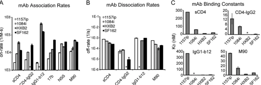
CD4-IgG2 bound poorly to 1157ip gp120 compared to 1084i in the ELISA (Fig. 8). These results were confirmed by SPR, which indicated that 1157ip had the weakest affinity for sCD4 and CD4-IgG2 of the four isolates, showing 5-fold- and 2-fold-weaker af-finities for sCD4 and CD4-IgG2, respectively, than 1084i (Fig. 9C
andTable 4). IgG1-b12 binding to 1084i fit poorly to a 1:1 binding
model, which complicated a direct comparison of 1084i and 1157ip binding to this antibody based on SPR. The weak affinity of 1157ip for CD4 binding site ligands was largely due to lower rates of association (Fig. 9A), although 1157ip also showed a slightly higher rate of dissociation from sCD4 compared to 1084i (Fig. 9B). Thus, it appears that the increased conformational flexibility of 1157ip is associated with slower association rates and an overall weaker affinity for CD4 mimetics (Fig. 9C). This effect appears to be most prominent in CD4 binding site ligands that require con-tact with both the inner and outer domains of gp120, because while 1157ip bound to CD4-IgG2 at a very low level in the ELISA, the potent and broad CD4 binding site antibody VRC01, which primarily makes contact with the gp120 outer domain (24), bound to 1157ip at only a slightly reduced level relative to 1084i (Fig. 8).
The few observable differences in the stability of peptides in-volved in the CD4 binding site across isolates (Fig. 7) suggest that differences in conformational stability of regions outside the CD4 binding site (for example, the gp120 inner domain) likely also
exert an influence on gp120’s affinity for CD4 and binding site-targeted antibodies. This model is supported by previous work that showed that layers 1, 2, and 3 of the inner domain, regions that are dramatically destabilized in 1157ip relative to the other isolates, significantly impact gp120 interactions with CD4 and CD4 mimetics (83–85).
Dynamics and the antigenicity of conformational inner do-main epitopes.Because isolate-specific differences in gp120
sta-bility were most apparent in the inner domain (Fig. 3and7B), we
measured binding of the four gp120s to antibodies that have been reported to target conformational epitopes within this region: M90, A32, and N5i5 (Fig. 7A). M90 is thought to recognize a conformational epitope involving layer 1 and the N and C termini
of gp120 (63, 86). N5i5 is a potent ADCC-active antibody that
binds to a CD4-induced, discontinuous epitope similar to that of A32 (61) that includes layers 1 and 2 of the inner domain; its contact residues are absolutely conserved in the isolates tested here (George Lewis and Marzena Pazgier, personal communica-tion). Because of the high level of sequence conservation within the epitopes recognized by these antibodies and in the N5i5 epitope, in particular, we were able to test the impact of gp120 dynamics on inner domain antigenicity with limited con-founding effects from amino acid variability in the antibody epitopes (Fig. 7C).
Antibodies M90, A32, and N5i5 bound well to 1084i, HXB2, and SF162 but only weakly to 1157ip gp120 in the ELISA (Fig. 8). These results were supported by the SPR data, which showed that
FIG 5Isolate-specific differences in sCD4-induced stabilization of gp120. (A) Difference heat maps show the spatial profile of sCD4-induced stabilization of gp120. The colors mapped onto the 3JWD structure indicate the quantitative differences in percent deuteration between unliganded and CD4-bound full-length gp120s. Darker blue areas correspond to greater CD4-induced stabilization. Missing peptide coverage is shown in gray. Difference heat maps for the 12-s, 5-min, and 4-h time points are shown. (B) Deuterium uptake plots (percent deuteration versus time) for identical or homologous peptides that are stabilized upon sCD4 binding. The peptide sequence and amino acid position (HXB2 numbering) are indicated for each peptide. The traces are color coded by isolates, as indicated in the symbol key. Stabilization as a result of sCD4 binding was observed as a downward shift in the deuterium uptake curve. Error bars reflect standard deviations, calculated as described in Materials and Methods.
Structural Dynamics and gp120 Antigenicity
on November 7, 2019 by guest
http://jvi.asm.org/
[image:9.585.111.471.67.335.2]1157ip bound to both N5i5 and M90 with the slowest on-rate of the four gp120s (Fig. 9A). Additionally, 1157ip had the fastest off-rate and weakest affinity for M90 of the four isolates (Fig. 9B
andCandTable 4; see also Fig. S21 in the supplemental material).
The rate of N5i5 dissociation was too slow to obtain reliable esti-mates of the off-rate or binding affinity, even when we used dis-sociation times of greater than 1 h (see Fig. S22 in the supplemen-tal material). Overall, the relatively high degree of disorder within the 1157ip gp120 inner domain appeared to correlate with a lower rate of association with and a weaker affinity for inner domain-specific, conformation-dependent antibodies.
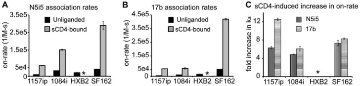
Antibodies recognizing CD4-induced epitopes bind slowly to 1157ip in both the unliganded and CD4-bound states.
HDX-MS analysis of sCD4-bound gp120 revealed that many re-gions of gp120 are stabilized as a result of sCD4 binding, including the bridging sheet microdomain and the gp120 inner domain (Fig. 5). Notably, these same regions are targeted by antibodies such as 17b and N5i5, which preferentially recognize CD4-bound gp120
(55,61,87). Because HDX-MS indicated that differences in
sta-bility are maintained in sCD4-bound gp120 (Fig. 5B), we sought
to test the antigenicity of CD4-induced epitopes and the antigenic consequences of sCD4 binding across isolates by using SPR.
As with N5i5, 17b off-rates were slow for all isolates (with the exception of 1084i), and we were limited to estimates of the on-rate for 1157ip, HXB2, and SF162 (see Fig. S23 in the supplemen-tal material). Notably, 1157ip had the lowest rate of association with 17b in its unliganded state and was poorly recognized by 17b in the ELISA (Fig. 8), despite having well-conserved 17b contact residues (Fig. 7C). Many peptides containing residues critical for 17b binding were saturated with deuterium at the earliest time
point (peptides 4, 5, and 10 inFig. 7B) or were similarly disordered
in the isolates for which data were available (peptide 8 inFig. 7B).
SF162 exhibited stabilization in peptide 9 (Fig. 7B), which con-tains a number of residues critical for 17b binding (Fig. 7C), and bound strongly to 17b in the ELISA and SPR relative to the other
isolates (Fig. 8and9). Conversely, peptide 6, which contains a
residue critical for 17b binding at position 262 (HXB2 number-ing) (55), was more disordered in 1157ip, as were peptides 2 and 3
(Fig. 7B), which are directly linked to-strands 2 and 3 of the
bridging sheet (highlighted as peptides 4 and 5, respectively, in
[image:10.585.111.472.68.227.2]FIG 6Linear epitope-specific antibody binding to a region of gp120 differentially stabilized across isolates. (A) Linear epitope-specific antibody (CA13, B18, or C4) binding to gp120 proteins measured by the ELISA. The curves are color coded by Env isolate as defined in the symbol key. Concentrations of monoclonal antibodies (mAb) are shown on thexaxis for C4 and B18, and the reciprocal dilution of antibody supernatant is shown on thexaxis for CA13. Curves are representative of at least two independent experiments; error bars indicate standard deviations from duplicate measurements. (B) The primary sequence recognized by antibodies CA13, B18, and C4 is absolutely conserved among the four gp120s. A dash indicates that the residue is conserved in a given isolate, and amino acid differences are indicated. This region is differentially stabilized in the four gp120s, based on HDX-MS (Fig. 3B) (C) Summary of SPR-derived binding constants for CA13 and C4 binding to the four gp120s. Raw and fitted SPR curves are shown in Fig. S20 in the supplemental material. The B18-gp120 SPR binding curves did not fit well to a 1:1 binding model; therefore, estimates of affinity are not available, although qualitative isolate-specific differences in binding to B18 were similar to those observed for CA13 and C4 (see Fig. S20). Error bars indicate standard deviations, calculated as described in Materials and Methods.
TABLE 3SPR binding parameters for linear epitope-specific antibodies
Antibody Parametera(units)
Affinity of antibody for the gp120 based on SPRb
1157ip 1084i HXB2 SF162
CA13 ka(M⫺
1s⫺1) 1.8 (1)⫻104 1.96 (9)⫻104 0.9 (1)⫻104 0.36 (1)⫻104 kd(s⫺
1) 5.31 (4)⫻10⫺4 4.3 (2)⫻10⫺4 2.03 (1)⫻10⫺4 4.32 (9)⫻10⫺4 KD(nM) 29 (2) 22 (2) 22 (3) 121 (1)
C4 ka(M⫺1s⫺1) 2.6 (2)⫻104 3.2 (1)⫻104 1.1 (1)⫻104 0.42 (5)⫻104 kd(s⫺1) 3.1 (2)⫻10⫺3 1.34 (1)⫻10⫺3 1.29 (3)⫻10⫺3 2.8 (1)⫻10⫺3 KD(nM) 118 (1) 42 (2) 110 (10) 600 (100)
a
ka, rate of association;kd, rate of dissociation;KD, equilibrium dissociation constant.
bValues are means⫾standard deviations. Values in parentheses are errors for the last significant figure; 1.9 (1)⫻104is equivalent to 19,000⫾1,000.
on November 7, 2019 by guest
http://jvi.asm.org/
[image:10.585.41.544.608.708.2]Fig. 7A). Given the few differences in stability observed for pep-tides containing residues critical for 17b binding, it seems plausi-ble that increased disorder observed in the inner domain of 1157ip is communicated to the bridging sheet, with implications for 17b binding to unliganded gp120.
Consistent with the stabilization of the 17b and N5i5 epitopes in sCD4-bound gp120 by HDX-MS (Fig. 5; see also Fig. S24 in the
supplemental material), we observed a dramatic increase in 17b and N5i5 binding to all four gp120s upon addition of sCD4 (Fig. 10). It is worth noting that, in addition to epitope stabilization, tertiary structural changes, not readily detectable by HDX-MS, are also likely to contribute to the increase in 17b and N5i5 binding to sCD4-bound gp120. This may be especially true for 17b, as the lateral movement of V1/V2 upon sCD4 binding appears to
coin-FIG 7Summary of isolate-specific differences in conformational dynamics and primary sequence for peptides involved in conformation-dependent antibody epitopes. (A) Colors and numbers mapped onto the 3JWD crystal structure indicate peptides within conformation-dependent antibody epitopes that can be tracked by HDX-MS. The structure is annotated to indicate the CD4 binding site (targeted by sCD4, IgG1-b12, and VRC01) as well as N5i5, A32, M90, and 17b epitopes. (B) Deuteration profiles (percent deuteration versus time) for peptides within conformation-dependent antibody epitopes. Plots are color coded and numbered as for panel A. (C) Amino acid alignment of the four isolates for regions of gp120 involved in conformation-dependent antibody binding. A dash indicates that the residue is conserved in a given isolate relative to HXB2; a period indicates an insertion in a separate sequence. N5i5 and A32 recognize a similar epitope involving layers 1 and 2 (61). Layer 1 and the C terminus are thought to be involved in the M90 epitope (63,86). Critical residues for CD4, IgG1-b12, VRC01, and 17b binding are indicated with dots, as shown in the symbol key, and are based on references21,24, and55. Peptides are indicated with colors and numbers as for panels A and B.
Structural Dynamics and gp120 Antigenicity
on November 7, 2019 by guest
http://jvi.asm.org/
[image:11.585.90.492.63.528.2]cide with ordering of the bridging sheet and formation of the 17b epitope (40).
We investigated whether the differences in 17b and N5i5 reac-tivity across isolates observed in unliganded gp120 would be sim-ilarly apparent in the context of sCD4-bound gp120. SPR was used to measure the rate of gp120 binding to 17b and N5i5 in the pres-ence of a 5-fold molar excess of sCD4. The rates of association with 17b and N5i5 increased for all isolates in the presence of sCD4, and the isolate rank order of highest (SF162) and lowest (1157ip) rates of association with the two antibodies was identical for
unligan-ded and sCD4-bound gp120 (Fig. 10AandBandTable 5).
Impor-tantly, the increase in the rate of 1157ip binding to 17b and N5i5 upon addition of sCD4 was similar to that of the other isolates (Fig. 10C), indicating that although 1157ip gp120 wasn’t as dra-matically stabilized by sCD4 as the other gp120s, it remained ca-pable of binding to sCD4 and undergoing a transition to a CD4-induced state. Overall, it appears that isolate-specific antigenic
differences within CD4-induced epitopes are maintained in the sCD4-bound state, in agreement with the conformational stability profiles observed for the sCD4-bound gp120s with HDX-MS.
Consideration of potential caveats of sCD4-induced stabili-zation of gp120 based on HDX-MS.Two factors we considered that could potentially complicate interpretation of the HDX-MS results for sCD4-bound gp120 were isolate-specific differences in kinetics and affinities of binding to sCD4 and isolate-specific vari-ability in the fraction of “active” gp120 within a purified gp120 preparation. Because 1157ip has the weakest affinity for sCD4, we considered the possibility that the greater levels of deuterium up-take by this isolate in the presence of sCD4 reflect a difference in the amount of time a molecule of 1157ip would spend in the sCD4-bound state or a lower level of sCD4 occupancy at equilib-rium. However, given the affinity values calculated by SPR (Table 4) and the concentration of sCD4 used in complex formation, we estimate that only 0.01% more 1157ip would remain unbound at
FIG 9Summary of conformation-dependent antibody binding to gp120 as measured by SPR. (A) Rate of association of the four gp120 proteins with immobilized sCD4 or captured antibodies. Because of slow dissociation rates for 17b and N5i5, only on-rates could be derived for these antibodies. mAb, monoclonal antibody. (B) Rates of dissociation of the four gp120s from immobilized sCD4 or captured antibody ligands. Reliable off-rates could not be determined for 17b or N5i5. (C) Summary of binding constants for gp120 binding to immobilized sCD4 or captured antibody ligands. LargeKDvalues reflect
weaker binding interactions. Raw data and fitted curves for gp120 binding to conformation-dependent ligands are presented in the supplemental material. *, the experiment could not be reliably performed or the signal could not be fit with a 1:1 binding model. Error bars indicate standard deviations, calculated as described in Materials and Methods.
FIG 8Conformation-dependent antibody binding to gp120s as determined by ELISA. Conformation-dependent antibodies specific for different regions of gp120 were tested: CD4 binding site ligands (CD4-IgG2, IgG1-b12, and VRC01), conformational inner domain antibodies (M90, N5i5, and A32), and the coreceptor binding site antibody (17b). Each graph corresponds to a different antibody binding to the four gp120s. The binding curves are color coded based on HIV isolate as shown in the symbol key. Antibody binding levels (measured as the absorbance at 450 nm) versus antibody concentrations are shown. Curves are representative of at least two independent experiments; error bars indicate standard deviations from duplicate measurements.
on November 7, 2019 by guest
http://jvi.asm.org/
[image:12.585.111.475.63.225.2] [image:12.585.77.510.513.655.2]equilibrium compared to 1084i (0.28% versus 0.27% unbound gp120 for 1157ip and 1084i, respectively), an insignificant differ-ence. Similarly, using a standard first-order rate equation to model the exponential decay of the gp120-sCD4 complex and the dissociation rates calculated by SPR (Table 4), after 12 s of deu-teration, only 0.3% more dissociation would occur for 1157ip compared to 1084i, yet we observed dramatic differences in deu-terium uptake at this time point (Fig. 5). Furthermore, we at-tempted to account for the weaker affinity of 1157ip for sCD4 by using a higher concentration of sCD4 in complex formation (5-fold molar excess) than with the other isolates (3-(5-fold molar ex-cess). The absence of additional stabilization in the 5-fold versus 3-fold molar excess samples suggests that sCD4 saturates the avail-able 1157ip gp120 population. Thus, differences in CD4 occu-pancy at equilibrium or kinetic differences in sCD4 dissociation are unlikely to explain the dramatic differences in stability ob-served for sCD4-bound 1157ip compared to the other isolates.
Similarly, if purified 1157ip gp120 contained a relatively large fraction of inactive gp120, as a result of heterogeneity in disulfide bonding or glycosylation, for example, then addition of sCD4 would only weakly stabilize gp120 at the population level (al-though it may be capable of dramatically stabilizing individual molecules of active gp120). However, there is no indication that
such a population exists in 1157ip; for example, Kratky plots of 1157ip indicate that the gp120s share a similar global structure (see Fig. S2). Furthermore, addition of sCD4 to 1157ip produces a similar increase in its rate of association with 17b and N5i5, as observed for the other gp120s (Fig. 10C). Because the rate of as-sociation is sensitive to the active concentration of analyte, this suggests that the fraction of gp120 that is active for sCD4 binding is similar among the four gp120s. Thus, we considered these ca-veats, but we concluded that they were unlikely to be responsible for the observed isolate-specific differences in the stability and antigenicity of sCD4-bound gp120. Instead, the differences ap-peared to reflect inherent structural features of each gp120.
DISCUSSION
[image:13.585.43.545.81.257.2]Our understanding of the structural basis for antigenic and immunogenic differences among gp120 proteins from diverse isolates has been hindered by our inability to extract detailed structural information for intact Env glycoprotein constructs con-taining the variable loops and glycans, which are major determi-nants of isolate antigenicity and neutralization sensitivity. Crystal structures of truncated core constructs provide valuable informa-tion about the conserved elements of gp120 and their recogniinforma-tion by receptor and antibodies, but they do not elucidate how the
TABLE 4SPR binding parameters for conformation-dependent antibodies
Antibody Parametera(units)
SPR-derived binding values for gp120-ligand interactionb
1157ip 1084i HXB2 SF162
sCD4 ka(M⫺
1s⫺1) 2.67 (1)⫻103 8.5 (2)⫻103 37 (5)⫻103 13.1 (4)⫻103 kd(s⫺
1) 7.41 (3)⫻10⫺4 4.9 (1)⫻10⫺4 3.3 (2)⫻10⫺4 2.46 (6)⫻10⫺4 KD(nM) 277 (1) 58 (2) 9 (2) 18.8 (2)
CD4-IgG2 ka(M⫺1s⫺1) 3.91 (8)⫻103 10.5 (1)⫻103 NDc 25 (4)⫻103 kd(s⫺1) 5 (2)⫻10⫺5 7.1 (6)⫻10⫺5 ND 0.9 (5)⫻10⫺5
KD(nM) 13 (4) 6.8 (6) ND 0.4 (3)
IgG1-b12 ka(M⫺
1s⫺1) 2.6 (1)⫻103 —d 430 (40)⫻103 150 (10)⫻103 kd(s⫺
1) 9.4 (5)⫻10⫺4 — 5.4 (1)⫻10⫺4 9.2 (2)⫻10⫺4
KD(nM) 362 (1) — 1.3 (1) 6.2 (6)
M90 ka(M⫺1s⫺1) 1.18 (4)⫻104 1.7 (3)⫻104 4.9 (4)⫻104 3.89 (7)⫻104 kd(s⫺1) 5.41 (7)⫻10⫺4 2.4 (1)⫻10⫺4 0.9 (2)⫻10⫺4 1.88 (5)⫻10⫺4 KD(nM) 45.7 (8) 14 (3) 1.9 (6) 4.8 (2)
a
ka, rate of association;kd, rate of dissociation;KD, equilibrium dissociation constant.
bValues are means⫾standard deviations. Values in parentheses are errors for the last significant figure; seeTable 3. c
ND, experiment not done.
d—, data fit poorly to a 1:1 binding model.
FIG 10Changes in CD4i antibody binding to gp120 in the presence of sCD4 by SPR. (A) On-rates of N5i5 binding to the four gp120s in the presence and absence of sCD4. (B) On-rates of 17b binding to the four gp120s in the presence and absence of sCD4. (C) Change in rates of association (ka) of 17b and N5i5 with the
gp120s as a result of sCD4 binding. Data are presented as the fold increase relative to the rate of association with unliganded gp120. *, the experiment could not be reliably performed. Error bars indicate standard deviations, calculated as described in Materials and Methods.
Structural Dynamics and gp120 Antigenicity
![FIG 1 Similar architecture of gp120 across isolates observed by SAXS. (A and B) SAXS curves (A) and pairwise distance distribution [P(r)] plots (B) show thatthe four full-length gp120s are similar in shape and size](https://thumb-us.123doks.com/thumbv2/123dok_us/148760.25904/5.585.137.450.64.291/similar-architecture-isolates-observed-pairwise-distance-distribution-similar.webp)