Ubiquitination Upregulates Influenza Virus Polymerase Function
James Kirui,a,bArindam Mondal,b Andrew Mehleb
Graduate Program in Cellular & Molecular Biology, University of Wisconsin—Madison, Madison, Wisconsin, USAa
; Medical Microbiology & Immunology, University of Wisconsin—Madison, Madison, Wisconsin, USAb
ABSTRACT
The influenza A virus polymerase plays an essential role in the virus life cycle, directing synthesis of viral mRNAs and genomes. It is a trimeric complex composed of subunits PA, PB1, and PB2 and associates with viral RNAs and nucleoprotein (NP) to form higher-order ribonucleoprotein (RNP) complexes. The polymerase is regulated temporally over the course of infection to ensure coordinated expression of viral genes as well as replication of the viral genome. Various host factors and processes have been implicated in regulation of the IAV polymerase function, including posttranslational modifications; however, the mechanisms are not fully understood. Here we demonstrate that ubiquitination plays an important role in stimulating polymerase activity. We show that all protein subunits in the RNP are ubiquitinated, but ubiquitination does not significantly alter protein levels. Instead, ubiquitination and an active proteasome enhance polymerase activity. Expression of ubiquitin upregulates polymerase function in a dose-dependent fashion, causing increased accumulation of viral RNA (vRNA), cRNA, and mRNA and enhanced viral gene expression during infection. Ubiquitin expression directly affects polymerase activity independent of nucleoprotein (NP) or ribonucleoprotein (RNP) assembly. Ubiquitination and the ubiquitin-proteasome pathway play key roles during multi-ple stages of influenza virus infection, and data presented here now demonstrate that these processes modulate viral polymerase activity independent of protein degradation.
IMPORTANCE
The cellular ubiquitin-proteasome pathway impacts steps during the entire influenza virus life cycle. Ubiquitination suppresses replication by targeting viral proteins for degradation and stimulating innate antiviral signaling pathways. Ubiquitination also enhances replication by facilitating viral entry and virion disassembly. We identify here an addition proviral role of the ubiqui-tin-proteasome system, showing that all of the proteins in the viral replication machinery are subject to ubiquitination and this is crucial for optimal viral polymerase activity. Manipulation of the ubiquitin machinery for therapeutic benefit is therefore likely to disrupt the function of multiple viral proteins at stages throughout the course of infection.
I
nfluenza viruses are a significant threat to human public health and a major cause of morbidity and mortality in both human and animal populations (1). Influenza A virus (IAV) is a member of theOrthomyxoviridaefamily, composed of eight minus-sense single-stranded RNA (⫺ssRNA) segments that make up its ge-nome. Each⫺ssRNA segment associates with a viral polymerase and oligomeric nucleoproteins (NPs) to form large ribonucleo-protein (RNP) complexes that direct expression of viral genes and replication and packaging of the viral genome (2,3). The ability of the virus to efficiently express its genes and replicate its genome is key to a successful infection as it enables modulation of the host cell environment by the virus to allow subsequent assembly of progeny virions. Transcription and replication of the genome are performed by the viral polymerase. The polymerase is a trimeric protein complex composed of the polymerase acidic (PA), poly-merase basic 1 (PB1), and PB2 proteins. Viral genes are tran-scribed by a “cap-snatching” process where the polymerase ac-quires 5=7-methylguanosine (7mG) caps from host mRNAs and uses them to initiate synthesis of viral messages. The 7mG cap is bound by the cap-binding domain in the PB2 subunit, and the mRNA is subsequently cleaved 11 to 15 nucleotides (nt) down-stream by the endonuclease domain in the PA subunit (4–6). The resulting short capped RNA is used to prime synthesis of viral mRNA by the polymerase catalytic site resident in the PB1 sub-unit. Transcription continues to the end of the template, where an iterative copy of a poly(U) tract appends a poly(A) tail to complete mRNA synthesis. Genome replication is performed by thepoly-merase during primer-independent synthesis of full-length plus-sense cRNA, which serves as a template for production of new genomic viral RNA (vRNA).
Influenza polymerase activity is impacted by the host cell en-vironment and temporally regulated during the course of infec-tion (7); i.e., it serves primarily as a transcriptase early in infection then is largely a replicase later in infection (8). Posttranslational modifications (PTMs) of viral proteins by host enzymes have been suggested to impact this change in activity. A number of PTMs have been reported to occur on NP and subunits of the viral poly-merase, including phosphorylation of PB1, PB2, PA, and NP (9), sumoylation of PB1 and NP (10,11), poly-ADP ribosylation of PB2 and PA (12), and ubiquitination of PB1 and NP (13,14). These modifications can impact multiple steps in the assembly and function of RNPs. Phosphorylation and sumoylation of NP have been reported to impact its localization to the nucleus or export to the cytoplasm, respectively (11,15,16).
Phosphoryla-Received9 September 2016Accepted20 September 2016
Accepted manuscript posted online28 September 2016
CitationKirui J, Mondal A, Mehle A. 2016. Ubiquitination upregulates influenza
virus polymerase function. J Virol 90:10906 –10914.doi:10.1128/JVI.01829-16.
Editor:S. Schultz-Cherry, St. Jude Children’s Research Hospital
Address correspondence to Andrew Mehle, amehle@wisc.edu. Copyright © 2016, American Society for Microbiology. All Rights Reserved.
on November 7, 2019 by guest
http://jvi.asm.org/
tion of NP has also been shown to regulate its homo-oligomeriza-tion and assembly of RNP complexes (17–19). Thus, the dynamic and reversible modification of proteins in the RNP is a likely reg-ulator of genome transcription and replication, yet the mecha-nisms underlying this regulation are not fully understood.
Ubiquitination involves the conjugation of the 8.5-kDa ubiq-uitin (Ub) protein to specific lysine residues on substrate proteins. This follows a cascade of reactions transferring ubiquitin from the E1 ubiquitin-activating protein to the E2 ubiquitin-conjugating protein, and ultimately the E3 ubiquitin ligase attaches ubiquitin to the substrate (20). Monoubiquitination attaches a single ubiq-uitin moiety to sites on a substrate protein, whereas polyubiquiti-nation attaches ubiquitin chains. The specific linkage between ubiquitin moieties in a chain determines the impact of polyubiq-uitination on the target protein. Ubiqpolyubiq-uitination regulates various cellular processes; however, polyubiquitin chains linked via lysine 48 (K48) within ubiquitin are most commonly associated with protein degradation via the proteasome, while chains linked by lysine 63 (K63) are involved in endocytic trafficking, potentially leading to lysosomal degradation, ribosomal modification, and DNA repair (20). The ubiquitin-proteasome pathway impacts multiple steps in the influenza virus cycle, and inhibition of this pathway is detrimental to the viral life cycle (21). During entry, the E3 ligase Itch is required for efficient release of virions from en-dosomes and the E3 Nedd4 is important for the turnover of the host protein IFITM3, an antiviral factor that blocks release from the endosome (22, 23). Following entry, free ubiquitin found within virions then targets the incoming viral core to the ag-gresome machinery to facilitate uncoating (24,25). Multiple E3 ubiquitin ligases have been identified as key host factors regulating the IAV infectious cycle, and ubiquitination of viral proteins has been shown to regulate their stability and function (12–14,22,23, 26,27). Indeed, ubiquitin-mediated proteasomal degradation of IAV protein has recently been shown to restrict infection (12,14). To establish the role of ubiquitination in the regulation of the IAV polymerase function, we investigated whether the RNP is ubiquitinated and how this might impact transcription, genome replication, and viral infection. We show here that all of the pro-tein components in the RNP are ubiquitinated during infection. This ubiquitination did not cause significant protein degradation, but rather resulted in a dramatic stimulation of polymerase activ-ity and enhanced gene expression during infection. Disrupting the ubiquitin pathway by inhibiting the proteasome reduced poly-merase activity. Primer extension assays demonstrated that ubiq-uitination is associated with increases in both transcription and replication by the IAV polymerase, which resulted in enhanced gene expression during infection. Our findings show that the ubiquitproteasome pathway plays diverse roles during viral in-fection, restricting infection in some cases, enhancing entry in others, and as our data show, stimulating gene expression and genome replication.
MATERIALS AND METHODS
Cells, antibodies, and reagents.HEK293T cells were maintained in com-plete Dulbecco’s modified Eagle’s medium (DMEM) supplemented with
10% fetal bovine serum (FBS). All cells were grown at 37°C in 5% CO2.
The antibodies used include M2 anti-FLAG (Sigma), anti-V5 (Bethyl), anti-RNP (BEI NR-3133), and anti-hemagglutinin (anti-HA [Roche]). MG132 was obtained from EMD Millipore.
Plasmids.The IAV RNP reconstitution plasmids used throughout encode polymerase proteins and NP derived from A/WSN/1933 (H1N1)
(WSN) and were described previously (28). Epitope-tagged PB2-FLAG
and NP-V5 were expressed as C-terminal fusions (28). The vNA-Luc
re-porter plasmids encode a minus-sense luciferase gene flanked by untrans-lated regions (UTRs) from neuraminidase (NA) that is expressed from a
polymerase I promoter and terminator (28). Mutants were created by
PCR-based strategies and confirmed by sequencing. The wild-type (WT) Ub and K48R and K63R ubiquitin plasmids used were N-terminally
tagged with HA-epitope (29) (a kind gift from D. Gabudza). A plasmid
expressing a 77-nt vNP microgene was constructed following previously
described microgenes (30). The influenza virus reverse genetics plasmids
include pTM⌬RNP, which expresses viral RNAs (vRNAs) for HA, NA, M,
and NS and pBD-PB1, -PB2, -PB2-FLAG, -PA, and -NP (28).
Viruses.Viruses stably encoding a C-terminal FLAG tag on PB2 re-quired duplication of a portion of the PB2 open reading frame to maintain a contiguous packaging signal and were engineered following the strategy
of Dos Santos Afonso et al. (31). WSN, WSN(PB2-FLAG), or reassortants
encoding PB2-FLAG, PB1, PA, and NP derived from A/green-winged teal/OH/175/1983 (S009) or A/New York/312/2001 (NY312) were res-cued in transfected HEK293T cells and amplified, and their titers were
determined as described previously (32).
Polymerase activity assay. HEK293T cells were transfected using TransIT-2020 (Mirus Bio) in triplicate with plasmids encoding PA, PB1, PB2-FLAG, NP, the vNA-Luc reporter plasmid, and increasing amounts of plasmid encoding HA-ubiquitin or an empty vector. The total DNA concentration for each transfection was kept constant. Cells were lysed 24 to 48 h posttransfection in cell culture lysis reagent (Promega), and lucif-erase activity was measured by using the luciflucif-erase assay system (Pro-mega). Expression of IAV RNP components and HA-ubiquitin was ana-lyzed by Western blotting.
Immunoprecipitations.HEK293T cells were transfected with plas-mids expressing PA, PB1, PB2-FLAG, NP, vNA-luciferase reporter, and HA-ubiquitin and lysed 24 to 48 h posttransfection. Alternatively, HEK293T cells were transfected with HA-ubiquitin plasmid, incubated for 16 to 18 h, and subsequently infected with PB2-FLAG virus at a mul-tiplicity of infection (MOI) of 1.0 for 16 to 18 h. Cells were lysed in buffer containing 50 mM Tris (pH 7.4), 150 mM NaCl, 1% NP-40, 0.5% deoxy-cholate, 0.1% SDS, 10% glycerol, and protease inhibitor cocktail. Clarified lysates were precleared and immunoprecipitated with M2 (anti-FLAG)-agarose beads (Sigma). The beads were washed extensively and then boiled in protein loading buffer, and immunoprecipitated complexes were analyzed by Western blotting.
Immunoprecipitation of denatured lysates.HEK293T cells were transfected with plasmids expressing either PA-FLAG, PB1-FLAG, PB2-FLAG, NP-V5, NP-V5 K184R, or an empty plasmid. After 24 to 48 h
posttransfection, the cells were lysed as described by Choo et al. (33).
Briefly, cells were lysed in a mixture of 2% SDS, 150 mM NaCl, 2 mM EDTA, 1% Triton, and protease inhibitor cocktails, clarified by centrifu-gation, and boiled for 10 min to completely denature the same. Lysates were then renatured by dilution with 9 volumes of 10 mM Tris HCl (pH 8.0), 150 mM NaCl, 2 mM EDTA, and 1% Triton. After renaturing, lysates were immunoprecipitated as described above.
RNA isolation and primer extension.Total RNA was extracted from transfected 293T cells using TRIzol reagent (Invitrogen). Seven nano-grams from each of the RNA samples was analyzed by primer extension.
RNA was mixed with an excess of32P-labeled DNA primers that
specifi-cally recognize plus-sense or minus-sense viral RNAs and 5S host RNA
(8). A different set of primers were used for the microgene primer
exten-sion analysis based on published work by Turrell et al. (30). Samples were
denatured by heating at 95°C for 2 min, snap chilled on ice, and incubated for 2 min at either 50°C for Superscript III reverse transcriptase (RT) (Invitrogen) reactions or 42°C for Moloney murine leukemia virus (MMLV) RT reactions prior to the addition of a transcription mix. The Superscript III RT transcription mix was added to create final conditions
on November 7, 2019 by guest
http://jvi.asm.org/
of 20 mM Tris (pH 8.4), 50 mM KCl, 0.5 mM deoxynucleoside
triphos-phate (dNTP) mix, 5 mM MgCl2, 10 mM dithiothreitol (DTT), 40 U
RNAseOUT, and 200 U Superscript III RT. The MMLV RT transcription was added to created final conditions of 50 mM Tris (pH 8.3), 75 mM KCl,
3 mM MgCl2, 0.5 mM dNTP mix, 40 U RNAseOUT, and 1l MMLV RT.
Reaction mixtures were incubated for 1 h, terminated by the addition of an equal volume of formamide, and denatured by heating at 95°C for 5 min. Transcription products were separated on denaturing polyacryl-amide gels, dried, and detected by autoradiography.
Viral gene expression assay.HEK293T cells were transfected with either empty plasmid or increasing amounts of HA-ubiquitin. The total amount of DNA transfected was kept constant. At 48 h posttransfection,
the cells were infected at an MOI of 0.1 with PASTN virus (34) for 0, 1, 2,
4, 6, or 8 h. At the end of the infection, viral gene expression was measured using the NanoGlo assay kit (Promega).
Statistics. Data were analyzed by one-way analysis of variance
(ANOVA).Pvalues of⬍0.05 are considered significant and are marked
with asterisks on some of the figures.
RESULTS
All proteins in the influenza A virus RNP complex are
ubiquiti-nated.While probing lysates from cells infected with influenza
virus, we detected unexpected higher-molecular-mass species of the polymerase subunits PB1 and PB2 (Fig. 1A). The shift in mo-lecular mass was consistent with posttranslational modification by the addition of an 8-kDa ubiquitin moiety. Since the ubiquitin proteasome pathway has been implicated in the regulation of the IAV life cycle, including the viral polymerase function (13,14,23, 24), we sought to establish whether these new species were ubiq-uitinated forms and if the IAV RNP complex undergoes ubiquiti-nation. The IAV RNP was reconstituted in cells expressing a green fluorescent protein (GFP)-based viral reporter gene, NP, and the polymerase subunits PB1, PB2, and PA. To detect ubiquitination, epitope tagged HA-ubiquitin or a control plasmid was included. The RNP was isolated by immunoprecipitation, and ubiquitina-tion was detected by Western blotting. A significant amount of HA-ubiquitinated protein coprecipitated with the IAV RNP, but not in its absence, suggesting that components of the RNP com-plex are ubiquitinated (Fig. 1B). Moreover, the high-molecular-mass smearing is indicative of polyubiquitination. Since the two major linkages present in polyubiquitin chains occur via conjuga-tion to lysine 48 (K48) or lysine 63 (K63) within Ub (4), we re-peated RNP immunoprecipitations from cells expressing wild-type (WT) or mutant forms of ubiquitin that cannot support chain elongation (Fig. 1B). Purified RNPs still displayed high-molecular-mass polyubiquitin smears in the presence of both Ub K48R and Ub K63R. Ubiquitination of the RNP was then tested during viral infections. Cells expressing WT or mutant HA-ubiq-uitin were infected with the WSN strain encoding a C-terminally FLAG-tagged PB2 (PB2-FLAG). RNPs were immunoprecipitated from infected cells and probed for ubiquitination. Polyubiquiti-nated proteins were immunoprecipitated from infected cells in the presence of WT or mutant ubiquitin, but not from mock-infected cells (Fig. 1C), validating results obtained from trans-fected cells. In agreement with data from transfections, ubiquiti-nation during infection was not solely dependent upon K48 or K63 linkages. We also detected ubiquitination of the RNP when cells were infected with virus encoding RNP subunits from pri-mary human (NY312) and avian (S009) isolates (Fig. 1D).
The RNP is composed of four different protein components: PB1, PB2, PA, and NP. Our data show that RNP-associated
pro-teins are ubiquitinated, but it is not clear which subunits are in-volved. Therefore, to establish which components are ubiquiti-nated, we individually expressed each protein with WT HA-ubiquitin or a control. We tested whether the RNPs are directly ubiquitinated by eliminating any possible contribution of copre-cipitating proteins by performing experiments with denatured ly-sates. Cells were lysed in denaturing buffer, boiled to disrupt pro-tein complexes, and then immunoprecipitated as previously described (33). Under these stringent conditions, ubiquitinated proteins were still detected in immunoprecipitates of PB1, PB2, and PA (Fig. 1E) and NP (Fig. 1F), providing further evidence that the polymerase proteins and NP are ubiquitinated. A monoubiq-uitination site had previously been mapped to NP K184 (13). Nonetheless, the NP mutant K184R remained ubiquitinated in our assays, suggesting additional lysines within NP can be used for ubiquitin conjugation (Fig. 1F). These findings indicate that IAV RNPs from multiple isolates are ubiquitinated. Further, they sug-gest that all of the RNP subunits have the potential to be ubiquiti-nated and that the RNP displays a complex polyubiquitination profile that is not solely associated with protein degradation via Ub K48-linked or K63-linked chains. The specific nature of this profile remains to be determined, but it may result from polyu-biquitination at one or more lysines, monoupolyu-biquitination at mul-tiple sites, or other less common modifications.
Host proteasome activity is essential for influenza virus
poly-merase function.Ubiquitination mediates multiple cellular
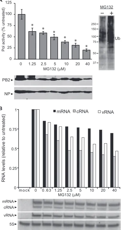
pro-cesses and is perhaps most commonly associated with protea-somal degradation (35). Ubiquitination had previously been associated with degradation of NP and free polymerase subunits (12,14,26). However, overexpression of WT or mutant forms of ubiquitin did not appreciably alter the steady-state levels of viral proteins (Fig. 1). Since IAV polymerase function is dependent on the stability of the RNP components, we sought to determine whether the proteasome had a significant effect on RNP steady-state levels and polymerase activity. IAV polymerase activity as-says were performed with cells treated with increasing concen-trations of proteasome inhibitor MG132. Polymerase activity decreased with increasing concentrations of MG132 (Fig. 2A). As expected, MG132 treatment stabilized proteins that would nor-mally be degraded by the proteasome, resulting in increased levels of total polyubiquitinated proteins (Fig. 2A, right). Nonetheless, there were no overt changes in steady-state levels for PB2 or NP upon MG132 treatment (Fig. 2A). Primer extension assays were then performed to analyze production of viral mRNA, the repli-cation intermediate cRNA, and the viral genome vRNA. These assays showed decreasing levels of all three RNA products with increase in MG132 concentration (Fig. 2B), indicating defects in both transcription and replication. These data suggest that host proteasome function is important for IAV polymerase activity in-dependent of the degradation of RNP components, providing a discrete step in the viral life impacted by the previously identified postfusion role of the ubiquitin-proteasome system (21).
Ubiquitination enhances influenza A virus polymerase
func-tion.Our data suggested that the IAV RNP components were
ubiquitinated during infection and that the host proteasome func-tion was essential for IAV polymerase activity, yet these processes did not have a major impact on protein steady state. This raised the question of whether ubiquitin exerts regulatory roles on the influenza polymerase function that are independent of protea-somal degradation, similar to other cellular activities that are
on November 7, 2019 by guest
http://jvi.asm.org/
ulated by ubiquitin and independent of proteasomal degradation, including protein-protein interactions, subcellular localization, and vesicular trafficking (35). Given that proteasomal activity maintains an equilibrium between free ubiquitin and conjugated ubiquitin, blocking proteasomal function would shift the equilib-rium and deplete the free ubiquitin pool (35). We therefore, hy-pothesized that maintaining a pool of free unconjugated ubiquitin
is necessary for IAV polymerase function given that inhibition of the proteasome decreased polymerase function (Fig. 2). To test this possibility, we performed polymerase activity assays of cells expressing increasing amounts of exogenous ubiquitin and dem-onstrated a strong correlation between enhanced polymerase ac-tivity and increased ubiquitin levels (Fig. 3A). Importantly, ubiq-uitin expression did not significantly alter the steady-state levels of
B A
PB2 Ub
Ub Ub
Ub
input
PB2 IP
150
100
75 50
250 150
100 75 75
50
37 150
100
75
FLAG IP
150
100
75 50
250 150 100 75 75
50
37
250
150
100
75
50
250
150
100
75
50
37 37
C
PB2 Ub
Ub
PB2 Ub
Ub
input
PB2 IP
input
PB2 IP
Ub RNP
WT WT K48R K63R Ub
RNP
WT WT K48R K63R
D Virus
Ub WSN WSN S009 NY312
E
control PA-FLAG PB1-FLAG PB2-FLAG
Pol F
control NP NP
K184R
NP Ub
Ub 150
100
75
150
100 75 75
50
37
100
75
input
NP
IP
input
75
50 75
50 100
75
100 150
75
PB1 PB1-Ub?
PB2 PB2-Ub? N40-PB1 C Infected
FIG 1All subunits of the influenza A RNP are ubiquitinated. (A) Lysates from control (lane C) or infected cells were probed for PB1 and PB2 by Western blotting. In addition to the expected proteins, additional higher-molecular-mass products were specifically detected in infected cells. Molecular masses (in kilodaltons) are indicated. (B) The IAV RNP complex expressing PB2-FLAG was reconstituted in HEK293T cells. Where indicated, cells coexpressed WT or mutant HA-tagged ubiquitin. The RNP complex was immunoprecipitated (IP) using anti-FLAG antibody. Input proteins and coprecipitating HA-ubiquitin were detected by Western blotting. (C) HEK293T cells and those expressing WT or mutant HA-ubiquitin were infected with WSN (PB2-FLAG) at an MOI of 1 for 16 h. Viral RNPs were immunoprecipitated as in panel B, and coprecipitating ubiquitin was analyzed by Western blotting. (D) HEK293T cells and cells expressing HA-ubiquitin were infected with either WSN PB2-FLAG or WSN-based PB2-FLAG reassortants carrying RNP genes from S009 or NY312. Viral RNPs were immunoprecipitated, and coprecipitating HA-ubiquitin was analyzed by Western blotting. (E) PA-FLAG, PB1-FLAG, and PB2-FLAG were individually coex-pressed with HA-ubiquitin in HEK293T cells and immunoprecipitated under denaturing conditions, and coprecipitating HA-ubiquitin was detected by Western blotting. (F) WT NP-V5 and the NP-V5 K184R mutant were coexpressed with HA-ubiquitin in HEK293T cells and immunoprecipitated under denaturing conditions, and coprecipitating HA-ubiquitin was detected by Western blotting.
on November 7, 2019 by guest
http://jvi.asm.org/
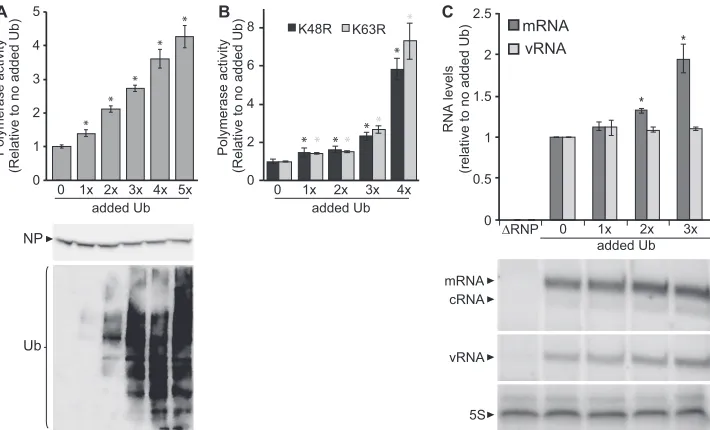
NP, nor did it cause a bulk shift of NP into higher-molecular-mass ubiquitinated species (Fig. 3A). It is possible that only a fraction of total NP is ubiquitinated. Similar results were obtained using a GFP reporter, excluding any reporter-specific effects (unpub-lished data). Polymerase activity assays were repeated using K48R and K63R ubiquitin mutants and again demonstrated that in-creasing levels of ubiquitin enhanced polymerase activity (Fig. 3B), suggesting that these linkages were not essential for enhance-ment of IAV polymerase function. Primer extension assays then delineated the impact of ubiquitin expression on polymerase function. RNPs reconstituted with a vNA template produced larger amounts of all viral RNA products in cells expressing in-creased amounts of ubiquitin (Fig. 3C). cRNA levels were too
close to the background level to be reliably quantified across all conditions, but they also showed a trend toward increased levels in the presence of excess ubiquitin. These results suggest that ubiq-uitin expression enhances global polymerase activity as we de-tected enhanced accumulation of all three RNA transcripts.
Ubiquitin mediated enhancement of IAV polymerase
func-tion is independent of NP.Ubiquitination of NP at K184 was
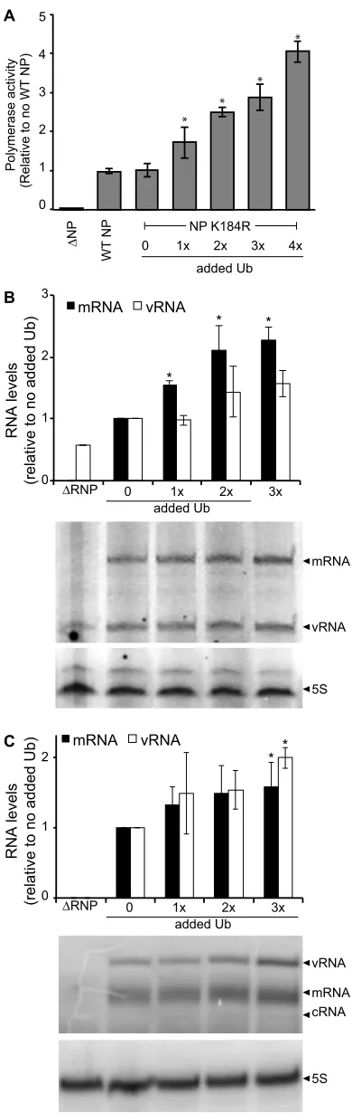
previously reported to enhance RNA binding and replication of the viral genome (13). To establish whether the ubiquitin-medi-ated enhancement of polymerase function that we have detected was based on NP K184 ubiquitination, we tested the effect of in-creasing ubiquitin expression on polymerase activity of RNPs re-constituted with NP K184R (Fig. 4A). Despite the absence of this well-characterized ubiquitination site, the polymerase activity of NP K184R RNPs was enhanced by increased ubiquitin expression. Our prior analyses had also shown that NP K184R is ubiquitinated and demonstrate ubiquitination patterns similar to that of WT NP (Fig. 1F). We performed primer extension assays to quantify the different RNA species generated by NP K184R RNPs. Ubiquitin expression increased levels of mRNA, cRNA, and vRNA (Fig. 4B), consistent with the results of our polymerase activity assays. The NP K184R mutation did, however, reduce cRNA levels to the background level in the absence of added ubiquitin, such that they could not be reliably quantified (Fig. 4B). This agrees with prior data showing that the NP K184R mutant exhibits selective defects in RNA synthesis resulting in reduced levels of cRNA and vRNA (13). To further test the requirement of NP for ubiquitin-medi-ated enhancement, we performed primer extension assays using micro-vRNPs in the presence of increasing ubiquitin expression. Micro-vRNA-like templates are short RNAs (⬃76 nt or less) con-taining viral UTRs that are replicated and transcribed by the poly-merase in the absence of NP (30,36). A 77-nt micro-vRNA gen-erated from the NP gene segment was used as a template during primer extension assays performed with increasing amounts of ubiquitin but without NP. These assays revealed higher levels of all RNA products in response to increased ubiquitin, even in the absence of NP (Fig. 4C), paralleling results obtained in the pres-ence of WT NP or the monoubiquitin-deficient NP K184R. cRNA is not routinely detected when using micro-vRNAs as a template (30) and thus could not be quantified here, even though we detect cRNA when polymerase activity is enhanced by ubiquitin expres-sion. Collectively, the primer extension assays demonstrated de-creased polymerase activity when the proteasome is inhibited (Fig. 2B) and increased activity in the presence of additional ubiquitin for WT RNPs (Fig. 3C), monoubiquitin-deficient NP K184R RNPs (Fig. 4B), and even for the NP-independent micro-vRNPS (Fig. 4C). Thus, it is likely that ubiquitination stimulates polymer-ase activity by directly impacting the polymerpolymer-ase, independent of NP or subsequent RNP assembly.
Ubiquitin expression enhances viral gene expression early
during infection.Our data have shown that ubiquitin expression
enhanced polymerase function, predicting that viral gene expres-sion during infection should also be enhanced under similar con-ditions. We therefore sought to establish whether ubiquitin ex-pression impacts polymerase activity not only in polymerase assays but also during infection. Cells expressing increasing amounts of ubiquitin were infected with our highly sensitive Na-noLuc reporter virus PASTN to quantify gene expression (34). We detected ubiquitin-dependent increases in viral gene expression as early as 2 h postinfection (hpi) that became more pronounced 0
25 50 75 100 125
0 1.25 2.5 5 10 20 40
Pol activity (% untreated)
MG132 (μM)
MG132 (μM)
NP PB2
A
B
mRNA cRNA
vRNA
5S 0 0.25 0.5 0.75 1
RNA levels (relative to untreated)
mRNA cRNA vRNA
m o c k 0 0 . 6 3 1.25 2.5 5 10 20 40 * *
*
* * *
MG132
Ub 250
150
100
75
50
37
FIG 2Host cell proteasome function is important for influenza A virus poly-merase activity. (A) Influenza A virus polypoly-merase activity assays were per-formed with HEK293T cells treated for 6 h with increasing concentrations of the proteasome inhibitor MG132 prior to lysis. NP protein expression was analyzed by Western blotting. Molecular masses in kDa are indicated. Data are
normalized to untreated cells (n⫽3⫾standard deviation). *,P⬍0.05 by
one-way ANOVA compared to untreated control. (B) Primer extension assays were performed to quantify RNP transcription (mRNA) and replication (cRNA and vRNA) in the presence of increasing concentrations of MG132. 5S rRNA served as a loading control. The viral RNA levels under each condition were normalized to polymerase activity in the absence of MG132.
on November 7, 2019 by guest
http://jvi.asm.org/
[image:5.585.63.267.72.456.2]throughout the time course (Fig. 5). These data reinforce results from our polymerase activity and primer extension assays, and combined they demonstrate stimulation of polymerase activity by elevated ubiquitin expression in both the replicon-based assays and during infection.
DISCUSSION
The IAV RNP complex is the minimal viral component capable of RNA synthesis during viral infection (2,3). The influenza virus polymerase transcribes viral mRNA, synthesizes the replication intermediate cRNA, and produces genomic vRNA. These pro-cesses are temporally regulated during the viral life cycle, in part by posttranslational modifications to NP and polymerase proteins. Here we show that ubiquitin and ubiquitination stimulate influ-enza virus polymerase activity and may be important regulators of polymerase activity during infection. RNPs from multiple viral strains were ubiquitinated, and all of the protein subunits in the RNP—PB1, PB2, PA, and NP—were subject to modification. Whereas ubiquitination is frequently associated with proteasomal degradation, we did not detect any notable change in protein steady-state levels either when ubiquitination of the viral proteins was increased or when the proteasome was inhibited. However, inhibition of the proteasome impaired polymerase function and alteration of ubiquitination changed production of viral mRNA, cRNA, and vRNA. These data suggest ubiquitination plays a reg-ulatory role that stimulates activity of the viral polymerase inde-pendent of its proteasomal degradation.
Ubiquitin and the ubiquitin-proteasome pathway impact key steps during the life cycle of many viruses (37). These include important roles for ubiquitination in viral entry, endosomal sort-ing, degradation of host restriction factors, regulation of innate antiviral signaling pathways, control of viral protein localization and stability, and finally trafficking and budding of progeny
viri-ons (37). During influenza virus infection, the E3 ubiquitin ligases Itch and Nedd4 are important during entry for release from the endosome (22,23), and ubiquitin found within virions is impor-tant for disassembly of incoming viral cores by the host aggresome (24). We have shown here that proteasome activity is important postentry for maximal activity of the viral polymerase (Fig. 2) and that ubiquitination of RNP subunits is associated with increased synthesis of all viral RNA products. Our data are supported by prior reports showing ubiquitination of individual subunits of the RNP (12–14,26).
Whereas our data demonstrate stimulatory effects of ubiquitin on polymerase activity, ubiquitination of NP and the polymerase proteins can direct their degradation by the proteasome. The E3 ligases TRIM22 and TRIM32 have been reported to be antiviral as they mediate proteasomal degradation of NP and PB1, respec-tively (14,26). Recent results have also shown an antiviral role for the ubiquitination and degradation of PB2 and PA, possibly asso-ciated with ADP ribosylation, another PTM these proteins un-dergo (12). It will be important to determine the E3 ligase(s) as well as the ADP ribosyltransferase(s) that target PB2 and PA.
Polyubiquitination of NP by the E3 ligase TRIM22 directs its degradation and restricts virus replication (26). In contrast, monoubiquitination at K184 is hypothesized to enhance replica-tion of the viral genome (13,26). Deubiquitination of NP K184 by the host ubiquitin-specific protease Usp11 decreased polymerase activity and reduced viral replication. We also detected NP ubiq-uitination in our assays; however, this was polyubiqubiq-uitination and was not dependent on K184, implicating additional ubiquitina-tion sites within NP. Moreover, increased ubiquitinaubiquitina-tion of NP did not reduce steady-state levels in our experiments, nor did we detect any increase in NP levels when we inhibited the protea-some, suggesting ubiquitination of NP might have additional im-pacts. We therefore sought to establish whether the enhanced
5
4
3
2
1
0
0 1x 2x 3x 4x 5x
Polymerase activity
(Relative to no added Ub)
Polymerase activity
(Relative to no added Ub)
added Ub
0 1x 2x 3x 4x
added Ub
Ub
K48R K63R
0 2 4 6 8
A B C
0 1x 2x 3x
RNA levels
(relative to no added Ub)
added Ub ΔRNP
mRNA cRNA
vRNA
5S *
* *
* *
* * *
* *
* * *
0 0.5 1 1.5 2 2.5
mRNA vRNA
* *
NP
FIG 3Increased ubiquitination enhances influenza A virus polymerase function. (A) Polymerase activity assays were performed with HEK293T cells expressing the IAV RNP and increasing amounts of cotransfected HA-ubiquitin. HA-ubiquitin and NP protein expression was analyzed by Western blotting. (B) Polymer-ase activity assays were performed as in panel A with increasing amounts of cotransfected K48R or K63R mutant HA-ubiquitin. Data are normalized to cells
lacking exogenous ubiquitin (n⫽3⫾standard deviation). *,P⬍0.05 by one-way ANOVA compared to cells without exogenous ubiquitin. (C) Primer extension
assays measured RNA transcripts generated in HEK293T cells expressing the IAV RNP, a vNA template, and increasing amounts of HA-ubiquitin. 5S rRNA served as a loading control. Quantified RNA levels were normalized to those present in cells lacking exogenous ubiquitin.
on November 7, 2019 by guest
http://jvi.asm.org/
[image:6.585.116.471.66.281.2]polymerase activity associated with increased ubiquitination was due to NP ubiquitination, as has been implicated before, or re-sulted from modification of the polymerase. When we tested the impact of increased ubiquitin expression on polymerase activity in the presence of the NP K184R mutant that lacks the monoubiq-uitination site, we still detected an enhancement in polymerase activity. Primer extension experiments on RNAs produced by RNPs containing the NP K184R mutant showed that mRNA, vRNA, and cRNA all increased with increasing ubiquitin expres-sion. Furthermore, we demonstrated that the increase in polymer-ase activity was largely independent of NP; increpolymer-ased ubiquitin expression enhanced RNA synthesis in the microgene system that does not require NP (30,36). Together, these data suggest that this increase in polymerase activity occurs via a different mechanism than the previously described monoubiquitination at NP K184 ubiquitination and is distinct from the antiviral ubiquitination catalyzed by TRIM22.
Since the IAV polymerase function is critical for the viral life cycle, we would expect that an enhancement of polymerase func-tion would cause enhanced viral gene expression in an infecfunc-tion setting. We employed our recombinant reporter virus to measure viral gene expression during infection in increased ubiquitin ex-pression (34). Our results showed that ubiquitin expression
in-Polymerase activity
(Relative to no WT NP)
0 2
1 4
3 5
Δ
NP
WT
NP NP K184R
added Ub
0 1x 2x 3x 4x
B
C A
0 3
2
1
0 1x 2x 3x
mRNA vRNA
RNA levels
(relative to no added Ub)
RNA levels
(relative to no added Ub)
added Ub
ΔRNP
mRNA
cRNA vRNA
vRNA
mRNA 5S
5S 0
2
1
0 1x 2x 3x
mRNA vRNA
added Ub
ΔRNP
* *
* *
*
*
*
* *
FIG 4Ubiquitin-mediated enhancement of influenza virus polymerase activ-ity does not require NP. (A) Polymerase activactiv-ity assays were performed with HEK293T cells expressing increasing amounts of HA-ubiquitin and IAV RNPs reconstituted with either WT NP, the mutant NP K184R, or empty plasmid
(⌬NP). Data are normalized to cells expressing WT NP in the absence of
exogenous ubiquitin (n⫽3⫾standard deviation). *,P⬍0.05 by one-way
ANOVA compared to NP K184R without ubiquitin. (B) Primer extension assays measured RNA transcripts generated in HEK293T cells expressing the IAV polymerase, mutant NP K184R, a vNA template, and increasing amounts of HA-ubiquitin. 5S rRNA serves as a loading control. RNA levels were nor-malized to those detected in cells expressing the RNP alone. (C) Influenza virus polymerase activity was reconstituted in the absence of NP using a 77-nt mi-crogene template. Activity was measured in the presence of increasing amounts of HA-ubiquitin. RNA products were detected by primer extension, quantified, and normalized to levels present in the absence of HA-ubiquitin.
0 1 2 4 6 8 hpi
Viral gene expression (RLU x 10
5) 5
4
3
2
1
0
Added Ub 3x
2x
1x 0
*
*
*
FIG 5Ubiquitination enhances viral gene expression during infection. HEK293T cells expressing increasing amounts of HA-ubiquitin were infected with PASTN virus at an MOI of 0.1 for 0, 1, 2, 4, 6, and 8 h. Following infection, gene expression was measured by a luciferase activity assay. RLU, relative light
units.n⫽3⫾standard deviation. *,P⬍0.05 by one-way ANOVA between
infection without added ubiquitin and cells treated with 3⫻ubiquitin.
on November 7, 2019 by guest
http://jvi.asm.org/
[image:7.585.320.525.68.266.2] [image:7.585.65.260.69.684.2]creased viral gene expression at early stages of infection; thus the enhanced polymerase function observed in replicon assays and primer extension is reflective of events that occur in a virus infec-tion setting. This is consistent with prior work showing that the proteasome and ubiquitination are important during postfusion and postnuclear entry steps (21) and combined with our replicon assays directly implicates the viral polymerase in this process. Changes in entry might also contribute to the enhanced gene ex-pression detected in infections with elevated ubiquitin levels. In-hibition of the proteasome blocks virion release from the endo-some, and the E3 ligase Itch plays a key role in this process (23,38). Elevated ubiquitin levels may increase both endosomal release and polymerase function. These enhancing activities of ubiquitin may be counteracted at other stages of infection when ubiquitination plays an antiviral role (e.g., the TRIM22-directed turnover of NP). The balance between these opposing functions may dictate how ubiquitination impacts the outcome of multicycle replications. The molecular mechanisms by which ubiquitination enhances polymerase function remain unclear. As all of the polymerase sub-units are subject to ubiquitination, and this does not appear to be related to proteasomal degradation as we detected no significant changes in protein steady-state levels, it is possible that these mod-ifications directly impact protein localization or protein-protein interactions. It has also been suggested that ubiquitination and the proteasome can have indirect effects that stimulate infection, such as activating the NF-B pathway and other host factors that are important for viral replication or degrading cellular factors that restrict infection (21). Thus, influenza virus exploits and relies on the ubiquitin-proteasome pathway at multiple steps during the early stages of infection and may be particularly sensitive to ther-apeutic interventions that disrupt these host processes.
ACKNOWLEDGMENTS
We thank D. Gabuzda for providing reagents and members of the Mehle laboratory for valuable contributions. Anti-RNP antibody (NR-3133) was obtained through the NIH Biodefense and Emerging Infections Research Resources Repository, NIAID, NIH.
This work was supported in part by the National Institutes of Health (R00GM088484 and R01AI125271), a Shaw Scientist award, and an American Lung Association Basic Research grant (RG-310016) to A.M.
FUNDING INFORMATION
This work, including the efforts of Andrew Mehle, was funded by Shaw Scientist Award. This work, including the efforts of James Kirui, Arindam Mondal, and Andrew Mehle, was funded by HHS | National Institutes of Health (NIH) (R00GM088484). This work, including the efforts of James Kirui, Arindam Mondal, and Andrew Mehle, was funded by HHS | Na-tional Institutes of Health (NIH) (R01AI125271). This work, including the efforts of James Kirui and Andrew Mehle, was funded by American Lung Association (RG-310016).
REFERENCES
1.Shaw ML, Palese P.2014. Orthomyxoviruses, p 1151–1185. InKnipe DM, Howley PM (ed), Fields virology, 6th ed. Lippincott Williams & Wilkins, Philadelphia, PA.
2.Mehle A, McCullers JA.2013. Structure and function of the influenza
virus replication machinery and PB1-F2, p 133–145. InWebster RG,
Monto AS, Braciale TJ, Lamb RA (ed), Textbook of influenza, 2nd ed. John Wiley & Sons, Ltd, Oxford, United Kingdom.
3.Fodor E.2013. The RNA polymerase of influenza a virus: mechanisms of
viral transcription and replication. Acta Virol57:113–122.http://dx.doi
.org/10.4149/av_2013_02_113.
4.Dias A, Bouvier D, Crepin T, McCarthy AA, Hart DJ, Baudin F, Cusack
S, Ruigrok RW.2009. The cap-snatching endonuclease of influenza virus
polymerase resides in the PA subunit. Nature458:914 –918.http://dx.doi
.org/10.1038/nature07745.
5.Guilligay D, Tarendeau F, Resa-Infante P, Coloma R, Crepin T, Sehr P, Lewis J, Ruigrok RW, Ortin J, Hart DJ, Cusack S.2008. The structural basis for cap binding by influenza virus polymerase subunit PB2. Nat
Struct Mol Biol15:500 –506.http://dx.doi.org/10.1038/nsmb.1421.
6.Yuan P, Bartlam M, Lou Z, Chen S, Zhou J, He X, Lv Z, Ge R, Li X, Deng T, Fodor E, Rao Z, Liu Y. 2009. Crystal structure of an avian influenza polymerase PA(N) reveals an endonuclease active site. Nature
458:909 –913.http://dx.doi.org/10.1038/nature07720.
7.Subbarao EK, London W, Murphy BR.1993. A single amino acid in the PB2 gene of influenza A virus is a determinant of host range. J Virol
67:1761–1764.
8.Robb NC, Smith M, Vreede FT, Fodor E. 2009. NS2/NEP protein regulates transcription and replication of the influenza virus RNA
ge-nome. J Gen Virol 90:1398 –1407. http://dx.doi.org/10.1099/vir.0
.009639-0.
9.Hutchinson EC, Denham EM, Thomas B, Trudgian DC, Hester SS, Ridlova G, York A, Turrell L, Fodor E.2012. Mapping the phosphopro-teome of influenza A and B viruses by mass spectrometry. PLoS Pathog
8:e1002993.http://dx.doi.org/10.1371/journal.ppat.1002993.
10. Pal S, Santos A, Rosas JM, Ortiz-Guzman J, Rosas-Acosta G. 2011. Influenza A virus interacts extensively with the cellular SUMOylation
sys-tem during infection. Virus Res158:12–27.http://dx.doi.org/10.1016/j
.virusres.2011.02.017.
11. Han Q, Chang C, Li L, Klenk C, Cheng J, Chen Y, Xia N, Shu Y, Chen Z, Gabriel G, Sun B, Xu K.2014. Sumoylation of influenza A virus nucleoprotein is essential for intracellular trafficking and virus growth. J
Virol88:9379 –9390.http://dx.doi.org/10.1128/JVI.00509-14.
12. Liu CH, Zhou L, Chen G, Krug RM.2015. Battle between influenza A virus and a newly identified antiviral activity of the PARP-containing
ZAPL protein. Proc Natl Acad Sci U S A112:14048 –14053.http://dx.doi
.org/10.1073/pnas.1509745112.
13. Liao TL, Wu CY, Su WC, Jeng KS, Lai MM.2010. Ubiquitination and deubiquitination of NP protein regulates influenza A virus RNA
replica-tion. EMBO J29:3879 –3890.http://dx.doi.org/10.1038/emboj.2010.250.
14. Fu B, Wang L, Ding H, Schwamborn JC, Li S, Dorf ME. 2015. TRIM32 senses and restricts influenza A virus by ubiquitination of
PB1 polymerase. PLoS Pathog11:e1004960.http://dx.doi.org/10.1371
/journal.ppat.1004960.
15. Zheng W, Li J, Wang S, Cao S, Jiang J, Chen C, Ding C, Qin C, Ye X, Gao GF, Liu W.2015. Phosphorylation controls the nuclear-cytoplasmic
shuttling of influenza A virus nucleoprotein. J Virol89:5822–5834.http:
//dx.doi.org/10.1128/JVI.00015-15.
16. Neumann G, Castrucci MR, Kawaoka Y. 1997. Nuclear import and
export of influenza virus nucleoprotein. J Virol71:9690 –9700.
17. Chenavas S, Estrozi LF, Slama-Schwok A, Delmas B, Di Primo C, Baudin F, Li X, Crepin T, Ruigrok RW.2013. Monomeric nucleoprotein
of influenza A virus. PLoS Pathog9:e1003275.http://dx.doi.org/10.1371
/journal.ppat.1003275.
18. Mondal A, Potts GK, Dawson AR, Coon JJ, Mehle A.2015. Phosphor-ylation at the homotypic interface regulates nucleoprotein oligomeriza-tion and assembly of the influenza virus replicaoligomeriza-tion machinery. PLoS
Pat-hog11:e1004826.http://dx.doi.org/10.1371/journal.ppat.1004826.
19. Turrell L, Hutchinson EC, Vreede FT, Fodor E.2015. Regulation of influenza A virus nucleoprotein oligomerization by phosphorylation. J
Virol89:1452–1455.http://dx.doi.org/10.1128/JVI.02332-14.
20. Komander D, Rape M.2012. The ubiquitin code. Annu Rev Biochem81:
203–229.http://dx.doi.org/10.1146/annurev-biochem-060310-170328.
21. Widjaja I, de Vries E, Tscherne DM, García-Sastre A, Rottier PJ, de Haan CA.2010. Inhibition of the ubiquitin-proteasome system affects
influenza A virus infection at a postfusion step. J Virol84:9625–9631.http:
//dx.doi.org/10.1128/JVI.01048-10.
22. Chesarino NM, McMichael TM, Yount JS.2015. E3 ubiquitin ligase NEDD4 promotes influenza virus infection by decreasing levels of the
antiviral protein IFITM3. PLoS Pathog11:e1005095.http://dx.doi.org/10
.1371/journal.ppat.1005095.
23. Su WC, Chen YC, Tseng CH, Hsu PW, Tung KF, Jeng KS, Lai MM. 2013. Pooled RNAi screen identifies ubiquitin ligase Itch as crucial for influenza A virus release from the endosome during virus entry. Proc
Natl Acad Sci U S A110:17516 –17521.http://dx.doi.org/10.1073/pnas
.1312374110.
on November 7, 2019 by guest
http://jvi.asm.org/
24. Banerjee I, Miyake Y, Nobs SP, Schneider C, Horvath P, Kopf M, Matthias P, Helenius A, Yamauchi Y.2014. Influenza A virus uses the
aggresome processing machinery for host cell entry. Science346:473– 477.
http://dx.doi.org/10.1126/science.1257037.
25. Shaw ML, Stone KL, Colangelo CM, Gulcicek EE, Palese P. 2008.
Cellular proteins in influenza virus particles. PLoS Pathog4:e1000085.
http://dx.doi.org/10.1371/journal.ppat.1000085.
26. Di Pietro A, Kajaste-Rudnitski A, Oteiza A, Nicora L, Towers GJ, Mechti N, Vicenzi E.2013. TRIM22 inhibits influenza A virus infection
by targeting the viral nucleoprotein for degradation. J Virol87:4523–
4533.http://dx.doi.org/10.1128/JVI.02548-12.
27. Watanabe T, Watanabe S, Kawaoka Y.2010. Cellular networks involved
in the influenza virus life cycle. Cell Host Microbe7:427– 439.http://dx
.doi.org/10.1016/j.chom.2010.05.008.
28. Mehle A, Doudna JA.2008. An inhibitory activity in human cells restricts the function of an avian-like influenza virus polymerase. Cell Host
Mi-crobe4:111–122.http://dx.doi.org/10.1016/j.chom.2008.06.007.
29. Mehle A, Strack B, Ancuta P, Zhang C, McPike M, Gabuzda D.2004. Vif overcomes the innate antiviral activity of APOBEC3G by promoting its
degradation in the ubiquitin-proteasome pathway. J Biol Chem279:7792–
7798.http://dx.doi.org/10.1074/jbc.M313093200.
30. Turrell L, Lyall JW, Tiley LS, Fodor E, Vreede FT.2013. The role and assembly mechanism of nucleoprotein in influenza A virus
ribonucle-oprotein complexes. Nat Commun4:1591.http://dx.doi.org/10.1038
/ncomms2589.
31. Dos Santos Afonso E, Escriou N, Leclercq I, van der Werf S, Naffakh N. 2005. The generation of recombinant influenza A viruses expressing a PB2
fusion protein requires the conservation of a packaging signal overlapping
the coding and noncoding regions at the 5=end of the PB2 segment.
Vi-rology341:34 – 46.http://dx.doi.org/10.1016/j.virol.2005.06.040.
32. Kirui J, Bucci MD, Poole DS, Mehle A.2014. Conserved features of the PB2 627 domain impact influenza virus polymerase function and
replica-tion. J Virol88:5977–5986.http://dx.doi.org/10.1128/JVI.00508-14.
33. Choo YS, Zhang Z.19 August 2009. Detection of protein ubiquitination.
J Vis Exphttp://dx.doi.org/10.3791/1293.
34. Tran V, Moser LA, Poole DS, Mehle A.2013. Highly sensitive real-time in vivo imaging of an influenza reporter virus reveals dynamics of
replica-tion and spread. J Virol87:13321–13329.http://dx.doi.org/10.1128/JVI
.02381-13.
35. Hershko A, Ciechanover A. 1998. The ubiquitin system. Annu Rev
Biochem 67:425– 479. http://dx.doi.org/10.1146/annurev.biochem.67.1
.425.
36. Resa-Infante P, Recuero-Checa MA, Zamarreno N, Llorca O, Ortin J. 2010. Structural and functional characterization of an influenza virus
RNA polymerase-genomic RNA complex. J Virol84:10477–10487.http:
//dx.doi.org/10.1128/JVI.01115-10.
37. Luo H.2016. Interplay between the virus and the ubiquitin-proteasome
system: molecular mechanism of viral pathogenesis. Curr Opin Virol17:
1–10.http://dx.doi.org/10.1016/j.coviro.2015.09.005.
38. Khor R, McElroy LJ, Whittaker GR.2003. The ubiquitin-vacuolar pro-tein sorting system is selectively required during entry of influenza virus
into host cells. Traffic4:857– 868.http://dx.doi.org/10.1046/j.1398-9219
.2003.0140.x.