Recombinant Isfahan Virus and Vesicular
Stomatitis Virus Vaccine Vectors Provide
Durable, Multivalent, Single-Dose
Protection against Lethal Alphavirus
Challenge
Farooq Nasar,
a*
Demetrius Matassov,
bRobert L. Seymour,
aTheresa Latham,
bRodion V. Gorchakov,
cRebecca M. Nowak,
bGrace Leal,
aStefan Hamm,
bJohn H. Eldridge,
bRobert B. Tesh,
aDavid K. Clarke,
bScott C. Weaver
aInstitute for Human Infections and Immunity, Center for Tropical Diseases, and Departments of Pathology and Microbiology and Immunology, University of Texas Medical Branch, Galveston, Texas, USAa; Profectus
BioSciences Inc., Tarrytown, New York, USAb; Department of Pediatrics, Section of Tropical Medicine, Baylor
College of Medicine, Houston, Texas, USAc
ABSTRACT
The demonstrated clinical efficacy of a recombinant vesicular stomatitis
virus (rVSV) vaccine vector has stimulated the investigation of additional
serologi-cally distinct
Vesiculovirus
vectors as therapeutic and/or prophylactic vaccine vectors
to combat emerging viral diseases. Among these viral threats are the encephalitic
al-phaviruses Venezuelan equine encephalitis virus (VEEV) and Eastern equine
encepha-litis virus (EEEV), which have demonstrated potential for natural disease outbreaks,
yet no licensed vaccines are available in the event of an epidemic. Here we report
the rescue of recombinant Isfahan virus (rISFV) from genomic cDNA as a potential
new vaccine vector platform. The rISFV genome was modified to attenuate virulence
and express the VEEV and EEEV E2/E1 surface glycoproteins as vaccine antigens. A
single dose of the rISFV vaccine vectors elicited neutralizing antibody responses and
protected mice from lethal VEEV and EEEV challenges at 1 month postvaccination as
well as lethal VEEV challenge at 8 months postvaccination. A mixture of rISFV
vec-tors expressing the VEEV and EEEV E2/E1 glycoproteins also provided durable,
single-dose protection from lethal VEEV and EEEV challenges, demonstrating the
po-tential for a multivalent vaccine formulation. These findings were paralleled in
stud-ies with an attenuated form of rVSV expressing the VEEV E2/E1 glycoproteins. Both
the rVSV and rISFV vectors were attenuated by using an approach that has
demon-strated safety in human trials of an rVSV/HIV-1 vaccine. Vaccines based on either of
these vaccine vector platforms may present a safe and effective approach to prevent
alphavirus-induced disease in humans.
IMPORTANCE
This work introduces rISFV as a novel vaccine vector platform that is
serologically distinct and phylogenetically distant from VSV. The rISFV vector has
been attenuated by an approach used for an rVSV vector that has demonstrated
safety in clinical studies. The vaccine potential of the rISFV vector was investigated
in a well-established alphavirus disease model. The findings indicate the feasibility of
producing a safe, efficacious, multivalent vaccine against the encephalitic
alphavi-ruses VEEV and EEEV, both of which can cause fatal disease. This work also
demon-strates the efficacy of an attenuated rVSV vector that has already demonstrated
safety and immunogenicity in multiple HIV-1 phase I clinical studies. The absence of
serological cross-reactivity between rVSV and rISFV and their phylogenetic
diver-gence within the
Vesiculovirus
genus indicate potential for two stand-alone vaccine
Received5 September 2016Accepted12
January 2017
Accepted manuscript posted online1
February 2017
CitationNasar F, Matassov D, Seymour RL,
Latham T, Gorchakov RV, Nowak RM, Leal G, Hamm S, Eldridge JH, Tesh RB, Clarke DK, Weaver SC. 2017. Recombinant Isfahan virus and vesicular stomatitis virus vaccine vectors provide durable, multivalent, single-dose protection against lethal alphavirus challenge. J Virol 91:e01729-16.https://doi.org/10.1128/ JVI.01729-16.
EditorStanley Perlman, University of Iowa
Copyright© 2017 American Society for
Microbiology.All Rights Reserved.
Address correspondence to Farooq Nasar, [email protected], or Scott C. Weaver, [email protected].
*Present address: Farooq Nasar, Virology Division, United States Army Medical Research Institute of Infectious Diseases, Frederick, Maryland, USA.
crossm
on November 7, 2019 by guest
http://jvi.asm.org/
vector platforms that could be used to target multiple bacterial and/or viral agents
in successive immunization campaigns or as heterologous prime-boost agents.
KEYWORDS
Eastern equine encephalitis virus, Isfahan virus, Venezuelan equine
encephalitis virus, vesicular stomatitis virus
T
he genus
Vesiculovirus
in the family
Rhabdoviridae
consists of arthropod-borne
viruses that are transmitted to susceptible vertebrate hosts through insect bites (1).
This genus consists of nine recognized virus species, including the prototypic species
vesicular stomatitis virus (VSV) and Isfahan virus (ISFV) (2). ISFV was isolated from
phlebotomine sandflies in Dormian, Isfahan Province, Iran, in 1975 and is endemic in
parts of Asia, including Iran, Turkmenistan, and the central Asian republics (3–5).
Transmission to susceptible hosts occurs via biting insects, most likely sandflies, and
although data indicate that ISFV can infect humans and domesticated animals, the
infection has not been linked to any illness (3, 6).
VSV is found only in the Americas and is an important pathogen of domesticated
animals, mainly ungulates (1). The virus was first reported during an outbreak in cattle
and horses in 1916 and was subsequently isolated from cattle in 1925 in Richmond, IN
(7). Infection in animals causes vesicular lesions at insect bite sites around the mouth,
nose, teats, and coronary bands on the hooves (8). The vesicular lesions may result in
lameness and weight loss due to difficulty in feeding but typically resolve in 7 to 10
days without serious consequences (8). Humans can also be infected with VSV at
mucosal surfaces as a result of either close contact with infected animals or accidental
exposure in the laboratory (9, 10). The resulting infection may either be subclinical or
produce mild flu-like symptoms that typically resolve in 5 to 7 days without
compli-cations.
As a prototype member of the genus
Vesiculovirus
, VSV has been extensively
studied, providing insights into many aspects of the virus replication cycle and
shed-ding light on the biology of other related viruses. The VSV and ISFV genomes consist
of a nonsegmented, single-strand, negative-sense RNA
⬃
11 kb in length containing five
genes in the order 3
=
-N-P-M-G-L-5
=
, encoding the N, P, M, G, and L proteins, respectively
(1, 11). The VSV particles are bullet-shaped (
⬃
80 nm by
⬃
200 nm) and comprise a
ribonucleoprotein core of genomic RNA and N (nucleocapsid) protein surrounded by a
matrix (M) protein layer, enveloped by the host cell plasma membrane containing the
transmembrane viral glycoprotein (G) (12). The phosphoprotein (P) and large (L)
polymerase protein associate to form the functional viral RNA polymerase, which
performs both mRNA transcription and genome replication from the nucleocapsid
template (13, 14). Viral mRNA transcription initiates at the 3
=
end of the genome and
proceeds with a pronounced 3
=
-to-5
=
gradient of gene expression, leading to abundant
N protein and successively decreasing levels of P, M, G, and L proteins in the infected
cell, providing virus proteins an optimal ratio for subsequent viral genome replication
and the assembly of mature virus particles (11, 15). Virus replication
in vitro
is rapid, and
virus progeny are detectable at 5 to 6 hours postinfection (hpi) in vertebrate cell lines.
The genomes of negative-strand viruses remained refractory to genetic
manipula-tion until 1994, when the recovery of infectious rabies virus from viral genomic cDNA
became possible (16). The development of similar rescue systems for other
negative-strand viruses, including vesicular stomatitis Indiana virus (VSIV), followed quickly (17,
18). The ability to manipulate the VSIV genome enabled the development of
recom-binant VSIV (rVSIV) as a vaccine vector (19). The immunogenicity and protective efficacy
of rVSIV vaccine vectors have been demonstrated for a range of human pathogens,
including influenza virus, human immunodeficiency virus (HIV), herpes simplex virus 2
(HSV-2), respiratory syncytial virus (RSV), severe acute respiratory syndrome-associated
coronavirus (SARS-CoV), chikungunya virus (CHIKV), and Ebola virus (EBOV), in
small-animal and nonhuman primate (NHP) disease models (20–31). However, the safety of
rVSIV vectors initially was a concern because both VSIV and the New Jersey serotype of
VSV (vesicular stomatitis New Jersey virus [VSNJV]) are known to have neurotropic
on November 7, 2019 by guest
http://jvi.asm.org/
properties in young mice and can cause neurological disease following intracranial (i.c.)
inoculation of cows and horses (32–35). A pilot NHP neurovirulence (NV) study
indi-cated that a prototypic rVSIV vector was insufficiently attenuated for clinical evaluation
(36–38). To reduce NV potential and enhance vector safety, a variety of attenuation
strategies were devised and tested in animal disease models (39–41). The most
prom-ising strategy was a combination of N gene translocation with a truncation of the
cytoplasmic tail (CT) of the G protein, enabling a very marked reduction of NV in young
mice. One of the most highly attenuated vectors (rVSIV-N4CT1HIVgag1) produced little
evidence of injury in the central nervous system (CNS) when NHPs were inoculated by
the intrathalamic route yet elicited an immune response that was similar to those
induced by more virulent vectors when inoculated by the intramuscular (i.m.) route
(39). These preclinical studies supported the evaluation of rVSIV-N4CT1HIVgag1 in
recently concluded phase I human clinical trials (HVTN 090 and HVTN 087), where the
rVSIV vector demonstrated safety at a dose as high as 10
8PFU and immunogenicity at
a dose as low as 10
4PFU (42).
An important target of biodefense vaccines are members of the genus
Alphavirus
of
the family
Togaviridae
, which is comprised of small, spherical, enveloped viruses with
genomes consisting of a single-strand, positive-sense RNA 11 to 12 kb in length (1).
Alphaviruses comprise 31 recognized species classified into 11 complexes based on
antigenic and/or genetic similarities (43, 44). The two aquatic alphavirus complexes are
not known to utilize arthropods in their transmission cycles, whereas all of the
remain-ing complexes (Barmah Forest virus, Ndumu virus, Middelburg virus, Semliki Forest
virus, Venezuelan equine encephalitis virus [VEEV], Eastern equine encephalitis virus
[EEEV], Western equine encephalitis virus [WEEV], Trocara virus, and Eilat virus) consist
of arboviruses that almost exclusively utilize mosquitoes as vectors (1, 45–47).
Mosquito-borne alphaviruses infect diverse vertebrate hosts, including equids, birds,
amphibians, reptiles, rodents, pigs, humans, and NHPs (1). The ability to infect both
mosquitoes and vertebrates enables the maintenance of alphaviruses in natural
en-demic transmission cycles that occasionally spill over into the human population and
cause disease. Infection with Old World alphaviruses such as CHIKV, o’nyong-nyong
virus (ONNV), Sindbis virus (SINV), and Ross River virus (RRV) are rarely fatal, and disease
is characterized by rash and debilitating arthralgia that can persist for months or years.
In contrast, New World alphaviruses such as WEEV, EEEV, and VEEV can cause fatal
encephalitis. Human case fatality rates for EEEV range from 40 to 80%, whereas those
for WEEV and VEEV infections range from
⬍
1 to 5% (1). In addition, all three viruses are
lethal (EEEV and WEEV) or highly debilitating (VEEV) in NHPs after aerosol infection
(48–50). Currently, there are no licensed antiviral treatments or vaccines for alphaviral
diseases, and the U.S. population remains vulnerable to natural disease outbreaks.
Here we report the first recovery of infectious rISFV from genomic cDNA and
demonstrate the immunogenicity and protective efficacy of attenuated forms of rISFV
and rVSV vector vaccines in mouse models of VEEV- and EEEV-induced disease.
RESULTS
Genetic and phylogenetic analysis of ISFV.
The available N, G, and L gene
sequences of currently recognized and tentatively classified members of the genus
Vesiculovirus
were downloaded from GenBank and aligned (Table 1). Genetic analysis
was performed by comparing both nucleotide and deduced amino acid sequences
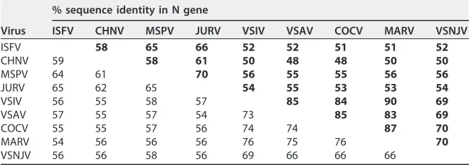
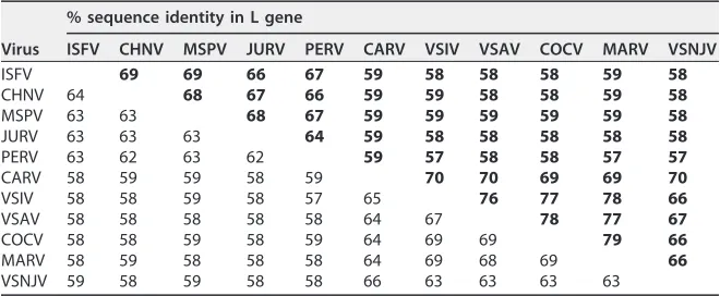
(Tables 2 to 4). Nucleotide and amino acid sequence identities of the ISFV genes with
other members of the genus ranged from 48 to 65% and 37 to 69%, respectively.
Isfahan virus genes consistently had higher nucleotide and amino acid sequence
identities with Chandipura virus (CHNV), Malpais Springs virus (MSPV), and Jurona virus
(JURV) than with VSIV, with N gene nucleotide and amino acid identities that were 2 to
8% and 6 to 14% higher, G gene nucleotide and amino acid identities that were 5 to
9% and 10 to 14% higher, and L gene nucleotide and amino acid identities that were
5 to 6% and 8 to 11% higher, respectively (Tables 2 to 4).
on November 7, 2019 by guest
http://jvi.asm.org/
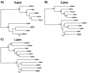
Phylogenetic trees were generated by utilizing the maximum likelihood (ML)
method to study the evolutionary relatedness between ISFV and other vesiculoviruses.
Based on comparisons of N, G, and L gene nucleotide sequences, the phylogenetic
analysis revealed similar evolutionary relationships. Two branching patterns were
observed: one main branch consisted of VSIV, its subtypes, and VSNJV, and another
branch consisted of ISFV, CHNV, JURV, Perinet virus (PERV), Piry virus (PIRV), and MSPV
(Fig. 1). Major branching nodes were statistically supported with bootstrap values of
100% in both analyses; however, statistical support for internal nodes was limited (Fig.
1). These analyses showed that ISFV is more closely related to CHNV, JURV, PERV, PIRV,
and MSPV than to VSIV and VSNJV.
In vitro
characterization of wild-type ISFV and VSIV.
The plaque phenotype and
one-step replication kinetics of wild-type (wt) isolates of ISFV and the VSIV San Juan
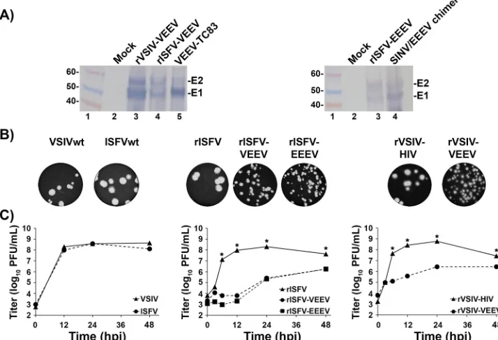
strain were compared (see Fig. 4B and C). Both isolates displayed similar plaque
phenotypes, reaching
⬃
3 to 4 mm in diameter at 48 hpi, and replication kinetics in Vero
cell monolayers were almost identical, with peak titers of 5
⫻
10
8PFU/ml being
achieved at 6 to 12 hpi.
[image:4.585.42.374.83.213.2]Generation of rISFV.
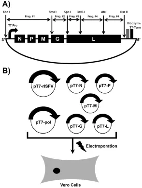
The assembly of a full-length rISFV genomic cDNA and
plasmids expressing the ISFV N, P M, G, and L proteins was carried out in a manner
similar to that used previously for the generation of rVSIV and mumps virus (MuV)
genomic cDNAs (16–18, 51). High-efficiency rescue of infectious rISFV from genomic
cDNA was achieved by the electroporation of Vero cells with plasmids encoding
full-length genomic cDNA and the N, P, M, G, and L proteins as described previously for
the rescue of rVSIV and other negative-strand RNA viruses (Fig. 2B) (52). The recovery
of rISFV was indicated by the induction of cytopathic effects (CPE) in Vero cells upon
passaging of the transfected cell supernatant onto fresh monolayers and confirmed
by full-length genomic sequencing of the resulting virus progeny. rISFV displayed
a plaque phenotype (
⬃
3 to 4 mm in diameter at 48 hpi), replication kinetics, and
TABLE 2Nucleotide and amino acid comparison based on full-length N gene sequences of members of the genusVesiculovirusa
Virus
% sequence identity in N gene
ISFV CHNV MSPV JURV VSIV VSAV COCV MARV VSNJV
ISFV 58 65 66 52 52 51 51 52
CHNV 59 58 61 50 48 48 50 50
MSPV 64 61 70 56 55 55 56 56
JURV 65 62 65 54 55 53 53 54
VSIV 56 55 58 57 85 84 90 69
VSAV 57 55 57 54 73 85 83 69
COCV 55 55 57 56 74 74 87 70
MARV 54 56 56 56 76 75 76 70
VSNJV 56 56 58 56 69 66 66 66
aPercent nucleotide identity is shown below the diagonal in lightface type. Percent amino acid identity is
[image:4.585.41.372.605.721.2]shown above the diagonal in boldface type.
TABLE 1List of rhabdovirus sequences utilized in analyses
Species Abbreviation GenBank accession no.
Carajas virus CARAV HW243161
Chandipura virus CHNV GU212858
Cocal virus COCV EU373657
Isfahan virus ISFV AJ810084
Jurona virus JURV HM566194
Malpais Spring virus MSPV KC412247
Maraba virus MARV HQ660076
Perinet virus PERV HM566195
Piry virus PIRV D26175
Vesicular stomatitis Alagoas virus VSAV EU373658 Vesicular stomatitis Indiana virus VSIV NC_001560 Vesicular stomatitis New Jersey virus VSNJV NC_024473
on November 7, 2019 by guest
http://jvi.asm.org/
a peak titer (
⬃
3
⫻
10
8PFU/ml) similar to those of wt ISFV (see Fig. 4B and C). These
findings confirmed the recovery of infectious rISFV from genomic cDNA and created
a pathway for further modification of rISFV as a vaccine vector.
In vitro
characterization of rISFV and rVSIV vectors encoding alphavirus
anti-gens.
Both the rISFV and rVSIV vectors were altered to express alphavirus E3-E2-6k/
TF-E1 (E3-E1) antigens. The E3-E1 genes of the North American lineage of EEEV strain
FL93-939 and VEEV subtype ID strain ZPC 738 (VEEV-ID), were inserted into the fifth
position of the rISFV genome(s) (Fig. 3), which also had the N gene translocated to
position 4 (Fig. 3), a strategy that was successful in attenuating rVSIV replication
in vitro
(35, 40, 53, 54). In addition, an rVSIV-N4CT1 vector, very similar in design to the vector
which demonstrated safety and immunogenicity in phase I clinical trials, was also
modified to express the VEEV-ID E3-E1 genes in the fifth position of the genome (Fig.
3) (42).
[image:5.585.40.372.93.228.2]All three vectors were characterized
in vitro
for the expression of alphavirus
antigens, plaque phenotype, and replication kinetics in Vero cells (Fig. 3 and 4); the
rISFV and rVSIV vectors demonstrated robust alphavirus antigen expression,
dis-played a small-plaque phenotype (
⬃
1 to 2 mm in diameter at 72 hpi compared to
⬃
3 to 4 mm at 48 hpi for wt viruses), and a significant reduction in replication
efficiency (
⬃
10- to 10,000-fold at 12 to 48 hpi) relative to those of unaltered rISFV
or rVSIV-HIV (Fig. 4A to C). These data indicated that, like rVSIV, rISFV can also be
attenuated by N gene shuffling and that both the attenuated rISFV and rVSIV
vectors can express abundant alphavirus antigens while achieving peak titers of
⬃
10
6PFU/ml on Vero cell monolayers.
TABLE 3Nucleotide and amino acid comparison based on full-length G gene sequences of members of the genusVesiculovirusa
Virus
% sequence identity in G gene
ISFV CHNV MSPV JURV PIRV PERV VSIV VSAV COCV MARV VSNJV ISFV 54 49 54 50 50 39 37 41 39 38 CHNV 56 48 52 51 51 39 40 39 40 37 MSPV 54 53 49 48 49 36 37 37 36 37 JURV 58 57 54 49 51 37 37 38 37 37 PIRV 55 55 55 56 57 39 38 38 39 37 PERV 55 56 55 56 60 38 40 38 39 38 VSIV 49 47 46 48 48 46 63 71 77 49 VSAV 48 49 47 48 48 49 64 66 64 48 COCV 49 47 46 48 47 46 69 65 73 47 MARV 49 48 45 48 48 48 71 65 69 50 VSNJV 48 48 47 49 47 46 55 55 53 55
aPercent nucleotide identity is shown below the diagonal in lightface type. Percent amino acid identity is
shown above the diagonal in boldface type.
TABLE 4Nucleotide and amino acid comparison based on full-length L gene sequences of members of the genusVesiculovirusa
Virus
% sequence identity in L gene
ISFV CHNV MSPV JURV PERV CARV VSIV VSAV COCV MARV VSNJV ISFV 69 69 66 67 59 58 58 58 59 58 CHNV 64 68 67 66 59 59 58 58 59 58 MSPV 63 63 68 67 59 59 59 59 59 58 JURV 63 63 63 64 59 58 58 58 58 58 PERV 63 62 63 62 59 57 58 58 57 57 CARV 58 59 59 58 59 70 70 69 69 70 VSIV 58 58 59 58 57 65 76 77 78 66 VSAV 58 58 58 58 58 64 67 78 77 67 COCV 58 58 59 58 59 64 69 69 79 66 MARV 58 59 58 58 58 64 69 68 69 66 VSNJV 59 58 59 58 58 66 63 63 63 63
aPercent nucleotide identity is shown below the diagonal in lightface type. Percent amino acid identity is
shown above the diagonal in boldface type.
on November 7, 2019 by guest
http://jvi.asm.org/
[image:5.585.41.371.585.721.2]Assessment of rISFV vaccine vector efficacy.
The efficacy of both rISFV-VEEV and
EEEV vaccine candidates was assessed by lethal alphavirus challenge in a murine model.
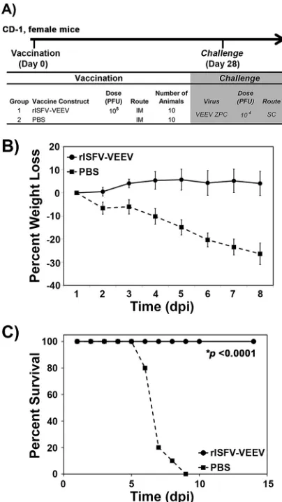
Cohorts of 10 CD-1 mice were vaccinated i.m. with 10
8PFU/mouse of rISFV-VEEV or
with a sham vaccine (Fig. 5). At 4 weeks postvaccination, animals were challenged
subcutaneously (s.c.) with 10
4PFU/mouse of VEEV-ID. Weight loss was observed by as
early as 2 days postchallenge in sham-vaccinated animals and increased to
⬎
20% of the
starting body mass at the time of euthanasia (Fig. 5B). By day 8 postchallenge, all mice
showed signs of severe illness and met euthanasia criteria (Fig. 5C). In contrast, no
weight loss or other signs of disease or lethality were observed in vaccinated mice (Fig.
5B and C).
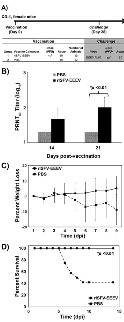
Similarly to the rISFV-VEEV study, cohorts of CD-1 mice were either sham vaccinated
or vaccinated i.m. with 10
8PFU/mouse of rISFV-EEEV (Fig. 6A). Neutralizing antibody
responses were measured via an 80% plaque reduction neutralization titer (PRNT
80) test
at 14 and 21 days postvaccination (Fig. 6A). All sham-vaccinated mice remained
seronegative (PRNT
80of
⬍
20), whereas 80% and 100% of the vaccinated mice had a
PRNT
80of
ⱖ
20 at 14 and 21 days postvaccination, respectively (Fig. 6B). At 28 days
postvaccination, mice were challenged s.c. with 10
4PFU/mouse of EEEV (Fig. 6A).
Sham-vaccinated mice lost weight beginning at day 5 postchallenge, continuing until
euthanasia criteria were met (Fig. 6C). Similarly to previous studies, weight loss was not
as pronounced as that with VEEV challenge, occurring closer to the time of euthanasia,
which was performed once mice became moribund. Nonetheless, the sham-vaccinated
mice succumbed to disease starting at day 6 postchallenge, and 60% met euthanasia
criteria (Fig. 6D), while none of the vaccinated mice showed any signs of disease. These
data demonstrated that an attenuated rISFV vector(s) expressing E3-E1 antigens could
protect mice against lethal VEEV and EEEV challenges following a single i.m.
vaccina-tion.
FIG 1Midpoint-rooted maximum likelihood tree based on nucleotide sequences of the N, G, and L genes. Bootstrap values of⬎75% are shown at internal nodes. The bar represents nucleotide substitutions per site.
on November 7, 2019 by guest
http://jvi.asm.org/
[image:6.585.40.407.69.377.2]Efficacy of bivalent rISFV-VEEV and -EEEV vaccines against lethal alphavirus
challenge.
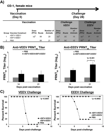
To assess the ability of rISFV vectors to provide protection against multiple
alphaviruses, the rISFV-VEEV and -EEEV vectors were blended into a bivalent
formula-tion, and cohorts of 20 CD-1 mice (VEEV) or 21 CD-1 mice (EEEV) were either sham
vaccinated or vaccinated i.m. with 10
8PFU/mouse of each vector, followed by lethal
VEEV and EEEV challenges (10 mice [VEEV] or 11 mice [EEEV] per challenge virus) (Fig.
7A). The neutralizing antibody response was measured via a PRNT
80assay at 14 and 21
days postvaccination. Similarly to the study described above, VEEV PRNT
80titers of
ⱖ
20
were detected in 60% and 80% of mice at days 14 and 21 postvaccination, respectively
(Fig. 7B). EEEV-neutralizing antibody responses were also detected at both time points
in all mice, whereas all sham-vaccinated mice had PRNT
80values below the limit of
detection (Fig. 7B). Following VEEV challenge, all 10 sham-vaccinated mice succumbed
to infection and were euthanized within 7 days of challenge, while vaccinated mice
remained free of detectable disease (Fig. 7C). Sensitivity to lethal EEEV infection was
increased by challenging mice with 10
5PFU/mouse via the intraperitoneal (i.p.) route.
Following EEEV challenge, the majority (9/11 animals) of sham-vaccinated mice
suc-FIG 2(A) Schematic diagram of the strategy utilized to generate the rISFV genomic cDNA clone. The full-length viral genome was cloned in 5 fragments flanked by unique restriction sites. The 3=leader and 5=trailer were flanked by the T7 RNA polymerase promoter and terminator, respectively. The hepatitis delta virus ribozyme sequence was used to generate a precise 5=trailer nucleotide sequence. (B) Outline of a helper-virus-free method of rescuing infectious rISFV. The T7 RNA polymerase and full-length and support rISFV plasmids were electroporated into Vero cells, and infectious virus was recovered at 5 to 10 days postelectroporation.
on November 7, 2019 by guest
http://jvi.asm.org/
[image:7.585.58.352.72.459.2]cumbed to infection and were euthanized within 7 days of challenge, while vaccinated
mice remained free of detectable disease (Fig. 7C). These data demonstrated that a
single dose of the blended rISFV-VEEV and -EEEV vaccines can provide complete
protection from lethal VEEV and EEEV challenges.
Short- and long-term efficacies of rVSIV and rISFV vaccine vectors against
lethal challenge.
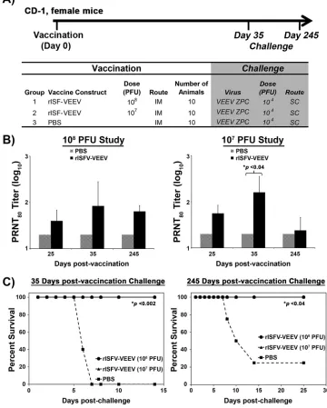
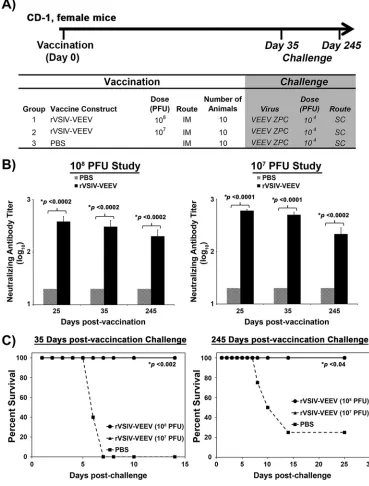
Short- and long-term protection induced by rISFV- and rVSIV-VEEV
vectors was assessed by vaccinating cohorts of 10 CD-1 mice with 10
8and 10
7PFU/mouse of each vaccine vector, followed by lethal VEEV challenge at days 35 and
245 postvaccination (Fig. 8A and 9A). Antibody responses were measured via a PRNT
80assay at days 25, 35, and 245 postvaccination. Neutralizing antibodies were detected in
FIG 3Schematic diagrams of the genetic organization and nomenclature of rISFV and rVSIV vectors. The N gene of rISFV-VEEV, rISFV-EEEV, and rVSIV-VEEV vectors was relocated to the fourth position of the genome. The rVSIV-VEEV vector also has a 28-amino-acid truncation in the cytoplasmic tail of the G protein. All foreign gene expression cassettes were added at genome position 5.
FIG 4In vitrocharacterization of rISFV vectors. (A) Western blot analysis of rISFV and rVSIV vectors expressing VEEV or EEEV E3-E1 proteins in Vero cells. Replicate Vero cell monolayers in six-well plates were infected at an MOI of 5 PFU/cell, and cell lysates were collected at 24 hpi. The E3-E1 proteins were detected with mouse polyclonal antisera against VEEV and EEEV. (B and C) Plaque phenotype (B) and replication kinetics (C) of rISFV and rVSIV vectors in Vero cells. The plaque phenotype was assessed in Vero cell monolayers infected with wt ISFV, wt VSIV, rISFV, and rVSIV vectors, and at 2 to 3 days postinfection, the cells were fixed and stained with crystal violet. Assays of replication kinetics (C) of all viruses were performed at an MOI of 10 PFU/cell in triplicate. Average titers⫾ standard deviations (error bars) are shown.*,Pvalues of⬍0.02.
on November 7, 2019 by guest
http://jvi.asm.org/
[image:8.585.49.365.71.206.2] [image:8.585.42.399.418.662.2]mice at all time points postvaccination regardless of the virus vector or dose. PRNT
80values of
ⱖ
20 were detected in 80 to 100% of vaccinated mice, and titers were
maintained for the duration of the study (Fig. 8B and 9B). Average PRNT
80values
induced by rISFV-VEEV following either dose at 25 and 35 days postvaccination ranged
from 40 to 160 and were reduced to 25 to 64 at day 245 (Fig. 8B and 9B), while
rVSIV-VEEV-induced PRNT
80titers ranged from 288 to 600 at days 25 and 35
postvac-cination and 304 to 360 at day 245 postvacpostvac-cination (Fig. 8B and 9B). All vaccinated mice
challenged at day 35 or 245 postvaccination with a lethal VEEV dose were protected
from disease by both vectors at both dose levels, whereas 100% (day 35) and 75% (day
245) of the sham-vaccinated mice succumbed to infection and were euthanized within
6 to 14 days of challenge (Fig. 8C and 9C). These data demonstrated that both the
attenuated rISFV and rVSIV vaccine vectors could elicit a durable neutralizing antibody
response and provided single-dose short- and long-term protection against lethal VEEV
challenge.
DISCUSSION
Various forms of rVSV have been developed as vaccine vectors and oncolytic agents,
made possible by the pioneering work of John Rose and colleagues; one such highly
attenuated rVSV vector recently demonstrated safety and immunogenicity in clinical
trials (42, 55–61). Here we report the rescue of rISFV from cDNA, modification of the
rISFV genome to attenuate potential virulence and to express VEEV and EEEV E3-E1
FIG 5Efficacy of the rISFV-VEEV vector in CD-1 mice. (A) Outline of the study design. (B and C) Percent weight loss (B) and survival (C) following lethal VEEV-ID challenge via the s.c. route. Average weight loss⫾standard deviations (error bars) are shown. dpi, days postinfection.
on November 7, 2019 by guest
http://jvi.asm.org/
[image:9.585.103.305.71.430.2]antigens, and the protective efficacy of the resulting rISFV vaccine vectors in lethal
mouse models of VEEV and EEEV disease. A highly attenuated rVSIV vector, based on
the rVSIV-N4CT1gag1 vector, which demonstrated safety and immunogenicity in phase
I clinical trials, was also modified to express VEEV and EEEV E3-E1 antigens. This vaccine
candidate was included in our studies as a comparator to the novel rISFV vector(s) and
as a candidate stand-alone vaccine(s) to prevent alphavirus-induced disease.
The primary reasons for selecting ISFV as a potential vaccine vector were the
FIG 6Immunogenicity and efficacy of the rISFV-EEEV vector in CD-1 mice. (A and B) Outline of the study design (A) and neutralizing antibody response measured by a PRNT80 assay following rISFV-EEEV vaccination (B). (C and D) Percent weight loss (C) and survival (D) following lethal EEEV-NA challenge via the s.c. route. Average PRNT80values and weight loss⫾standard deviations (error bars) are shown.
on November 7, 2019 by guest
http://jvi.asm.org/
[image:10.585.102.303.65.586.2]phylogenetic distance from VSIV and known serological differences from VSIV and
other vesiculoviruses (2, 3). The inability of serum from VSV-infected mice to neutralize
ISFV and the divergence of the protein sequences between both viruses, reducing the
likelihood of cross-reactive T cell epitopes, should allow rISFV vaccine vectors to elicit
immune responses when used either as a booster after priming with rVSIV expressing
autologous antigens or as a stand-alone vaccine vector for protection against
heterol-ogous disease to be used in people previously vaccinated with rVSIV vectors (2). The
studies described here clearly demonstrate that rISFV vectors can be utilized as a
stand-alone vaccine platform, and we are in the process of investigating the
immuno-genicity and protective efficacy of rISFV vectors in the context of preexisting rVSV
immunity. Preliminary prime-boost studies with rVSV and rISFV vectors expressing an
immunodominant HIV-1 Gag epitope suggest that immune responses elicited by rISFV
vectors will not be blunted in mice with preexisting rVSIV immunity (our unpublished
FIG 7Immunogenicity and efficacy of blended rISFV-VEEV/rISFV-EEEV vectors. (A) Outline of the study design in CD-1 mice. (B) Neutralizing antibody response measured by a PRNT80assay following vaccination. (C) Survival following lethal VEEV-ID or EEEV-NA challenge via the s.c. or i.p. route, respectively. Average PRNT80values⫾ standard deviations (error bars) are shown.
on November 7, 2019 by guest
http://jvi.asm.org/
[image:11.585.41.402.72.528.2]data). Also, preliminary neurovirulence studies in young mice indicated that
unmodi-fied wt ISFV was 1,000-fold more attenuated than wt VSIV, based on the amount of
infectious virus required to provide a measureable 50% lethal dose (LD
50), presumably
due to inherently lower-virulence properties and likely reducing the need for extensive
attenuation compared to rVSV vectors (our unpublished data). The latter is also
supported by the absence of any clinical signs of disease after vaccination of mice. The
significance of this natural murine attenuation for humans is unknown, but greater
sensitivity to innate immune responses
in vivo
could be responsible, given that
un-modified rISFV and rVSV grew robustly and equally well in Vero cell monolayers. The
anticipated safety of rISFV and attenuated rISFV vectors is supported by the
serocon-version of people in regions of endemicity, presumably as a result of being bitten by an
infected insect, without reported illness (3–5, 62).
The recovery of virus entirely from cDNA enabled the evaluation of rISFV as a new
FIG 8Dose titration and duration of efficacy of the rISFV-VEEV vector. (A) Outline of the study design in CD-1 mice. Animals were vaccinated with 108or 107PFU and challenged with lethal VEEV-ID at 35 and 245 days postinfection. (B) Neutralizing antibody response measured by a PRNT80assay following vaccination. (C) Survival following lethal VEEV-ID challenge via the s.c. route. Average PRNT80values⫾standard deviations (error bars) are shown.
on November 7, 2019 by guest
http://jvi.asm.org/
[image:12.585.39.399.80.529.2]vaccine vector. The alphaviruses VEEV and EEEV were selected as vaccine targets
because both viruses cause serious and sometimes fatal disease in humans, and there
are currently no licensed vaccines available to protect against either virus. Although
rISFV was believed to be less virulent than rVSIV
in vivo
based on results from a
preliminary neurovirulence study in young mice, additional attenuation was sought by
N gene translocation, as previously reported for rVSIV (35, 40). The reduction of
in vitro
plaque sizes and the significant reduction in the level of progeny virus in replication
kinetics studies indicated that moving the N gene to position 4 in the genome (N4)
attenuates rISFV growth, although the potential contribution of VEEV/EEEV E3-E1
expres-sion to attenuation was not formally tested. However, the shuffling of the rISFV N gene
FIG 9Dose titration and duration of efficacy of the rVSIV-VEEV vector. (A) Outline of the study design in CD-1 mice. Animals were vaccinated with 108or 107PFU and challenged with lethal VEEV-ID at 35 and 245 days postinfection. (B) Neutralizing antibody response measured by a PRNT80assay following vaccination. (C) Survival following lethal VEEV-ID challenge via the s.c. route. Average PRNT80values⫾standard deviations (error bars) are shown.
on November 7, 2019 by guest
http://jvi.asm.org/
[image:13.585.40.409.75.561.2]produced results similar to those previously obtained for rVSIV vectors, with and
without the expression of a foreign gene, that greatly attenuate vectors both
in vitro
and
in vivo
(35, 40, 41). Genes encoding the VEEV/EEEV E3-E1 proteins were chosen for
expression by rISFV vectors because they contain major virus neutralization epitopes
(1). The complete E3-E1 polyprotein was expressed from a single rISFV transcription unit
in each case to allow natural proteolytic processing, E2/E1 heterodimer folding, and,
presumably, authentic structural presentation on the surface of infected cells and rISFV
particles. The additional expression cassette was positioned in the fifth position of the
rISFV vector in each case to limit the amount of the E3-E1 protein expressed and
thereby to reduce the possibility of toxicity to rISFV replication, as has been observed
with some other foreign proteins expressed by rVSIV. The highly attenuated rVSIV
vector expressing VEEV E3-E1 was based on the rVSIV-N4CT1gag1 vector design, which
contains two major attenuating mutations, N gene translocation to genome position 4
(N4) and a truncation of the G protein cytoplasmic tail (CT) from 29 amino acids to a
single amino acid (CT1), and has demonstrated safety and immunogenicity in phase I
clinical trials (39–42). The attenuation of rISFV vectors may produce lower yields of
rISFV-based vaccines; however, strategies have been developed to enhance the yields
of highly attenuated rVSIV vectors during good manufacturing practice (GMP)
produc-tion of clinical trial materials (CTM), and it is very likely that similar strategies can be
utilized to overcome any low-yielding rISFV vectors.
Western blot analysis indicated that the level of rISFV-VEEV E3-E1 protein expression
was lower than that expressed by rVSIV-VEEV; the reason for this is not clear but could
involve reduced transcription initiation efficiency at the ISFV 3
=
promoter relative to
that of VSIV or differences in the attenuation of transcription across intergenic regions.
However, a single i.m. dose of either rISFV-VEEV or rISFV-EEEV elicited neutralizing
antibody responses and provided complete protection from disease following lethal
challenge with VEEV or EEEV, respectively. Complete protection was also observed
when both the rISFV-VEEV and rISFV-EEEV vaccine vectors were blended together and
administered as a bivalent vaccine formulation, indicating little or no interference
between vectors and demonstrating the potential for a multivalent alphavirus vaccine.
Neutralizing antibodies are generally considered to be the correlate of protection
against alphavirus infection, and our results are in agreement with data from previous
reports. Several studies have suggested the potential role of nonneutralizing antibodies
and/or T cell-mediated immunity; however, their role is not well studied or understood
(63–66). Consequently, we did not perform analyses of formal correlates of protection
and used neutralizing antibody levels as a measure of immunogenicity. Both the rISFV
and rVSIV vectors were able to induce neutralizing antibody responses when
admin-istered as either a single or a blended regimen. The PRNT
80values were similar when
rISFV vectors were administered as single or blended vaccines, although VEEV titers
were overall slightly lower when administered as a blended vaccine. The
coadminis-tration of multiple alphavirus vaccines can result in interference between vaccine
constituents, influencing the immunological readout for each vaccine component, and
the very modest reduction in titers observed here may be a reflection of this
phenom-enon (64, 67–70). The rISFV and rVSIV dose-down study (10
7and 10
8PFU) indicated that
a more durable neutralizing antibody response may be elicited by a higher vaccine
dose, but protection from disease was still achieved following challenge at 245 days
postvaccination, when neutralizing antibody levels had decreased in the cohort
receiv-ing 10
7PFU to levels indistinguishable from those in unvaccinated mice, presumably
due to the rapid expansion of E3-E1-specific memory B cells after challenge.
The rVSIV-VEEV vector also demonstrated 100% protection of mice from lethal VEEV
challenge following a single i.m. vaccine dose. The more robust expression of the VEEV
E3-E1 protein observed by Western blot analysis may explain the induction of a greater
neutralizing antibody response by rVSIV-VEEV than by rISFV-VEEV, although rVSIV may
also be an inherently more immunogenic vector than rISFV.
In summary, we have introduced rISFV as a new vaccine vector platform and
demonstrated that rISFV may be attenuated by one of the strategies used for the
on November 7, 2019 by guest
http://jvi.asm.org/
attenuation of rVSIV. The attenuated rISFV-VEEV, rISFV-EEEV, and rVSIV-N4CT1 VEEV
vectors provided multivalent, single-dose, durable protection of mice from disease
following challenge with a lethal dose of EEEV or VEEV. Either of these vectors may
provide the pathway to a safe and efficacious vaccine(s) to combat serious disease
caused by the encephalitic alphaviruses.
MATERIALS AND METHODS
Viruses and cells.The VSIV San Juan isolate, ISFV, VEEV ZPC 738, and EEEV FL93-939 were obtained from the World Reference Center for Emerging Viruses and Arboviruses at the University of Texas Medical Branch (UTMB).
The Vero cell line was obtained from the American Type Culture Collection (Bethesda, MD). Vero cells were propagated at 37°C with 5% CO2in Dulbecco’s minimal essential medium (DMEM) containing 10% (vol/vol) fetal bovine serum (FBS), sodium pyruvate (1 mM), 1% (vol/vol) nonessential amino acids, and gentamicin (50g/ml).
Phylogenetic analysis.Sequences of the N, G, and L genes were downloaded from GenBank (Table 1) and aligned in SeaView by utilizing the MUSCLE algorithm (71, 72). The sequences were aligned by using the deduced amino acid sequence from open reading frames (ORFs) and then returned to nucleotide sequences for subsequent analyses. Maximum likelihood (ML) analysis was performed by utilizing the PHYLIP package (73). Modeltest in PAUP was used to identify the best-fit nucleotide substitution model, the GTR⫹I⫹G model (where GTR is general time reversible, I refers to invariant sites, and G is the gamma distributed rate variation among sites) (74). The robustness of the ML phylogeny was evaluated by bootstrap resampling with 500 replicates.
Generation of rISFV genomic cDNA.An infectious rISFV cDNA clone was generated by amplifying the genome in five DNA fragments flanked by compatible unique restriction enzyme sites (Fig. 2A). The full-length ISFV genome nucleotide sequence was determined from viral RNA isolated from the cell culture supernatant by using the QIAamp viral RNA minikit (Qiagen, Alameda, CA); cDNA for nucleotide sequencing and assembly of a full-length genomic cDNA was generated by reverse transcription (RT) utilizing the Superscript III system (Invitrogen, Carlsbad, CA), using random hexamers. PCR products were generated from this cDNA with Phusion high-fidelity DNA polymerase (NEB) by utilizing ISFV-specific primers. Amplified PCR products were purified by using a QIAquick gel extraction kit (Qiagen) and sequenced by using ABI Prism BigDye Terminator 1.1 cycle sequencing kits, and the reaction products were analyzed on an ABI Prism 3700 DNA analyzer (Invitrogen). Each cDNA fragment was cloned into pBluescript (Invitrogen) in a stepwise manner to generate full-length genomic cDNA. The cDNA nucle-otide sequence was verified after each successive round of cloning and after the assembly of a complete genome cDNA, to ensure a match to the viral genome consensus sequence. All full-length cDNA clones and viruses were verified via Sanger sequencing using ABI Prism BigDye Terminator 1.1 cycle sequencing kits on an ABI Prism 3130 or 3700 DNA analyzer (Invitrogen).
Generation of attenuated rISFV and rVSIV vectors.A method for the generation of the highly attenuated N4CT1gag1 vector was described in detail previously (52). By using the rVSIV-N4CT1gag1 cDNA as the starting template, the positions of the N gene and the GCT-1gene were switched, and the expression cassette in position 1, expressing the HIVgaggene, was relocated to position 5 between the N and L genes of this vector. These manipulations preserved the N4CT1 attenuating mutations while creating an expression cassette at position 5 for the insertion of the VEEV E2/E1 ORF in place of thegaggene. The rISFV genomic cDNA was also manipulated in a similar fashion to translocate the N gene from position 1 to position 4 in the genome, and an expression cassette was then inserted at position 5 between the N and the L genes to accommodate VEEV and EEEV E2/E1 ORFs.
Generation of rISFV and rVSIV vectors expressing alphavirus antigens.As outlined above, cDNA clones of rISFV-N4 and rVSIV-N4CT1 were engineered to contain discrete transcriptional start and stop signals flanked by XhoI and NotI restriction sites, respectively, in the fifth position of the viral genome. The E3-E2-6k-E1 ORFs of the North American-lineage EEEV strain FL93-939 (EEEV-NA) and VEEV subtype ID strain ZPC 738 (VEEV-ID) were inserted into the fifth position of rISFV-N4, and the E3-E2-6k-E1 ORF of VEEV subtype ID was inserted into the fifth position of rVSIV-N4CT1 (Fig. 3).
Vaccine vector nomenclature.Diagrams of rISFV and rVSIV vector genomes are shown in Fig. 3. The positions of N gene and alphavirus E3-E1 ORFs are given relative to those of the wild-type virus genome. The vaccine vectors are referred to throughout the manuscript as rISFV-EEEV (rISFV-N4 EEEV NA FL93-939), rISFV-VEEV (rISFV-N4 VEEV subtype ID strain ZPC 738), HIV (HIVgag5), and rVSIV-VEEV (rVSIV-N4CT1 rVSIV-VEEV subtype ID strain ZPC).
Rescue of rISFV and rVSV vectors.All rISFV and rVSIV vectors were rescued by electroporation of cDNA plasmids into Vero cells (Fig. 2B). Plasmid DNAs encoding T7 RNA polymerase or rVSIV or rISFV full-length genomic RNA and support plasmids encoding viral N, P, M, G, and L proteins were combined as follows for each electroporation: 50g T7, 12g genomic cDNA, 12g N, 4g P, 2g M, 2g G, and 1g L. The plasmid DNA mixture was ethanol precipitated, collected by centrifugation, washed with 70% ethanol, dried, and resuspended in 250l of sterile deionized water.
In preparation for electroporation, Vero cells were seeded overnight to obtain ⬃95% confluent monolayers in a T-150-cm2flask for each electroporation. The monolayers were then trypsinized into single-cell suspensions, followed by the addition of 5 ml of 0.1% (wt/vol) soybean trypsin inhibitor (Invitrogen). Cells were collected by centrifugation and then washed with 20 ml of 1⫻DMEM containing 10% (vol/vol) fetal bovine serum (FBS), sodium pyruvate (1 mM), 1% (vol/vol) nonessential amino acids, and 0.4% 2-mercaptoethanol (Invitrogen). Cells were collected by centrifugation, resuspended, and
on November 7, 2019 by guest
http://jvi.asm.org/
washed in 20 ml of 1⫻DMEM containing sodium pyruvate (1 mM), 0.4% 2-mercaptoethanol (Invitrogen), 1% (vol/vol) nonessential amino acids, and dimethyl sulfoxide (DMSO) (Corning). Cells were collected by centrifugation and resuspended at⬃107cells per 700l in 1⫻DMEM containing sodium pyruvate (1 mM), 0.4% 2-mercaptoethanol, 1% (vol/vol) nonessential amino acids, and DMSO. The DNA mixture (250 l) and Vero cells (700l) were then mixed by gentle pipetting, and 750l of the mixture was placed into 4-mm electroporation cuvettes and immediately electroporated (BTX, ECM-830 Electro Square Porator; Harvard Apparatus Inc., Holliston, MA). Electroporation was performed at 140 V, the pulse length was 70 ms, the interval between pulses was 500 ms, and the number of pulses was 4. Following electroporation, cells were washed and resuspended in 20 ml of 1⫻DMEM containing 10% (vol/vol) FBS, sodium pyruvate (1 mM), and 1% (vol/vol) nonessential amino acids. Cells were incubated at 37°C with 5% CO2for 3 h, followed by a heat shock at 43°C with 5% CO2for 4 h. Following heat shock, cells were placed at 37°C with 5% CO2, and at 24 h postelectroporation, medium was replaced with Vero cell growth medium. Medium was replaced at 2 days postelectroporation with 1⫻DMEM containing 2% (vol/vol) FBS, sodium pyruvate (1 mM), 1% (vol/vol) nonessential amino acids, and gentamicin (50g/ml). Flasks were monitored for CPE for 14 days postelectroporation. For each rISFV and rVSIV construct, 6 to 12 electroporation replicates were performed.
Virus propagation, purification, and titration.Virus was routinely amplified and titrated on Vero cell monolayers. For virus amplification, cells were infected at a multiplicity of infection (MOI) of 0.5 PFU/cell. The virus inoculum was adsorbed for 15 min at room temperature, followed by 30 min at 37°C. Additional growth medium was then added, and cells were incubated at 37°C until CPE was observed. The infected cell supernatant was clarified by centrifugation at 3,000 ⫻g for 10 min at 4°C. The supernatant was then flash frozen in an ethanol– dry-ice bath and stored at⫺80°C prior to titration. Virus was further purified from the infected cell supernatant by centrifugation through 10% (wt/vol) sucrose in 1⫻phosphate-buffered saline (PBS). Briefly, 20 ml of the clarified cell supernatant was underlaid with 14 ml of 10% (wt/vol) sucrose in a Beckmann Ultraclear tube, followed by centrifugation at 28,000 rpm in a Beckmann SW-28 rotor for 1.5 h at 4°C. Following centrifugation, supernatants were aspirated, and the virus pellet was resuspended in PBS and then flash frozen and stored at⫺80°C prior to plaque assays. Virus titration was performed on 100% confluent Vero cell monolayers seeded overnight in six-well plates. Duplicate wells were infected with 0.1-ml aliquots of serially 10-fold-diluted virus in growth medium, 0.4 ml of growth medium was also added to each well to prevent cell desiccation, and virus was adsorbed for 15 min at room temperature and then for 30 min at 37°C. Following adsorption, the virus inoculum was aspirated, and cell monolayers were overlaid with 3 ml of 0.4% (wt/vol) Sea-Plaque agarose (Cambrex Bio Science) in growth medium. After 10 min at room temperature to allow the agarose to set, cells were incubated at 32°C with 5% CO2for 1 to 3 days to allow plaque development. Following incubation, the overlay was removed, and cells were stained with 2% crystal violet in 70% methanol for 5 min at room temperature; excess stain was removed under running water, and plaques were counted.
One-step growth kinetics. Virus growth kinetics were assessed on 100% confluent Vero cell monolayers seeded overnight in T-25-cm2 tissue culture flasks. Virus infections were performed in triplicate at an MOI of 10 PFU/cell, and virus was adsorbed for 15 min at room temperature, followed by 30 min at 37°C. Following incubation, the inoculum was removed, monolayers were rinsed five times with room-temperature DMEM to remove any unbound virus, and 5 ml of growth medium was added to each flask; 0.5-ml samples were taken immediately afterwards, designated “T 0” samples, and replaced with 0.5 ml of fresh medium. Flasks were incubated at 37°C, and additional samples were taken at 6, 9, 12, 24, and 48 hpi. All samples were flash frozen in ethanol-dry ice and stored at⫺80°C.
Western blotting.Replicate confluent Vero cell monolayers in six-well plates were infected at an MOI of 5 PFU per cell. The virus inoculum was adsorbed for 15 min at room temperature, followed by 30 min at 37°C. Additional growth medium was then added, and cells were incubated at 37°C under 5% CO2for 24 h. At 24 hpi, cells were scraped into the suspension and collected by centrifugation at 3,000⫻gfor 10 min at 4°C. The supernatant was removed, and cell pellets were treated with 0.5 ml of lysis buffer (0.05 M Tris-HCl [pH 7.5], 0.01 M NaCl, 1⫻Triton X-100). Cell lysates were then diluted 1:1 in Laemmli sample buffer (Bio-Rad) and heated at 90°C for 5 min to denature proteins. Samples were electrophoresed on 4 to 12% Bis-Tris-polyacrylamide gel electrophoresis gels (NuPAGE) with a Precision Plus protein standard (Bio-Rad), and proteins were then transferred onto a nitrocellulose membrane by using the iBlot system (Invitrogen). Mouse polyclonal antisera against VEEV and EEEV were obtained from the World Reference Center for Emerging Viruses and Arboviruses at the University of Texas Medical Branch. Antisera were diluted 1:1,000 in diluent provided in the Western Breeze anti-mouse chromogenic kit (Invitrogen). Color was allowed to develop after the addition of a chromogenic substrate provided in the Western Breeze kit, and the nitrocellulose membrane was then rinsed with distilled H2O and air dried.
Plaque reduction neutralization test.Serum samples were heat inactivated at 56°C for 30 min. Samples were serially diluted 2-fold in a solution containing DMEM, 2% FBS, and gentamicin (50g/ml); mixed with an equal volume of 2,000 PFU/ml of VEEV ZPC or EEEV FL93; and incubated for 1 h at 37°C. Vero cell monolayers in 6-well plates were then inoculated with 100l of the serum-virus mixture in triplicate. Anti-VEEV- or EEEV-positive sera, and medium-only controls, and virus-only controls were included in the assay. Plates were incubated at 37°C with 5% CO2for 3 days and then fixed and stained with crystal violet as described above. PRNT80values were calculated and expressed as the reciprocal of the serum dilution yielding a⬎80% reduction (PRNT80) in the number of plaques.
Mouse vaccination and challenge.All studies were carried out in accordance with the recommen-dations in theGuide for the Care and Use of Laboratory Animalsof the National Institutes of Health and were approved by Institutional Animal Care and Use Committee protocol 02-09-068 at UTMB (75). CD-1 outbred female mice, 4 to 6 weeks old, from Charles River (Wilmington, MA) were utilized in the studies.
on November 7, 2019 by guest
http://jvi.asm.org/
Mice received 108or 107PFU of either the rVSIV or rISFV vaccine vector in 50l of PBS by i.m. injection. Vaccinated mice were challenged with a lethal dose (104PFU) of either VEEV-ID or EEEV-NA by s.c. injection. Based on the outcome of the first pilot EEEV vaccine study, mice were subsequently challenged with 105PFU by i.p. injection. Mice were observed daily for signs of illness and were euthanized with the onset of neurological disease, consistent with previous VEEV and EEEV challenge studies (76, 77).
Statistics.RStudio (version 0.97; RStudio, Boston, MA) running R (version 3.0.1; R Development Core Team, Vienna, Austria) software was used for statistical analysis. Significant differences in mean titers between wt viruses and attenuated vaccine constructs were determined by using two-way analysis of variance (ANOVA) for all viruses, followed by a Tukey test. A two-tailed Fisher exact test was performed to determine significant differences in survival rates.
ACKNOWLEDGMENTS
This work was supported by National Institutes of Health grant AI120942.
F.N., D.M., R.L.S., T.L., R.V.G., R.M.N., S.H., J.H.E., R.B.T., D.K.C., and S.C.W. have a patent
pending on an Isfahan virus vaccine platform.
The views expressed in this article are those of the authors and do not reflect the
official policy or position of the U.S. Department of Defense or the Department of the
Army.
REFERENCES
1. Knipe DM, Howley PM, Cohen JI, Griffin DE, Lamb RA, Martin MA, Racaniello VR, Roizman B (ed). 2013. Fields virology, 6th ed, vol I. Lippincott Williams & Wilkins, Philadelphia, PA.
2. Tesh RB, Travassos Da Rosa AP, Travassos Da Rosa JS. 1983. Antigenic relationship among rhabdoviruses infecting terrestrial vertebrates. J Gen Virol 64(Part 1):169 –176.
3. Tesh R, Saidi S, Javadian E, Loh P, Nadim A. 1977. Isfahan virus, a new vesiculovirus infecting humans, gerbils, and sandflies in Iran. Am J Trop Med Hyg 26:299 –306.
4. Gaidamovich S, Obukhova VR, Sveshnikova NA, Cherednichenko IUN, Kostiukov MA. 1978. Natural foci of viruses borne by Phlebotomus papatasi in the USSR according to a serologic study of the population. Vopr Virusol 1978:556 –560. (In Russian.)
5. Gaidamovich S, Altukhova LM, Obukhova VR, Ponirovskii EN, Sadykov VG. 1980. Isolation of the Isfahan virus in Turkmenia. Vopr Virusol 1980:618 – 620. (In Russian.)
6. Wilks CR, House JA. 1986. Susceptibility of various animals to the ve-siculoviruses Isfahan and Chandipura. J Hyg (Lond) 97:359 –368.https:// doi.org/10.1017/S002217240006544X.
7. Cotton WE. 1926. The causal agent of vesicular stomatitis proved to be a filter-passing virus. J Am Vet Med Assoc 23:168 –179.
8. Letchworth GJ, Rodriguez LL, Del Cbarrera J. 1999. Vesicular stomatitis. Vet J 157:239 –260.https://doi.org/10.1053/tvjl.1998.0303.
9. Johnson KM, Vogel JE, Peralta PH. 1966. Clinical and serological response to laboratory-acquired human infection by Indiana type vesicular sto-matitis virus (VSV). Am J Trop Med Hyg 15:244 –246.
10. Fields BN, Hawkins K. 1967. Human infection with the virus of vesicular stomatitis during an epizootic. N Engl J Med 277:989 –994.https://doi .org/10.1056/NEJM196711092771901.
11. Ball LA, White CN. 1976. Order of transcription of genes of vesicular stomatitis virus. Proc Natl Acad Sci U S A 73:442– 446.https://doi.org/ 10.1073/pnas.73.2.442.
12. Ge P, Tsao J, Schein S, Green TJ, Luo M, Zhou ZH. 2010. Cryo-EM model of the bullet-shaped vesicular stomatitis virus. Science 327:689 – 693.
https://doi.org/10.1126/science.1181766.
13. Emerson SU, Yu Y. 1975. Both NS and L proteins are required for in vitro RNA synthesis by vesicular stomatitis virus. J Virol 15:1348 –1356. 14. Emerson SU, Wagner RR. 1972. Dissociation and reconstitution of the
transcriptase and template activities of vesicular stomatitis B and T virions. J Virol 10:297–309.
15. Abraham G, Banerjee AK. 1976. Sequential transcription of the genes of vesicular stomatitis virus. Proc Natl Acad Sci U S A 73:1504 –1508.https:// doi.org/10.1073/pnas.73.5.1504.
16. Schnell MJ, Mebatsion T, Conzelmann KK. 1994. Infectious rabies viruses from cloned cDNA. EMBO J 13:4195– 4203.
17. Lawson ND, Stillman EA, Whitt MA, Rose JK. 1995. Recombinant vesicular stomatitis viruses from DNA. Proc Natl Acad Sci U S A 92:4477– 4481.
https://doi.org/10.1073/pnas.92.10.4477.
18. Whelan SP, Ball LA, Barr JN, Wertz GT. 1995. Efficient recovery of
infec-tious vesicular stomatitis virus entirely from cDNA clones. Proc Natl Acad Sci U S A 92:8388 – 8392.https://doi.org/10.1073/pnas.92.18.8388. 19. Schnell MJ, Buonocore L, Whitt MA, Rose JK. 1996. The minimal
con-served transcription stop-start signal promotes stable expression of a foreign gene in vesicular stomatitis virus. J Virol 70:2318 –2323. 20. Chattopadhyay A, Wang E, Seymour R, Weaver SC, Rose JK. 2013. A
chimeric vesiculo/alphavirus is an effective alphavirus vaccine. J Virol 87:395– 402.https://doi.org/10.1128/JVI.01860-12.
21. Cobleigh MA, Buonocore L, Uprichard SL, Rose JK, Robek MD. 2010. A vesicular stomatitis virus-based hepatitis B virus vaccine vector provides protection against challenge in a single dose. J Virol 84:7513–7522.
https://doi.org/10.1128/JVI.00200-10.
22. Kahn JS, Roberts A, Weibel C, Buonocore L, Rose JK. 2001. Replication-competent or attenuated, nonpropagating vesicular stomatitis viruses expressing respiratory syncytial virus (RSV) antigens protect mice against RSV challenge. J Virol 75:11079 –11087.https://doi.org/10.1128/JVI.75.22 .11079-11087.2001.
23. Kapadia SU, Rose JK, Lamirande E, Vogel L, Subbarao K, Roberts A. 2005. Long-term protection from SARS coronavirus infection conferred by a single immunization with an attenuated VSV-based vaccine. Virology 340:174 –182.https://doi.org/10.1016/j.virol.2005.06.016.
24. Matassov D, Marzi A, Latham T, Xu R, Ota-Setlik A, Feldmann F, Geisbert JB, Mire CE, Hamm S, Nowak B, Egan MA, Geisbert TW, Eldridge JH, Feldmann H, Clarke DK. 2015. Vaccination with a highly attenuated recombinant vesicular stomatitis virus vector protects against challenge with a lethal dose of Ebola virus. J Infect Dis 212(Suppl 2):S443–S451.
https://doi.org/10.1093/infdis/jiv316.
25. Mire CE, Matassov D, Geisbert JB, Latham TE, Agans KN, Xu R, Ota-Setlik A, Egan MA, Fenton KA, Clarke DK, Eldridge JH, Geisbert TW. 2015. Single-dose attenuated Vesiculovax vaccines protect primates against Ebola Makona virus. Nature 520:688 – 691. https://doi.org/10.1038/ nature14428.
26. Natuk RJ, Cooper D, Guo M, Calderon P, Wright KJ, Nasar F, Witko S, Pawlyk D, Lee M, DeStefano J, Tummolo D, Abramovitz AS, Gangolli S, Kalyan N, Clarke DK, Hendry RM, Eldridge JH, Udem SA, Kowalski J. 2006. Recombinant vesicular stomatitis virus vectors expressing herpes sim-plex virus type 2 gD elicit robust CD4⫹Th1 immune responses and are
protective in mouse and guinea pig models of vaginal challenge. J Virol 80:4447– 4457.https://doi.org/10.1128/JVI.80.9.4447-4457.2006. 27. Palin A, Chattopadhyay A, Park S, Delmas G, Suresh R, Senina S, Perlin DS,
Rose JK. 2007. An optimized vaccine vector based on recombinant vesicular stomatitis virus gives high-level, long-term protection against Yersinia pestis challenge. Vaccine 25:741–750.https://doi.org/10.1016/j .vaccine.2006.08.010.
28. Roberts A, Kretzschmar E, Perkins AS, Forman J, Price R, Buonocore L, Kawaoka Y, Rose JK. 1998. Vaccination with a recombinant vesicular stomatitis virus expressing an influenza virus hemagglutinin provides complete protection from influenza virus challenge. J Virol 72: 4704 – 4711.