Submitted28 June 2018 Accepted 22 August 2018 Published17 October 2018
Corresponding author Cecília Pascelli,
Academic editor Mya Breitbart
Additional Information and Declarations can be found on page 15
DOI10.7717/peerj.5625
Copyright 2018 Pascelli et al.
Distributed under
Creative Commons CC-BY 4.0
OPEN ACCESS
Morphological characterization of
virus-like particles in coral reef sponges
Cecília Pascelli1,2,3, Patrick W. Laffy1, Marija Kupresanin4, Timothy Ravasi4and
Nicole S. Webster1,3,5
1Australian Institute of Marine Science, Townsville, Queensland, Australia
2James Cook University, Townsville, Queensland, Australia
3AIMS@JCU, Australian Institute of Marine Science and James Cook University, Townsville, Queensland,
Australia
4KAUST Environmental Epigenetic Program (KEEP), Division of Biological and Environmental Sciences &
Engineering, King Abdullah University of Science and Technology, Thuwal, Kingdom of Saudi Arabia 5Australian Centre for Ecogenomics, University of Queensland, Brisbane, Queensland, Australia
ABSTRACT
Marine sponges host complex microbial consortia that vary in their abundance, diversity and stability amongst host species. While our understanding of sponge-microbe interactions has dramatically increased over the past decade, little is known about how sponges and their microbial symbionts interact with viruses, the most abundant entities in the ocean. In this study, we employed three transmission electron microscopy (TEM) preparation methods to provide the first comprehensive morpho-logical assessment of sponge-associated viruses. The combined approaches revealed 50 different morphologies of viral-like particles (VLPs) represented across the different sponge species. VLPs were visualized within sponge cells, within the sponge extracellular mesohyl matrix, on the sponge ectoderm and within sponge-associated microbes. Non-enveloped, non-tailed icosahedral VLPs were the most commonly observed morphotypes, although tailed bacteriophage, brick-shaped, geminate and filamentous VLPs were also detected. Visualization of sponge-associated viruses using TEM has confirmed that sponges harbor not only diverse communities of microorganisms but also diverse communities of viruses.
SubjectsBiodiversity, Marine Biology, Microbiology, Virology, Biological Oceanography Keywords Transmission Electron Microscopy, Virus, TEM, VLP, Marine sponges, Great Barrier Reef, GBR, Red Sea
INTRODUCTION
environments (Pile & Young, 2006), especially in oligotrophic ecosystems such as coral reefs (De Goeij et al., 2013).
Sponges form intimate partnerships with diverse microbial consortia, and these relationships range from mutualism to commensalism to parasitism (Webster & Taylor, 2012; Thomas et al., 2016). The sponge microbiome is often highly conserved across individuals of the same sponge species but varies considerably across species (Thomas et al., 2016). It is because of these functionally important symbiotic partnerships that sponges are considered a typical example of a marine ‘holobiont’, an organism comprised of various ‘bionts’,living in symbiogenesis (Margulis & Fester, 1991; Webster & Thomas, 2016). However, while the symbiotic association between sponges and their bacterial/archaeal symbionts has been extensively studied (Schmitt et al., 2012;Thomas et al., 2016;Pita et al., 2018), the role of viruses in the sponge holobiont remain largely unknown, despite TEM images from the 1970s alluding to viral-infected sponge cells (Vacelet & Gallissian, 1978), a demonstration of phage infection in a sponge-associated bacterium (Lohr, Chen & Hill, 2005), and a few recent metagenomic studies providing insights into sponge virus diversity and function (Butina et al., 2015;Laffy et al., 2016;Laffy et al., 2018).
Viruses are the most abundant biological agents in marine ecosystems, with about 1010 viruses per liter of surface seawater and 1010 per gram dry weight of marine sediment (Suttle, 2007;Danovaro et al., 2011). Importantly, viruses have the ability to regulate the prokaryotic and eukaryotic populations responsible for maintaining metabolic cycling in complex ecosystems such as coral reefs (Seymour et al., 2005;Thurber & Correa, 2011;Mojica & Brussaard, 2014). Viruses modulate microbial-driven processes through mortality, horizontal gene transfer and metabolic reprogramming by viral-encoded auxiliary metabolic genes (AMGs) (Bergh et al., 1989;Rohwer & Thurber, 2009;Danovaro et al., 2011;Hurwitz et al., 2014;Breitbart et al., 2018). Recent years have seen an increased focus on the diversity and function of viruses associated with reef invertebrates including sea anemones (Wilson & Chapman, 2001); starfish (Hewson et al., 2018); scleractinian corals and their associated microbial communities (Patten, Harrison & Mitchell, 2008; Weynberg et al., 2014;Weynberg et al., 2017a;Laffy et al., 2018). However, while viruses have been described as essential components of coral reef ecosystems, capable of controlling microbial community dynamics, playing a role in coral bleaching/disease, and mediating reef biogeochemical cycling (Thurber et al., 2017), there is a paucity of research exploring viruses associated with ecologically important reef sponges.
microenvironments were also evident, with viral auxiliary genes being differentially represented across sponge species (Laffy et al., 2018).
While molecular approaches have substantially improved our understanding of viral-host interactions (Breitbart et al., 2002;Rosario & Breitbart, 2011;Laffy et al., 2016), biases associated with DNA/RNA extraction methods (Wood-Charlson et al., 2015) and the limited genomic resources available for most environmental viruses (Roux et al., 2015) can still constrain our understanding of host-associated viral ecology. Transmission electron microscopy (TEM) is a powerful approach that has helped to reveal the morphology and distribution of virus-like particles (VLPs) in many marine hosts as well as deciphering patterns of host-viral interactions (Wilson & Chapman, 2001;Patten, Harrison & Mitchell, 2008;Brum, Schenck & Sullivan, 2013;Pollock et al., 2014;Weynberg et al., 2017b). Here we use TEM to provide the first morphological characterization of viruses associated with 15 different coral reef sponge species and confirm the spatial localization of these VLPs within the sponge holobiont.
MATERIALS AND METHODS
Sponge collection and identification
Sampling was conducted on coral reefs of Orpheus Island, Great Barrier Reef, Australia (18◦3503400S, 146◦2805300E) and Al Fahal, Red Sea, Saudi Arabia (22◦1309500N, 39◦0108100E), between December 2015 and February 2016. Sampling in Australia was conducted under the Great Barrier Reef Marine Park Authority permit G12/35236.1, and sampling in Saudi Arabia was authorized by the Saudi Arabian coastguard as the study did not involve endangered or protected species.
Triplicate specimens of 15 sponge species were collected by scuba diving between three and 15 m depth. Two sponge species,Stylissa carteriandCarteriospongia foliascenswere found at both locations, and sampling was performed in triplicate at both sites. Sponge specimens were photographed in situbefore being individually placed within sterile FalconR
tubes and kept on ice until processing. All sampling materials were sterilized prior to and between each sampling. Morphological characterization of sponge species was performed as described in (Hooper & Van Soest, 2002) and DNA barcoding was additionally performed using mitochondrial cytochrome oxidase I (COI) gene primers and internal transcriber spacer 2 (ITS2) region of nuclear ribosomal DNA as described in (Erwin & Thacker, 2007;Andreakis, Luter & Webster, 2012;Wörheide et al., 2012). Sponge species are described inTable 1and can be seen inFig. S1.
Table 1 Collection details for all sponge species examined by TEM.GBR refers to the Great Barrier Reef collection site and RS refers to the Red Sea collection site.
Sponge species Location Depth (m)
Carteriospongia foliascens,P.S. Pallas (1766) GBR, RS 3–10
Stylissa carteri,A. Dendi (1889) GBR, RS 10–15
Xestospongiasp. GBR 5–15
Lamellodysidea herbacea,C. Keller (1889) GBR 5–10
Cymbastela marshae,J.N.A.Hooper & P.R. Bergquist (1992) GBR 10–15
Cinachyrella schulzei,C. Keller (1891) GBR 3–7
Pipestela candelabra, B. Alvarez et al. (2008) GBR 7–15
Echinochalina isaaci,J.N.A. Hooper (1996) GBR 7–15
Xestospongia testudinaria,J.B.P. Lamarck (1815) RS 7–15
Amphimedon ochracea,C. Keller (1889) RS 7–15
Hyrtios erectus,C. Keller (1889) RS 5–15
Crella (Grayela) cyathophora,H.J. Carter (1869) RS 7–15
Mycalesp. RS 5–15
Preparation of ultrathin sections of sponge tissue
Histological sections were prepared from fresh sponge tissue based on standard procedures for TEM (Cheville & Stasko, 2014). Briefly, each fragment of approximately 1 mm3was fixed in 2.5% glutaraldehyde in 0.2M cacodylate buffer and kept at 4◦C for 2–24 h. After fixation, samples were immersed in 1% osmium tetroxide in 100 mM phosphate buffer for 1–2 h, washed in distilled water and stained in the dark with 2% aqueous uranyl acetate for 2 h at 4◦C. Stained tissue was dehydrated through a series of ethanol and propylene oxide then embedded in epoxy resin. Ectosome-choanosome oriented sections (about 65 nm thick) were prepared using a Leica EM UC7 ultramicrotome and placed on TEM copper grids.
Viral purification via density gradient solution
Viral purification was performed according to the fraction separation method by sedimentation in density gradients (Meselson, Stahl & Vinograd, 1957) following the pre-processing approach established to isolate viruses from coral and sponge tissue (Weynberg et al., 2014;Laffy et al., 2018). In order to eliminate contaminants present in the aquiferous system, sponges were partially dried via repeated gentle squeezing alternated with rinses of filtered (0.02µm) seawater. Sponge tissue was then dissected into small pieces (∼5 mm3) and covered with 15µL of 0.02µm filter-sterilized (Anotop, Whatman)
SM buffer (100 mM NaCl, 8 mM MgSO4, 50 mM Tris pH 7.5), then homogenized with a Craig’s HS30E homogenizer (Witeg, Germany) for 5 to 10 min (min). Tissue homogenate was filtered through a FalconR
centrifugal spin columns (Millipore, EUA) and centrifuged at 4,000 g for 30 min at 4◦C. This process was repeated four–six times per sample. Filter- sterilized SM Buffer was added to the concentrate and all flow-through was discarded. The concentrate was fixed in 0.5% glutaraldehyde and kept at 4◦C until TEM analysis. TEM preparation involved applying a droplet of sample onto a TEM Copper grid, rinsing with sterile water, staining with 1% uranyl acetate for one min, washing with sterile water, followed by removal of excess liquid from the grid by touching filter paper to the edge.
Viral purification via filtration of sponge mucus
To describe the VLPs associated with sponge mucus and the external ectoderm, the sponge surface was carefully scraped with a sterile scalpel blade followed by rinsing three times with filtered (0.02µm) seawater. This TEM preparation method was based on a viral purification method described for marine hydras (Grasis et al., 2014). Extracted mucus was added to filtered (0.02µm)Milli-QR
water (1:4) and centrifuged at 4,000 g for 10 min. Mucus supernatant was filtered through 0.45 µm filters (EMD Millipore, Burlington, CA, USA) and fixed in 1.5% glutaraldehyde. TEM imaging of mucus preparations was performed as described above for CsCl purified samples.
RESULTS
Sponge associated viruses
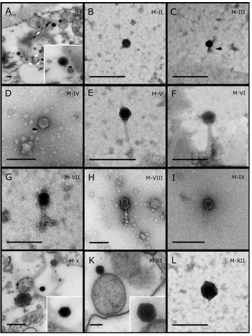
TEM analysis revealed that viral particles are diverse constituents of the sponge holobiont. Fifty VLP morphotypes (Figs. 1–5; Table S1; Morphotypes: M-I–M-L) were found in association with eight coral reef sponge species from the Great Barrier Reef: Carteriospongia foliascens,Stylissa carteri, Xestospongia sp., Pipestela candelabra,
Lamellodysidea herbacea, Cymbastella marshae, Echinochalina isaaci and Cinachyrella schulzei; and seven sponge species from the Red Sea:Carteriospongia foliascens,Stylissa carteri,Xestospongia testudinaria,Hyrtios erectus,Mycale sp.,Amphimedon ochraceaand
Crella cyathophora.VLPs were observed within sponge cells, in the extracellular mesohyl matrix, in the mucus/surface biofilm and within sponge-associated microbes. A diverse range of viral morphologies were observed, including hexagonal (tailed and non-tailed), spherical, filamentous, brick-shaped, beaded and geminate VLPs. While we detected numerous viral morphotypes, most were rare and often obscured by vesicles, cell debris and particulate organic matter.
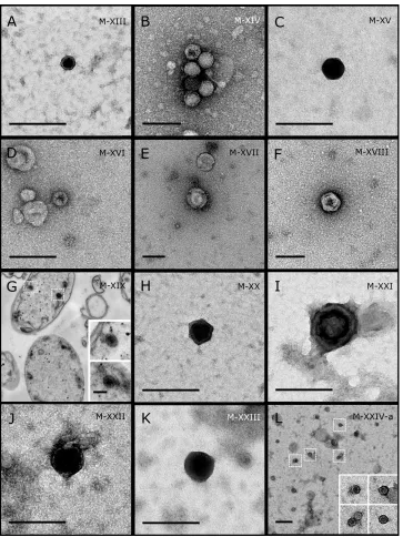
Most sponge-associated VLP morphotypes possessed an icosahedral/polyhedral symmetry (∼75%), ranging from 60–205 nm in diameter (Figs. 1– 3,4A). Tails were
evident on some VLPs, confirming the presence of viruses from the bacteriophage order
Caudovirales. Tailed VLPs were tentatively assigned to the three Caudoviralesfamilies based on their capsid symmetry and tail size/shape. VLPs characteristic of thePodoviridae
presented a short tail attached to a non-enveloped icosahedral capsid and these VLPs were observed in the spongesC. foliascens(Fig. 1A),Xestospongiasp. (Fig. 1B),E. isaaci(Fig. 1C) andS. carteri(Fig. 1D). VLPs characteristic of theSiphoviridaepresented an icosahedral head with a long non-contractile tailand these VLPs were detected in the surface biofilm of
Figure 1 Representative morphotypes of virus-like particles associated with GBR and Red Sea sponges.GBR sponge species: (A, J, K, L)C. foliascens,(B)Xestospongia sp., (C, F, G)E. isaaci, (E)
C. schulzei.Red Sea sponge species: (D, H)S. carteri, (K)Amphimedon ochracea. TEM preparation method: (A, J, K) ultrathin sections of sponge tissue, (B–I, L) viral purification via filtration of sponge mucus. Scale bar: 200 nm. Black arrows indicate the viral tail and white arrows indicate the VLPs.
Figure 2 Representative morphotypes of virus-like particles associated with GBR sponges.Sponge species: (A)C. foliascens,(B, C) Stylissa carteri, (D)Xestospongiasp., (E–H)Pipestela candelabra, (I–K)
Lamellodysidea herbacea, (L)C. schulzei.TEM preparation method: (A, H–K) viral purification via filtra-tion of sponge mucus, (B–F, L) viral purificafiltra-tion via CsCl gradient centrifugafiltra-tion, (G) ultrathin secfiltra-tions of sponge tissue. Scale bar: 200 nm.
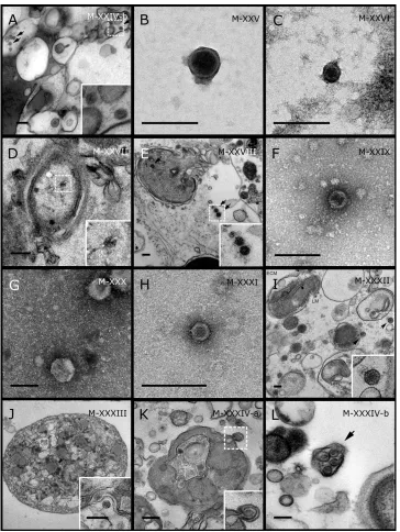
Figure 3 Representative morphotypes of virus-like particles associated with GBR and Red Sea sponges.GBR sponge species: (A, B)C. schulzei,(C)Cymbastella marshae.Red Sea sponge species: (D, E)C. foliascens, (F–H)S. carteri, (I)Xestospongia testudinaria, (J–L)Hyrtios erectus. TEM preparation method: (A, D–E, I–L) ultrathin sections of sponge tissue, (B, C, F–H) viral purification via filtration of sponge mucus. Scale bar: (A–C, E–L) 200 nm, (D) 500 nm. Black arrows indicate the VLPs.
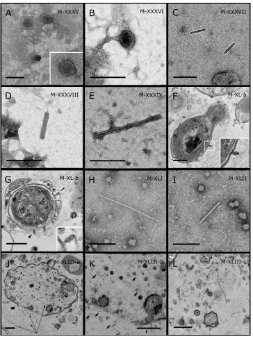
Figure 4 Representative morphotypes of virus-like particles associated with GBR and Red Sea sponges.GBR sponge species: (A)Mycalesp., (B)C. foliascens, (C, D)Xestospongiasp., (E)C. schulzei. Red Sea sponge species: (F, G)C. foliascens, (H, I)S. carteri, (J–L)Xestospongia testudinaria. TEM preparation method: (A, B, D, E, H, I) viral purification via filtration of sponge mucus, (C) viral purification via CsCl gradient centrifugation, (F, G, J–L) ultrathin sections of sponge tissue. Scale bar: (A–E, H, I) 200 nm, (F, G, J–L) 500 nm.
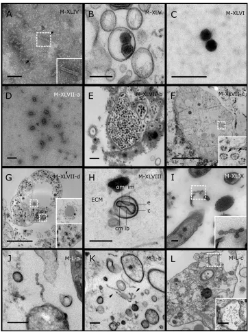
Figure 5 Representative morphotypes of virus-like particles associated with GBR and Red Sea sponges.GBR sponge species: (C)Lamellodysidea herbacea, (I)C. foliascens.Red Sea sponge species: (A, B, H)Crella cyathophora, (D–G)Amphimedon ochracea, (J–L)Hyrtios erectus. TEM preparation method: (A, C, D) viral purification via filtration of sponge mucus, (B, E–L) ultrathin sections of sponge tissue. Scale bar: (D) 100 nm, (A–C, E, H–L) 200 nm, (F, G) 5µm. ECM: External Cell Matrix, om: outer membrane, im: inner membrane, cm: core membrane, lb: lateral bodies; c: core, e: external membrane; b: bacterium. Black arrows indicate the VLPs.
and a longcontractile tailand these VLPs were observed in the spongesE. isaaci(Figs. 1F, 1G),S. carteri(Fig. 1H) andA. ochracea(Fig. 1I).
Non-tailed icosahedral/polyhedral VLPs were observed using all three TEM preparation methods. Particle sizes ranged from 60 to 205 nm in diameter and some presented an electron dense core inside the viral capsid (35–124 nm in diameter). The majority of VLPs did not show an envelope outside the capsid, however an envelope was observed in association with a small proportion of VLPs. (Figs. 2E,3K–3L,5H). A typical example of an enveloped VLP was observed inHyrtios erectuswhere a group of four virions were observed within a vacuole in the mesohyl matrix (Fig. 3L) and another free virion was captured merging its envelope into the cell membrane of the host (Fig. 3K).
In addition to the polyhedral VLPs, eight morphotypes of filamentous virus-like particles (FVLPs) were observed in the sponge mucus, mesohyl matrix, within sponge cells and associated with sponge-associated microorganisms (Figs. 4C–4L;5A). These morphotypes varied greatly in size (100–1300 nm length, 12–60 nm width) and shape. Rod-shaped FVLPs were detected in the CsCl purified viral fraction ofXestospongiasp. (Figs. 4C,4D) and the mucus ofS. carteri(Fig. 4I). Although similar, theS carteribacilliform VLPs were longer than those observed in Xestospongiasp. (230 nm long, 19 nm wide inS, carteri; 120–130 nm long, 18 nm wide inXestospongiasp.). InC. foliascens, a FVLP was frequently observed attached to cyanobacteria and within the sponge mesohyl, (Figs. 4F–4G). This FVLP resembled viruses of the familyInoviridaedue to their shortened body (100–130 nm length, 50–60 nm width) and electron-translucent core with outer membrane structures consistent with a glycoprotein coat surrounding the entire membrane (Ploss & Kuhn, 2010). In X. testudinaria, a FVLP morphotype was observed within cells and dispersed throughout the mesohyl (Figs. 4J–4L). This thin, elongated FVLP (340–1,300 nm long and 15–30 nm wide) was observed at high abundance inside some choanocyte cells and lysed cells releasing virions were also evident (Figs. 4J–4L). Another distinct FVLP morphotype was evident in the sponge mucus ofC. cyathophora(Fig. 5A). It presented a tube-like shape indicating helical symmetry and size ranging from 150–154 nm in length and 22–25 nm in width.
Geminate VLPs were observed inC. cyathophoramesohyl matrix (Fig. 5B), inL. herbacea
mucus (Fig. 5C), and found infecting filamentous cyanobacteria associated with the sponge
A. ochracea(Figs. 5D–5G). The cyanobacteria associated VLPs shared morphological traits with viruses from the familyGeminiviridae (Li, Ou & Zhang, 2013) and were typically twinned (81–95 nm long, 37–48 nm wide), comprising two quasi-isometric particles (34–45 nm length). The VLPs were spread across the cytoplasm, thylakoid lumen, and vacuoles of the cyanobacterial cells and were often at high abundance surrounding the stellar bodies (Fig. 5F).
A beaded VLP was observed in sections of the spongesC. foliascens(Fig. 5I) andH. erectus
(Figs. 5J–5L). InC. foliascens,the branched VLP was 340 nm long, and was comprised of six beads, each measuring 30–35 nm in diameter. InH. erectus,the VLPs varied from 80 to 350 nm in length and were composed of 2–8 aligned beads with diameters ranging from 36–42 nm. This morphotype was observed as isolated VLPs, attached to extracellular vacuole membranes in the sponge mesohyl, and within intracellular vacuoles of archaeocyte cells.
DISCUSSION
Sponges are complex holobionts that host a diverse array of bacteria, archaea, and eukaryotic microorganisms (Fan et al., 2012;Fan et al., 2013;Webster & Thomas, 2016). Whilst previous publications have alluded to the potential importance of viruses in sponges (Claverie et al., 2009;Webster & Taylor, 2012;Laffy et al., 2016;Laffy et al., 2018), including in sponge disease (Luter, Whalan & Webster, 2010), this study provides the first visual evidence that viruses are diverse components of the sponge holobiont. The broad range of VLP morphologies visualised across the 15 different sponge species is consistent with recent molecular data showing sponges harbour diverse communities of viruses (Laffy et al., 2016;Laffy et al., 2018).
The frequent detection of multiple viral morphotypes within a single sponge species most likely reflects the large number of potential hosts within the sponge holobiont (sponge cells, bacteria, archaea, microeukaryotes). However, it is also possible that multiple viruses infect the same host, as has been observed in some bacterioplankton (Holmfeldt et al., 2007) and corals (Thurber & Correa, 2011). Similarly, the same viral morphotype may infect multiple hosts within the holobiont, as recently highlighted from phage-bacteria network analyses (Flores et al., 2011;Flores, Valverde & Weitz, 2013). This is particularly relevant considering the role of viruses in lateral gene transfer between hosts and their subsequent effects on host metabolism (Breitbart et al., 2018). Observed viral morphotypes may also not be native to the holobiont, as some may have been extracted from the virioplankton by the sponge’s aquiferous system. Although the isolation methods employed in this study unveiled a wide range of VLP morphotypes, no quantitative assessments were undertaken. To further our understanding of viral dynamics within the sponge holobiont, quantitative studies that count the number of VLPs per known tissue area, perform quantitative transmission electron microscopy (qTEM) (Brum, Schenck & Sullivan, 2013), flow cytometry (Brussaard, 2004;Pollock et al., 2014) or fluorescent staining (Leruste, Bouvier & Bettarel, 2012;Pollard, 2012) should also be performed.
In this study, TEM analysis revealed a prevalence of polyhedral VLPs with characteristic bacteriophage morphology, consistent with what has been described for other marine invertebrates (Wilson et al., 2005;Davy et al., 2006;Davy & Patten, 2007;Patten, Harrison & Mitchell, 2008). The presence ofCaudovirales-like morphotypes highlights the potential for these VLPs to target sponge symbionts and ultimately control microbial population dynamics within the sponge holobiont. Amongst them, aSiphoviridae’’-VLP detected in the surface biofilm ofC. schulzeipresented similar morphology, although slightly smaller, to the previously described sponge-associated Phage8JL001 (Lohr, Chen & Hill, 2005).
Surprisingly, relatively few tailed bacteriophage were detected within the reef sponges, despite the dominance ofCaudoviraleswithin the assigned sponge viromes (Laffy et al., 2018). Although the dominance of tailed viruses in aquatic ecosystems is well characterised (Mizuno et al., 2013; Weynberg et al., 2017a;Weynberg et al., 2017b;Thurber et al., 2017; Laffy et al., 2018), results from morphological analysis of uncultivated viruses vary with respect to the relative dominance of tailed (Cochlan et al., 1993;Colombet et al., 2006; Dutova & Drucker, 2013) versus non-tailed (Bergh et al., 1989; Wommack et al., 1992; Auguet, Montanié & Lebaron, 2006;Brum, Schenck & Sullivan, 2013) VLPs. The reduced number of tailed VLPs in morphological descriptions has been attributed to the destruction of the delicate VLP structures during centrifugation and TEM sample preparation (Cochlan et al., 1993; Proctor, 1997). However,Brum, Schenck & Sullivan (2013)have shown that sample preservation and preparation do not alter the morphological characteristics of seawater derived VLPs (Brum, Schenck & Sullivan, 2013) and non-tailed VLP have therefore been proposed as the dominant viral group in aquatic ecosystems (Brum, Schenck & Sullivan, 2013;Kauffman et al., 2018). Nevertheless, in this study, tailed VLPs were almost exclusively detected in samples purified via filtration of mucus or scraping of the external biofilm, the least disruptive of the three TEM preparation methods. This suggests that tailed VLPs are either more abundant on the external surface of the sponge or that the TEM preparation method could bias the detection of tailed VLPs in sponges by mechanically damaging or distorting viral structures.
Filamentous viral-like particles (FVLP) were detected in both prokaryotic and eukaryotic cells within the sponge holobiont. InC. foliascens, multiple individualInoviridae-like VLPs were observed attached to the surface of cyanobacteria, although no virions were observed inside the cells. The absence of intracellular FVLPs combined with the absence of a dense core in these morphotypes provides further support for their classification as putative
Inoviridae, as the replication mechanism of this viral family often relies on the virus injecting its DNA into the host cell and getting extruded without inducing cell lysis (Bayer & Bayer, 1986; Russel, 1991; Ploss & Kuhn, 2010). A previous study demonstrated that temperate viruses are relatively less abundant within host cells at high density (McDaniel et al., 2002).
FVLPs with helicoidal symmetry resemblingSpiraviridaewere detected in the spongeC. cyathophora, with this viral family known to infect Archaea (Mochizuki et al., 2012). FVLPs were also observed infecting eukaryotic cells inX. testudinaria. Abundant elongated and flexible FVLPs were also detected in the archaeocytes and extracellular mesohyl matrix of
cell releasing virions into the extracellular matrix (Figs. 4J–4K), characteristic of typical lytic viral infection (Dyson et al., 2015). Morphologically similar filamentous VLPs have been detected in coral mucus and associatedSymbiodiniumand were characterised as a coral-infecting RNA virus (Davy et al., 2006;Weynberg et al., 2017b). There is a general lack of studies investigating filamentous viruses in marine invertebrates, although metaviromic sequencing recently detected sequences assigned as filamentous viruses of the family Inoviridae in Great Barrier Reef sponges (Laffy et al., 2018).
VLPs morphologically consistent with viruses from the family Geminiviridae were observed in association with cyanobacteria in the spongeA. ochracea.Geminiviridae-like viruses have been isolated from infected freshwater cyanobacteria (Li, Ou & Zhang, 2013), and, with the exception of being slightly smaller (79 ±5 nm in length, 28 ±3 nm in
diameter), the geminate VLPs from A. ochracea were morphologically similar. Most infected cyanobacterial cells had dense populations of these VLPs (Figs. 5D–5G), although no lysed cells or free geminate VLPs were observed in the sponge mesohyl. However, several extracellular vesicles containing VLPs were observed, indicating that VLPs could use cell extrusion as part of their reproductive cycle. A geminate VLP has previously been isolated from mucus secreted by scleractinian corals (Davy & Patten, 2007), however the morphology differs from theA. ochraceaVLP, since it is notably bigger (about 145 nm in length, 82 nm width), with each isomer being wider than they are long, contrasting with the isomer dimensions in theA. ochraceaVLP. Beaded VLPs were also detected in sponges and their non-isomeric particles comprising a flexible filament strongly resembled the beaded VLPs previously reported from scleractinian corals (Davy & Patten, 2007;Lawrence et al., 2015).
Brick-shaped VLPs closely resembling viral morphotypes from the familyPoxviridae
were observed within the mesohyl ofCrella cyathophora, a (Fig. 5H). Typical of enveloped viruses, poxviruses use their envelopes to connect and fuse with their host membrane so that the viral capsid is injected directly into the host cell (Moss, 2012). Poxviruses are notable pathogens, infecting a wide host range among vertebrate and invertebrate taxa (Bracht et al., 2006;Grasis et al., 2014;Haller et al., 2014). In the marine environment, they have been reported associated with cetaceans and pinnipeds (Bracht et al., 2006) and more recently, analysis of sponge metaviromes detected sequences affiliated toPoxviridae in
Amphimedon queenslandicaandIanthella basta(Laffy et al., 2018).
CONCLUSION
most effective at preserving delicate viral structures and minimizing the amount of cellular debris, however, it was restricted to recovering VLPs associated with the sponge mucus or ectoderm.
This first morphological characterisation of sponge-associated viruses revealed a wide diversity of VLPs infecting both the sponge cells and symbiont compartments of the holobiont. By confirming that viruses are a significant component of the sponge holobiont, this work paves the way for future metaviromic and cell culturing analyses that can characterise the taxonomy and function of the sponge viral community.
ACKNOWLEDGEMENTS
The authors wish to acknowledge Dr. Karen Weynberg for support with the microscopy sample preparation and Dr. Rachid Sougrat and Dr. Ptissam Bergam for support with the operation of the TEM.
ADDITIONAL INFORMATION AND DECLARATIONS
Funding
Nicole S. Webster was funded through an Australian Research Council Future Fellowship FT120100480 and Cecília Pascelli was funded through a PhD Scholarship from The CAPES Foundation and Science without Borders. The funders had no role in study design, data collection and analysis, decision to publish, or preparation of the manuscript.
Grant Disclosures
The following grant information was disclosed by the authors: Australian Research Council Future Fellowship: FT120100480. The CAPES Foundation and Science.
Competing Interests
The authors declare there are no competing interests.
Author Contributions
• Cecília Pascelli conceived and designed the experiments, performed the experiments,
analyzed the data, contributed reagents/materials/analysis tools, prepared figures and/or tables, authored or reviewed drafts of the paper, approved the final draft.
• Patrick W. Laffy analyzed the data, authored or reviewed drafts of the paper, approved
the final draft.
• Marija Kupresanin performed the experiments, authored or reviewed drafts of the paper,
approved the final draft.
• Timothy Ravasi contributed reagents/materials/analysis tools, authored or reviewed
drafts of the paper, approved the final draft.
• Nicole S. Webster conceived and designed the experiments, analyzed the data,
Field Study Permissions
The following information was supplied relating to field study approvals (i.e., approving body and any reference numbers):
Sampling in Australia was conducted under the Great Barrier Reef Marine Park Authority permit G12/35236.1
Data Availability
The following information was supplied regarding data availability: The raw data are provided inTable S1.
Supplemental Information
Supplemental information for this article can be found online athttp://dx.doi.org/10.7717/ peerj.5625#supplemental-information.
REFERENCES
Andreakis N, Luter HM, Webster NS. 2012.Cryptic speciation and phylogeographic relationships in the elephant ear sponge Ianthella basta (Porifera, Ianthellidae) from northern Australia.Zoological Journal of the Linnean Society166:225–235 DOI 10.1111/j.1096-3642.2012.00848.x.
Auguet JC, Montanié H, Lebaron P. 2006.Structure of virioplankton in the charente estuary (France): transmission electron microscopy versus pulsed field gel elec-trophoresis.Microbial Ecology51:197–208DOI 10.1007/s00248-005-0043-0.
Bayer ME, Bayer MH. 1986.Effects of bacteriophage fd infection on Escherichia coli HB11 envelope: a morphological and biochemical study.Journal of Virology
57:258–266.
Bergh Ø, BØrsheim KY, Bratbak G, Heldal M. 1989.High abundance of viruses found in aquatic environments.Nature340:467–468DOI 10.1038/340467a0.
Bracht AJ, Brudek RL, Ewing RY, Manire CA, Burek KA, Rosa C, Beckmen KB, Maru-niak JE, Romero CH. 2006.Genetic identification of novel poxviruses of cetaceans and pinnipeds.Archives of Virology151:423–438DOI 10.1007/s00705-005-0679-6.
Breitbart M, Bonnain C, Malki K, Sawaya NA. 2018.Phage puppet masters of the ma-rine microbial realm.Nature Microbiology3:754–766DOI 10.1038/s41564-018-0166-y.
Breitbart M, Salamon P, Andresen B, Mahaffy JM, Segall AM, Mead D, Azam F, Rohwer F. 2002.Genomic analysis of uncultured marine viral communities.Proceedings of the National Academy of Sciences of the United States of America99:14250–14255 DOI 10.1073/pnas.202488399.
Brum JR, Schenck RO, Sullivan MB. 2013.Global morphological analysis of marine viruses shows minimal regional variation and dominance of non-tailed viruses.The ISME Journal7:1738–1751DOI 10.1038/ismej.2013.67.
Baikal sponge Lubomirskia baicalensis.Russian Journal of Genetics51:313–317 DOI 10.1134/S1022795415030011.
Cheville NF, Stasko J. 2014.Techniques in electron microscopy of animal tissue.
Veterinary Pathology51:28–41DOI 10.1177/0300985813505114.
Claverie JM, Grzela R, Lartigue A, Bernadac A, Nitsche S, Vacelet J, Ogata H, Abergel C. 2009.Mimivirus and Mimiviridae: giant viruses with an increasing number of potential hosts, including corals and sponges.Journal of Invertebrate Pathology
101:172–180DOI 10.1016/j.jip.2009.03.011.
Cochlan WP, Wikner J, Steward GF, Smith DC. 1993.Spatial distribution of viruses, bacteria and chlorophyll a in neritic, oceanic and estuarine environments.Marine Ecology Progress Series92:77–87.
Colombet J, Sime-Ngando T, Cauchie HM, Fonty G, Hoffmann L, Demeure G. 2006.
Depth-related gradients of viral activity in Lake Pavin.Applied and Environmental Microbiology72:4440–4445DOI 10.1128/AEM.00021-06.
Danovaro R, Corinaldesi C, Dell’Anno A, Fuhrman JA, Middelburg JJ, Noble RT, Suttle CA. 2011.Marine viruses and global climate change.FEMS Microbiology Reviews
35:993–1034DOI 10.1111/j.1574-6976.2010.00258.x.
Davy JE, Patten NL. 2007.Morphological diversity of virus-like particles within the surface microlayer of scleractinian corals.Aquatic Microbial Ecology47:37–44 DOI 10.3354/ame047037.
Davy S, Burchett S, Dale A, Davies P, Davy J, Muncke C, Hoegh-Guldberg O, Wilson W. 2006.Viruses: agents of coral disease?Diseases of Aquatic Organisms69:101–110 DOI 10.3354/dao069101.
De Goeij JM, Van Oevelen D, Vermeij MJA, Osinga R, Middelburg JJ, De Goeij AFPM, Admiraal W. 2013.Surviving in a marine desert: the sponge loop retains resources within coral reefs.Science342:108–110DOI 10.1126/science.1241981.
Dutova NV, Drucker VV. 2013.Viral community of biofilms forming on different substrates under natural conditions of Lake Baikal.Doklady Biological Sciences
451:238–240DOI 10.1134/S0012496613030113.
Dyson ZA, Tucci J, Seviour RJ, Petrovski S. 2015.Lysis to kill: evaluation of the lytic abilities, and genomics of nine bacteriophages infective for gordonia spp. and their potential use in activated sludge foam biocontrol.PLOS ONE10:e0134512 DOI 10.1371/journal.pone.0134512.
Erwin PM, Thacker RW. 2007.Phylogenetic analyses of marine sponges within the order Verongida: a comparison of morphological and molecular data.Invertebrate Biology
126:220–234DOI 10.1111/j.1744-7410.2007.00092.x.
Fan L, Liu M, Simister R, Webster NS, Thomas T. 2013.Marine microbial symbiosis heats up: the phylogenetic and functional response of a sponge holobiont to thermal stress.The ISME Journal7:991–1002DOI 10.1038/ismej.2012.165.
Flores CO, Meyer JR, Valverde S, Farr L, Weitz JS. 2011.Statistical structure of host-phage interactions.Proceedings of the National Academy of Sciences of the United States of America108:E288–E297 DOI 10.1073/pnas.1101595108.
Flores CO, Valverde S, Weitz JS. 2013.Multi-scale structure and geographic drivers of cross-infection within marine bacteria and phages.The ISME Journal7:520–532 DOI 10.1038/ismej.2012.135.
Grasis JA, Lachnit T, Anton-Erxleben F, Lim YW, Schmieder R, Fraune S, Franzenburg S, Insua S, Machado G, Haynes M, Little M, Kimble R, Rosenstiel P, Rohwer FL, Bosch TCG. 2014.Species-specific viromes in the ancestral holobiont hydra.PLOS ONE9(10):e109952DOI 10.1371/journal.pone.0109952.
Hadas E, Marie D, Shpigel M, Ilan M. 2006.Virus predation by sponges is a new nutrient-flow pathway in coral reef food webs.Limnology and Oceanography
51:1548–1550DOI 10.4319/lo.2006.51.3.1548.
Haller SL, Peng C, McFadden G, Rothenburg S. 2014.Poxviruses and the evolu-tion of host range and virulence.Infection, Genetics and Evolution21:15–40 DOI 10.1016/j.meegid.2013.10.014.
Hewson I, Bistolas KSI, Quijano Cardé EM, Button JB, Foster PJ, Flanzenbaum JM, Kocian J, Lewis CK. 2018.Investigating the complex association between viral ecology, environment, and northeast Pacific sea star wasting.Frontiers in Marine Science5DOI 10.3389/fmars.2018.00077.
Holmfeldt K, Middelboe M, Nybroe O, Riemann L. 2007.Large variabilities in host strain susceptibility and phage host range govern interactions between lytic marine phages and their Flavobacterium hosts.Applied and Environmental Microbiology
73:6730–6739DOI 10.1128/AEM.01399-07.
Hooper JNA, Van Soest RWM. 2002.Systema Porifera. A guide to the classification of sponges.Invertebrate Systematics18:233–234DOI 10.1007/978-1-4615-0747-5_1.
Hurwitz BL, Westveld AH, Brum JR, Sullivan MB. 2014.Modeling ecological drivers in marine viral communities using comparative metagenomics and network analyses.
Proceedings of the National Academy of Sciences of the United States of America
111:10714–10719DOI 10.1073/pnas.1319778111.
Kauffman KM, Hussain FA, Yang J, Arevalo P, Brown JM, Chang WK, Van Insberghe D, Elsherbini J, Sharma RS, Cutler MB, Kelly L, Polz MF. 2018.A major lineage of non-tailed dsDNA viruses as unrecognized killers of marine bacteria.Nature
554:118–122DOI 10.1038/nature25474.
Laffy PW, Wood-Charlson EM, Turaev D, Jutz S, Pascelli C, Botté ES, Bell SC, Peirce TE, Weynberg KD, van Oppen MJH, Rattei T, Webster NS. 2018.Reef invertebrate viromics: diversity, host specificity and functional capacity.Environmental Microbiol-ogy20(6):2125–2141DOI 10.1111/1462-2920.14110.
Lawrence JE, Steward GF. 2010. Purification of viruses by centrifugation. In: Wilhelm SW, Weinbauer MG, Suttle CA, eds.Manual of aquatic viral ecology. American Society of Limnology and Oceanography, 166–181
DOI 10.4319/mave.2010.978-0-9845591-0-7.166.
Lawrence S, Wilkinson S, Davy J, Arlidge W, Williams G, Wilson W, Aeby G, Davy S. 2015.Influence of local environmental variables on the viral consortia associated with the coral Montipora capitata from Kaneohe Bay, Hawaii, USA.Aquatic Microbial Ecology74:251–262DOI 10.3354/ame01743.
Leruste A, Bouvier T, Bettarel Y. 2012.Enumerating Viruses inCoral Mucus.Applied and Environmental Microbiology78:6377–6379DOI 10.1128/AEM.01141-12.
Li S, Ou T, Zhang Q. 2013.Two virus-like particles that cause lytic infections in freshwa-ter cyanobacfreshwa-teria.Virologica Sinica28:303–305DOI 10.1007/s12250-013-3339-0.
Lohr JE, Chen F, Hill RT. 2005.Genomic analysis of bacteriophage JL001: insights into its interaction with a sponge-associated alpha-proteobacterium.Applied and Environmental Microbiology71:1598–1609DOI 10.1128/AEM.71.3.1598-1609.2005.
Luter HM, Whalan S, Webster NS. 2010.Exploring the role of microorganisms in the disease-like syndrome affecting the sponge ianthella basta.Applied and Environmen-tal Microbiology76:5736–5744DOI 10.1128/AEM.00653-10.
Margulis L, Fester R. 1991.Symbiosis as a source of evolutionary innovation: speciation and morphogenesis. Cambridge: MIT press.
McDaniel L, Houchin LA, Williamson SJ, Paul JH. 2002.Lysogeny in marine Syne-chococcus.Nature415:496–496DOI 10.1038/415496a.
Meselson M, Stahl FW, Vinograd J. 1957.Equilibrium sedimentation of macromolecules in density gradients.Proceedings of the National Academy of Sciences of the United States of America43:581–588.
Mochizuki T, Krupovic M, Pehau-Arnaudet G, Sako Y, Forterre P, Prangishvili D. 2012.Archaeal virus with exceptional virion architecture and the largest single-stranded DNA genome.Proceedings of the National Academy of Sciences of the United States of America109:13386–13391DOI 10.1073/pnas.1203668109.
Mojica KDA, Brussaard CPD. 2014.Factors affecting virus dynamics and microbial host-virus interactions in marine environments.FEMS Microbiology Ecology
89:495–515DOI 10.1111/1574-6941.12343.
Moss B. 2012.Poxvirus cell entry: how many proteins does it take?Viruses4:688–707 DOI 10.3390/v4050688.
Patten NL, Harrison PL, Mitchell JG. 2008.Prevalence of virus-like particles within a staghorn scleractinian coral (Acropora muricata) from the Great Barrier Reef.Coral Reefs27:569–580DOI 10.1007/s00338-008-0356-9.
Patterson MR, Chernykh VI, Fialkov VA, Savarese M. 1997.Trophic effects of sponge feeding within Lake Baikal’s littoral zone. 1. Insitu pumping rates.Limnology and Oceanography 42:171–178DOI 10.4319/lo.1997.42.1.0171.
Pita L, Rix L, Slaby BM, Franke A, Hentschel U. 2018.The sponge holobiont in a chang-ing ocean: from microbes to ecosystems.Microbiome 6:46
DOI 10.1186/s40168-018-0428-1.
Ploss M, Kuhn A. 2010.Kinetics of filamentous phage assembly.Physical Biology
7:45002DOI 10.1088/1478-3975/7/4/045002.
Pollard PC. 2012.Enumerating viruses by using fluorescence and the nature of the non-viral background fraction.Applied and Environmental Microbiology78:6615–6618 DOI 10.1128/AEM.01268-12.
Pollock F, Wood-Charlson E, van Oppen M, Bourne D, Willis B, Weynberg K. 2014.
Abundance and morphology of virus-like particles associated with the coral Acrop-ora hyacinthus differ between healthy and white syndrome-infected states.Marine Ecology Progress Series510:39–43DOI 10.3354/meps10927.
Proctor LM. 1997.Advances in the study of marine viruses.Microscopy Research and Technique37:136–161
DOI 10.1002/(SICI)1097-0029(19970415)37:2<136::AID-JEMT3>3.0.CO;2-M.
Rohwer F, Thurber RV. 2009.Viruses manipulate the marine environment.Nature
459:207–212DOI 10.1038/nature08060.
Rosario K, Breitbart M. 2011.Exploring the viral world through metagenomics.Current Opinion in Virology1:289–297 DOI 10.1016/j.coviro.2011.06.004.
Roux S, Enault F, Hurwitz BL, Sullivan MB. 2015.VirSorter: mining viral signal from microbial genomic data.PeerJ3:e985 DOI 10.7717/peerj.985.
Russel M. 1991.Filamentous phage assembly.Molecular Microbiology 5:1607–1613 DOI 10.1111/j.1365-2958.1991.tb01907.x.
Schmitt S, Tsai P, Bell J, Fromont J, Ilan M, Lindquist N, Perez T, Rodrigo A, Schupp PJ, Vacelet J, Webster N, Hentschel U, Taylor MW. 2012.Assessing the complex sponge microbiota: core, variable and species-specific bacterial communities in marine sponges.The ISME Journal6:564–576DOI 10.1038/ismej.2011.116.
Seymour J, Patten N, Bourne D, Mitchell J. 2005.Spatial dynamics of virus-like particles and heterotrophic bacteria within a shallow coral reef system.Marine Ecology
Progress Series288:1–8DOI 10.3354/meps288001.
Suttle CA. 2007.Marine viruses—major players in the global ecosystem.Nature Reviews Microbiology5:801–812DOI 10.1038/nrmicro1750.
Thomas T, Moitinho-Silva L, Lurgi M, Björk JR, Easson C, Astudillo-García C, Olson JB, Erwin PM, López-Legentil S, Luter H, Chaves-Fonnegra A, Costa R, Schupp PJ, Steindler L, Erpenbeck D, Gilbert J, Knight R, Ackermann G, Victor Lopez J, Taylor MW, Thacker RW, Montoya JM, Hentschel U, Webster NS. 2016.Diversity, structure and convergent evolution of the global sponge microbiome.Nature Communications7:11870DOI 10.1038/ncomms11870.
Thurber RV, Payet JP, Thurber AR, Correa AMS. 2017.Virus—host interactions and their roles in coral reef health and disease.Nature Reviews Microbiology15:205–216 DOI 10.1038/nrmicro.2016.176.
Vacelet J, Boury-Esnault N. 1995.Carnivorous sponges.Nature373:333–335 DOI 10.1038/373333a0.
Vacelet J, Gallissian M-F. 1978.Virus-like particles in cells of the sponge Verongia cavernicola (demospongiae, dictyoceratida) and accompanying tissues changes.
Journal of Invertebrate Pathology31:246–254DOI 10.1016/0022-2011(78)90014-9.
Van Soest RWM, Boury-Esnault N, Vacelet J, Dohrmann M, Erpenbeck D, De Voogd NJ, Santodomingo N, Vanhoorne B, Kelly M, Hooper JNA. 2012.Global diversity of sponges (Porifera).PLOS ONE7:e35105
DOI 10.1371/journal.pone.0035105.
Webster NS, Taylor MW. 2012.Marine sponges and their microbial symbionts: love and other relationships.Environmental Microbiology14:335–346
DOI 10.1111/j.1462-2920.2011.02460.x.
Webster NS, Thomas T. 2016.The sponge hologenome.mBio7:e00135–16 DOI 10.1128/mBio.00135-16.
Weynberg KD, Laffy PW, Wood-Charlson EM, Turaev D, Rattei T, Webster NS, van Oppen MJH. 2017a.Coral-associated viral communities show high levels of diversity and host auxiliary functions.PeerJ5:e4054DOI 10.7717/peerj.4054.
Weynberg KD, Neave M, Clode PL, Voolstra CR, Brownlee C, Laffy P, Webster NS, Levin RA, Wood-Charlson EM, van Oppen MJH. 2017b.Prevalent and persistent viral infection in cultures of the coral algal endosymbiont Symbiodinium.Coral Reefs
36:773–784DOI 10.1007/s00338-017-1568-7.
Weynberg KD, Wood-Charlson EM, Suttle CA, van Oppen MJH. 2014.Generating viral metagenomes from the coral holobiont.Frontiers in Microbiology5:1–11 DOI 10.3389/fmicb.2014.00206.
Wilson WH, Chapman DM. 2001.Observation of virus-like particles in thin sections of the plumose anemone, Metridium senile.Journal of the Marine Biological Association of the UK 81:879–880DOI 10.1017/S0025315401004726.
Wilson WH, Dale AL, Davy JE, Davy SK. 2005.An enemy within? Observations of virus-like particles in reef corals.Coral Reefs24:145–148DOI 10.1007/s00338-004-0448-0.
Wommack KE, Hill RT, Kessel M, Russek-Cohen E, Colwell RR. 1992. Distribu-tion of viruses in the Chesapeake Bay.Applied and Environmental Microbiology
58:2965–2970.
Wood-Charlson EM, Weynberg KD, Suttle CA, Roux S, van Oppen MJH. 2015.
Metagenomic characterization of viral communities in corals: mining biological signal from methodological noise.Environmental Microbiology 17:3440–3449 DOI 10.1111/1462-2920.12803.
Wörheide G, Dohrmann M, Erpenbeck D, Larroux C, Maldonado M, Voigt O, Borchiellini C, Lavrov DV. 2012.Deep phylogeny and evolution of sponges (Phy-lum Porifera).Advances in Marine Biology61:1–78