Structure deter.mination of bovine
S-lactoglobulin variants
A
and
B
The structural consequences of
point mutations
The structural basis of
the Tanford transition
The structural influence of
ligand on bovine
BLGA
Submitted to Massey University as
partial fufilment of the requirements
for the degree of
Doctor of Philosophy in Chemistry
Bin Qin
Spring, 1998
Abstracts, Acknowledgements, Table of contents
Abstract
F rom the structure determination of bovine is-lactoglobulin variants A and B, the structural consequences of point mutations, the structural basis of the Tanford transition and mode of ligand binding by fatty acids have been elucidated.
Bovine is-lactoglobulin was isolated first in 19 3 4 by Palmer from skim milk. Bovine is-lactoglobulin contains 162 amino acid residues with 8 major is-strands which fold forth and back to form a is-barrel ----creating a cup-shaped molecule or calyx. As a member of the lipocalin superfamily, this protein demonstrates the ability to bind a variety of small hydrophobic molecules, the most notable of which is retinol. Bovine is lactoglobulin has at least six variants and undergoes pH-dependent conformational changes. Around pH 7, the conformational change is named as the Tanford transition and is characterized by the exposure of a buried COOH group above pH 7.
Even though bovine is-lactoglobulin has a long history of structural studies, several regions of this protein molecule have remained poorly characterized. We applied X-ray diffraction techniques and successfully determined the structures of bovine is-lactoglobulin for variant A in lattice Z at pH 6.2, 7.1 and 8.2, for variant B in lattice Z at pH 7.1, and for variant A in lattice Z at pH 7.3 with the ligand 12-bromododecanoic acid (BrC12) bound. The structures have resolutions of 2.56, 2.24, 2.46, 2.22, and 2.23
A,
respectively. The corresponding values of R (Rf) are 19 .19 % (24.0 3 %) , 23 .3 5% (27.9 3 %) , 23.16% (27.69 %) , 23 .9 3 % (28 . 62)%, and 23.23% (27. 9 3%) . The C and N termini, as well as two disulfide bonds, are clearly defined in these models.Bovine is-lactoglobulin can be divided into several portions: the flexible top region which involves loops AB, CD, EF, and GH; the more rigid bottom region comprising loops BC, DE and F G; the calyx handle region which partially covers the is-barrel; and the is-barrel which is formed by is sheets I and II. While the major portions of bovine is-lactoglobulin remain unchanged over the pH range 6.2 to 8.2, the loop EF, which contains Glu89 , experiences a critical conformational change. This transition causes the side chain of Glu89 , which is buried at pH 6.2, to become
exposed at pH 7.1 and 8 . 2 . This conformational change provides a structural basis for a variety of pH-dependent phenomena which are collectively known as the Tanford transition.
Bovine S-lactoglobulin variant A differs from variant B at two point mutation sites: D64G and V118A. The first point mutation occurs in a rather mobile region of bovine S-lactoglobulin, loop CD, and results in the side chain of Glu65 adopting a different orientation. The second point mutation occurs in a very rigid region of bovine S-lactoglobulin, S-sheet II, and results in no significant conformational difference between two variants . While the maj or portions of the structures of variants A and B at pH 7 . 1 remain very similar, the loop EF adopts a different conformation. At pH 7.1, loop EF of variant B is closed, whereas that of variant A is opened.
The crystal structure of the complex of bovine S-lactoglobulin variant A with 12-bromododecanoic acid (BrC12) reveals that the primary binding site for an aliphatic acid is in the interior of the calyx. The ligand (BrC12) has limited influence on the structure of bovine S-lactoglobulin A . Compared with unliganded BLGA at pH 7 . 1, one hundred and fifty six ca atoms (9 6.3% out of total of 162 ca atoms) have a displacement smaller than 0 . 5
A;
among these ca atoms, the average displacement is only 0 .169A.
The region of major disturbance is loop GH, which has the maximumdisplacement of ca, 1.3 8
A.
Comparison of the structures of bovine S-lactoglobulin (BLG) which crystallised in lattices X (triclinic) , Y (orthorhombic) , Z (trigonal) reveals that the core portion of bovine S-lactoglobulin, which includes the "bottom" region, the S-sheets I and II, is relatively more rigid than other portions of bovine S-lactoglobulin molecule. The ca atom trace of the structure of bovine S-lactoglobulin in lattice X is closer to that in lattice Z than that in lattice Y. The "lock and key" interface of bovine BLG uniquely exists in lattice Z, whereas the dimer interfaces of bovine BLG are found in all three lattices . Bovine BLG in lattice X and in lattice Y have similar "bottom:bottom" interface.
Comparison of the binding protein,
structure of bovine S-lactoglobulin with odorant-binding protein and bilin-binding
Byqin. Spring
of1998,
Masseyretinol protein
Abstracts. Ackl1owledf!ements. Table of contents
reveals a similar fold, typical of the lipocalin family: an eight S stranded S-barrel to which is attached a three-turn a-helix. However, the S-barrel of bovine S-lactoglobulin is potentially relatively flexible, as it is comprised of two S-sheets . This flexibility may reflect the fact that bovine S-lactoglobulin is able to bind a variety of small and not-so-small hydrophobic molecules .
The conformational transition of loop EF may be relevant to the physiological function of S-lactoglobulin . The acid- and proteinase resistant bovine S-lactoglobulin may hold and protect its ligand inside the calyx in the acidic conditions of stomach . After the holo bovine S lactoglobulin passes the stomach, the basic conditions of the intestine cause the conformational change of loop EF, which opens the "door" to release the ligand. This property of bovine S-lactoglobulin suggests a potential pharmaceutical application ---- as a shuttle to convey acid sensitive medicines .
Acknowledgements
people have g me r t and advice during my
Ph.D period and if I do not mention your name please don't
be offended as I you.
The ggest acknowl goes t
my
supervisors Prof. TedN. Baker and Assoc. Pro�. Geo�� B. James who gave endless
support guidance a long and fficult Ph.D
period. contribution was s1..:rable both
edge
id
so IVlrs Rea c forher crystallization ski s ki ear '",ii:1en
req1..: Dr. ria BevJley who p lded me specia
during che initial stage of re lnement.
I should say sDeclal t to ��ssoc. Prof. chae
lS the first person I met at Massey Gniversi
years ago, Dr. Ka Ki t son 'vJho the first
contact with severa staff of Massey Universi
To t members of
Anderson, Ross
lab: Peter I'1et cal f ,
f etc.
th,
To Dr. Lawrie Creamer and NZDRI for the protein samples and
he ful discussions.
I am grateful to the New Zealand
allowance and to Prof. Ted
assistance he arranged.
for its student
Baker for the financial
Last I d like to thank my family who provide their
support through my time at Massey Universi
Abstracts, Acknowledgements, Table of contents
Table of Contents
Abbreviations List of Tables List of F igures Related publications
Chapter 1 Chapter 2
Introduction Review
2 . 1 Discovery and early research history
2.
Discovery and of bovine is- �in2.1.2
P�ri fioation o f the Pa1mer's crystalline protein2 . 2 Terminology
2 . 3 Biological properties of S-lactoglobulin
.3.1
is-Lactog1ob�lin and 1 lins2.3.2
Gene struoture o f two 1 II ns: BLG and i1UP.3
.3 BO'vine 1 ins2. .4
Distrib'Jtion o f is- lob'J1in in 2 . 4 Isolation of bovine S-lactoglobulin 2 . 5 Properties of bovine S-lactoglobulin2.5.
ica properties2.5.2
B propertiesBiochemical properties and potential functioYl
2.5.4
Gelation of is-lactoglobulin2.5.5
Resistance tc gastric environment2.5.6
Ef fects of bovine is- lob�linical properties
lO
on lcal
2.5.7
Interaction of bovine is- 1in with proteins 2 . 6 Crystal forms2.7
Conformational changes of bovine S-lactoglobulinin solution
2.7.1
pH Induced reversible con formational2.7.2
Heat-induced con formational change 2 . 8 structural knowledge2 . 9 Goals of the project Chapter 3 Principles
B yqin, Spring of
1 998,
Masseyxi Xll xv xx
1
4
4
4
5
8
1 '7
1
2
21
22
22
23
24
25
28
29
31
34
3 . 1 Principles of single-crystal X-ray diffraction
3 . 2 Relationship between crystal and diffraction pattern 3.3 F rom diffraction data image to molecular structure image 3.3.1 Phase angle o f di f fraction
3.3. 2 Patterson function
3. .3 I replacement methods 3.3.4 Molecular replacement methods 3.4 structure refinement
Chapter 4 Experimental procedures 4 . 1 Source of material
4.1.1 Source o f bovine Z- lobu1in 4.1.2 Preparations o f
4.1.3 Isolation o f bovine Z- lobulin A cmd E 4 . 2 Evaluation of the purity and concentration of
protein samples
4.2.1 P::inc e o f SDS el :.s 4.2 . 2 Procedure o f SDS el SlS 4.2.3 Purity f samj::les or. the SDS P.=-.GE 4.2 .4 Optical spec�ra of bovine
4 . 3 F inal sample preparation
4 . 4 Crystallization and pH measurements .4.1 Crystall ization o f bovine BLG 4.4. Crystall ization screen and tria s
l obulin samples
4 .5 Diffraction system and data collection 4.5.1 Data col l ection system
.5.2 Cryscal mount and data collection 4 . 6 Processing of diffraction data
4.0.1 and o f refl ections
4.0.2 Data process for BLGA at pH 8.2 Lt � • 0 r Cl . � Data process for BLG.=-. a:. pH .1
4.0.4 Data processing fer BLGA at pH 6.2 4.0.5 Data process for BLGB at pH 7.1
34
J1 n "::tv 40 42 3 45 45 48 4 48 49 50
51 5 51
54 55 56
o (I 62
6 65 67 69 4.0.6 Data processing for complex o f BLGA with BrC12 at pH � .3 71
4.0.7 Summary o f data processing 73
4 . 7 Initial structures for bovine BLG
4. � .1 / Structure determinations o f BLG molecular replacement 74 4. 7 .2 Holecu1ar replacement for BLGA at pH 8.2 74 4.7 .3 Holecular replacement for BLGA at pH 7 .1 75
Abstracts, Acknowledgements, Table of contents
4. .4 Molecular replacement for BLGA at pH 6.2 � . 5 Mclecular replacement for BLGB at pH 7.1 '± .
.6 Molecular for BLGA-BrC 12 at pH 7 .3
4 .8 Structure refinement 4.8. 1 X-PLOR and TURBO
4. .2 Structure refinement o f BLG? .. at pH . L ') .8 . 3 Structure refinement o f BLGA at pH 7 . 1 4.8.4 Structure re finement o f BLGA at pH 6.2 4.8. 5 Structure re finement o f BLGB at pH . 1 4.8 . 6 Structure ref inernent o f BLG_�-BrC12 Chapter 5 Results
5.1 Validation of Models
5.1. 1 Quality of reflection data sets 5.1.2 Qual ty o� reodels
5.2 General description of bovine BLGA and BLGB in lattice Z 5. . 1 Core structure o f la
5.2.2 of the main-chain bonds
5.2. Sub- c f 13, lin
5.2.4 Average B factors -� bcvine BLG
5.2.5 Charac�eristics o f the surfaces o f ELGA and BLGE 5.3 Top region of S-lactoglobulin
5.3 . 1 General descr ion of the top 5.3.2 Loop AB
Loop CD 5.3 .4 Loop EF 5.3. 5 Loop GH
5.4 Bottom region of S-lactoglobulin 5.5 H andle of the BLG calyx
5.5. 1 General descr ion o f the handle region of the BLG calyx
5.5.2 N-terminus and b-O 5.5.3 C-terminus and u-3 5.5.4 U-H and b-I
5 . 6 S-Sheet I of the calyx 5 . 7 S-Sheet I1 of the calyx
5 . 8 Highlights of selected residues 5.8 . 1 Cysteines and methionines
5.8.2 Aromatic residues: Tyr, Trp, Phe and His
Byqin. Spring of
1 998,
Massey76 7 7; 79
8 1
3 84 5 oc.: u _, 5 E
9
94 oc: JJ le; 1 1
lJ
10 0:
1 12 1 12
1 1 5 1 17 20 123 123 124
5.8.5 Polar residues: Thr, Ser, Asn, and GIn 5.8.6 Prolines and ines
5.9 Molecular packing in lattice Z 5.9.1 Dimer interface
5.9.2 "Look and key" inter faoe 5.9.3 Locp interface
5 . 1 0 Ligand binding sites of BLG
5.1 .1 Proposed retinol site
5.10 . 2 I1eroury site 5.10 . 3 B or ratty- acids
5. .4 B of 12-bro�ododeoanoio aoid to BLGA a� pH , . 5 Chapter 6 Discussion
6 .1 structural basis of the Tanford transition 6 . . 1 Tan ford (10 <=> Ri transition
6.1 . 2 cs el f EF
6.1.3 Con formational and in fra-red spectroscopy 6 . 1. Reacti7ity o f Cys121 dif ferent p E
6.1. 5 Sedimentation coe f fio 'Cs
6 . . 6 Thermal beha7ior of BLGA in the Tanford transition O . L . ; Conser7ation o f residue 89 in all BLG
6.1.8 Functional 'ications of the Tanford transition
127 128 128 129 13 0
1 3 13 3 13 4 :3 5
13 1 8 1 _
141 142 14
6 . 2 structural basis of ligand (fatty acid) binding to bovine BLG 4 5
6.2.1 Interaction o f b07ine g - lobul 14 5
6.2 . 2 The BLG as the 1 site
6.2 . 3 In fluence o f BrC12 on the nati7e structure o f BLGA 1 5 6.2.4 The potential role o f aoid
lobulin 1 52
6 . 3 structural consequences of point mutations of BLG 1 53
6 . 3 .1 C rystal structure o f BLGA and BLGB 1 53
6 . 3 .2 Displacements and torsional changes due to
point mutations 1 54
6.3 .3 BLG ca and handle 1 58
Top region o f BLG moleoule 1 58
6 . 4 structural comparisons of BLG in lattices X, Y and Z 160
6.4.1 Crystal parameters and moleoular 160
Abstracts. Acknowledgements. Table of contents
6.4.2 The BLG 163
6.4.3 Handle region 164
6.4.4 Top 165
6.4. 5 Disul fide bonds 1 6 6
6 .S structural relationships of BLG with other lipocalins 166 6 . 5.1 Source o f tert structures and crystal parameters
6. 5.2 Pr structure lS
6. 5.3 The 1 in fold
6. 5.4 L sites
r: � :::
0 . ....: . ....: Bottom and handl e region
6 . 5 . 6 Disul fide bonds
Chapter 7 Conclusions and future prospects 7.1 Summary of current research on bovine BLG 7 . 2 F uture research
References Appendix
I . Script files
11. List of structure factor and coordinate files Key word index
Byqin. Spring of
1 998.
Massey" r r ..LO O 167 168 1 9 17 1 7 1
2 ..L ! I 5 l 9
1
Abbreviations
5-DSi". 5 lstearic acid 12-DS.Z.. 12
16-DSi". 16 cx2-UG rat apo-D apo-l
BBP
bi inBLG
fs-BLGi".
bovinefS-BLGB
bovinefS-stearic aCld stearic acid
protein In protein
lobulin variant A lobulin variant
B
B:::Cl
12-bromododecanoic acidESP rat � secre�ory protein
FFA free fatty acid
HC inter-a-�rpsin inhibitor 1 chain precuscl: ECHU HC pro�ein frc� human
HEPES N-( 1) p ine-N' 2-ethanesu1fonic acid)
tobacco horn�orm insecti
HUP
mouse urea proteinOBP
pyrazine protein, odorantPCHB
-benzoic aCldFBP
retinol proteinENS root-mean-square
SDS sodium dodecanoic su1fric acid SDS-P.2\GE sodium
TAPSO 3 (N-trisi
Byqin, Spring of
1 998,
Massey1 sulfate po 1-3
protein
lamide ge electrophoresis sulfonic acid
Abstracts, Acknow!edf!ements, Table of contents
List of tables
Table 2 3 2 -1 : Similar gene structure of two lipocalins Table 2 3 3 -1 : Sequence alignment of structurally defined
bovine lipocalins
Table 2 3 4 -1 : Distribution of g-lactoglobulin
Table 2 3 4 -2 : Sequence alignment of g-lactoglobulins Table 2 3 5-1: The known mutation sites for bovine BLG
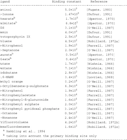
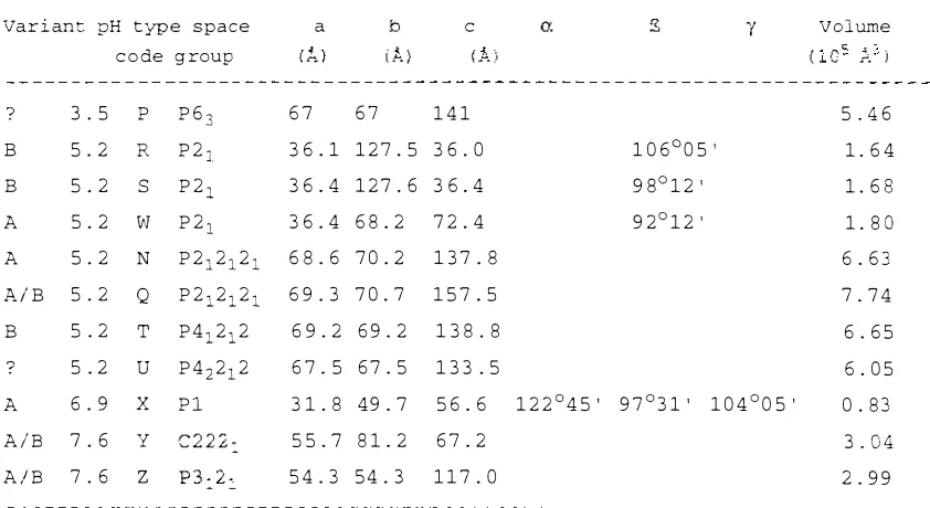
Table 2 51 -1 : Physical properties of bovine g-lactoglobulin Table 2 52 -1 : Ligand binding to bovine g-lactoglobulin Table 2 6 -1 : Crystal forms of bovine g-lactoglobulin Table 4 2 2 -1 : Resolving-gel and stacking-gel recipes Table 4 2 2 -2 : stock solutions for SDS electrophoresis Table 4 4 -1 : Stock solutions of screen solutions Table 4 4 -2 : crystallization screen matrix I Table 44 -3 : crystallization screen matrix 11
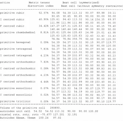
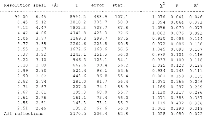
Table 4 6 2 -1 : Auto-indexing by DENZO for BLGA at pH 8.2 Table 4 6 2 -2 : Completeness of data for BLGA at pH 8.2 Table 4 6 2 -3 : Data merging statistics for BLGA at pH 8.2 Table 4 6 3 -1 : Auto-indexing by DENZO for BLGA at pH 7.1 Table 4 6 3 -2 : Completeness of data for BLGA at pH 7.1 Table 4 6 3 -3 : Data merging statistics for BLGA at pH 7.1 Table 4 6 4 -1 : Auto-indexing by DENZO for BLGA at pH 6 .2 Table 4 6 4 -2 : Completeness of data for BLGA at pH 6 .2 Table 4 6 4 -3 : Data merging statistics for BLGA at pH 6 .2 Table 4 6 5-1 : Auto-indexing by DENZO for BLGB at pH 7.1 Table 4 6 5-2 : Completeness of data for BLGB at pH 7.1 Table 4 6 5-3 : Data merging statistics for BLGB at pH 7.1 Table 4 6 6 -1 : Auto-indexing by DENZO for BLGA-BrC12 Table 4 6 6 -2 : Completeness of data for BLGA-BrC1 2 Table 4 6 6 -3 : Data merging statistics for BLGA-BrC1 2 Table 4 6 7-1 : Data sets and backup tape name
Table 4 6 7-2 : Crystal parameters
9
1 0 l1 ,1 2 1 3 ,1 4 ,1 5
1 6 1 8 1 9 2 5 52 52 57 58 58 6 4 6 4 ,6 5
6 5 6 6 6 6 ,6 7 6 7 6 8 6 8,6 9
6 9 70 70 71 71,72 72 72 73 73 Table 4 72 -1 : Molecular replacement results for BLGA at pH 8.2 75 Table 4 72 -2 : Translation and rotation matrices for BLGA at pH 8.2
75 Table 4 73 -1 : Molecular replacement results for BLGA at pH 7.1 75
Table 473-2: Translation and rotation matrices for BLGA at pH 7.1 76 Table 474-1: Molecular replacement results for BLGA at pH 6 .2 76 Table 474-2: Translation and rotation matrices for BLGA at pH 6 .2
76 Table 475-1: Molecular replacement results for BLGB at pH 7.1 77 Table 475-2: Translation and rotation matrices for BLGB at pH 7.1
77 Table 476 -1: Molecular replacement results for BLGA-BrC1 2 78 Table 476 -2 : Translation and rotation matrices for BLGA-BrC1 2 79 Table 511-1: Data collection statistics for bovine BLG crystals 85 Table 51 2 -1: Refinement statistics for structures of bovine BLG
in lattice Z 86
Table 52 4-1: Average B factors in different regions of BLG 95 Table 525-1: Accessible surface areas of bovine BLGA
and BLGB
Table 53 2 1: Accessible surface area of residues in
loop AB
(A?)
Table 53 3 -1: Accessible surface area of residues in
loop CD
(t,2j
Table 534-1: Accessible surface area of residues in
loop EF
(A2)
Table 534-2: comparison of
�/o/
torsion angles of residues in loop EFTable 53 5-1:
� /V
torsion angles of residues in loop GH Table 5 3 5-2: Accessible surface area of residues inloop GH
<A2)
Table 54-1: Accessible surface area of residues in the
bottom region
<A2)
Table 54-2: Hydrogen-bond network involving conserved residues of the bottom region
Table 552-1:
�i\)f
torsion angles of residues in strand s-o for BLGA at pH 7.1Table 552-2: Accessible surface area of residues at the
N-terminus and in strand S-O
(A2)
Table 553 -1: Accessible surface area of residues in the
C-terminus and in helix a-3
(A2)
Byqin, Spring of
1 998, Massey
1 0 0
1 0 2
104
1 05
1 0 5 1 0 8
1 0 8
1 1 1
1 1 1
1 1 3
1 1 3
1 1 4
Abstracts, Acknowledgements, Table of contents
Tab l e 554 -1 : Acces s ible surface area of res idue s in the
CX-H region
(A.2)
Tab l e 56 -1: Acce s s ib l e surface area of r e s idues of
S-shee t I
(11.2)
Tab l e 57-1: Acces s ible surface area of res idues of
S-sheet I1
(:112)
Tabl e 57-2 : Hydrogen bond network stab i l i s ing t h e y-turn
1 1 6 ,1 1 7
1 1 9
1 2 1 ,1 2 2
at Tyr9 9 1 2 2
Tab l e 583 -1: B fact ors o f the atoms o f arginine r e s idu e s 1 2 6 Tab l e 583 -2 : B fac t ors o f the atoms o f lys ine res idues 1 2 6 Tabl e 59 1 - 1 : Acce s s ible surface area of res idues of
strand S-I
(A2)
1 3 0Tab l e 59 2 - 1 : Hydrogen bonding i n the "lock and key" inter face 1 3 1 Tab l e 6 4 1- 1 : crystal parameters o f BLG i n lat t ic e s X, Y and Z 1 6 0 Tabl e 6 51 - 1 : Crystal paramet ers o f se lected l ipoc a l in s 1 6 7 Tab l e 6 52 - 1 : Structural a l i gnment of four l ipocal in s w i t h
respect to s ec ondary structure 1 6 7
List of figures
Figure 231-1: The lipocalin fold, exemplified by REP, etc. 7
Figure 252-1: Conformation of all-trans retinol molecule 20
Figure 271-1: Titration behaviour of a BLGA and BLGB mixture 26
Figure 271-2: Conformational changes of bovine S-lactoglobulin
monitored by optical rotation 27
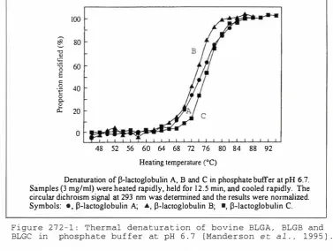
Figure 272-1: Thermal denaturation of BLGA, BLGB and BLGC in
phosphate buffer at pH 6.7 28
Figure 28-1: Bovine BLGA packed in lattice Z, as determined
by Monaco et al. (1987) 30
Figure 31-1: Scattering phase-shift in a two-electron system 34
Figure 31-2: X-ray scattering by an atom 35 Figure 31-3: X-ray scattering by a unit cell 36
Figure 31-4: Reflection condition, illustrated in an
Argand diagram 37
Figure 32-1: Two-dimensional example of lattice plane formed
by the end points of vectors a/h, and b/k 38
Figure 32-2: International Tables representation of
space group P3221 39
Figure 32-3: Ewald construction, showing conditions for
diffraction 40
Figure 332-1: Vector relationships in a two-dimensional unit
cell with two atoms 42
Figure 333-1: Change in structure factor when a heavy atom is
attached to protein molecule
Figure 333-2: Determination of the phase angle for
reflection (hkl)
Figure Figure
Figure
Figure
41-1: Dairy herd of New Zealand
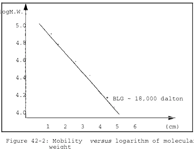
42-1: SDS-PAGE of bovine S-lactoglobulin
42-2: Mobility versus logarithm of molecular weight 42-3: UV spectrum of bovine S-lactoglobulin
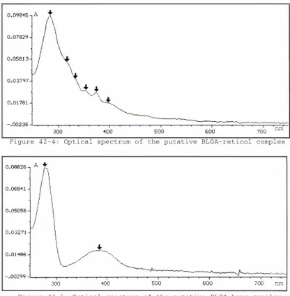
Figure 42-4: Optical spectrum of the putative
BLGA-retinol complex
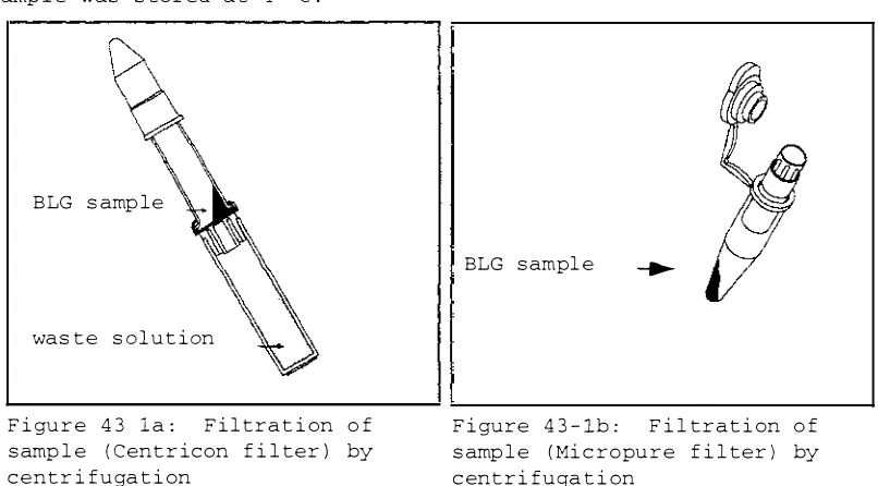
Figure 42-5: Optical spectrum of the putative
BLGA-heme complex
Figure 43-1a: Filtration of sample (centricon filter) by
Byqin, Spring of 1998, Massey
43
44
48
53
53
54
55
55
Abstracts. Acknowledtyemems. Table of contents
centri f ugat ion
F i gure 43-1b: F i ltrat ion of samp le ( M icropure f i l t er) by centri fugat ion
F i gure 44-1: Sketch of BLGA c rystals in lat t i c e Z F i gure 44-2: Sketch o f BLGA-SDS c rystals
F i gure 45-1: Photograph showing crystal mount ed on the goniometer
F igure 45-2: Repre s entat ive X-ray d i f fract ion image
F i gure 476-1: D i f ferenc e e l ectron den s i t y map o f BLGA-BrC12 F igure 4B2-1: Progre s s o f re f inement for BLGA at pH B.2 F i gure 483-1: Progr e s s o f r e f inement for BLGA at pH 7.1 F igure 484-1: Progre s s o f re f inement for BLGA at pH 6.2 F i gure 485-1: Progre s s of ref inement for BLGB at pH 7.1 F i gure 486-1: Progre s s of re f inement for BLGA-BrC12 F i gure 521-1: Cartoon p i c ture of the bovine BLG calyx
F i gure 521-2: The ext ended twisted B- sheet of the BLG dimer F i gure 521-3: superpos i t ion of the ca trac e s of BLGA in
latt i c e Z at pH 6.2, 7.1 and 8.2
F i gure 521-4: Superpos i t ion of the ca t rac e s of BLGA and BLGB at pH 7.1 in lat t ice Z
F igure 522-1: Topology diagram of main chain hydrogen bonds o f BLGA at p H 7.1
F i gure 522-2: Topology diagram of main chain hydrogen bonds of BLGA at pH 6.2
F i gure 522-3: Topology diagram of main chain hydrogen bonds of BLGA at pH 8.2
F i gure 522-4: Topology diagram of main cha in hydrogen bon d s of BLGB at pH 7.1
F i gure 523-1: D i sa s s embly of BLGA into sub-regions F i gure 525-1: Surface charge on the top region of BLGA
in lat t ice Z at pH 7.1
F i gure 525-2: Surface charge on the top region of BLGA in lat t ic e Z at pH B.2
F i gure 525-3: Surface charge on the t op region of BLGA in lat t ice Z at pH 6.2
F i gure 525-4: Surface charge on the t op region of BLGB in l at t ice Z at pH 7.1
F i gure 525-5: Surface charge on the bottom region of
Byqin. Spring of
1 998.
Massey56
56 59 59
60 61 78 80 81 82 83 84 87 88
89
89
90
91
92
93 94
96
97
97
98
BLGA a t pH 7.1
F i gure 525-6: Sur f a c e charge in the v i c inity of the c av i ty by Glu44 of BLGA at pH 7.1
F i gure 525-7: Re lat ive pos it ion of monomer s
F i gure 532-1: 2Fo-Fc e lectron dens ity map, showing two hydrogen bonds in loop AB
F i gure 533-1: 2Fo-Fc e le c t ron dens ity map, showing loop CD o f 98
99 99
101
BLGA in l a t t ice Z at pH 7.1 103
F i gure 533-2: Orien t a t ion of Glu65 i n BLGA and BLGB at pH 7.1 103 F i gure 534-1a: 2Fo-Fc e lec tron den s i ty map, showing c on format ion
of loop EF of BLGA at pH 7.1 10 6
F i gure 534-1b: 2Fo-Fc e lec tron dens ity map, showing con f ormat ion of l oop EF of BLGA at pH 6.2
F i gure 534-1c: Superpos it ion of loop EF of BLGA at pH 6.2, pH 7.1 and pH 8.2
F i gure 534-2: Hydrogen bond between Glu89 and Serl16 in BLGA at pH 6.2
F i gure 534-3: Superpo s i t ion of loop EF of BLGA at pH 6.2 and BLGB at pH 7.1
F i gure 535-1: 2Fo-F c e lectron dens ity map, showing conf ormat ion of loop GH at pH 7.1
F i gure 54-1: 2Fo-Fc e lectron den s i ty map, showing conformat ion of l oop OA
F igure 54-2: Hydrogen bond network of bott om region
F i gure 552-1: 2Fo-F c e lectron den s i ty map, showing s a l t bridge between N-terminus and Glu108
F i gure 553-1: Loca t ion of aromatic s ide cha ins of BLG
F i gure 554-1: Orient a t i on of O',-H he l ix with respect to c alyx F i gure 554-2: Amphipathic nature of helix CX-H
F igure 554-3: Smal l cavity formed by the CX-H he l ix F igure 554-4 : Sur f a c e charge of BLGA at pH 7.1 in the
vicinity of cx-H he l ix F i gure 56-1: Twi s t e d S-sheet I F i gure 56-2: S-turn in loop BC F i gure 57-1: S-sheet II
F i gure 57-2: 2Fo-F c e lectron dens ity map o f BLGA at pH 7.1 showing mut at ion s it e Val118A l a
F i gure 581-1: Superpos i t ion of the two d i su l f ide bonds of BLG,
B yqin, Spring of 1998, Massey
106
106
10 7
10 7
108
109 110
112 114 115 116 116
116 118 119 122
123
Abstracts, Acknowledgements, Table of contents
s howing the F igure 583-1: Locat ion o f F i gure 584-1: Locat ion o f F i gure 585-1: Location o f F i gure 586 -1: Locat i on o f
d i f f erent conformat i ons Glu and Asp s i de chain
al iphat i c s ide chain res idues
po lar uncharged s ide chain r e s idu e s prol ine s and g ly c i n e s in BLGB
124 125 127 127 128 F i gure 59 - 1: Unit c e l l c ontent s for bovine BLG in latt i c e Z 129 F igure 591-1: The dimer interface, showing sma l l cavity
near Cys 121 130
F i gure 592 -1: The "lock and key" inter f ac e 131
F i gure 592 -2: The z i g -zag chain of bovine BLG in lat t ice Z 132
F i gure 593-1: The loop interface 132
F i gure 5101-1: Ret ino l mode l led in the propo s ed
l i gand - b inding s it e 133
F i gure 5101-2: 2Fo - F c e l e c t ron den s ity map o f str ing o f wat e r mol e c u l e s ins ide t h e calyx o f BLGA a t pH 6.2 134
F i gure 5102-1: The mercury-binding s i t e o f BLG 135
F i gure 5103-1: Mod e l o f palmi t i c acid binding ins ide the BLG calyx
F igure 5104-1: D i f f erence e lectron density map of BLGA-BrC12, showing BrC12
Figure 611-1: Titrat ion curve o f bovine BLGA and BLGB F i gure 613-1: Inf rared spectra o f BLGB a s a f unc t ion F i gure 614-1: React ivity o f Cys121 at d i f ferent pH
o f
F igure 615-1: Sedimentat ion c oe f f ic i ent s o f BLG versus pH
pH F i gure 616 -1: Thermal graros o f BLGA at pH 6.75 and pH 8.0 5 F i gure 617 -1: Locat i on o f con s erved r e s idue s of bovine BLG F i gure 618-1: Mode l f or mo lecu lar transport by BLG
F i gure 622-1: Propo s ed retinol -binding region of bovine BLG, [Monaco et
al.,
1987]F i gure 622-2: BLG and the van der Waa l s surface o f ret i n o l, ac c ording t o the surface b inding mode l
135
136 137 139 140 142 143 143 144
147
147 F i gure 622-3: S - Sheet I and the van der Waa l s surface o f BrC12 149 F igure 622 -4: S- Sheet II and the van der Waal s
sur face o f BrC12
F i gure 623-1: Compar i s on o f ca p o s i t ions and
�!�
value s f o r BLGA and BLGA -BrC12Figure 623- 2: Superpos i t ion of BLGA (thin l ines) and BLGA-BrC12 (thick lines)
Byqin, Spring of 1 998, Massey
149
150
151
F igure 623-3: Structural change of l oop GH a f t er
l i gand b inding 152
F igure 631-1: Displacement s of ca atoms for the superpo s it i on
of BLGA and BLGB at pH 7.1 154
F i gure 632-1: 2Fo-Fc e le c t ron den s i t y map of loop CD of BLGB 155 F igure 632-2: 2Fo-Fc e le c t ron dens ity map around A118 o f BLGB 155 F i gure 632-3: Conformat ional propert i e s of BLGB compared
with BLGA in lat t ice Z at pH 7.1
( a) Ramachandran p lot of BLGB and BLGA in latt ice Z at pH 7.1 156 ( b)
�
val u e s of BLGA and BLGA in lat t i c e Z at pH 7.1 157 ( c )�
value s of BLGA and BLGA in l at t i c e Z at pH 7.1 157 F i gure 634-1: Loop EF in BLGA ( thin l ines) and BLGB ( th i n l ine s )at pH 7.1, showing change s in conformat ion F i gure 634-2: Opt ical rotation spe c t ra of BLGA and BLGB Figure 641-1: Linear array of BLG in t r i c l in i c lat t i c e X F igure 641-2: Dif f erenc e s in the dimer interface of BLG for
lat t i c e s X, Y and Z
Figure 642-1: Disp lacement p lot s of ca atom of bovine BLG molecules in d i f ferent lat t i c e s
Figure 643-1: Pos i t ion o f a-H he l ix in three lat t i c e s, re lat i ve to superpo s i t ion of the whole molecule
158 159 161
162
163
164 Figure 644-1: Loop EF of BLG in lat t i c e s X ( thin) and Y (thi ck) ,
superpo s i t ion based on the whole molecu l e F i gure 653-1: OBP dimer, showing non-convent ional
lipocal in fold F i gure 654-1: Bil in-BBP complex F i gure 654-2: RBP-ret inoid comp lex F i gure 654-3: OBP-pyrazine comp lex F i gure 654-4: BLG-BrC12 complex
F i gure 655-1: CPK mode l of the 8-barr e l o f OBP
Byqin, Spring of
1998,
Massey
165
168 169 169 170 1 70 171
Abstracts, Acknowledgements, Table of contents
Related publications
1. Structu r al bas i s o f the Tan f ord tr an s i ti on o f bov i n e g -lactog1obu l i n f r om cry s tal s tructures at thr e e p H val u e s
Author s : B i n Y . Qi n , Mar i a C . B ewley , L a\vrence K . Cr e ame r , H e athe r M . B ake r , Edward N. Baker , Geo f B . Jame s on
B i ochem i s try, i n pre s s
2. Structu r al con s e que nce s o f the b ov i n e g - l actoglobu l in
mutati on s o f var i ants A and B o f
l\.uthor s : B i n Y. Lawre n ce K . Cre amer , Maria C . B ake r , G e o f B . Jame s on
Pro te i n Scie n ce , accepted
3. B r omo-dodecano i c aci d binds ins i de the ca l actogl obu l i n
E dvJard N .
o f bov" i n e beta
?utho r s : B i n Y . Qi n , Lavire nce K . Cre ame r , Ed'vlard N. B ake r , G eo f B . Jame s on
FEBS l e tter s , in pre s s
4.
Bovine beta- l acto g l obu l i n and i ts var i ants : a thr e e d imens i onal s tructural p e r s p e ctiveAutho r s : Mar i a C . B e vll e y, B i n Y . Qin, G e o f f r e y B . Jame s on , L i n d s ay Sa'/Ty e r and Edward N . Baker
I n tl . Dai ry F e d . Bu l l . Spe c i al I s su e . "Mi l k Prote i n P o lymorph i s m , · IDF , B ru s s e 1 s : 1 9 9 8 , 1 0 1 - 1 0 9
Chapter 1
Introduction
tJiamlua 1 s re on m i l k t o nou r i sh t he i r newborn o f f sp r i ng . I n the course of evo l u t i on , i t can be ant i cipated t ha t indiv i du a l spe c i e s may vary the
components o f t h e i r mi l k to achieve the best surviva l strat egy f or t h e i r
o f f sp r ing . C ompar ison o f the compos i t ion o f the mi l k o f human b e i n gs and
cows reve a l s a or d i f f er ence : G-Iact oglobu l i n ( abbr e v i a t ed BLG) ,
wh i ch is abundant in cow's m i l k , i s absent in human's [ Hamb l i n g et al . ,
1992 ]. 'iioat i s G- lact o g l obu l in? vmat is the f unct i on of bovine G - l a c t og l obu:;' i n? 'l1hat f a c t ors are r esponsi b l e f or the ex i st ence of BLG
in cow but n o t in human m i l k?
Bovine G- l ac t og lcbu l in i s a sma l l p ro t e i n mo l ecu l e w i t h 162 r e s i du e s . To b i ochemists , toe ab"clndar;ce and easy preparat i on o f bov i n e
G-l i n make it a good candida t e as a research e c t . t o 1 97 5 ,
lobu l in was one o f the most invest p ro t e i ns and
was cons i d e r e d to be
HacG i bbon , 1 996] . In-v-est
ca l o f most p r o t e ins [ Cr eame r and
i ons of bov ine g-l act og l obu l i n r e ve a l that
it may be invo l ved in t he t ranspor t a t i on of r e t ino l and / o r t he metabol ism
o f a l i phat i c acids [ P�rez and C a lvo , 1 9 9 5 ] . Howeve r , t he exact
i o l o g i c a l funct i on r ema i ns unce r t a i n a f t e r mor e t han s
extens i ve r esearch . I t lS obv i ous t ha t bett e r know 1
:i-e a rs o f
o f g
-l i n is essen t i a -l t o unde rstand i ts b i o -l og i ca -l funct i on .
I n f act , much b i ochemica l r esearch on bovine g- l actog l o bu l i n has c r e a t e d
m o r e puzz l es than exp l anat i ons conce r n i ng its b i o l o g i c a l f u n c t i on . One
d ist inct i ve b i ochemica l f eature o f bovine g- l :1 is that i t undergoes p H - dep endent conf orma t iona l changes , t h e most Orle
around pH 7 ca l l ed t he N <=> R o r the Tan ford t rans i t i on [ Ta n f o r d et a l . , 1 9 5 9J . Anot h e r d ist inct i ve f e ature o f bovine g - l a c t o g 1 obu l i n i nvo l ves
t he wide r ang e of li gands t hat b ind qu i t e st rongly [ O'Ne i l l and K inse l la ,
1 9 8 7 ] , such as ret i no l , a l iphat i c acids , porphyr in spec i es, e t c . To dat e ,
no c ryst a l l og raph i c evidence exists t o show exa c t whe r e t h e b in d i ng
s i t e i s .
One o f the n i ce charact e r i st i cs o f bovine g-lac t og lobulin i s t ha t t h i s
pro t e i n c ryst a l l ises r e l a t ive ly eas ily i n mu l t ip l e cryst a l f o rms . Th i s
i s a n encourag i ng aspect o f bovine G- l a c t og l obu l i n s t ruct u r a l resea r ch
Chapter 1
Introduction
activities. X-ray diffraction studies ha'Ie pro'Jided the evidence which ascribes this protein to the lipocalin super-fami I1embers of this super fami have a calyx-like (or cup-like) fold and are capable of binding small hydrophobic molecules. Unfortunate X-ray diffraction studies have not answered all the puzzles of g- in, One very recent ication has described this protein as "still an enigmatic lipocalin" [Bro"vvnlo':J et
al., 997J
To add to the complexity, bovine g- lin has mult le 'Jariants. Some authors [Green and Aschaffenburg,
1959J
have suggested that it isnot to distinguish the different bovine g- in
'Iariants ions of the biochemical characteristics,
HO"vJever, it has been observed that milk dominated different variants Hill et
al., 1996]
behaves different industrial process
This is else that structura
The previous research history of bovine g difficulties ln the studies of bovine
g-sts can address,
in has demonstrated in _ To achiev'"e
scientific advances, we can that the more structural information we
obtain for g- lobulin, the more kno';!l we have about
g-lactoglobulin, and the more we can answer the queries on the
structure-function relat of g-lactoglobulin. The structure determinations
of bovine g-lactoglobulin A and B in lattice Z at a variety of conditions a:::-e the focus s of this research ect.
obulin research is reviewed in chapter 2. The history of its discovery is described first. FoIl the history, the biochemical and biological properties of bO'v'ine g-lactoglobulin are discussed,
vrith its nomenclature, isolation methods, sequence ana eSI structure information in solution, and previous diffraction studies. Chapter 2 ends with a statement of the aims of this ect.
The pri"ciples of X-ray diffraction techniques are reviewed in chapter 3. The principle of diffraction of X-rays from a crystal, Fourier
transformations, the Patterson function, and the principles of
refinement are discussed. On the basis of chapter
3,
the details of the \vhole experimental procedure are described in chapter 4, 'iflhich includes the source of protein material, purification procedure, crystallizationtrials, data collection and process refinement.
, and structure determination and
The results are presented in 5, included four ful determined bovine g-lactoglobulin struotures, and one structure 1;Jith an aliphatic acid bound to bovine g-lactoglobulin. The structures of bovine g
lobulin are disassembled into sub-regions and examined. The four major are the top region, bottom region. handle region. and the g-barrel which is further segregated into g-sheets I and 11. The structures are described based on these
the ligand binding site are examined further.
ions Some residues and
The structures are discussed in detail in chapter 6 . The structural basis cf the Tanford transition has been elucidated with the structural
disturbance of bovine
BLG
after 1 the structuralconsequences of point mutations of
BLG,
the structural differences among bovineBLG
in different lattice types, and the structural relationship of bovineBLG
with 1 ins.In chapter 7 , the final conclusions are made about current research, and suggestions are offered for future research.
Chapter
2Chaper
2
Review2 . 1 Discovery and early re search hi s t ory
Review
2 . 1 . 1 D i s c overy and naming of bovine B-lac toglobul in
The mai n whey pro t e ins o f ma ture bovine milk lactog l obu l i n ( - 0 . 3 g / l O O mL ) , a-lac talbumin albumin ( - 0 . 0 4 g/ 1 0 0
roL)
and immunoglobul inare [ Be l l , 1 9 6 4 ) : B ( - 0 . 1 g / l O O mL ) , serum
( - 0 . 0 8 g / 1 0 0 mL ) . B-Lac toglobul i n i s , there fore , the ma j or c omponent of whey protein . The
compo s i t i on o f mi lk was unc lear in the 1 9 3 0s . Albumin was recognised as
the maj or component o f the skim mi l k o f cow and was the general subj e c t
o f much research . Palmer ( 1 9 3 4 ) f o l l owed S j ogren and Svedberg's ( 1 9 3 0 )
lac talbumin preparat i on procedure t o i s olate albumin , but c l a imed a new globular protein had been crys t a l l i z ed . Subs equently i t wa s referred t o
a s Palmer ' s c rystal l ine protein .
Palmer d i d not name h i s dis covery . A f ew years later , Cannan e t a l . ( 1 9 4 1 ) sugges ted a nomenc lature " B - lactoglobul in" for Palmer's c rys tal l i ne protein . The Greek pre f ix der i ved from Pedersen's publ ica t i on ( 1 9 3 6 a ) , where three maj or components o f s kim mi lk were dis t ingu i shed based on ultracentr i fugation s tudies . Palme r ' s crys t a l l ine protein corresponded to the s e c ond component of the s kim milk . Thi s t erm a l s o i dent i f i es the source ( lacto for mi l k ) and the bas ic charac ter i s t i c ( gl obul in f or globular protein) o f Palmer's c rys t a l l ine protein , yet di s t ingui shes i t from the c l a s s ical globul in frac t i on o f mi lk .
2 . 1 . 2 Pur i f i cation o f Palmer's c rys t a l l ine protein
Palmer's c rys tal l ine protein was regarded as a pur i f ied protein samp l e f o r a l ong t ime . Obs ervati ons i n support o f thi s conc l u s i on incl uded that : ( i ) thi s prote in was pur i f ied by the method of crys t a l l i za t i o n ; ( i i ) t h e free boundary elect rophores i s experiment conduc t ed by Pedersen ( 1 9 3 6b ) s howed homogenei ty o f t h i s protein , and ( i i i ) S$rens en and Palmer ( 1 9 3 8 ) s howed that i t s solubi l i ty wa s independent o f preparat i on .
However , the fact that d i f ferent values [ Peders en , 1 9 3 6 a ; Bul l and Curry , 1 9 4 6 a ) had been repor ted for the mol ecular weigh t of Palmer ' s crys t a l l ine
protei n , as wel l as the di f ferent c ontents o f c ertain amino ac i ds [ Brand et al . , 1 9 4 5 ; Chibnal l , 1 9 4 6 ] sugges ted that thi s c rys tal l i ne samp l e might n o t b e a pure subs tance . Choh Hao L i ( 1 9 4 6 ) f ir s t provided the evidence by means of high res o l u t i on elec trophores i s that showed that Palmer's crys t a l l ine pro t e in was in fact het erogeneous . No mat ter how many t imes Palmer's crys ta l l i ne protein was re- crys tal l i zed , there were at l e a s t three electrophores i s boundar ies . Thi s obs erva t i on was supported by the i s o l a t i on of the s ub-components of Palme r's crys tal l i ne pro t e i n in 1 9 5 0 . Po l i s e t al . ( 1 95 0 ) demons trated that at leas t two d i f ferent components exi s t ed in the B- lactoglobul in prepared accordi ng to Palmer's method . They named them B1 and B2 . The s o lubi l i ty of the B1 component i s obvious ly di f ferent from Palmer's crys t a l l ine protein . The relat ive amount of the two sub-components was cons tant in general . The notable except i on appeared in the sample from the f i rs t - day colos trum of one c ow [ Po l i s et al . , 1 9 5 0 ) . Only a s i ngle component was observable f r om this sample .
Exper imental techniques as well as biological theory evolved drama t i c a l ly in the 1 9 5 0 s . Free -boundary e l e c trophore s i s was replaced by s o l i d - s upport electrophore s i s . By paper e l e c trophores i s , Ascha f f enburg and Drewry (1 9 55 ) s eparated Palmer's crys t a l l ine protein c l early i nto Bl- l a c t oglobu l in and B2 - lactog l obu l i n . In the mid 1 9 5 0 s , the origin of prote ins was related to the expres s i on of individual gene s . The i r results a l s o demons trated the re l a t i onship between the occurrenc e of Bl or B2-lactoglobul i n and the genetic type o f cow . From the i n forma t i on wh ich the i r publ i c a t ion provided , wi th the aid o f modern gene t ic theory , we can c onclude that bovine B- l actoglobul in i s cont r o l l ed by a s ingle gene s i te on one of the autosome s . No more than two vari ants o f B lactoglobul in can occur i n the m i l k f rom a s ingl e cow . However , i f B lactoglobul in is a prote in whi c h enj oys s ome f lexibi l i ty during the cour s e o f evolut i on , more var i ants c an be ant ic ipated from bovine gene pool .
2 • 2 Terminology
Bovine B - l a c t oglobulin i s only one member o f the widely d i s t ributed B
lac t o g l obul in fami ly . To c l as s i fy B- lactoglobul ins , we need to indi c ate
the m i l k s ource (secreted by whi ch animal ) , gene s e r i e s (some animals
Chapter
2 Reviewmay have more than one �- lactog l obu l in gene , usual ly label led as I , 11, etc . ) , and the variant type ( each gene may have s everal point mutants ,
labe l l ed as A , B , C , etc . ) . Compared t o many other macromo l ecules , bovine �- lac t o g l obul in crys ta l l i s e s rather r eadi ly in a var iety of crys tal forms [ Ascha f fenburg e t al . , 1 9 6 5 ] . Thes e crys tal f o rms have been ass igned a lat t i c e type code . Ther e f ore , for research involving X- ray di f fract ion experiment s , the lattice type code of �-lactog1obul i n c rys tals i s a l s o required , whi c h spec i f ies t h e crys tal cell dimens i ons and the space group . Note that bovine �-lac toglobulin i s r e f erred t o as bovine BLG throughout thi s thes i s .
2 . 3 The biological propert ies of &-lactoglobulin
2 . 3 . 1 �- lactoglobul in and l ipocal ins
The s e quence homo logy o f �- l ac toglobu l in wi t h o ther proteins has been
noted by s everal authors . Pervai z and Brew ( 1 9 8 5 ) noted the sequence
homo logy among human protein HC ( al s o known as a- 1-mi c roglobul i n , or
inter - a - t ryps i n inhibi tor l i ght chain precusor ( protein HC ) from human ,
abbreviated as HCHU ) , RBP and �-lac toglobu l i n . Drayna ( 19 8 6 ) inc luded
human apo - l ipoprotein ( apo-D ) , tobacco hornworm i n s ec t i cyanin ( INCYN ) or
b i l in-binding protein ( BB P ) , rat urinary protein ( a2 -UG) and rat
androgen- dependent epididymal secre tory pro t e i n ( ES P ) as prote ins whi ch
share amino acid sequence s imi l a r i ty wi th �- lactoglobul in . Lee et al.
( 1 9 8 7 ) added a frog prote in BG to this l i s t , where BG i s a protein
corresponding to an abundant mRNA in the cel l s o f Bowman's gl and in the
ol f a c tory t i s sue of f rog .
Member s o f this potent i a l protein fami ly , inc luding BLG , have a mas s around 2 0 , 0 0 0 Dalton , and are able to bind sma l l hydrophobic mo l ecul es . Later thi s protein f ami ly was class i f ied as a protein super - f ami ly and t i t l ed as "l ipocal i n " [ Perva i z and Brew , 1 9 8 7 ] . Now , at least 6 8 individual l ipocalins can b e browsed in the SWI SS-PROT data base . Whi l e the b inding o f hydrophobic molecules i s the bas i c proper ty o f member s o f t h i s super - fami ly, the func t i onal importance o f binding by individua l proteins i s qui te vague . For ins tance , the phys i o l ogical func t i on o f � lactoglobul in remains uncertain after many years o f research .
L ipocal ins gener a l ly exhibi t only a low l eve l o f res idue i dent i ty , o f ten no more than 2 0 % between pairs of s equences . This is bel ow the 2 5 % l evel where s t ructural s im i l a r i ties frequently go undetected [ S cader and Schne i der , 1 9 9 1 ] . In cases of such low s im i l a r i ty , the sequence a l i gnment
can lose s ta t i s ti ca l s i gni f i cance . I dent i fying the exi s tence o f
l ipocal ins by sequence inf orma t i on i s prac t i ca l ly based o n the mot i f s o f
protein s equence [ F lower et a l . , 1 9 9 3 ] . The heart o f the mo t i f - based sequence analys i s ( Parry- Smi th and Attwood , 1 9 9 2 ) 1S that a protein family can be charac teri zed by one or more cons erved mot i f s , that the
order of mo t i f s within a sequence is ma intained, and , opti ona l l y , that
the maximum d i s tance ( in res idues ) be tween the mot i fs i s res tricted .
r ---- ---1
,---,
F i gure 2 3 1 - 1 : The l ipocal in f o l d , exemp l i f ied by RBP , e t c . F i gure prepared by MOLSCRI PT [ Kraul i s , 1 9 9 1 ] .
L ___________________________ _
[image:28.561.95.512.246.745.2]Chapter
2 ReviewThe mot i f bas -X-X-G-X-W-aro ( bas=ba s i c r e s i due , aro=aroma t i c r e s i due ) occurs near res i due 2 0 ( in the bovine BLG sequenc e ) . A second mot i f T D-Y-X-X-aro appears around residue 1 0 0 ( in the bovine BLG sequenc e ) . Between the s e two segment s i s a cys teine that may form a disul f i de bond with another cys teine at the C - terminal end . A third mot i f R-X- P occurs around res i due 1 2 4 . Each mo t i f corresponds to an important s t ruc turally conserved region of the l ipoca l i n fold . Under such c r i teria , we are encounter ing a rapidly burgeoning protein fami ly . The analys i s shows the l ipocal in protein fami ly to be composed of a core s e t of qui te cl osely related proteins , the kernel l ipoca l ins , whi ch form by far the large s t sel f-cons i s t ent subse t wi thin the who le set o f related s equences . The
kernel l ipocal i ns share the three conserved sequence mo t i f s . A smaller number o f more divergent sequences , the out l i er l i pocal ins , share only two of the three mot i fs . Bovine g- lac toglobu l in has a l l three mo t i f s , which are " KVAGTWY " , " TDYKKY" and " RTP" , respec t ively . A dis ul f ide bond
is formed between res i due 6 6 and res i due 1 6 0 . Theref ore , bovine g lactoglobu l i n belongs to the kerne l part of the l ipoc a l i n super - fami ly .
The s truc tures of s everal l ipocal ins have been determined by X - ray
di f frac t i on techniques : plasma RBP [ Cowan et al . , 1 9 9 0 ; Zanot t i et a l . , 1 9 9 3 ) , BBP [ Huber e t a l . , 1 9 9 2 ) , OBP [ Tegoni e t a l . , 1 9 9 6 ) , BLG [ Green et a l . , 1 9 7 9 ; Brownlow et al . , 1 9 9 7 ; Qin et al . , 1 9 9 8 ) , MUP [ Bocskei e t a l . , 1 9 9 2 ) , ERBP [ Newcomer , 1 9 9 3 ] and NPl protein [We i chs e l , e t a l . , 1 9 9 8 ) . The s imilar i ty in three- dimens i onal s truc ture i s obv i ous enough
to include them into a s i ngle protein fami l y , whi ch is charac t e r i zed by
a common f o l d : a calyx - l ike central 8 - s t randed g-barre l , plus one long
three- turn a-hel ix . The calyx fo lds of the l ipoca l ins RBP , MUP , NP1 and
BLG are d i s p l ayed in F i gure 2 3 1 - 1 .
2 . 3 . 2 Gene s truc ture o f two l ipoca l ins : BLG and MUP
Analys i s o f the gene s t ructure of g-lactoglobul in from sheep a l s o support s t h e idea that these related prote ins form a protein family [ A l i and Clark , 1 9 8 8 ) . The pr imary s t ruc tures o f sheep g- lactoglobul in and mouse maj or urinary protein ( MUP , Unterman e t a l . , 1 9 8 1 ) have l im i t ed amino - a c i d s equence homo l ogy . However , the genes which encode these two proteins have s imi lar intron and ex on pat terns . The analogous introns are in pha s e wi th respect to the reading frame . B o th of these genes encode
a 1 6 2 - res i due mol ecu l e wi th s ix exons w i th one unt rans l a t ed exon . The anal ogous exons in the genes have s imi lar s i ze and func t i on ( s ee Table 2 3 2 - 1 ) .
Tab l e 2 3 2 - 1 : S imi lar gene s tructure o f two l ipocal i n s
BLG MUP
- - --- -- --- - -- -- - - --- -- - -- - - - -- -- --- -- - -- - --- -- - --- - - -
-Exon I 1 3 6 bp 1 6 1 bp
Exon I I 1 4 0 bp 1 3 4 bp
Exon I I I 7 4 bp 7 4 bp
Exon IV 1 1 1 bp 1 1 1 bp
Exon V 1 0 5 bp 1 0 2 bp
Exon VI S top codon S top c o don
Exon VI I Untrans lated Untran s l ated
2 . 3 . 3 B ovine l ipocal ins
Ret inol -binding protein ( RB P ) and odorant-binding prote i n ( OBP ) are two s t ruc tura l ly de f ined l ipocal ins which exi s t in c ow . Re t inol - binding protein [ Zanot t i et al . , 1 9 9 3 ] is the highly speci f i c carr i e r of vi tamin A i n vi vo . It is bi o synthes i zed i n the l iver and c i rculated i n the blood ,
where i t forms a comp l ex wi th trans thyretin ( al s o c a l l e d p r e - a l bumin ) . Thi s mac romol ecul e makes the inso lubl e r e t inol mo l ecul e t rans f erabl e in the blood and del ivers ret inol to the cel l s with appropr i a t e receptors . RBP i s very s ens i t ive t o the c on f o rmat ion o f retino l , binding only the a l l -t rans c o nforma t i on of retinol [ Jang and Swa i s good , 1 9 9 0 ] . RB Ps a r e f ound not only i n bovine bloo d , but a l s o i n the b l o o d o f human [ Rask e t al . , 1 9 7 9 ] , rat and rabbi t [ Sunde l i n , 1 9 8 5 J , whi ch are s pe c i es wi thou t g- lac toglobu l i n . The s equence s o f thes e RBPs demonstrate h i gh ident i ty , and mos t o f the s equence changes are concentrated i n res tr i c ted regi ons of the mo lecule [ Pe l os i , 1 9 8 9 ) .
OBP ( a l s o called pyrazine -binding protein ) i s i s o la t ed in h i gh c onc entra t i on in the nasal muco s a homogenat e s o f cows [ B i gnetti e t a l . , 1 9 8 5 J . Thi s protein was f i r s t i dent i f i ed by i t s abi l i ty to bind 2 -i s obutyl - 3 -methoxy-pyraz -ine [ Pe l o s -i , 1 9 8 9 ] . Subsequently -i t was found that many o ther hydrophobic vol a t i l e mol ecules c an be bound to i t s act i ve s i t e [ Pe l o s i , 1 9 8 9 ; Pevsner , 1 9 9 0 ] . Thi s protein i s a dimer wi th a s ingle binding s i te per monomer f or hydrophobic l igands . A f t e r bovine OBP [ Ti r inde l l i et al . , 1 9 8 9 ] and mous e OBP [ Pevsner and Reed , 1 9 8 8 J pr imary
Chapter
2 Reviews truc tures were determined , the s equence homol ogy wi th g- lac togl obu l i n sugges te d that OBP bel ongs t o t he RBP- BLG protein f ami ly [ Lee ,
1 9 87 J .
The s ub s e quent tertiary s truc ture determina t i ons o f t he s e three prote ins support thi s i dea [Monaco and Zanot t i ,1 9 9 2 J .
The s e quence alignmen t o f the s e three proteins provides usefu l informa t i on , a s summarised i n Tabl e
2 3 3 - 1 .
W e c an trace t h e l ipoca l i n mot i f s on both B- lac t og l obu l i n and retino l -binding p r o t e in . Indeed , both o f them are f avourabl e retinol carriers . Comparing the s equence i den t i ty o f OBP t o these two proteins , we f ound that OBP has a c l o s e relationship to RBP . Two of the l ipocal i n moti fs ( TDYxxaro and RXP ) di s appear in the OBP . But l ipocal ins are bas e d on t he i r folding s imi l a r i ty more than the ir sequence i dent i ty .Tabl e
2 3 3 - 1 :
Sequence a l i gnment o f s t ructura l ly de f ined bovine lipoc a l ins by MULTALIN [ Corpet ,1 9 8 8 ]
1 * 3 4
BLG MKCLLLALAL TCGAQJ".L IVT QTMKGLDIQK VAGTWYSLAM AAS D I SLLDA RBP . . . ER DCRVSSFRVK EN . . . FDKPR FAQTNYp� KDPEGLFLQD OBP . . . AQEEEp£ QNLSEL . . . SGPWRTVYI GSTNPEK I QE C ons ensus . c . aq . . . v . qn . . . ld . . . . aGtWy .
.
a ..
..
. .. . l q .3 5 8 3
BLG QSAP . LRVYV EELKPTPEGD LEILLQKvffiN GECAQKKI IA EKTKI PAVFK RBP NIVAEFS,lDE NGHMSATAKG RVRLLNNWDV . . CADMVGTF TDTEDPAKFK OBP NGP . . FRTYF RELVFDDEKG TVDFYFSVKR DGKWKNVHVK ATKQDDGTYV C on s ensus n . . . . frvy . .e l . . . . ekg . v . l l . .w . . . . ca . . v . . . t . dpa . fk
8 4 1 2 2
BLG IDALNENKVL . . . VL DTDYKKYLL . F . CMENSAEP E QSLACQ . CL RBP MKx�GVASFL QKGNDDHWI I DTDYETFAVQ Y SCRLLNLDG TCADSYSFVF OBP ADY . . . EGQNVFKIV SLS . RTHLVA HNINVDKHGQ TTELTELFV . C onsensus . dy . . . l . . g .
.
..
. i . dtdy . t . lv . .. c . . ..
. . . t . . l . . . fv .1 2 3 1 6 2
BLG VRTPEVDDEA LEKFDKALKA . . . LPMHIRL SFNPT . . QLE EQCHI . RBP ARDPSGFSPE VQKIVRQRQE ELCLARQYRL I PHNGYCDGK SERNIL OBP . . KLNVEDED LEKF . WKLTE DKGIDKKNVV . . . . NFLENE DHPHPE Consensus . r . p . v . de . l eK f . .
.
l . e . .. 1
. . . . rl . ..... .
. . e . . . hi .* S e quence numbers f o r the mature ( p o s t - t rans lat ion ) form o f bovine BLG are g iven . The l ipoca l in mot i fs are underl ined .
2 . 3 . 4 D i s t r ibut i on o f g - l a ct og l obul in
S ince the 1 9 6 0 s , the exi s t ence o f g- l ac togl obu l in in o ther anima l s has been inve s t i ga t ed in s everal l aboratories [ Lys ter e t al . , 1 9 6 6 ; L i beratori e t a l . , 1 9 7 9 b ; Perva i z and Brew, 1 9 8 6 ; e tc . ] . Accumu lated data demons Lrate that this secreted g l obular protein is widel y di s tributed , as indi cated in Tabl e 2 3 4 - 1 . Mos t g- lactoglobu l ins occur a s dimers , whi l e monome r i c g - l a c t oglobul in s exi s t i n s ome non- ruminant species such a s p i g , hor s e , cat , etc . No s imilar macromolecul e has been f ound i n rodents , s uch as mouse and rabbi t . I t i s bel i eved that no BLG exi s t s in human mi l k
[ Be l l and McKenz ie , 1 9 6 4 ; Liberatori e t al . , 1 9 7 9 a l .
Table 2 3 4 - 1 : D i s tribution o f g - lactoglobulin among di f f erent species
Species
Cow ( Bos t a urus ) Oxen ( Bos javan i cus )
Yak ( Bos grunn i ens )
Z ebu ( Bos i ndi cus)
Bu f f a l o ( B uba l i s buba l i s l
B i s on ( B i s on bi son )
Mus k ox ( Ovibos moscha t u s ) E l and ( Ta uro tragus oryx)
Goa t ( Capra h i rcus )
Sheep ( Ovi s ari es )
Mou f l on ( Ovi s ammon musimon ) Red deer(Cervus e l aph us
L . )
European e l k ( A l ees al e esL . )
Reindeer( Rangi fer t arandus L . ) Whi te - ta i l ed deer( Odocoi l e u s virginianu s )
Fal l ow deer ( Dama dama ) Cari bou ( Rangi fer arc t i cus )
Gira f f e ( Gi ra ffa camel oparda l i s )
Okapi( Okap i a johns t on i )
Byqin, Spring of 1 998, Massey
Type Reference
dimer dimer dimer dimer dimer dimer dimer dimer dimer dimer dimer dimer dimer dimer
dimer dimer dimer dimer dimer
Bul l , 1 9 4 6 a , b Bel l , 1 9 8 1
Gro s c l aude , 1 9 6 6 Lys ter , 1 9 6 6 Lys ter , 1 9 6 6 Lys ter , 1 9 6 6 Lys ter , 1 9 6 6 Lys ter , 1 9 6 6 Askonas , 1 9 5 4 Bel l , 1 9 6 7
Liberator i , 1 9 7 9b Lys ter , 1 9 6 6 Lys ter , 1 9 6 6 Lys ter , 1 9 6 6
Lys ter , 1 9 6 6 Lys t er , 1 9 6 6 Lys ter , 1 9 6 6 Lys ter , 1 9 6 6 Lys ter , 1 9 6 6
( c on ti nued)
Chapter
2 ReviewTable 2 3 4 - 1 : D i s t ribution o f g - l actoglobul in among d i f f erent spec ies
Spec i e s
Prong- horn ante l ope
( An t i l ocapra ameri cana ) Camel ( Camei us dromedari us ) Peccary ( Pecari t ajac u ) P i g ( Sus scrofa domes t i ca ) Horse ( Equus caba l l us ) Zebra ( Equ us quagga )
Rhinoceros ( Di ceros bi corn i s ) Donkey(Equinus asinus )
Dolphin( Turs i ops trunca t u s ) Manatee
( Tri chechus manatus l a t i ros tri s ) Beagl e ( Can i s famil iar i s )
Cat ( Fel i s c u t u s )
Grey kangaroo
( Ma cropus gigan teus )
Red kangaroo ( Ma cropus ru fus ) L lama ( Lama gl aba L . )
Mous e ( Mus muscul us ) Rat ( Ra t us norvegi cus )
Guinea - p i g ( Ca vi a porcel l us )
Rabbi t ( Oryc t o l agus c uni cul u s ) Human ( Homo sapiens )
Type dimer monomer monomer monomer monomer monomer monomer monomer monomer monomer monomer monomer monomer monomer none none none none none none Reference
Lys ter , 1 9 6 6 Liberat o r i , 1 9 7 9 Lyster , 1 9 6 6 Liberatori , 1 9 7 9 b
Liberator i , 1 9 7 9 b Lys ter , 1 9 6 6 Lys ter , 1 9 6 6
Godovac - Z i mmermann , 1 9 8 8 Perva i z , 1 9 8 6
Perva i z , 1 9 8 6
Perva i z , 1 9 8 6 Hal l i day , 1 9 9 1
McKenz i e , 1 9 8 3
McKen z i e , 1 9 8 3 Fernandez , 1 9 8 8
C l ar k , 1 9 8 7
Henn ighaus e n , 1 9 8 2
Brew , 1 9 6 7
Lyster , 1 9 6 6
Be l l , 1 9 6 4
The ruminant anima l s have dimeric g- lactogl obu l in and show very h i gh sequence iden t i ty to bovine g- lactoglobul ins . Genet i c polymorphi sm o f g l ac toglobul i n l S more common in ruminant anima l s than in the o ther spec ies . Cow has several vari ants as mentioned before . Donkey pos s e s s
two var i ants [Godovac - Z immermann e t al . , 1 9 9 0 ] , whereas sheep have var i ants A , B and C [ Erhardt e t al . , 1 9 8 9 ] , whi c h are di f ferent from the A, B and C variants for cows . Horse [ C lark et a l . , 1 9 8 7 ; Hal l i day et al . ,
1 9 9 1 ] , cat [ Hall iday e t a l . , 1 9 9 0 ] and dog [ Ha l l i day e t al . , 1 9 9 3 ] have more than one form o f the g-lact oglobulin gene and they are no t the produc t s of an autos omal a l lele (a pair of genes in the c e l l i rrespect ive of the s ex ) In other words , they are not phenotypes o f one gene . The
SWI S S - PROT p r o t e i n data bank now has at leas t 1 4 s equenc e s ava i l abl e f o r B- l a c t og l obu l i n , exc l uding var i an t s .
S equence a l i gnment o f avai l abl e B - l a c t oglobu l i n s i s di sp layed in Tab l e
2 3 4 - 2 . The f i rs t 1 6 r e s i dues o f bovine B- l ac t o g l obu l i n a r e t h e s i gna l p ep t ide , whi ch do not appear i n the mature pro t e i n . In c on t ras t to c ow ,
p i g ' s B- l ac t oglobul i n r e t a ins the s i gnal p ep t i de . The s equence mo t i f s
unde r l ined are charac t e r i s t i c o f the l ipoc a l i n super- f ami ly . The high
s equence i dent i ty among B- l ac t og l obulins � s obvious , except f or
Kangaro o . In the cons ensus s e quence , there a r e 2 1 per f e c t matched r e s i dues ( 1 1 . 3 5 % ) . The two di sul f ide bonds between res idues 6 6 - 1 6 0 and 1 0 6 - 1 1 9 are c ons erved in a l l B- l a c toglobul ins . Re s i due 8 9 ( bo l d l e t t er ) ,
a glutamic a c i d , i s a l s o conserved throughout a l l B - l a c t ogl obul ins .
F i gure 6 1 7 - 1 d i sp l ays the loc a t i on o f the cons e rved res idue s o f BLG in a s ter eodi agram .
Tab l e 2 3 4- 2 : Sequence a l i gnment o f B- l a c t og l obu l ins , by CLUSTAL [ Thomp s on et al . , 1 9 9 4 J
ID
LACB_BUBAR LACB_CAPHI
LACA_CANFA LACC_CANFA LACB_FELCA Ll-.CC_FELCl-. LACB_EQUAS LACB_HORSE LACA_EQUAS LACA_HORSE LACA_FELCA LACB_MACEU LAC B_MACG I Q 2 9 1 4 6 Consensus b
1 1 0 2 0 3 0
MKCLLLALAL T - -CGAQALIVTQTMKGLDI QKVA�SLAMAASDI S LLDAQSAPLR-VY - - - I IVTQTMKGLDI QKVA�SLAMAASDISLLDAQSAPLR-VY MKCLLLALGLALACGIQAI IVTQTMKGLDIQKVA�SLAMAASD I S LLDAQSAPLR-VY MKCLLLALGLALACGVQAI IVTQTMKGLDIQKVA�SLAMAASDI SLLDAQSAPLR-'JY MRCLLLTLGLALLCGVQAVEVTPIMTELDTQKVA�TVAMAVSDVSLLDAKSS PLK-AY - - - IVVPRTMEDLDLQKVA�Sl��SDI S LLDSETAPLR-VY - - - IVI PRTMEDLDLQKVA�SMAMAASDI SLLDSETAPLR-VY - - - -ATVPLTMDGLDLQKVAGMNHSMAMAASDISLLDSETAPLR-VY - - - -ATVPLTMDGLDLQKVA�SMAMAASD I SLLDSEYAPLR-v{ - - - -TNI PQTMQDLDLQEVAGKWHSVAMAASDISLLDSEEAPLR-VY MKCLLLALGLALMCGIQATNI PQTMQDLDLQEVA�SVAMAASD I S L LDSESAPLR-VY - - - -TDI PQTMQDLDLQEVA�SVAMVASD I SLLDSESAPLR-VY MKCLLLALGLSLMCGNQATDI PQTMQDLDLQEVA�SVAMVASDI SLLDSESVPLR-VY - - - -ATLPPTMEDLDIRQVA�SMAMAASD I S LLDSETAPLR-VY MKFLLLTVGLAL IGAIQAVENI RSKNDLGVEKFV�LREAAKT- - - - MEF S I - PLFDMD - - - -VENI RSKNDLGVEKFV�LREAAKT - - - -MEFS I - PLFDMD MKFLLLTVGLTS I CAIQAIENIHSKEELVVEKL I�RVEEAKA- - - -MEFS I - PLFDMN
* * * * *
(con tinued)