DISSERTATION ON
BACTERIOLOGICAL PROFILE, ANTIBIOGRAM AND RISK FACTORS
OF SURGICAL SITE INFECTIONS IN A
TERTIARY CARE HOSPITAL
Dissertation submitted in partial fulfillment of the Requirement for the award of the Degree of M.D. MICROBIOLOGY (BRANCH IV)
TRICHY SRM MEDICAL COLLEGE HOSPITAL AND RESEARCH CENTRE IRUNGALUR, TRICHY- 621 105
Affiliated To
CERTIFICATE
This is to certify that the dissertation entitled,
“
BACTERIOLOGICAL PROFILE, ANTIBIOGRAM AND RISK FACTORS OF SURGICAL SITEINFECTIONS IN A TERTIARY CARE HOSPITAL
”
by
Dr.G.DHANALAKSHMI,
Post graduate in Microbiology (2016-2019), is a bonafide research work carried
out under our direct supervision and guidance and is submitted to The Tamilnadu
Dr. M.G.R. Medical University, Chennai, for M.D. Degree Examination in
Microbiology, Branch IV, to be held in May 2019.
Guide: Professor and Head:
Dr. A. Uma M.D, Dr. A. Uma M.D,
Professor and Head, Professor and Head,
Department of Microbiology, Department of Microbiology,
CMCH&RC. CMCH&RC.
Dean:
Dr. A. Jesudoss M.S., D.L.O.,
Trichy SRM Medical College Hospital and Research Centre,
Irungalur,
DECLARATION
I solemnly declare that the dissertation titled
“Bacteriological Profile,
Antibiogram and Risk Factors of Surgical site infections in a Tertiary care
hospital”
is bonafide record of work done by me during the period ofMay 2017 to April 2018 under the guidance of Professor and HOD DR.A.UMA, M.D., Department of Microbiology, Trichy SRM Medical College Hospital and Research Institute, Trichy.The dissertation is submitted to The Tamil Nadu Dr.M.G.R Medical
University in partial fulfillment of the requirement for the award of M.D Degree
(Branch IV) in Microbiology.
Place: Trichy
Date
:
Dr. G. DHANALAKSHMI
,
Post Graduate Student,
M.D Microbiology,
Trichy SRM Medical College Hospital and Research Centre
Irungalur,
CERTIFICATE – II
This is to certify that this dissertation work titled
“
BACTERIOLOGICAL PROFILE, ANTIBIOGRAM AND RISK FACTORS OFSURGICAL SITE INFECTIONS IN A TERTIARY CARE HOSPITAL
”
of the
candidate
Dr. G. DHANALAKSHMIwith registration Number
201614602is
for the award of
M.D.MICROBIOLOGYin the branch of IV. I personally
verified the urkund.com website for the purpose of plagiarism Check. I
found that the uploaded thesis file contains from introduction to
conclusion pages and result shows
7%
percentage of plagiarism in the
dissertation.
ACKNOWLEDGEMENT
I humbly submit this work to ALMIGHTY, who has given me the strength, endurance and ability to overcome the difficulties encountered in the process of
compilation of my dissertation work.
I wish to express my sincere thanks to our DEAN Dr. A. Jesudoss M.S., D.L.O., Trichy SRM Medical College Hospital and Research Centre, Trichy, for permitting me to use the resources of this Institution for my study.
Firstly, I would like to express my sincere gratitude to Dr. A. Uma my beloved Prof & Head of Microbiology, Trichy SRM Medical College Hospital and Research Centre for her timely suggestions and valuable guidance during my work. She
has a great role in improving my ability to analyze the study.
I would like to whole heartedly thank Dr.Thirumalaikolundu Subramanian,
Professor of Medicine for his valuable suggestion for completion of my dissertation
work.
I express my thanks and heartfelt gratitude to my co guide Dr.G.Vazhavandal,
Associate Professor, Department of Microbiology, Trichy SRM Medical College
Hospital and Research Centre, for her valuable technical support and constant
I would like to whole heartedly thank all Assistant Professors
Dr.R.Saraswathy, Dr.A.Anupriya, Dr.J.Lalithambigai and Dr.DiegoEdwin,
Department of Microbiology, Trichy SRM Medical College Hospital and Research
Centre, for their voluntary valuable assistance and encouragement during the study
period.
Special thanks to my senior postgraduates Dr. Shalini, Dr. Jahappriya and
Dr. Jane esther and my friend Dr. Anand who helped in my works and for their support. I
would also wish to thank my junior postgraduates for their help.
I would like to thank all staff of Department of Microbiology, Trichy SRM
Medical College Hospital and Research Centre.
Finally, I am indebted to my family members for their everlasting support,
encouragement and heartfelt blessings throughout the study without whom this blue print
CONTENTS
S.NO TITLE PAGE NO.
1 INTRODUCTION 1
2 AIMS AND OBJECTIVES 5
3 REVIEW OF LITERATURE 6
4 MATERIALS AND METHODS 29
5 RESULTS 48
6 DISCUSSION 66
7 SUMMARY 77
8 CONCLUSION 80
APPENDIX-I ABBREVATIONS
ANNEXURE-I CERTIFICATE OF APPROVAL ANNEXURE-II STUDY PROFORMA
ANNEXURE-III PATIENTS CONSENT FORM ANNEXURE-IV MASTER CHART
APPENDIX – I
ABBREVIATIONS
SSI - Surgical site Infections
NNIS - National Nosocomial infection surveillance program PBP - Penicillin binding protein
MMM - Mannitol motility medium TSI – Triple sugar iron
IEC – Institutional ethical committee
CLSI – Clinical Laboratory Standard Institute CoNS – Coagulase negative Staphylococcus
MR – Methyl Red test VP – VogesProskauer test
ATCC -American Type Culture Collection MHA - Muller Hinton Agar
MRSA – Methicillin resistant Staphylococcus aureus
MSSA – Methicillin sensitive Staphylococcus aureus
MBL – Metallobetalactamases
MRM – Modified Radical Mastectomy
LSCS - Lower Segment Caesarean Section
ORIF – Open Reduction and Internal Fixation
IOL – Intraocular lens implantation
URSL - Uretheroscopic lithotripsy
TURP - Transurethral Resection of Prostate
ASA – American society of anesthesiologists
CDC – Center for Disease Control
SENIC – Study on Efficacy of Nosocomial Infection Control
HIV - Human Immunodeficiency Virus
SHV - sulphhydryl in variable
MIC – Minimum Inhibitory Concentration
CTX - Cefotaxime
CTR - Ceftriaxone
CAZ - Ceftazidime
EDTA – Ethylenediaminetetraacetic acid
MHT - Modified Hodge Test
ESBL - Extended spectrum betalactamase MDR -Multi Drug Resistance
Contents of Tables
S.no Contents
Page
No
1
Common causes of SSIs
2
2
Historical perspectives of SSIs
7
3
ASA score based on physical status of the patient
16
4
List of causative agents of SSI
17
5
Antibiotic prophylaxis for surgical procedure
20
6
Evolution of drug resistance
22
7
Biochemical reactions and isolation of microbes
38
8
List of antibiotics tested
41
9
Disk diffusion- CLSI guidelines for carbapenems
45
10
Types and of surgeries carried out
49
11
Comparison between site of surgery andorganism isolated
50
12
Distribution of cases in relation to gender
50
13
Distribution of SSI and age group
51
14
Distribution of SSI and category of surgery
52
15
Distribution of risk factors among SSIs
54
16
Distribution of ASA score along with SSIs
55
17
Distribution of SSIs in relation to prophylactic antibiotics
55
18
Day of sampling and surgical infections
56
19
Distribution of pus cells and culture positivity
57
20
Association between gram stain and culture positivity
57
22
Antimicrobial susceptibility pattern in gram positive cocci
60
23
Antimicrobial susceptibility pattern in gram negative bacilli
61
24
Prevalence of SSIs in different regions
67
Table of figures
S.no
Contents
Page
No
1
Cross section of abdominal wall depicting CDC classification of SSI
10
2
Common pathogens causing SSIs
18
3
Cefoxitin disc diffusion for detection of MRSA
42
4
Combined disc test for ESBL producers
43
5
Amp C disc test
44
6
Modified Hodge test
46
7
Ten disc procedure
47
8
Age wise distribution of SSIs
51
9
Comparison of Elective vs Emergency surgeries
52
10
Distribution of SSIs and nature of wound
53
11
Distribution of SSIs and extent of wound
54
12
Distribution of gram positive and gram negative organisms in SSIs
59
13
Distribution of no of organisms in SSIs
59
14
Frequency of MRSA in SSIs
61
15
Distribution of ESBL producers in SSIs
62
16
Distribution of Amp C producers in SSIs
63
17
Distribution of MBL producers in SSIs
63
1
1.0.
INTRODUCTION
The infection of a wound can be defined as the invasion of organisms
through tissues following a breakdown of local and systemic host defences, leading to
cellulitis, lymphangitis, abscess and bacteraemia. Infections of surgical wounds are
called as surgical site infections (SSIs).1
SSIs are defined as infections occurring within 30 days after a surgery or
within one year if an implant is left in place after the procedure and affecting either
the incision or deep tissue at the operation site2.
According to the National Nosocomial Infection Surveillance program
(NNIS), it is classified into superficial, deep, organ/space infections3.
Source of SSIs include the patient‘s own normal flora, organisms present
in the hospital environment that are introduced into the patient by medical procedures,
specific underlying disease, trauma or burns which may cause a mucosal or skin
surface interruption.4
SSIs are serious operative complications that occur in approximately 2% of
surgical procedures and account for 20% of health care-associated infections. Many
studies reported that SSIs rank third among common nosocomial infection next only
tourinary tract and respiratory tract infections.2,6
Recent studies reported that SSI rate ranges from 19.4% to 36.5% 7all over
the world, whereas in India it ranges from 3% to 12%.8,9
SSI remains a common and widespread problem that contributes to
significant morbidity and mortality, prolongs hospital stay and consequently
2
Factors which promote SSIs include length of hospital stay, Obesity,
Diabetes mellitus, smoking etc..The development of a post operative wound infection
depends on the complex interplay of many factors. Most postoperative wounds are
endogenous. Exogenous infections are mainly acquired from the nose or skin flora of
the operating team and transmitted through the hands of the surgeon or improper
operation theatre steriliation10which includes pre operative, intra operative and post
operative care
Some significant factors that can influence the incidence of subsequent
infection are surgical techniques, skin preparation, timing, method of wound closure
and antibiotic prophylaxis after certain types of surgery. Also many other factors have
been identified as having an effect on the potential for infection and these should be
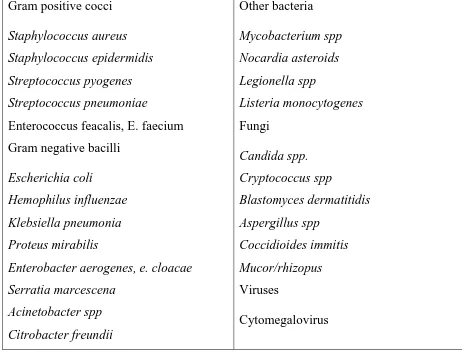
[image:16.595.71.532.480.648.2]considered by the healthcare professionals before, during and after surgery.11
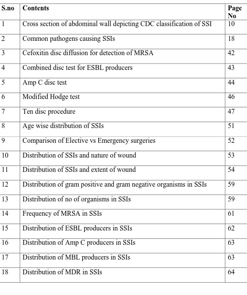
Table no.1. Common causes of SSIs:
Gram positive organisms Gram negative organisms
Staphylococcus aureus CONS
Enterococci
Eschericia coli Klebsiella spp Proteus spp Enterobacter spp Pseudomonas spp Acinetobacter spp
The resistance offered by a microbe to antimicrobial agent that is used in the
prevention or treatment of infections is called antimicrobial resistance.12Beta -lactams
are the most widely used antibiotics for treatment of postoperative woundsdue to their
3
different mechanisms which cause resistance to beta lactams namely a reduction in the
affinity of the drug targets (penicillin binding proteins) via amino-acid substitution, a
phenomenon occurring in both gram positive and gram negative bacteria. Gram
negative species, alteration in outer-membrane permeability that prevents passage to
the beta lactams and in both Gram-positive and Gram-negative bacteria, the
production of beta lactamase that inactivate the drug through hydrolysis of the beta
lactam ring. Hence widespread use of these groups of antibiotics has lead to
emergence and rapid spread of resistance.14
Among the members of the Enterobacteriaceae family, resistance to β
lactams has been reported to be associated with ESBL and Amp C β- lactamase.15
ESBL producing organisms hydrolyze oxyamino β- lactams like Cefotaxime,
Ceftriaxone, Ceftazidime and Monobactams but have no effect on Cephamycins,
Carbapenems and related compounds.16
Production of β- lactamase is frequently plasmid encoded and bears
clinical significance. Plasmids responsible for ESBL and Amp C β- lactamase
production frequently carry genes encoding resistance to other drugs also and
therefore antibiotic options in the treatment of β- lactamase producing organisms are
extremely limited.17
Data from last few decades show an increasing resistance for drugs that
were considered as the first line of treatment for post-operative wound
infections.18The most frequent co-resistances which are found in ESBL producing
organisms are amino glycosides, tetracyclines, chloramphenicol,
4
policies should be implemented to reduce hospital length of stay, morbidity and
expenditure per day in the hospital.19
The carbapenemases are betalactamases that are capable of inactivating
or hydrolyzing the carbapenem group of betalactam antibiotics. This is the main cause
of carbapenem resistance in gram negative bacilli. Hyperproduction of enzymes called
Amp C betalactamases can also result in resistance to carbepenem.20
The isolates which showed resistance to at least three or more than three
groups of antibiotics were considered as multi drug resistant (MDR).
The prevalence of antimicrobial resistance pattern may vary between
geographical areas. However, the publications available on the susceptibility pattern
of bacterial isolates causing SSI and ESBL prevalence in South India are minimal.
Hence, the present study is under taken at Trichy SRM Medical College and Research
Centre situated at Irungalur, Trichy in India, which is a tertiary care hospital serving
rural population mostly, prevalent bacteria and their susceptibility pattern, risk factors
5
2.0. AIMS AND OBJECTIVES
1. To find out the prevalence of SSI in this hospital.
2. To elicit the association between bacterial isolates and anatomical site of
infection.
3. To identify the probable risk factors for development of surgical site infections
4. To isolate and identify aerobic pathogenic bacteria from surgical site infections
(SSI).
5. To determine the antimicrobial sensitivity pattern of pathogens.
6
3.0. REVIEW OF LITERATURE
Surgical site infection (SSI) has always been one of the major
complications in surgical patients. It has been first mentioned even around BC. They
have been described and documented since ancient times (4000-5000 years) and
considered as one of the important nosocomial infections worldwide.
In 1846, Ignaz Semmelweis noticed that the mortality from puerperal
fever was much higher in teaching ward. He also made interesting observation that
women who delivered before arrival in the teaching ward had a negligible mortality
rate. The tragic death of a colleague due to overwhelming infection after a knife
scratch received during an autopsy of awomen who died of puerperal sepsis led Ignaz
to observe that pathologic changes in his friend were identical. Then, he hypothesized
that puerperal fever was caused by putrid material transmitted from patients by
carriage on examining fingers of medical students and physicians who frequently went
from autopsy room to the wards. He posted a notice on the door to the ward requesting
all caregivers to rinse their hands thoroughly in chlorine water before entering the
area. This simple intervention reduced mortality of puerperal fever to 1.5%.21
In 19th century, Louis pauster proposed germ theory. His work in
humans followed experiments identifying infectious agent in silk worms. He stated
that contagious diseases are caused by specific microbes and that microbes are foreign
7
In 1904, William Osler discovered the first cytokines which began to allow
insight into organism‘s response to infection, and led to the explosion in our
understanding of host inflammatory response.22
The word ‗Hospitalism‘ was introduced by Sir James Simpson to describe what we
now call hospital acquired surgical site infections. The following table describes the
[image:21.595.75.555.292.736.2]Historical background of surgical site infections.
Table no.2: Historical Perspectives of Surgical site infections:23 S.No Contributors Period Contributions
1 Hippocrates BC 460 – 375 Used wine & vinegar for simple wound irrigation
2 Galen 130-200 Recognized localization of infection (suppuration) in wounds inflicted in the
gladiatorial arena often heralded
recovery, particularly after drainage.
3 Theodoric of Cervia Ambroise Pare Guy de Chaulic
1210-98?1298-1368
1510-90
Observed clean wounds, closure of
wounds favours healing without
localization/infection/suppuration
4 Ignac Semmelweis 1818-65 Introduced hand washing technique & proved reduction of puerperal sepsis
(10% to 2%) by simple hand washing
8
5 Joseph Lister 1827-1912 Pioneer of antiseptic surgery. Introduced carbolic acid to clean
wounds and for sterilizing surgical
instruments.
6 Alexander Fleming 1881-1955 Introduced chemotherapeutic agents like sulphonamides and penicillin
3.1. CLASSIFICATION OF SURGICAL WOUNDS:
The risk of infection varies by type of surgical incision site. Invasive
procedures that penetrate bacteria-laden body sites, especially the bowel, are more
prone to infection. The theoretical degree of contamination, proposed by the National
Research Council(USA) over 40 years ago, relates well to infection rates.23 The
traditional wound classification system designed by the CDC stratifies the increased
likelihood and extent of bacterial contamination during the surgical procedure into
four separate classes of procedures24
Based on degree of microbial contamination.25
Clean wound:
Elective, not emergency, non-traumatic, primarily closed; no signs of acute
inflammation;
Clean wound
Clean-contaminated wound
Contaminated wound
9
No break in technique;
Respiratory, gastrointestinal, biliary and genitourinary tracts not entered
Clean-contaminated: A number of studies carried out in India indicate an overall SSI rate of 4.04 to 30% for clean surgeries and 10.06 to 45% for clean-contaminated
surgeries. 26, 27
Emergency case that is otherwise clean
Elective opening of respiratory, gastrointestinal, biliary or genitourinary tract with
minimal spillage (e.g. appendectomy) not encountering infected urine or bile
Minor break in technique.
Contaminated:
Acute, non-purulent inflammation
Gross spillage from gastrointestinal tract and entry into biliary or genitourinary tract in
the presence of infected bile or urine.
Major break in technique
Penetrating trauma of less than 4 hours
Chronic open wounds to be grafted or covered
Dirty or Infected:
Purulent inflammation of the wound (e.g. abscess);
10
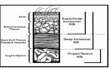
3.2. CLASSIFICATION OF SURGICAL SITE INFECTION:
The CDC Guideline for prevention of surgical site infection, published in 1999
defining an SSI
Superficial incisional SSI
Deep incisional SSI
Organ/ Space SSI
[image:24.595.73.519.268.545.2]11
Superficial incisional SSI:
Infection occurs within 30 days of surgery and infection involves only skin or
subcutaneous tissue of the incision and patient must present with atleast one of the
following criteria:
Purulent discharge with or without laboratory confirmation.
Organism isolated from aseptically obtained culture of fluid or tissue from the
superficial incision.
At least one of the following signs of inflammation: pain or tenderness,
localized swelling, redness or heat and superficial incision deliberately opened
by a surgeon unless incision is culture negative.
Diagnosis of superficial incisional SSI by the surgeon.
Excluding stitch abscess, infected burn wounds.
Deep incisional SSI:
Infection involves incision site that extend into the fascial and muscle layers and
patient must present with atleast one of the followingcriteria:
Purulent discharge
Deep incision spontaneously dehisces or deliberately opened by a surgeon and
is culture positive or not cultured when the patient has any of the signs and
symptoms of inflammation.
Evidence of infection by direct examination, during reoperation, or by
12
Diagnosis of deep incisional SSI by the surgeon.
Organ/ Space SSI:
Infection involves any part of anatomy (organs / spaces) other than the incision.
Purulent discharge from drain that is placed through a stab wound into organ/
space.
Evidence of infection by direct examination, during reoperation, or by
laboratory confirmation, histopathological and radiological examination.
Diagnosis of Organ/ Space SSI by the surgeon or attending physician.2
3.3. PATHOPHYSIOLOGY:29 Normally entry of microorganism is prevented by the intact epithelial surfaces. Apart from this there are also other protective mechanism in
the host namely
➢Cellular: Phagocytic cells, macrophages, polymorphonuclear cells and killer lymphocytes.
➢Humoral: Antibodies against the microorganisms, complement and opsonins
➢Chemical: Acidic pH of the stomach
Reduced host response to infection may be due to:
➢ Metabolic: Malnutrition, Diabetes mellitus, Uremia, Jaundice.
➢ Cancer, Acquired Immune Deficiency Syndrome (AIDS)
➢ Iatrogenic: Chemotherapy, radiotherapy and steroids.
13
3.4. Pathogenesis of surgical site infections:
3.5. Risk factors of SSI:
Kowli et al. (1985) found an infection rate of 17.4% when preoperative stay was 0-7 days, and an infection rate of 71.4% with a preoperative stay of more
than 21 days.12Nichols et al (1997) in his study on Prolonged postoperative hospitalization, which is a major concern of most of the hospitals, has been evident in
patients developing surgical site infection.30Anvikar et al. (1999) established that preoperative hospital stay predisposed an individual to 1.76% risk of nosocomial
infection. With an increase in preoperative stay, the risk increased proportionally. A
preoperative stay of one week increased the risk rate to 5% 31.
Contamination
•exogenous/ endogenous/hematogenous
Proliferation of bacteria
Induce inflammation,signs & symptoms
Identified or unidentified
14
A mean postoperative stay in patients who developed infection was almost
three times as compared to patients who did not develop SSI. The results indicated
that 12% of patients undergoing surgery developed SSI.31
In 1988 Lilienfeld et al published reports have demonstrated that patients with diabetes mellitus and obesity are more susceptible to wound infection because of
impaired neutrophil chemotaxis and phagocytosis.
Malnutrition has long been identified as a risk for nosocomial infections,
including SSI, among patients undergoing any type of surgery.32
Clip the hair immediately before an operation also has been shows a lower
risk of SSI than shaving or clipping the night before an operation (SSI rates
immediately before = 1.8% vs night before = 4.0%). Dessie et al reported emergency
surgeries more prone to SSIs. Dirty and contaminated surgeries are more likely to
develop SSIs.32a,b,c,e
The risk for developing SSI is a complex interaction between the patient, the
procedure and environmental factors which have been listed in the boxes given below.
15
Environment factors:
In 1964, Altemeir and Culbertson conceptualized the pathogenic relationship, key
factors of SSIs and also stated that risk of SSIis directly proportional to the microbial
Host related factors:
Age
Obesity
Severity of disease
ASA score(American society of
anesthesiologist)
Nasal carriers of MRSA
Remote infection
Duration of preoperative
hospitalization
Malnutrition
Diabetes mellitus
Malignancy
Immunosuppressive therapy
Procedure related factors:
Type of procedure
Preoperative hair removal
Antibiotic prophylaxis
Duration of surgery
Skin disinfection
Trauma to tissue
Foreign materials
Drains
Blood transfusion
Emergency surgery
Improper post-operative wound care
Length of post-operative stay
Uncontrolled blood glucose
16
contamination of the operative wound and to virulence of the microorganism and
inversely proportional to the integrity and resistance of the host defenses.
Risk of SSI= Dose of bacterial contamination x Virulence of microorganism
Resistance of patient defence
As per American Society of Anesthesiologists (ASA), SSI has been scored based on
preoperative physical status of the patient and shown in Table 2
Table no.3: American Society of Anesthesiologists score based on physical status
ASA Score Patient‘s preoperative physical status
1 Normally healthy patient
2 Patient with mild systemic disease
3 Patient with severe systemic disease that is not incapacitation
4 Patient with incapacitation systemic disease that is constant threat to life
5 Moribund patient who is not expected to survive 24hrs with or without
surgery
ASA score is an index to assess overall physical status of patient before
operation ranging from 1 to 5. It has been shown highly predictive for development of
SSI.36
CDC has developed National Nosocomial Infections Surveillance System
17
Efficacy of Nosocomial Infection Control) risk index which ranges from 0 to 3 points
and is defined by three independent and equally weighted variables.
One point is scored for each of the following if present:
• ASA physical status score >2
• Either contaminated or dirty/infected wound classification
• Length of operation > T hours (where T is approximate 75th percentile of duration
of the specific operation being performed.38
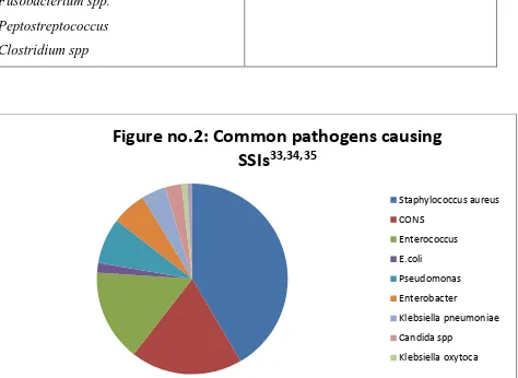
[image:31.595.64.526.413.769.2]3.6. Causative Agents:22
Table no.4: Causative agents of SSIs:
Gram positive cocci
Staphylococcus aureus Staphylococcus epidermidis Streptococcus pyogenes Streptococcus pneumoniae
Enterococcus feacalis, E. faecium Gram negative bacilli
Escherichia coli
Hemophilus influenzae Klebsiella pneumonia Proteus mirabilis
18
Pseudomonas aeroginosa Xanthomonas maltophilia
Anaerobes
Bacteroids spp. Fusobacterium spp. Peptostreptococcus Clostridium spp
Epstein –Barr virus Hepatitis A,B,C Herpes simplex virus HIV
[image:32.595.74.548.176.522.2]Varicella zoster virus
Figure no.2: Common pathogens causing
SSIs
33,34,35Staphylococcus aureus CONS
Enterococcus E.coli
Pseudomonas Enterobacter
Klebsiella pneumoniae Candida spp
19
3.7. Historical Aspects of antibiotic prophylaxis:
Experimental studies published during the early 1960s helped clarify
many of these problems and resulted in a more scientifically accurate approach to
antimicrobial prophylaxis. Most important was the report by Burke 39, which
demonstrated the crucial relationship between timing of antibiotic administration and
its prophylactic efficacy. His experimental studies showed that to greatly reduce
experimental skin infection produced by penicillin-sensitive S. aureus, the penicillin
had to be in the skin shortly before or at the time of bacterial exposure. This study and
others fostered the attitude that to prevent subsequent infection the antibiotic must be
in the tissues before or at the time of bacterial contamination. This important change
in strategy helped correct the common error of first administering the prophylactic
antibiotic in the recovery room.
As early as 1964, Bernard and Cole40 reported on the successful use of
prophylactic antibiotics in a randomized, prospective, placebo-controlled clinical
study of abdominal operations on the gastrointestinal tract. The success of antibiotic
prophylaxis noted in this early study was clearly due to the authors' appropriate patient
selection and wise choice of available agents, as well as the timing of administration.
Further advances in understanding of antibiotic prophylaxis in abdominal
surgery occurred in the 1970s. During this decade, the qualitative and quantitative
nature of the endogenous gastrointestinal flora in health and disease was appropriately
defined 41. Many prospective, blinded clinical studies in the 1980s and 1990s
prompted definitive recommendations concerning the proper approaches to antibiotic
20
3.8. Table no.5: Antibiotic prophylaxis for surgical procedure42,33
Surgical procedures Antibiotics
Cardiac surgery Cefuroxime 1.5g 8 hourly
Neurosurgery Cefuroxime 1.5g single dose
Head and Neck Cefuroxime 1.5g and metronidazole
500mg 8 h(single dose) involving mucous, and upto 3 doses if membrane and deep tissue involved
Biliary tract surgery Cefuroxime 1.5g single dose
Endoscopic retrograde
cholangiopancreatography
Cefuroxime 1.5g single dose
Gastroduodenal Cefuroxime 1.5g single dose
Appendectomy Cefuroxime 1.5g/ gentamycin 2-3mg/kg
and metronidazole 500mg (single dose)
Colorectal surgery Cefuroxime 1.5g/ gentamycin 2-3mg/kg
and metronidazole 500mg (single dose)
Orthopaedic surgery Cefuroxime 1.5g single dose
Lower limb amputation Benzylpenicillin 2mega units IV 6 h; metronidazole /clindamycin for patient allergic to penicillin
All antibiotic should be given for 24 h duration
Peripheral vascular surgery Cefuroxime 1.5g 8 hourly (3 doses)
Urological surgery IV antibiotic depends upon urine
21
Hysterectomy Cefuroxime 1.5g and metronidazole
500mg or amoxiclav 1.2g alone(single dose)
Caesarean section Cefuroxime 1.5g or amoxiclav 1.2g IV after umbilical cord is clamped (single )
3.9. Prevalence of SSIs:
It is estimated that 234 million major surgical procedures are performed
annually worldwide.43 Among all types of Health care associated infections, SSI
varies from 2.5% to 41.9% all over the world44,45. They are associated with longer
post-operative hospital stays, additional surgical procedures, treatment in intensive
care units and higher mortality.46Many studies reported that it varies from hospital to
hospital based on infection control measures and antibiotic policy. One review study
reported that SSI develops around 1 in 20 surgical patients in hospitals47
Suchithra et al observed that the prevalence of SSIs was 12%; and the
common etiologic agents are gram-positive organisms like Staphylococcus aureus and
22
for SSIs.49Mortality rate of appendectomy is 0.7% and 2.4% in patients without and
with perforation50
The modern surgeon cannot escape the responsibility of dealing with
infections and when dealing with them, should have knowledge of the appropriate use
of aseptic and antiseptic technique, proper use of prophylactic and therapeutic
antibiotics and adequate monitoring and support with novel surgical and
pharmacological modalities, as well as nonpharmacological aids50.
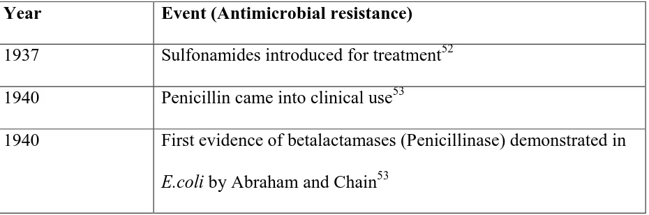
3.10. Antimicrobial Resistance in surgical site infections
Antibiotic era started with discovery of penicillin by Alexander Fleming in
1928 58. Use of Penicillin started in 1941. Emergence of penicillin resistance is identified in Staphylococcus aureus due to plasmid encoded β-lactamase. First
plasmid mediated β-lactamase in gram negative organisms- TEM-1 was described in
early 1960‘s58
. It was first isolated in Escherichia coli from a patient Temoniera in Greece and the gene responsible for it was named after him. It spread to other genera
[image:36.595.66.531.589.743.2]soon. Evolution of drug resistance is shown in table no.6 given below
Table no.6: Evolution of drug resistance
Year Event (Antimicrobial resistance)
1937 Sulfonamides introduced for treatment52
1940 Penicillin came into clinical use53
1940 First evidence of betalactamases (Penicillinase) demonstrated in
23
1940 Tetracycline came into clinical use54
1953 First tetracycline resistance was reported in Shigella dysentria54
1970s Plasmid mediated β-lactamases assumed importance in
Enterobacteriaceae and other gram negative bacteria54 1972 First epidemic of Chloramphenicol resistant Salmonella in
Kerala reported by Paniker et al.55
1989 MDR S.Typhi outbreaks resistant to Chloramphenicol, Ampicillin, Trimethoprim, Streptomycin, Tetracycline and
Sulfonamides were reported in India and Pakistan55
1992 S.Typhi resistant to Ciprofloxacin was first reported in UK.55 1970-80s Development of broad spectrum Cephalosporins, Cephamycins,
Monobactams and Carbapenems53
1990 Inducible chromosomally mediated β-lactamases among gram
negative bacteria53
Beta lactamases:
Enzymes which inactivate betalactam antibiotics by hydrolysing the nitrogen
carbonyl bond in their betalactam ring are collectively known as betalactamases. They
are members of a super family of active site serine proteases and act by cleaving an
amide bond of beta- lactam ring to form an acyl-enzyme complex. They can be
plasmid mediated or chromosomal .These β-lactamases are secreted as exozymes in
gram positive bacteria and within the periplasmic space in bacteria that are gram
24
Methicillin resistant Staphylococcus aureus (MRSA):
Methicillin was the first penicillinase resistant penicillin and has been widely
used in testing susceptibility of S. aureus to penicillinase resistant β-lactam agents. Hence, despite the fact that methicillin is no longer available and oxacillin and
cefoxitin have replaced it for susceptibility testing, resistant strains are commonly
known as MRSA.
MRSA strains are a continuing and increasing problem in healthcare settings,
with outbreaks now occurring in the community. Screening for MRSA provides a
means of identifying patients and staff who may be at risk of infection and/or involved
in transmission of the organism.
MRSA were first described in the 1960s 67. During the late 1970s and early
1980s, strains of S. aureus resistant to multiple antibiotics including methicillin and gentamicin were increasingly responsible for outbreaks of hospital infection
worldwide and several clonal types have shown extensive international spread 68,69,70
In England and Wales, the spread of MRSA was well controlled until the 1990s.
Between 1989 and 1991 only 1.6% of S. aureus bacteraemia isolates were methicillin resistant 71. However, methicillin resistance rates increased steadily throughout the 1990s, there were also significant increases in the percentages of isolates resistant to
erythromycin, clindamycin, ciprofloxacin, gentamicin, trimethoprim and rifampicin72.
MRSA reached in excess of 40% in several regions in 2001 which triggered the
introduction of mandatory surveillance of MRSA bacteraemia73. In 2005, trusts were
tasked with reducing the number of cases of MRSA and since that time cases have
25
are isolated are colonised rather than infected with the organism 76. Factors
predisposing to superficial colonisation include procedures involving ―hands on‖ care
especially in acute surgical, renal dialysis and critical care units 77. The risk of
colonisation resulting in infection is increased in the presence of any breach in the
skin, such as surgical wounds and devices penetrating the skin, for example prostheses
and catheters, which provide a portal of entry for bacteria 77. MRSA and MSSA are
similar in virulence and this is often connected to mobile genetic elements the
presence or absence of which determines the clinical outcome 78
Extended spectrum of β-lactamase: (ESBL)
The ESBL enzymes are plasmid - mediated enzymes capable of hydrolyzing
and inactivating a wide variety of β-lactams (oxyimino side chain). These
cephalosporins include cefotaxime, ceftriaxone, and ceftazidime, as well as the
oxyimino-monobactamaztreonam. 57
Another common plasmid mediated β-lactamase gene found in Klebsiella pneumonia and Escherichia coli are SHV-1 (SulphHydryl in Variable). Over the last 20 years many new β - lactam antibiotics have been developed which were resistant to
hydrolytic action of β - lactamases but, because of indiscriminate use, these antibiotics
alsobecame resistant. To overcome it, around 1980, 3rd generation cephalosporins also called broad spectrum Cephalosporins were introduced. Because of their
extensive use, they also became resistant. Widespread use of third generation
cephalosporins and aztreonam is believed to be the major cause of the mutations in
26
Various classification schemes have been proposed by many researchers
since 1968.60However, a more modern scheme based on molecular structure
classification was proposed by Ambler especially of only those enzymes that have
been characterized.
All ESBLs have serine at their active sites except for a small (but rapidly
growing) group of metallobetalactamases belonging to class B. They share several
highly conserved amino acid β sequences with penicillin binding proteins (PBPs)61
β--lactamases attack the amide bond in the betalactam ring of penicillins and
cephalosporins, with subsequent production of pencillinoic acid and cephalosporic
acid, respectively, ultimately rendering the compounds antibacterially inactive 62 .
Plasmids responsible for ESBL production tend to be large (80 Kb or more in size)
and carry resistance to several agents, an important limitation in the design of
treatment alternatives 63. The most frequent coresistances found in ESBL producing
organisms are aminoglycosides, fluoroquinolones, tetracyclines, chloramphenicol and
sulfamethoxazole-trimethoprim 59.
1. Impermeability of the Membrane mediated by both chromosome and plasmid.
2. Alteration of target protein e.g., Penicillin binding protein.
3. Increased efflux of the drug from the periplasmic space.
Characteristics of ESBLs: 56
They are mostly class- A Cephalosporinases carried on plasmids.
They are more common in Klebsiella species followed by Escherichia coli described first in Germany and France.
27
2) Imipenem and Cefoxitin not hydrolysed.
3) Comparative activity against Cefotaxime and Ceftazidine varies with enzymes.
4) Some enzymes active against Aztreonam.
5) Inhibition of activity by β-lactamase inhibitors can be demonstrated.
Major risk factors for ESBL production:
Risk factors are prolonged stay in ICU, long term use of antibiotics, nursing
home residency, severe illness, high rate of use of Ceftazidime and other Third
Generation Cephalosporins and use of life lines
Medical significance of detection of ESBL:
Patients having infections caused by ESBL – producing organisms are at
increased risk of treatment failure with expanded spectrum β-lactam antibiotics. So, it
is recommended that if an organism is confirmed to produce ESBL it is considered as
resistant to all 3rd Generation Cephalosporins.
Many ESBL isolates will not be phenotypically resistant; even through their
MIC is so high. ESBL producing strains have been established in many hospitals
producing epidemic diseases especially in Intensive Care Units.64 Failure to control
outbreaks has resulted in new mutant types in some institution.
Staphylococcus aureus was the most frequently isolated pathogenic bacteria from post-operative wounds. A majority of the isolates were methicillin resistant
28
cephalosporins. Rest had Enterobacteriaceae, either extended-spectrum β-lactamase (ESBL) producers or Amp-C hyperproducers. Indiscriminate use of antibiotics is a
major problem predisposing patients to harm by multi-resistant pathogens.
Carbapenems were in use nowadays, but the selection pressure exerted by
cephalosporins, suggesting a role of single plasmid carrying resistance genes to
multiple classes.66
Carbapenemases:
Carbapenemases are beta lactamases that cause resistance to carbapenem,
the β-lactam group with the broadest spectrum of antibacterial action. Carbapenems
were less susceptible to the inactivating activity of many betalactamases till the recent
past. But now, even these efficient antibiotics are becoming susceptible to the
enzymatic inactivation by betalactamases.
The enzymes hydrolysing carbapenems can be grouped into classes A or B
by molecular analysis. The former has serine as the active site member and the latter
has zinc at the active site. Since these enzymes are dependent on zinc, a metal, they
are called Metallobetalactamases. Some class C cephalosporinases can
hydrolyse/inactivate carbapenems and result in carbapenem resistance, but they are
not called carbapenemases because they are not carbapenem specific.
Antibiotic resistance is rising to dangerously high levels in all parts of the
world. New resistance mechanisms are emerging and spreading globally, threatening
29
4.0. MATERIALS AND METHODS:
This was a Hospital based Prospective Cross sectional study and carried
out at the Department of Microbiology, Trichy SRM Medical College Hospital and
Research Centre, Irungalur, Trichy, Tamilnadu. The study was carried out over a
period of one year (May 2017 to April 2018).
4.1. Materials:
Consecutive cases of both sexes and all adults belonging to various
surgical wards and underwent surgical procedure during the study period comprising
of elective as well as emergency were considered for the present study.
Patients belonging to anyone of the following were excluded.
1. Paediatric cases.
2. Cases taken for second surgery at the same site for any reason.
3. Patients on immunosuppressant or with immunodeficiency status.
4. Patients on antibiotics already for any other infections.
5. Presence of infection elsewhere in the body or focal sepsis.
The work was carried out after getting approval from Institutional
research board and Institutional ethics committee (copy enclosed – Annexure –I).
Informed consent (in vernacular) was obtained from every case (model copy of
30
4.2. Patient history
Age, sex demographic details, clinical details including name of the
procedure, date and duration of surgery, experience of surgeons, preoperative hospital
stay, nature of surgery, antibiotic prescribed (prophylactic/post operative), post
operative hospital stay, risk factors, onset of illness and other relevant history were
collected and recorded in a proforma (copy enclosed - Annexure- II).
4.3 Specimen collection and transport
After 48 hours of surgery, dressings on the surgical wounds were removed.
Evidence of wound infection was considered if the patient had local inflammatory
changes such as edema, redness, warmth or discharge from wound site. These were
looked into each case and the changes were documented. If there was any discharge,
samples were collected before dressing of the wounds. If only inflammatory changes
were present without any discharge, the wounds were monitored till discharge of the
patient and for development of discharge from wound. If no inflammatory signs were
noticed within 48 hrs, cases were followed up with the help of respective surgeons.
The surgeons incharge of the case was requested to inform/call the postgraduate
scholar doing this work whenever he/she suspected signs of SSIs in the form of fever
and local signs of inflammation. In addition, these patients were educated and
followed up through mobile phone for the development of SSIs over the period of 30
days.
4.3.1 Pus swab and aspirate:
Preparation of wound site– The suspected as well as overt infected areas were
31
was collected using sterile swab. Two swabs were taken from the depth of the wound
or lesion and aspirates were collected in a sterile disposable syringe and transported to
the laboratory within two hours.79The color, consistency and odor of the samples were
observed and recorded.
4.4. Laboratory works: Gram stain:
Direct thin smear was made from each wound swab and/or aspirates on a
clean grease free glass slide and was air dried. It was then heat fixed and Gram
staining was done with positive and negative control (ATCC Staphylococcus aureus
25923 and E.coli 25922). The presence of pus cells and microorganisms was observed under the oil immersion (100 X) objective.
The samples were cultured onto Nutrient agar, 5% Sheep blood agar and Mac
Conkey agar plates by adopting standard microbiological techniques. After 24 hrs of
incubation aerobically at 37°c, plates were read and the isolates were identified based
on colony morphology, Gram stain, motility and biochemical tests. Antibiotic
sensitivity test (AST) was performed by Kirby-Bauer disc diffusion method for all
isolates according to the CLSI 2017 guidelines. Repeat subculture was carried out on
next day for samples showing no growth on plates on first day and were processed
further80. All the isolates were identified by colony morphology, microscopic
32
A) Identification of Gram positive cocci:
Staphylococcus aureus, Enterococci and Micrococci were identified by colony morphology, Gram staining and biochemical test as per standard
microbiological procedures.
i) Staphylococcus aureus, was identified based on the following characteristics i.e; gram positive cocci in clusters on Grams staining, golden yellow pigment on
Nutrient agar plate, positive for catalase and tube coagulase test and showing
fermentative pattern in Oxidative Fermentative (OF) test of Hugh and Leifson.
ii) All coagulase negative gram positive clusters were considered as CoNS.
iii) Micrococci were identified based on grams staining and oxidative pattern in
OF test and excluded as commensal.
iv) Enterococci were identified based on microscopic morphology i.e; gram positive cocci in diplos, negative for catalase, positive for bile esculin hydrolysis, heat
tolerence property and mannitol fermentation80 .
Biochemical tests:81
Catalase test:
It was performed by Tube test with controls.
A small portion of colony was transferred from the Nutrient agar plate by a clean
platinum wire or glass rod into a tube containing 3% hydrogen peroxide.
Positive control: Staphylococcus aureus
33
Interpretation:
Positive - Evolution of effervescence within 10 seconds
Negative – no or delayed effervescence
Coagulase test:
This was performed by slide test (for detecting bound coagulase) and tube test (for
detecting free coagulase). Slide Coagulase Test:
The suspected Staphylococcal colony was emulsified in a drop of water on a
microscope slide. A flamed and cooled straight inoculating wire was dipped into the
undiluted plasma at room temperature, the adhering traces of plasma was stirred into
the Staphylococcal suspension on the slide with control.
Positive – Coarse visible clumping within 10 seconds
Negative - Absence of clumping in less than 10 seconds.
Tube coagulase test:
A 1/6 dilution of the plasma was prepared in normal saline (0.85%Nacl) and 1ml
volume of the diluted plasma was taken in a small tubes. A colony of Staphylococcus
was emulsified in a test tube with diluted plasma. It was incubated at 37ºC for up to 4
hours. The tubes were examined at 1, 2 and 4 hours for clot formation by tilting the
tube through 90º. The negative tubes were left at room temperature overnight and
re-examined.
34
Interpretation:
Positive - Any degree of clot formation
Negative - If the plasma remained liquid or showed only a flocculent or ropy
precipitate.
Bile Esculin hydrolysis:
One to two colonies from an 18 to 24 hours growth on nutrient agar plate was
inoculated on to the surface of the bile esculin agar slant. It was incubated at 35ºC in
ambient air for 48 hours.
Positive control: Enterococcus spp
Negative control: Viridans streptococcus Interpretation:
Positive - Blackening of the agar slant
Negative - no colour change.
B) IDENTIFICATION OF GRAM NEGATIVE BACILLI (GNB)
The gram negative bacilli were identified based on the colony morphology, motility,
catalase test, oxidase test, indole test, Methyl red, Voges Proskauer, triple sugar iron
agar, citrate utilisation and urease production.
Oxidase test:
It was performed by picking a colony using platinum loop or glass rod. The colony
was tested on freshly prepared solution of 1% oxidase reagent (tetra methyl
paraphenylene diaminedihydro chloride) with control.
35
Interpretation:
Positive – deep purple colour change within 10 seconds.
Negative – colour change after 10 seconds.
Indole test:
The organism was inoculated into peptone water and incubated for 24 hrs. Later,
Kovacs reagent was added. If the color changed to red on the top of the test tube it
was considered as positive.
Positive control: Escherichia coli ATCC 25922 Negative control: Klebsiella pneumoniae
Interpretation:
Positive – Red coloured ring
Negative – Yellow coloured ring
Methyl red test (MR):
The gram negative bacteria from a 24 hrs growth culture was inoculated in glucose
phosphate broth and incubated at 35ºC to 37ºC for 48 to 72 hrs aerobically. Then 5 to
6 drops of 0.04% solution of Methyl red was added. The results were read
immediately after mixing well.
Positive control: Escherichia coli ATCC 25922 Negative control: Enterobacter aerogenes
Interpretation:
36
Negative – no colour or intermediate orange colour change.
Voges Proskauer test (VP):
The test organism was inoculated in glucose phosphate broth and incubated at 35°C to
37ºC for 48 to 72 hours. 6 drops of solution A (alpha naphthol) and 2 drops of solution
B (KOH) were added to 1 ml of the broth and was observed after mixing well for 5
minutes.
Positive control: Enterobacter aerogenes
Negative control: Escherichia coli ATCC 25922
Interpretation:
Positive - Red color within 15 minutes or more after addition of reagent.
Negative – no colour change or copper colour after 1 hour.
Citrate utilization test:
Bacterial colony was picked by touching the tip of the needle on the colony that was
18 to 24 hrs old and inoculated into solid (Simmon‘s) media with indicator
bromothymol blue, lightly on the slant and incubated at 37ºC. Then it was observed
for development of blue color and growth.
Positive control: Enterobacter aerogenes
Negative control: Escherichia coli ATCC 25922
Interpretation:
Positive - Intense blue color and/ or growth on the slant.
37
Christensen‟s urease test:
The test was done by using Christensen‘s medium. The organism was inoculated on
the entire slope of the medium and overnight incubated at 37°C for up to 7 days.
Positive control: Proteus spp
Negative control: Escherichia coli ATCC 25922
Interpretation:
Positive – Pink Colour
Negative – Pale yellow colour
Triple sugar iron (TSI) test:
The medium was inoculated with bacterial culture using a straight wire (Stab culture)
and then streaked on the slant. It was incubated at 37°C 24 to 48 hours.
Interpretation:
Acid / Acid with gas – Glucose and Lactose/ Sucrose fermenter
Alkaline / Acid– Glucose fermentor
Alkaline / Acid with abundant black colour – Glucose fermentor with Hydrogen
sulphide production
Alkaline / Alkaline – Non fermenting GNB
Nitrate reduction test:
The test organism was inoculated with one drop from a 24 hrs nitrate broth culture
which was incubated at 35ºC for 48 – 72 hrs. It was then examined for nitrogen gas in
the inverted Durham tubes and 5 drops of nitrate reagent A and B (sulphanilic acid
and α–naphthylamine) were added. It was observed for 3 min for red color to develop.
38
Negative control: Acinetobacter baumannii
Interpretation:
Positive - Red color change within 30 seconds
[image:52.595.25.572.250.578.2]Negative – no colour change
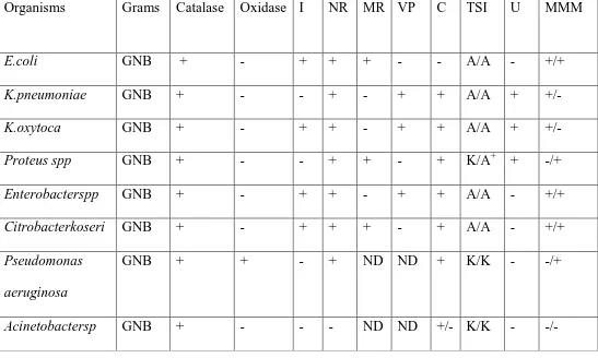
Table no.7: Biochemical reactions and isolation of microbes81:
GNB-Gram negative bacilli, I – Indole, MR – Methyl Red, VP- VogesProskauer, C- Citrate, U- Urease, MMM- mannitol motility medium, NR – Nitrate
Reduction, TSI –Triple Sugar Iron, A- Acid, K- alkaline, + Hydrogen sulphide production, ND- not done.
Organisms Grams Catalase Oxidase I NR MR VP C TSI U MMM
E.coli GNB + - + + + - - A/A - +/+
K.pneumoniae GNB + - - + - + + A/A + +/-
K.oxytoca GNB + - + + - + + A/A + +/-
Proteus spp GNB + - - + + - + K/A+ + -/+
Enterobacterspp GNB + - + + - + + A/A - +/+
Citrobacterkoseri GNB + - + + + - + A/A - +/+
Pseudomonas aeruginosa
GNB + + - + ND ND + K/K - -/+
39
4.5. ANTIMICROBIAL SENSITIVITY TESTING80
The antimicrobial sensitivity testing for all the isolates was done on Muller
Hinton Agar by Kirby – Bauer disc diffusion method as per CLSI 2017 guidelines
using antibiotic discs (Himedia, Mumbai)
I. Kirby Bauer Disk Diffusion Test: Preparation of turbidity standard:
McFarland 0.5 standard was prepared by adding 99.55 ml of 1% Suphuric
acid and 0.5 ml of 1.175 % barium chloride. This solution was dispersed into tubes
comparable to those used for inoculum preparation. It was sealed tightly and stored in
the dark at room temperature. The McFarland 0.5 standard provides turbidity
comparable to that of a bacterial suspension containing approximately 1.5 X 10⁸
CFU/ml.
Preparation of Inoculum:
In order to prepare the inoculum, about 3-5 representative colonies were picked up and inoculated in 4 - 5 ml of peptone water and incubated at 37ºC for 2 – 6
hrs to attain 0.5 McFarland‘s standard and if it was found more turbid, then some
more quantity of peptone water was added and adjusted to 0.5 McFarland‘s standard
by comparing against a card with white background and contrasting black lines.
Inoculation of Muller Hinton Agarplates:
Within 15 minutes of adjusting the turbidity of the inoculum suspension, a
sterile cotton swab was dipped into broth and rotated several times. During this
process, the swab was pressed firmly on the inside wall of the tube above the fluid
40
Hinton agar plate was inoculated by streaking the swab over the entire sterile agar
surface. This procedure was repeated by streaking two more times by rotating the
plates at an angle of approximately 60ºc to ensure an even distribution of inoculum
and finally, the rim of the agar was swabbed. The plate was closed and left for 3-5
minutes to allow any excess surface moisture to be absorbed before applying
antibiotic impregnated discs.
Application of discs to inoculated agar plates: Disc container was taken out from refrigerator one or two hours before use and brought to room temperature. Once a
cartridge of discs has been removed from its sealed package, it was replaced in a
tightly sealed dry container after use in refrigerator. The entire discs were placed on
agar plates and pressed down to ensure complete contact with the agar surface. Discs
were distributed evenly so that they were not closer than 25 mm from centre to centre
of the disc and incubated at 37º C for 16 – 18 hrs.
Reading and interpretation of results:
After 16-18 hrs of incubation, each plate was examined for satisfactory streaking with
confluent lawn of growth uniformly and circular zones of inhibition. The diameter of
the zones of complete inhibition including the diameter of the discs was measured.
The zones were measured to the nearest millimeter using a ruler that was held on the
back by inverting Petri plate. The Petri plate was held a few inches above a black, non
reflecting background and illuminated with reflected light. The zone margin showing
no obvious visible growth that could be detected with unaided eyes was considered as
41
standards and reported as ‗susceptible‟, „intermediate‟ or „resistant‟ to the drugs that were tested.
A bacterium can be
Susceptible – when it is inhibited by the concentration of the drug usually used
Intermediate – when it is susceptible to drug at higher than normal dosages
Resistant – when it is not inhibited by the drug82
Control strains used with each batch:
i. Escherichia coli ATCC 25922
ii. Pseudomonas aeruginosa ATCC 27853
iii. Staphylococcus aureus ATCC 25923 iv. Enterococcus faecalis ATCC 29212
Table no.8: List of antibiotics tested:
As per CLSI 2017 guideliness83
Gram positive cocci Gram negative bacilli
Penicillin(10U) Ampicillin (10 μg), Erythromycin (15 μg), Clindamycin (2 μg), Gentamicin (10 μg),
Co-trimoxazole (1.25/ 23.75 μg), Tetracycline (30 μg),
Ciprofloxacin (5 μg)
High level gentamycin(120 μg) Linezolid (30μg))
Ampicillin (10 μg) Amoxclav(20/10μg) Amikacin (30 μg) Gentamycin(10μg) Ciprofloxacin (5 μg)
Trimethoprim/sulfoethoxazole (1.25/23.75μg)
Ceftriaxzone (30 μg) , Cefotaxime (30 μg) Ceftazidime (30μg) Cefepime (30μg )
42
4.6. Detection of MRSA:
MRSA isolates were detected by standard disc diffusion method using
Cefoxitin (30µg). Cefoxitin is considered as a better inducer of mec-A gene than
oxacillin or methicillin, and can be used to screen heterogeneous MRSA populations.
As per CLSI 2017 guidelines, zone of inhibition ≤ 21 mm was considered as
Methicilin resistant isolates.84
Fig 3 - Cefoxitin disc diffusion method for detection of MRSA ZOI ≤ 21 mm.
4.7. Detection of Extended Spectrum Betalactamases:
As per CLSI 2017 guidelines, the test isolates which showed an inhibition
zone of ≤27mm for cefotaxime (CTX), ≤25mm for Ceftriaxone(CTR) and ≤ 22mm for Ceftazidime (CAZ) were considered as presumptive ESBL producer. All these
isolates were further tested for phenotypic confirmation test for ESBL.
Phenotypic Confirmation Test:
Antibiotic susceptibility testing was done on Muller Hinton Agar with 0.5
43
Lawn culture of the organism was made and 3rd generation cephalosporin,
Ceftazidime and Cefotaxime (30μg) disc was tested alone and along with their
combination for 10µg of Clavulanic acid. Organisms with 5mm increase in zone of
inhibition for Ceftazidime and Cefotaxim / Clavulanic acid (30μg/10μg) are
confirmed as ESBLs. 86,87.
Indicators of ESBLs: 5 mm increase in diameter of inhibition zone when using disc diffusion method with 3rd generation Cephalosporin and Clavulanic acid combined
disc.
Figure no.4: Combined disc test of ESBL producers 4.8. DETECTION OF AMP C PRODUCERS
As per CLSI 2017 guidelines, the test isolates which showed an inhibition zone
of ≤ 18 mm for Cefoxitin disc (30µg) were considered as presumptive Amp C producer. All these isolates were further tested by Amp C disk test.
AMP C DISK TEST:
All the Cefoxitin resistant strains were subjected to Amp C disk test to detect
the production of Ambler class C β-lactamase.88
Cefotaxime + clavulanic acid Cefotaxime
Ceftazidime
44
An overnight culture suspension of ATCC E.coli25922 was prepared in
peptone water, matched to 0.5 McFarland turbidity standards and inoculated as
lawn culture over a 90mm MHA plate as for routine disk diffusion procedure.89
A Cefoxitin disk with a potency of 30 microgram was placed over the lawn.
An empty disk moistened with sterile saline and inoculated with the test
organism was placed at the vicinity of the Cefoxitin disk almost touching it.
The culture plate was kept in the incubator for overnight incubation at 37° C.88
Blunting of the zone of inhibition of cefoxitin near the test strain inoculated
disc was taken as indicative of the strain being a producer of Ambler class C
betalactamase, as shown in Fig no.5.
The results were recorded and tabulated.
Figure no.5: Amp C disc test
4.9. Detection of Carbapenemase producing organisms:
As per CLSI 2017 guidelines, the test isolates which showed an inhibition zone
45
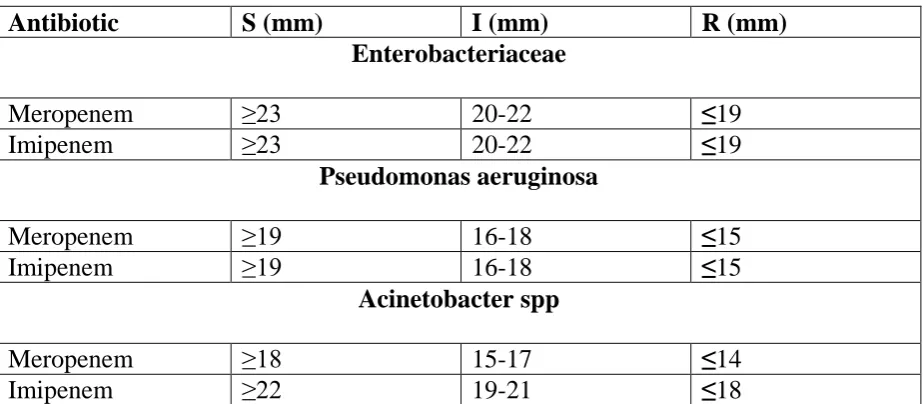
Table no.9: Disc diffusion - CLSI guidelines for Carbapenems:
Antibiotic S (mm) I (mm) R (mm)
Enterobacteriaceae
Meropenem ≥23 20-22 ≤19
Imipenem ≥23 20-22 ≤19
Pseudomonas aeruginosa
Meropenem ≥19 16-18 ≤15
Imipenem ≥19 16-18 ≤15
Acinetobacter spp
Meropenem ≥18 15-17 ≤14
Imipenem ≥22 19-21 ≤18
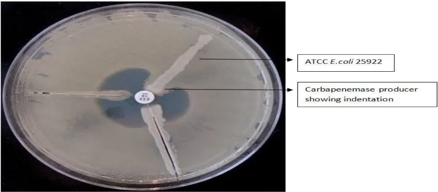
4.10. MODIFIED HODGE TEST:
An overnight culture suspension of ATCC E.coli25922 was prepared in
peptone water, matched to 0.5 McFarland turbidity standards, diluted to one in
ten and inoculated as lawn culture over a 90mm MHA plate as for disk
diffusion.90
After waiting for 3-5 mins for drying, a Meropenem disc was placed at the
centre of the plate.
Using a loop which can deliver 10 microlitre, the test organism was taken and
streak inoculated from the disk edge towards all four directions. 4 isolates were
tested in a plate with a single Meropenem disc. The plate was incubated at
37°C for 16-20 hrs.
The plates were examined the next day for enhanced growth around the test
organism and the zone of inhibition giving a clover leaf appearance, which was
indicative of Carbapenemase production90 as shown in Figure no.6. The results
46
Figure no.6. Modified Hodge test
4.11. Ten disc method:91
This procedure helps in screening of a bacterial isolate for all β-lactamases
(ESBLs, AmpC and Carbapenemases). Aztreonam (30μg), Cefotaxime (30μg),
ceftazidime (30μg), Cefotaxime + clavulanic acid(30/10), ceftazidime + clavulanic
acid(30/10μg), Ceftriaxone (30μg), Cefoxitin (30μg), Cefepime, Imipenem(10μg),
Imipenem + EDTA are the drugs for which the sensitivity of the organisms is detected
, by using Kirby Bauer disc diffusion assay.
Detection of ESBLs:
Ceftazidime or cefotaxime discs with and without clavulanic acid are used to detect
ESBLs. If the zone increases by 5mm or above with clavulanic acid combination, the