BENEFITS OF HYPOFRACTIONATION IN POST
OPERATIVE BREAST CANCER PATIENTS
INSTITUTION
DEPARTMENT OF RADIOTHERAPY
MADRAS MEDICAL COLLEGE
RAJIV GANDHI GOVERNMENT GENERAL HOSPITAL
CHENNAI - 600003.
Dissertation Submitted in Partial Fulfillment of
MD BRANCH IX RADIOTHERAPY EXAMINATION
MAY 2019
CERTIFICATE
This is to certify that the dissertation entitled
“BENEFITS
OF HYPOFRACTIONATION IN POST OPERATIVE BREAST
CANCER
PATIENTS”
submitted
by
Dr.JAGADESH
KUMAR.S.N, in partial fulfillment for the award of the degree of
Doctor of Medicine in Radiotherapy by the Tamil Nadu Dr.MG.R.
Medical University, Chennai is a bonafide record of the work done
by him in the Department of Radiotherapy, Madras Medical College
during the academic year 2016-2019.
DEAN,
Madras Medical College,
Rajiv Gandhi Government General Hospital
Chennai - 600 003.
PROFESSOR & HOD,
Department of Radiotherapy,
Madras Medical College,
CERTIFICATE OF THE GUIDE
This is to certify that the dissertation entitled
“BENEFITS
OF HYPOFRACTIONATION IN POST OPERATIVE BREAST
CANCER
PATIENTS”
submitted
by
Dr.JAGADESH
KUMAR.S.N, in partial fulfillment for the award of the degree of
Doctor of Medicine in Radiotherapy by the Tamil Nadu Dr.MG.R.
Medical University, Chennai is a bonafide record of original work
done by him under my guidance and supervision in the Department
of Radiotherapy, Madras Medical College during the academic year
2016-2019.
CERTIFICATE
This is to certify that the dissertation entitled
“BENEFITS
OF HYPOFRACTIONATION IN POST OPERATIVE BREAST
CANCER
PATIENTS”
of
the
candidate
Dr.JAGADESH
KUMAR.S.N, with Registration Number
201619004
for the award
of
M.D. Degree
in the Branch of
Radiotherapy
. I personally
verified the urkund.com website for the purpose of plagiarism
check. I found that the uploaded thesis file contains from
Introduction to Conclusion pages and result shows
19 Percentage
of Plagiarism in the Dissertation.
ACKNOWLEDGEMENT
I thank THE LORD ALMIGHTY, for his eternal grace and guidance in helping me finish this study.
I express my sincere gratitude to Prof.Dr.R.JAYANTHI MD., FRCP, Dean, Madras Medical College, Chennai - 03, who has been a continuous source of encouragement. I am grateful to h er for permitting me to conduct this study.
I express my sincere gratitude to Prof.Dr.SUDHA SESAIYAN M.D., Vice Principal, Madras Medical College, Chennai – 03 for her kind words of encouragement.
I express my gratitude to Chairman Dr.C.RAJENDRANM.D and the members of the Ethical Committee Council of Madras Medical College and Rajiv Gandhi Govt. General Hospital, Chennai - 03, affiliated to The Tamil Nadu Dr. M. G. R Medical University, Chennai - 32 for having approved my study and for his valuable suggestions.
case discussions I am gratified to Prof.Dr.GIRIDHARAN., M.D, DMRD, Additional Professor, for his practical understanding of the subjects, the assignments he gave which helped me to improve my knowledge about the subject and he plays a major role in instituting the habit of daily reading. The feedback what he gives for the assignment and project paved way to explore many areas of the subject.
I am grateful to Prof.Dr.MUTHUVEL., Ph.D., Professor & HOD, Department of Radiological Physics, for the support, encouragement and motivation rendered throughout the study period.
I wish to express my sincere gratitude to all the Assistant professors of our department (past and present) for guiding me during my study period. They guided me in acquiring the cases, planning the treatment, executing the chemotherapy and radiotherapy, manage the side effects during the treatment and much more.
Dr.P.K.BASKAR, M.D.R.T, Dr.S.MADHUMATHI, M.D.R.T,
Dr.SUNDARESAN, M.D.R.T, Dr.SANJAL KUMAR, M.D.R.T,
Dr.VIJEY KARTHIK, M.D.R.T, Dr.POONKODI, M.D.R.T,
Dr.JEYASHANKAR, M.D.R.T, Dr.SENTHIL KUMARAN, M.D.R.T,
guidance during difficult situations, Mrs.A.KOPPERUNDEVI, Mr.SURENDRAN.
My words of appreciation and gratitude also extends to our department radiographers Mr.PURUSHOTAM, Mr.VIVEK and their team of radiographers and students for their sincere execution of the treatment, kind support and service rendered for the patients in this study.
My sincere gratitude goes out to my fellow post graduates and friends of our department for the magnanimous assistance offered to me throughout the study period.
DECLARATION
I solemnly declare that the dissertation titled “BENEFITS OF HYPOFRACTIONATION IN POST OPERATIVE BREAST
CANCER PATIENTS” was done in department of radiotherapy,
Madras Medical college and Rajiv Gandhi Government General Hospital, Chennai during July 2017 to July 2018 under guidance and supervision of Prof.Dr.R.GIRIDHARAN, MD.
The dissertation is submitted to The Tamil Nadu Dr.M.G.R Medical UNIVERSITY towards the fulfilment for the award of M.D. Degree (Branch-IX) in Radiotherapy.
Dr.S.N.JAGADESH KUMAR
M.D. Radiotherapy Post graduate,
Madras Medical College Place : Chennai
CONTENTS
Sl. No. TITLE PAGE No
1. INTRODUCTION 1
2. LITERATURE REVIEW 5
3. AIM OF THE STUDY 47
4. MATERIALS AND METHODS 49
5. RESULTS AND ANALYSIS 62
6. DISCUSSION 73
7. CONCLUSION 78
INTRODUCTION
Carcinoma Breast is a complex heterogenous disease and is the most
common cancer in women globally ,based on different cancer registries data,
accounting for approximately 1.4 million cases each year, with more than half
of the 400,000 deaths caused by carcinoma Breast occurring in lower -middle
income Countries. It has surpassed Carcinoma Cervix to become the leading
cause of Cancer-related mortality among women living in a developing
country like India, having placed incidenc e of the disease at 30 to 33 per 1,
00,000 women in urban India.
The number of cases of Carcinoma Breast in India is about 100,000
women each year and the count will be approximately 2, 50,000 new cases of
breast cancer in India in upcoming decade. The Incidence of Carcinoma
Breast has doubled in India over the last few decades.
The increasing incidence could be attributed to the increasing working
women population which may increase the probability of exposure to various
risk factors.
Data at BIR & O shows that about 20 %-25% of all cancers registered
are female carcinoma breast, the incidence of male Carcinoma Breast is about
1%.
The standard of care for Carcinoma Breast is multi modality which is
diagnosis followed by Radiotherapy as indicated since there is a higher
incidence of local recurrence followed by Hormonal therapy depending on the
receptor status and menstrual status.
Majority of the cases presented in BIR & O have either neoadjuvant
chemotherapy followed by Modified radical mastectomy followed by adjuvant
chemotherapy and referred to our Department for feasibility of post
mastectomy radiotherapy, whereas a small percentage usually less than 20%
are prescribed palliative Radiotherapy to chest wall and whole breast
irradiation.
Many studies have confirmed the rationale for post mastectomy
radiation which is the prevention of a local-Regional recurrence.The
theoretical idea is that clinically occult persistent disease maybe present in
the operative site and draining nodes which may act as a source of metastases,
thus targeting operative site through radiotherapy which may have a
beneficial effect on the patients.
The conventional radiotherapy dose is 200CGy delivered over 25
fractions for a total dose of 50Gy for a duration of 5 weeks including the
posterior axillary boost so which accounts for a duration of 70 days minimum
since it includes the time for chemotherapy and surgery and clinical analysis
which causes a loss in Disability adjusted life years and physical ,emotional
burden and social isolation both for the patient and the family.Thus in turn the
hospital setup and staff undergoes extra work which puts up a challenge for
worker especially the supporting workforce( physicians and physicist and the
radiographers).
Many top centres around the world nowadays practice a sound
established, hypofractionated protocol for pmrt which reduces the total
number of fractions and in turn the treatment duration.When it comes to the
protocol prescription dose which is 4000 CGy delivered over 15 fractions
with 2.67Gy per fraction is being practiced and has been considered as a fair
standard dosage world over after stringent trials.
The hypofractionation in cancer treatment has been studied extensively
and is considered a routine in many parts of the world to use this approach in
palliative care. Different schedules have been developed to minimize the
burden on machines and operators. The data obtained has induced interest
about hypofractionation in curative setting and its possible reduction on the
load to all concerned.The protocols tried have been recorded to show good
LITERATURE REVIEW
EPIDEMIOLOGY
Ca Breast is the most challenging disease because it is the most
frequently diagnosed cancer and presents in multiple ways, and it is the
leading cause of cancer death in female‟s worldwide accounting for 23%
of the total new cancer cases and 14% of the total cancer deaths.
Economically insufficient countries account for about half the Ca Breast
cases and over 60% of the deaths occur in these regions. incidence rates
are high in Western and Northern Europe, Australia/New Zealand, and
North America; intermediate in South America, the Caribbean, and
Northern Africa; and low in Sub-Saharan Africa and Asia.
Geographical spread of the disease is complex with the highest
incidences seen in the more developed regions of the world and the
lowest incidences observed in the least developed regions .according to
SEER data which says that around 1 in 8 U.S. women will develop
invasive Ca Breast over her entire lifetime.
The factors that contribute to the international variation in
incidence rates largely arise from differences in reproductive and
hormonal factors which are causative factors and the availability of
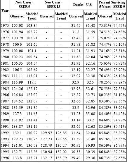
In 2015, an 242,476 new cases of invasive Ca Breast were
diagnosed in women in the U.S., A man‟s lifetime risk of Ca Breast is
[image:16.595.80.515.203.763.2]about 1 in 1,000.
Table 1:Surveillance, Epidemiology and end Results registry
Year
New Cases - SEER 9
New Cases -
SEER 13 Deaths - U.S.
Percent Surviving 5 Years - SEER 9
Observed Modeled
Trend Observed
Modeled
Trend Observed
Modeled
Trend Observed
Modeled Trend
1975 105.08 103.34 - - 31.45 31.48 75.31% 74.47%
1976 101.94 102.77 - - 31.8 31.59 74.51% 74.68%
1977 100.79 102.21 - - 32.48 31.7 75.02% 74.89%
1978 100.6 101.65 - - 31.73 31.82 74.47% 75.10%
1979 102.08 101.1 - - 31.21 31.93 74.16% 75.31%
1980 102.23 100.54 - - 31.68 32.04 74.96% 75.51%
1981 106.35 104.54 - - 31.92 32.16 75.65% 75.72%
1982 106.5 108.69 - - 32.19 32.27 76.49% 75.92%
1983 111.11 113.01 - - 32.07 32.38 76.43% 76.12%
1984 115.99 117.5 - - 32.9 32.5 78.25% 77.89%
1985 124.26 122.17 - - 32.98 32.61 78.53% 79.55%
1986 126.84 127.02 - - 32.87 32.73 80.23% 81.10%
1987 134.52 132.07 - - 32.66 32.85 83.30% 82.55%
1988 131.39 131.85 - - 33.2 32.96 84.58% 83.90%
1989 127.3 131.63 - - 33.23 33.08 84.40% 84.42%
1990 131.92 131.41 - - 33.14 33.2 84.68% 84.92%
1991 133.87 131.19 - - 32.69 32.62 85.26% 85.41%
1992 132.1 130.97 129.97 126.85 31.64 32.04 85.84% 85.89%
1993 129.23 130.75 127.23 128.55 31.39 31.48 85.76% 86.35%
1994 131.01 130.53 128.79 130.27 30.92 30.93 86.59% 86.79%
1995 132.71 132.85 130.94 132.02 30.55 30.39 86.84% 87.23%
Year
New Cases - SEER 9
New Cases -
SEER 13 Deaths - U.S.
Percent Surviving 5 Years - SEER 9
Observed Modeled
Trend Observed
Modeled
Trend Observed
Modeled
Trend Observed
Modeled Trend
1997 138.1 137.62 136.15 135.58 28.21 28.36 88.43% 88.06%
1998 141.49 140.07 139.07 137.4 27.54 27.4 89.55% 88.45%
1999 141.52 142.56 138.59 139.24 26.61 26.9 89.64% 88.83%
2000 136.62 139.3 134.29 136.17 26.64 26.41 90.25% 89.20%
2001 138.86 136.12 135.9 133.17 26.01 25.93 89.55% 89.56%
2002 135.89 133.01 132.9 130.24 25.62 25.46 90.31% 89.91%
2003 127.13 129.97 124.29 127.37 25.27 25 89.86% 90.25%
2004 128.26 127 125 124.56 24.49 24.55 90.04% 90.57%
2005 126.72 127.4 124.53 124.81 24.14 24.1 90.65% 90.89%
2006 126.44 127.79 122.96 125.06 23.56 23.66 90.87% 91.20%
2007 128.33 128.19 126.2 125.3 22.96 23.23 91.26% 91.13%
2008 128.53 128.59 126.38 125.55 22.55 22.81 90.76% 91.06%
2009 130.97 128.99 127.89 125.8 22.24 22.4 91.39% 90.98%
2010 127.07 129.39 123.7 126.05 21.92 21.99 90.90% 90.91%
2011 130.46 129.79 127.16 126.3 21.54 21.59 - 90.84%
2012 130.15 130.19 126.72 126.55 21.27 21.2 - 90.77%
2013 131.03 130.6 127.1 126.8 20.74 20.81 - 90.70%
2014 131.19 131 126.08 127.05 20.55 20.43 - 90.62%
2015 131.1 131.41 127.45 127.3 20.31 20.06 - 90.55%
But the surprsing news is from 1999 to 2005, Ca Breast incidence
rates in the U.S. decreased by about 2% per year. The decrease was seen
only in women aged 50 and older. The theoretical statement is that this
decrease was partially due to the reduced use of hormone Replacement
therapy (HRT) by women after the results of a large study called the
These results suggested a connection between HRT and increased
Ca Breast risk and which led to scientific discussions. though death rates
have been decreasing since 1990 – especially in women under 50 years
of age
These decreases are thought to be the result of treatment
advances, earlier detection through screening, and increased awareness.
In 2011, there were more than 2.6 million Ca Breast survivors in the US.
But when it comes to India The number of women estimated to be dying
of Ca Breast every year has been steadily mounting and rai sing concerns
among the healthcare fraternity. Data records tells us that around 48,170
women who died of Ca Breast in 2007, this slowly increased and
reached the 50,000 mark in 2010; now at present it is 87090 ,with a
ranking of number 1.
India has a rich mix of culture practices,tradition which has led to
a period of complex social and economic change. Cancer is now the
second leading cause of death in Indians after cardiovascular disease,
Women diagnosed with Ca Breast in India are on average 10 years
younger than the Western countries.
In India, a majority of the Carcinoma Breast cases present as late
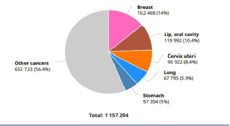
advanced stages. "The lifetime probability of developing Ca Breast in
developed countries. In India, the number of new Ca Breast cases is
162,468 cases per year and this is expected to rise and surpass others.
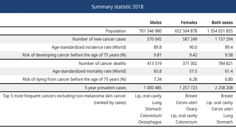
The age standardised mortality rate for Ca Breast in India is 19
per 100000 (24.7per 100000 globally). In common with other
developing regions mortality rates for Ca Breast in India are high in
comparison to incidence rates. This explains the poor survival rate
which is attributed to the lack of access or limited access to early
[image:19.595.98.501.433.655.2]detection and treatment.
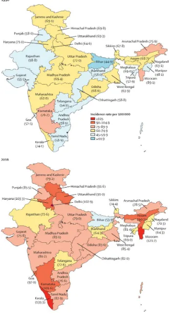
Table 3:Statistics of cancer cases in India
[image:20.595.100.497.372.588.2]I. RISK FACTORS AND CAUSES
Both in the developing and developed nations,Carcinoma Breast
accounts for higher incidence rates because of the many risk factors and
causative factors.
1) Gender: Woman is the main risk for Ca Breast. While men
occasionally get the disease; it is many times more common in
women than in men.
2) Age: Older women have a higher chance of getting Carcinoma
Breast and about two-third of the women with invasive Ca Breast
are 55 or older when the cancer is detected
3) Genetic mutations: About 5% to 10% of Ca Breasts are due to
gene mutations. The most common gene mutations are those of
the BRCA1 and BRCA2 genes Women with these gene changes
Have up to an 80% chance of getting breast cancer during their
lifetimes.
4) Race: Overall,white women are slightly more prone to get Ca
Breast than African-American women. But in less than 45 years of
age ,African American women,
5) Family history: Breast cancer risk is higher among women whose
with breast cancer the risk is 1.7 to 2.5 and aunt or grandmother
with breast cancer ,the risk is 1.5
6) Personal history of breast cancer: A woman with cancer in one
breast has a greater chance of getting contralateral breast cancer
or in another quadrant.
7) Menstrual history: Women who attained menarche early (before
age 12) or who attain menopause after the age of 55 have a
slightly increased risk of breast malignancy.
8) Dense breast tissue: Dense breast tissue means there is more
gland tissue and less fatty tissue .Thus Women with denser breast
tissue have a higher risk of Ca Breast
9) Lobular carcinoma in situ: Women with lobular carcinoma in
situ (LCIS) have a 7 to 11 time‟s greater risk of developing cancer
in either breast.
10) Nulliparous women or Not having children or delayed child
birth: Women who have no children, or who had their first child
after age 30, have a slightly higher risk of Ca Breast
11) Alcohol: The use of alcohol is clearly linked to an increased risk
of getting Ca Breast. Those who have 60ml to 150ml drinks daily
12) Body Mass Index: Based on BMI overweight or obese is linked
to a higher risk of Ca Breast, especially for women after
menopause or if the weight gain took place during adulthood.
13) Breast radiation early in life: Women who have had radiation
treatment to the chest area (as treatment for other conditions like
hodgkins lymphoma and diagnostic fluoroscopy) earlier in their
lifehood have a greatly increased risk of Ca Breast. The risk from
chest radiation is highest if the radiation were give n during the
adolescence period,because this gives a latent period of 10 - 15
years that is when the breasts were still developing.Radiation
treatment after age 40 does not seem to increase Ca Breast risk.
14) Treatment with Diethylstilbestrol: In the past, some pregnant
women were given this drug.Studies have shown that these
women and the children exposed in the womb have a slightly
increased risk of getting Carcinoma Breast.
15) Recent use of birth control pills: Studies have found that women
who are using birth control pills have a slightly greater risk of Ca
Breast than women who have never used them
16) Using hormone therapy after menopause: This has been proven
by many studies and its withdrawal and stoppage of this therapy
17) Combined Hormonal therapy: Use of combined Hormonal
therapy after menopause increases the risk of getting Ca Breast.It
may also increase the chances of dying from Ca Breast. Ca Breast
in women taking hormones may also be found at a more advanced
stage
18) Not breast-fed: Some studies have shown that breast-feeding
slightly lowers Ca Breast risk, especially if the breast -feeding
lasts 1½ to 2 years.This decreased 4.3% of the relative risk of
breast cancer for 12 months of breast feeding.
19) Estrogen Replacement Therapy: some studies have found that
Estrogen replacement therapy increases the risk of ovarian and Ca
Breast, if used for more than a decade.
II. PATHOLOGY
A. INVASIVE BREAST CANCER
1) Invasive (Infiltrating) Ductal Carcinoma (IDC): It is the most
common type of Ca Breast.3/4ths of the cases of invasive Ca
Breasts are classified as invasive ductal carcinoma
2) Invasive (Infiltrating) Lobular Carcinoma: Lobular Carcinoma
originates from the milk- producing lobules where it extends into
the surrounding adipose tissue of the breast.It is relatively
compared with IDC, patients with ILCs often have bilateral
disease
3) Tubular Carcinoma is a highly differentiated invasive carcinoma
and pure tubular carcinoma has limited metastatic potential and
better than average prognosis.
4) Medullary Carcinoma is a relatively uncommon type of invasive
carcinoma, accounting for less than 5% to 7% of all invasive Ca
Breasts. The prognosis for this type of Ca Breast is quite
favourable when compared to the other types.
5) Mucinous Carcinoma, also called colloid carcinoma, is an
invasive form of Ca Breast characterized by large amounts of
extracellular mucin production. This histopathological type
confers a relatively favourable prognosis.
6) Metaplastic Carcinoma is uncommon, representing less than 5%
of all Ca Breasts. Prognostic implications of this type of breast
cancer are unpredictable.
7) Invasive Cribriform Carcinoma is a well- differentiated cancer
comprised of Small and uniform cells. It shares some features
with tubular carcinoma and is also associated with better than
8) Invasive Micropapillary Carcinoma is a distinct but poorly
recognized variant of breast cancer, usually presenting as a firm,
Immobile mass. Pure micro papillary carcinoma is uncommon,
limited research suggests that this type of cancer may be
associated with a relatively poor prognosis
9) Invasive Papillary Carcinoma is very rare comprising less than
1% to 2% of invasive breast cancers and a predominant
presentation in postmenopausal women , it is characterized by
nodular densities that may be Multiple and are frequently
lobulated. Available data suggests relatively favourable prognosis
OTHER BREAST CANCER TYPES
1) Inflammatory Breast Cancer is a unusual form of locally
advanced breast cancer. Patients present acute onset of clinical
features including signs of inflammation, warmth, thickening or
dimpling ,peau d' orange and a clinically palpable ridge at the
margin of induration . Inflammatory breast cancer is relatively
rare, representing about 1% to 5% of all Ca Breasts and has a less
favourable than Average prognosis
2) Paget’s Disease of the Nipple presents either in situ or invasive
cancer;but has an excellent prognosis rate when ass ociated with
3) Phyllodes Tumors can present as benign, borderline or
malignant. Rare presentation is malignancy. Phyllodes tumors are
biphasic and composed of benign epithelial elements and Cellula r
connective tissue stroma.They can grow to a relatively large size
within a few months, although rapid growth does not necessarily
indicate malignancy
TUMOR BIOLOGY: BREAST CANCER
Genetic changes that occur in cancer include mutation of key
regulatory genes, changes in protein products, and changes in the
amount of product produced by genes (gene expression). As changes
accumulate, cells become more abnormal and cancer progresses
Some of the genetic elements that have been shown to be
important in the development of Ca Breast are listed below:
1) BRCA1 and BRCA2 Genes-tumour suppressor genes,thus
germline mutations increases risk of breast cancer by 65% -85%
2) Estrogen and Progesterone receptor-attributed to increase
exposure to hormones
3) HER-2/neu Gene-determines the prognosis and metastasis
4) TP53 Gene and Li-Fraumeni Syndrome-guardian of the genome
5) PTEN Gene and Cowden Syndrome-autosomal dominant and
benign and malignant growth.
6) ATM Gene and Ataxia-Telangiectasia-autosomal recessive and
features neurological clinical features.
HORMONE RECEPTOR STATUS IN INDIA
Desai et al. reported on 798 patients where the median age was 48
years and found 33% were ER+ and 46% were PR+, Data on combined
receptor profiles found 25% were ER+/PR+,
7% were ER+/PR−
21% were ER−/PR+ and
47% were ER−PR−.
Twenty percent of patients were found to be Her-2 positive.
Table 5 :Comparision of cases based on histopathology
Author No. of Patients
Mean Age
Median Size
Histology Grade
LN ER PR IDC ILC 1 2 3
Saxena
etal 569 88% 4% 80%
Dinshaw
NATURAL COURSE AND CLINICAL FEATURES
As the cancer grows, it breaches the basement membrane and invades
the adjacent lobules, ducts and presents as lump in the breast or discharge
from nipple with associated with no or mild pain or discomfort.
The tumour may spreads into deep lymphatics of the dermis and
produces oedema of the skin called Peau d „orange and a common feature.The
tumour may involve the skin directly and cause ulceration or Nodules in the
skin.Posterior extension of the tumor to pectoralis fascia and muscle may
cause fixity which can be detected by contracting the muscle. More advanced
cases can cause chest wall invasion i.e. involvement of ribs or intercosta l
muscle and or serratus anterior produce fixity of the tumour when these
muscles are at action or at rest.
T1 and T2 Ca Breast have pathological evidence of axillary nodal
metastasis which is approximately 10 – 40% of newly diagnosed stage and in
T3 and T4 disease the percentage is higher.
Metastasis to the internal mammary nodes is an entity and is often
correlated with tumour size, lesions in the inner and central quadrant and
when the axillary nodes are involved. Supraclavicular nodal involvement is
common when there is axillary and IMN involvement or when there is direct
infiltration of skin of upper half of the breast by the tumour.
Haematogenous spread to the distant sites is very common in cancer
breast even when the tumour size is small. The common est sites are lung,
Molecular profiling has led to classification of Ca Breast into the
following five distinct subtypes.
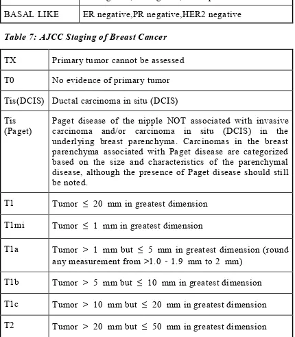
Table 6:Molecular Classification of Breast Cancer subtypes
Luminal A ER positive,PR positive,HER2 negative
Luminal B ER positive,PR positive,HER2 positive
HER 2 LIKE ER negative,PR negative,HER2 positive
[image:31.595.84.514.237.728.2]BASAL LIKE ER negative,PR negative,HER2 negative
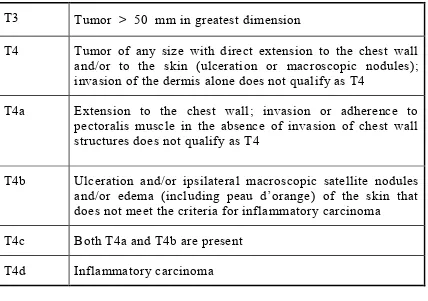
Table 7: AJCC Staging of Breast Cancer
TX Primary tumor cannot be assessed
T0 No evidence of primary tumor
Tis(DCIS) Ductal carcinoma in situ (DCIS)
Tis (Paget)
Paget disease of the nipple NOT associated with invasive carcinoma and/or carcinoma in situ (DCIS) in the underlying breast parenchyma. Carcinomas in the breast parenchyma associated with Paget disease are categorized based on the size and characteristics of the parenchymal disease, although the presence of Paget disease should still be noted.
T1 Tumor ≤ 20 mm in greatest dimension
T1mi Tumor ≤ 1 mm in greatest dimension
T1a Tumor > 1 mm but ≤ 5 mm in greatest dimension (round any measurement from >1.0‐1.9 mm to 2 mm)
T1b Tumor > 5 mm but ≤ 10 mm in greatest dimension
T1c Tumor > 10 mm but ≤ 20 mm in greatest dimension
T3 Tumor > 50 mm in greatest dimension
T4 Tumor of any size with direct extension to the chest wall and/or to the skin (ulceration or macroscopic nodules); invasion of the dermis alone does not qualify as T4
T4a Extension to the chest wall; invasion or adherence to pectoralis muscle in the absence of invasion of chest wall structures does not qualify as T4
T4b Ulceration and/or ipsilateral macroscopic satellite nodules
and/or edema (including peau d‟orange) of the skin that
does not meet the criteria for inflammatory carcinoma
T4c Both T4a and T4b are present
[image:32.595.86.514.70.371.2]T4d Inflammatory carcinoma
Table 8:Survival by Presenting Stage of Breast Cancer
Stage 5 Year survival (%)
0 100%
I 98%
IIA 88%
IIB 76%
IIIA 56%
IIIB, IIIC 49%
Table 9: Incidence of Lymph Node Metastasis in Invasive Breast Carcinoma
Tumour size (cm) <0.5 0.5-1.0 1.1-2.0 2.1-3.0 3.1-5.0
Incidence (%) 3-7% 12-17% 20-30% 35-45% 40-60%
WORK UP
1) Physical examination - loco regional extent of tumour i.e.
size,ulceration,nodules,peau d orange,dimpling of the skin and
examination of potential sites of spread.
2) Laboratory studies include a complete blood cell cou nt, serum
bichemistry profile, and liver function tests.
3) If liver function values are abnormal, a computed tomography
(CT) scan of the abdomen should be obtained plus skeletal survey
and bone scan should be done.
4) If anaemia, leukopenia, or thrombocytopenia is present, bone
marrow biopsy might be necessary.
5) Radiographic studies include chest x-ray, bone scans, and plain
radiographs of symptomatic regions or suspicious areas of
increased uptake on bone scans .
6) CT scans of the chest and abdomen are obtained ro utinely.
8) If neurologic symptoms suggest brain metastases, a contrast
-enhanced CT scan or gadolinium--enhanced magnetic resonance
imaging scan of the brain should be obtained; magnetic resonance
imaging is preferred if leptomeningeal carcinomatosis is
suspected .
TREATMENT OF BREAST CANCER
Multimodality integrated treatment has been proved the best
tumour control and survival for early stages, breast conservative
treatment includes a combination of the three treatment modalities
surgery, radiation therapy, and chemotherapy have shown the
effectiveness required for survival.For advanced stages, the primary
treatment includes a combination of surgery and chemotherapy, with
selective use of postmastectomy radiation therapy in certain cases.
SURGERY
Lumpectomy is appropriate for DCIS, and stages T1, T2 ,Invasive
Ductal or lobular Ca Breast. Mastectomy is indicated in all patients who
are not Suitable for breast conservation. Breast conservation surgery
(BCS) may consist of (in order of decreasing tissue removed) :
quadrantectomy, wide excision, lumpectomy (local excision) .
Indications for surgery
T3,T4 stage
CONTRAINDICATIONS FOR SURGERY
Metastatic disease
VARIATIONS OF MASTECTOMY
1) Radical Mastectomy: removal of breast, pectoralis minor and
major muscles, axillary LN dissection (ALND) (levels I–III)
2) Modified Radical Mastectomy removal of breast to the level of
pectoralis minor muscle, ALND (levels I–II) while the spared one
is pectoralis major
3) Total (Simple) Mastectomy is removal of breast to the level of
pectoralis minor muscle with no lymph node dissection
4) Skin Sparing Mastectomy: preserves skin of the breast for
enhanced reconstructive cosmetic outcomes
Total Skin Sparing Mastectomy preserves skin and nipple/
areolar complex for enhanced reconstructive outcome for invasive
cancers, sentinel axillary lymph node sampling with or without
subsequent Axillary dissection for positive sentinel node is routinely
completed reconstructive options postmastectomy include delayed vs.
Immediate, and autologous tissue vs. expander/implant.
SYSTEMIC THERAPY
30%, respectively. This translated into a 10% absolute improvement in
15-year survival (HR = 42% vs. 32%). For women aged 50 to 69 years,
the annual risk of relapse or death from Ca Breast was decreased by
19% and 12%, respectively. This translated into a 3% absolute gain in
15-year survival (HR = 50% vs. 47%). The absolute gain in survival for
polychemotherapy versus no adjuvant therapy in Women younger than
50 was twice as great at 15 years as it was at 5 years(10% vs. 4.7%),
while the main effect on disease recurrence was present in the first 5
years.
Anthracyclines (doxorubicin)-based regiments (± taxanes for
high-risk disease) have been associated with superior out comes as
compared to nonanthracycline containing regimens with recent evidence
augmenting the increased DFS and OS with taxane-based therapy as
compared to anthracyclin-based therapy
Neoadjuvant chemotherapy is considered standard of care in high
-risk populations such as young patients and/or advanced- stage disease,
and has been evaluated in Stage II– IIIa Ca Breast in large number of
randomized trials
Neoadjuvant chemotherapy converts 20–30% of patients initially
ineligible for BCT to eligible category patients. For advanced -stage
HORMONAL THERAPY
Hormonal therapy has been indicated in almost many patients.It is
usually with tablet Tamoxifen or Tablet Letrozole and the post
menopausal women Ca Breasts are more ER / PR positive, they respond
well with hormonal therapy in the form of loco regional control and
disease progression.In case of pre menopausal women hormonal therapy
is indicated only if there is positive ER / PR status.
RATIONALE FOR USING POST MASTECTOMY
RADIATION THERAPY
Virtually every Post Mastectomy Radiation Therapy randomized
trial to date has demonstrated a reduction in the risk of loco regional
recurrence with the use of comprehensive Radiotherapy.Prevention of
Loco regional failure is an important part in oncology m anagement and
loco regional recurrences can be subsequently controlled by Post
Mastectomy Radiation Therapy to improve survival.Radiotherapy must
be able to sterilize residual loco regional disease which, if left untreated,
could lead to disease progression.The patients who would potentially
benefit are those without micro metastatic disease at presentation or
patients with micro metastatic disease effectively treated by systemic
therapy. Randomized trials have shown inconsistent reduction in the
rates of Loco Regional Failure with systemic therapies alone and the
RADIOTHERAPY TREATMENT TECHNIQUES
Early Breast Cancer Trialists‟ Collaborative Group published
many meta-analysis which shows that usually the post mastectomy
irradiation of the chest wall is done in all T3, T4 disease, and positive
margin and in case of > 4 nodes positive for tumor. The Principle behind
is to sterilize the micro metastasis presents locally and regionally.
The technique of the treatment is patients positioned usually in
supine position with ipsilateral arm abducted to 90º at shoulder level and
the elbow flexed at 90º and the head is turned towards opposite side.
The treatment consists of two parts , flap and drainage field. The target
volume of the Chest wall field includes chest wall with a small portion
of underlying lung in the Irradiated volume .When it is combined with a
supraclavicular portal, the upper margin of the portal is placed at the
second intercostals space (the angle of Louis).
The medial margin, if no internal mammary portal is used, should
be at or 1 cm across the Midline. If an internal mammary field is used,
the medial margin is 3cm across the midline ,covering the first three
intercostals spaces. The lateral margin is usually near the mid-axillary
line. The inferior margin is drawn 2 to 3 cm below the the
inframammary fold . The portals used are medial and lateral tangential
If only the apex of the axilla is treated (after modified radical
mastectomy or axillary Dissection), the inferior border of the
supraclavicular field is the first or second intercostal space . The medial
border is 1 cm across the midline, extending upward, following the
medial border of the sternocleidomastoid muscle to the thyrocricoid
groove.
The lateral border is a vertical line at the deltoid insertion with
the humeral head blocked as much as possible without compromising
coverage of the high Axillary lymph nodes. This field is angled
approximately 15 to 20 degrees laterally to spare the spinal cord . The
portal is single AP given at D-max level or at 3 cm depth.
DOSE PRESCRIPTION IN PMRT
The conventional radiotherapy dose followed for postmastectomy
radiotherapy across many radiation oncology centres is 50 GY given i n
25 fractions over a period of 5 weeks including PA boost.Thus resulting
a lot of burden on the existing infrastructure and manpower which are
already stretched because of ever growing patients who are in need of
RT, a hypofractionated Protocol i.e. reducing both the number of
fractions and total treatment duration is being evalauted .
For a number of years, centres in the United Kingdom and Canada
fraction given over a shorter period of time is just as effective as the
more traditional longer schedule . Schedules used have ranged from 40
to 45 Gy in 15–20 fractions, administered over 19–22 days with fraction
sizes of 2.3–2.7Gy.
The use of a 15-fractions, instead of a 25-fractions regime, for
instance , would save 1000 treatment sessions per 100 patients (2500 -
1500 = 1000).
This corresponds to an additional 66 (1000:15) patients who could
be treated with the same number of fractions. This would result in
substantial economic benefit as Carcinoma Breast patients represent the
majority of patients treated in Radiotherapy departments.
In a well-known randomized trial from Canada, Whelan et al
reported equivalent results for a 10 year follow up (regarding local
control, survival, and post-radiation effects) between the standard
fractionation schedule of 50 Gy in 25 fractions and a hypofractionation
scheme of 42.5 Gy in 16 fractions over 22 days for women with node
-negative early Ca Breast.
The START A trial randomized 2236 patients from 17 centres
across the UK and reported that 41.6 Gy/13 fractions or 39 Gy/13
fractions are similar to 50 Gy/25 fractions in terms of local -regional
The START A trial showed that after a median follow -up of 5.1
years, the rate of local-regional tumor relapse at 5 years was 3.6% [95%
confidence interval (CI): 2.2%-5.1%] after 50 Gy, 3.5% (95% CI:
2.1%-4.3%) after 41.6 Gy, and 5.2% (95% CI: 3.5%-6.9%) after 39
Gy. The estimated absolute differences in 5-year local-regional
relapse rates compared with 50 Gy were 0.2% (95% CI: -1.3%-2.6%)
after 41.6 Gy and 0.9% (95% CI: -0.8%-3.7%) after 39 Gy.
The START B trial randomized 2215 patients from 23 centre s
across the UK and reported that a RT schedule of 40 Gy/15 fractions
offers equivalent results to the standard schedule of 50 Gy/25 fractions.
After a median follow-up of 6.0 years, the rate of Local-regional tumour
relapse at 5 years was 2.2% (95% CI: 1.3%-3.1%) in the 40 Gy group
and 3.3% (95% CI: 2.2%-4.5%) in the 50 Gy group. Photographic and
patient self assessments indicated lower rates of late adverse effects
after 40 Gy than after 50 Gy.
Another short RT schedule, 40 Gy in 15 fractions, has been us ed
traditionally at Christie Hospital in Manchester, UK; the reported results
of 2159 treated patients are Comparable to those reported from other
centres, this schedule is now becoming the “standard” in the UK,
Institute of Nuclear Medicine & Oncology (INMOL) Pakistan,
published a Randomised result of three hypofractionation protocols in
postmastectomy carcinoma breast in terms of local control, toxicity and
work load .A total of three hundred patients with stage T2-4, N any were
randomized into three arms after mastectomy. All the patients were
treated with four fields on Co60 and were randomized into three arms
i.e. 2700 CGy in 5 fractions (one week) arm A,3500 CGy in 10
fractions(2 weeks)arm B and 4000 CGy in 15 fractions (3 weeks) arm C
Skin, cardiac, pulmonary and haematological toxicities and
lymphoedema, and workload were studied. The loco regional relapses
were 11%, 12% and 10% in arms A, B and C respectively. 26%, 24%
and 28% patients developed metastatic disease and 17%, 18% and 20%
died in the three arms . G3 and G4 skin toxicities were 37%, 28% and
14%. G2 and G3 lymphedoema was 21%, 22% and 27% . Cardiac
toxicity was 5%, 6% and 5% while pulmonary toxicity was 4%, 5% and
5% respectively . All the differences except skin toxicity were
statistically Insignificant . There were no cases of haematological
depression or rib fractures all the three short protocols were equally
effective in loco regional disease control and toxicity was also
comparable.
An Indian Study from January 1989 to June 1992 on 108 Ca
Breast patients at Pt. B. D. Sharma Post Graduate Institute of Medical
randomly divided into 2 groups (A&B), 54 patients in Group A a nd 54
patients in Group B. Group A patients were given external radiotherapy .
40 Gy in 17 fractions over 3.2 weeks and Group B patients were given
45 Gy in 20 fractions over 4 weeks Results of treatment in Group A
versus Group B were as follows; chest wall failure 5/50 (10%) versus
3/54 (5.6% ), axillary lymph node failure 3/50 (6%) versus 4/ 54 (7%),
distant metastasis 16/50 (32%) versus 15/54 (28%). Most of the patients
in both the groups had no evidence of disease at last follow -up i.e. 26/50
(52%) in Group A and 32/54 (59%) in Group B. There was no
statistically significant difference in local control and efficacy of these
two radiation schedules in post mastectomy carcinoma of the breast.
Gary M Freedom et al four week course of radiation for Ca Breast
using hypofractionated intensity modulated radiation therapy (IMRT)
with an incorporated electron boost. A total of 75 pts (age > 18 yrs)
treated with 45 Gy to the whole breast and the lumpectomy bed 56Gy in
20 treatments over 4 wks The treatment was feas ible and is associated
with acceptable acute skin toxicity. This may represent an alternative to
longer 6-week standard radiotherapy
“LIMITS OF HYPOFRACT
IONATION IN BREAST RT: FAST
This trial tested 30 Gy in 5 fractions (6 Gy/fraction) over 35 d (5
35 d (control arm) No recurrences were seen after a median follow -up of
[image:44.595.91.502.170.349.2]3 years.
Table 10: Comparision of hypofractionation schedules
Trial Dose No. of
Weeks %CRF
% Good Excellent Cosmesis
Follow-up (Yr)
RMH/ GOC
50Gy/2Gy/25 5 12.1 47* 10act
39Gy/3Gy/13 5 14.8 42*
42.9Gy/2Gy/25 5 9.6 44*
Canadian 50Gy/ 2Gy/25 5 6.7 70 10act
42.5Gy/ 2.66Gy/16
3.1 6.2 71
Start-A 50Gy/2Gy/25 5 3.6 59* 5
41.6Gy/3.2Gy/13 5 3.5 59*
39Gy/3Gy/13 5 5.2 70*
Start-B 50Gy/2Gy/5 5 3.3 57* 5
RADIOBIOLOGY
The Optimization of a RT schedule for individual patients with
regards to Total dose and number of fractions and overall time of the
radiotherapy course keeping in thought about Tumour Control
probability (TCP) and normal tissue Complication probability (NTCP) is
possible. The response depends not only on dose but also on individual
Radio sensitivity, timing, fraction size, other agents given concurrently.
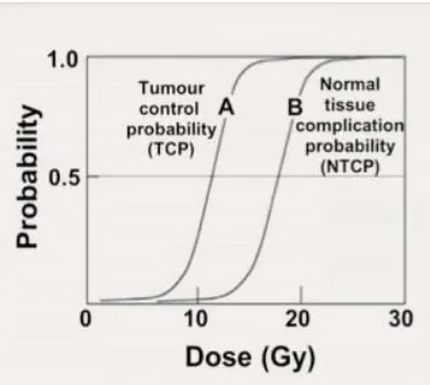
NORMAL AND TUMOUR CELLS: THERAPEUTIC RATIO
The principle of radiotherapy is demonstrated by plotting two
[image:45.595.200.395.390.565.2]sigmoid curves.
Figure 2: graph showing therapeutic ratio
The principle of therapeutic ratio, Curve A represents the TCP,
curve B the Probability Of Complications, the total clinical dose is
usually delivered in 2 Gy fractions. Thus optimum choice of radiation
maximizes the TCP and simultaneously minimizes the NTCP for a
typical good radiotherapy treatment, TCP ≥ 0.5 and NTCP < 0.05
The further curve B (NTCP) is to the right of curve A (TCP) the
easier it is to achieve the radio therapeutic goal, the larger is the
therapeutic ratio and the less likely will it be that the treatment causes
complications. The therapeutic ratio generally refers to the ratio of the
TCP and NTCP at a specified level of response (usually 0.05) for normal
tissue.
CELL SURVIVAL CURVE
The surviving fraction of cells, i.e., the fraction of irradiated cells
that maintain their reproductive integrity (clonogenic cells). Several
factors can modify the cells less radio-sensitive:
Removal of oxygen to create a hypoxic state.
Use of low dose rates or multi-fractionated irradiation
Addition of chemical radical scavengers
Synchronization of cells in the late S phase of the cell cycle.
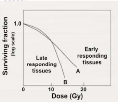
PROPERTIES OF CELL SURVIVAL CURVES
late responding tissues indicate the survival curves are more
In early effects the ratio α/β is large; for late effects it is small
and α dominates at low doses.
For late effects β has an influence at doses lower than for early
responding tissues
The α and β components of mammalian cell killing are equal at
the following doses:
o α/β ≈ 10 Gy for early responding tissues
[image:47.595.199.394.370.540.2]o α/β ≈ 3 Gy for late responding tissues
Figure 3: graph showing early vs late responding tissue in terms of dose and surving fraction
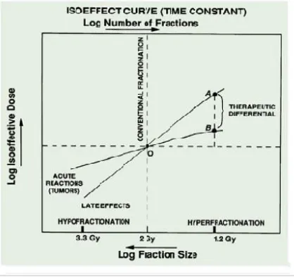
ISOEFFECT FORMULA
The effect of changes of dose fractionation schedule on the total
dose required to produce a certain level of biologic effect is
Fowler and Stern.These observations led to the formulation of Normal
Standard Dose (NSD) formula of Ellis
D ≈ N0.24
x T0.11
[image:48.595.193.402.226.423.2]Where, N = Number of dose fractions, T = Overall time.
Figure 4:Isoeffect curve on effects and therapeutic potential
This formula is purported to represent the effect of changing
fractionation parameters on the tolerance of normal con nective tissue,
which are considered to impose the ultimate dose limitation for all
radiation therapy. From the NSD concept, tolerance dose formula and
the cumulative radiation effect (CRE) formula were derived Later, it was
recognized that the exponents N and T used in NSD formula was not
appropriate for all tissue types . In general, Isoeffect curves for late
smaller components of T. This led to the modification of the formula on
Tissue specific basis.
Thames et al stated that curvier survival curve would be expected
for target cells in tissues in which isoeffect dose increased more steeply
with Decreasing dose per fraction and that this steepness could be
quantified by the ratio α/β in case the LQ model correctly described
target cell response
THE LINEAR-QUADRATIC MODEL
Is most often used in describing the cell surviving fraction S(D),
with the assumption that there are two components to cell kill by
radiation (linear and quadratic):
Equation 1
α is a constant describing the initial slope of the cell survival
curve
β is a smaller constant describing the quadratic component.
The currently used model for describing the cell survival curve is
the the linear quadratic model with constants α and β. The ratio α/β
Figure 5 linear quadratic model
The LQ formalism is now almost universally used for calculating
Radio therapeutic isoeffect (31),In summary, LQ has the fol lowing
useful properties for predicting isoeffect useful properties for predicting
isoeffect doses
1) A mechanistic, biologically-based model
2) Has sufficiently few parameters to be practical
3) Most other mechanistic models of cell killing predict the same
fractionation dependencies as does LQ fourth, it has well
documented predictive properties for fractionation/dose - rate
effects in the laboratory
4) Well validated, experimentally and theoretically, up to about 10
Gy / fraction, and would be reasonable for use up to about 18 Gy
RELATIVE BIOLOGICAL EFFECTIVENESS
Relative biological effectiveness (RBE) compares the dose of
radiation to the dose of standard radiation to produce the same
biological effect.
Dose from standard radiation to produce a given biological effect RBE = ---
Dose from test radiation to produce the same biological effect
Equation 2
The RBE varies with
1) Type of radiation
2) Type of cell/ tissue
3) Biologic effect under investigation
4) Dose rate
5) Fractionation
The RBE increases with the LET to reach a maximum RBE of 3–8
depending on the level of cell kill) at LET 200 keV/m and then
decreases because of energy overkill, an increase in the RBE in itself
offers no therapeutic advantage unless there is a differential effect
making the RBE for normal tissue smaller than that for the tumour,
Increasing the relative level of tumour cell killing and the therapeutic
The isoeffect curves vary for different tissues. A biolo gically
equivalent dose (BED) can be obtained using this formula
Equation 3
Use of BED to calculate equivalent doses when changing
fractionation, comparison of equivalent regimens and determination of
the isoeffective single dose in groups of tumours wit h similar kinetics
(α/β ratio) is possible. (33)
Equation 4
n = standard number of fractions, n 1 = equivalent number of
fractions in altered schedule, d = standard dose/fraction, d 1= desired
dose/fraction
/
FOR BREAST TISSUE
Low α/β ratio for breast cancer has drawn a growing interest for
exploring Hypofractionation for breast irradiation, Post operative
radiotherapy (RT) is widely used to reduce local recurrence of
carcinoma Mammary Gland. Conventionally, a dose per fraction of 1.8–
used. Recently, it has been argued that the α/β ratio for carcinoma
Mammary Gland may be lower than previously Considered .
Based on large randomized clinical trials of whole breast
irradiation, the analysis from multiple institutions yields ratios in the
range of 0.75–5.01 Gy 95% CL, supporting that carcinoma Mammary
Gland has a low α/β ratio. Such a low α/β ratio suggests that
hypofractioated radiotherapy regimens may be advantageous for
carcinoma Mammary Gland.
Using the low α/β ratio , selected possible fractionated regimens
that are radio biologically equivalent to the conventional 2.0 Gy* 25 # ,
regimens were calculated and were studied in many studies with a
[image:53.595.97.502.513.643.2]standard value of 3 for delayed effects and 10 for early reactions
Table 11: Radiobiological parameters of hypofractionated trials
CONVENTIONAL FRACTIONATION
It is the application of daily doses of 1.8 to 2 Gy and 5 fractions
HYPOFRACTIONATION
Hypofractionation means the use of a reduced number of
fractions, or a larger dose Per Fraction, if the dose per fraction is
increased above the reference value of 2 Gy, then the total dose (for an
[image:54.595.172.421.253.422.2]isoeffect) must be reduced as indicated in this graph
.
Figure 6:comparision based on α/β
Compared with a reference treatment using 2 Gy per fraction, the
diagram shows how the total dose must be changed in order to maintain
a constant level of effect when dose per fraction is modified. Full lines
are for low α/β ratios (as in late-responding normal tissues), and broken
lines are for early-responding normal tissues or for most tumours
For tumours with lower α/β values compared with the
Surrounding normal tissues, hypofractionation may be a viable option to
hypofractionation may also be a convenient way to accelerate the
treatment, thereby counteracting repopulation
Most tumors are actively proliferating and rapidly responding, and
their fractionation response is similar to that of acute -responding normal
tissues, although there are exceptions. one of the main principles of
radiobiology is that the late effects of normal tissues are stron gly
dependent on the size of dose per fraction, so that the higher the dose
per fraction the greater the susceptibility of healthy tissues to radiation.
This is known as “fractionation sensitivity
Fractionation sensitivity of tissues is quantified, in term s of linear
quadratic (LQ) isoeffect formulation, by the α/β ratio the higher the
sensitivity to the size of dose per fraction, the lower the α/β ratio is
Additionally the only drawback of applying a Hypofractionation Scheme
AIMS AND OBJECTIVES
PRIMARY OBJECTIVE
To assess the loco regional tumour control between conventional
fractionation radiotherapy and hypofractionation radiotherapy in post
operative carcinoma breast patients.
SECONDARY OBJECTIVE
To evaluate the toxicities and quality of life and compliance of
MATERIALS AND METHODS
RECRUITMENT
All The patients in this study were registered in the Department of
Radiotherapy,BIRO,Rajiv Gandhi Government General Hospital.
Assessment was done for the requirement criteria of the study
The study period was for the period of July 2017-June 2018
60 patients were recruited,with 30 each in conventional arm
<ARM A> and 30 each in hypofractionated arm<ARM B>
DATA COLLECTION
Personal and clinical data of the patients were collected such as –
Name, Age, Sex, Address, Occupation, Presenting symptoms, Past
medical / surgical history, Co-Morbidities, family history, General
examination,Local examination.
Their treatment plan was completing chemotherapy,surgery and
then radiotherapy <i.e. Post Mastectomy Radiation Therapy>
RANDOMIZATION OF THE PATIENTS
60 patients were recruited and then divided 30 into each arm,
hypofractionated and conventional arm. This selection was randomized
INCLUSION CRITERIA
Histologically proven malignant tumour of the breast
Tumour staging-T2/T3/T4,Nodal Staging-N0/N1/N2/N, Metastasis
- MO STAGING
Underwent Modified Radical Mastectomy
EXCLUSION CRITERIA
Metastatic breast cancer patients
History of non malignant disorder <severe cardiac or severe
pulmonary disease>
Psychiatric illness
Co mordibities – uncontrolled diabetes/uncontrolled hypertension
LIST OF INVESTIGATIONS DONE BEFORE THE STUDY
Lab Investigations
Metastatic survey
Pulmonary function test
ECHO
TREATMENT PLAN
2D planning
External beam radiotherapy-Post Mastectomy Radiotherapy
Chest wall-2 tangential fields
Drainage - Single anterior field
Posterior axillary boost
Machine: Cobalt 60 <Theratron Phoenix 60 >
Position: Supine with shoulder flexed at 90degree and elbow
flexed at 90 degree
Shielding: Humerus and trachea shielding
[image:61.595.81.522.519.649.2]DOSE
Table 12:Conventional and Hypofractionation arm schedules
Arm A Arm B
Dose per Fraction 2Gy 2.67Gy
No. Of Fractions 25# 15#
Total Dose 50Gy 40Gy
Duration 5 weeks 3 weeks
Arm A- conventional arm , 2Gy/25#/50Gy
Field borders and isocentres was marked with permanent marker
for each patient,based on anatomical landmarks.Patient was positioned
and instructed to maintain the same position till treatment is over.The
position for all patients was supine with the arm abducted at 90degree
and elbow flexed at 90 degree and the head turned to t he opposite side.
CHEST WALL FIELD
Upper border- second intercostal space
Lower border-2cm below the inframammary fold
Medial border – midline
Lateral border- mid axillary line
Two tangential portals were used to treat the chest wall
While marking the field borders,the scar and the drain site was
also covered in the field.
Chest wall contour was taken and drawn in the paper and target
volume was marked.
Chest wall thickness was determined using ultrasound technique
Figure 7:Field borders
ASSESSMENT
Before starting on radiotherapy
Skin assessment
Lymph edema
PFT
ECHO
At regular intervals, that is weekly, monthly, 3 and 6 monthly
periods –assessment of the following was done
Skin toxicity
Lymph edema
PFT
ECHO
Table 13: ECOG grading
Grade ECOG Performance Status
0 Fully active, able to carry on all pre-disease performance without restriction
1 Restricted in physically strenuous activity but ambulatory and able to carry out work of a light or sedentary nature, e.g., light house work, office work
2 Ambulatory and capable of all selfcare but unable to carry out any work activities; up and about more than 50% of waking hours
3 Capable of only limited selfcare; confined to bed or chair more than 50% of waking hours
4 Completely disabled; cannot carry on any selfcare; totally confined to bed or chair
[image:64.595.86.517.414.751.2]5 Dead
Table 14:Skin toxicity grading(CTCAEv5)
Grade 1 Faint erythema or dry desquamation
Grade 2
Moderate to brisk erythema; patchy moist desquamation, mostly confined to skin folds and creases; moderate edema
Grade 3
Moist desquamation in areas other than skin folds and creases; bleeding induced by minor trauma or abrasion
Grade 4
Life-threatening consequences; skin necrosis or ulceration of full thickness dermis; spontaneous bleeding from involved site; skin graft indicated
Table 15:Grading of Radiation pneumonitis (CTCAEv5)
Grade 1 Asymptomatic; clinical or diagnostic observations only; intervention not indicated
Grade 2 Symptomatic; medical intervention indicated; limiting instrumental ADL
Grade 3 Severe symptoms; limiting self care ADL; oxygen indicated
Grade 4 Life-threatening respiratory compromise; urgent intervention indicated (e.g., tracheotomy or intubation)
Grade 5 Death
Table 16:Lymphedema (CTCAEv5)
Grade 1 Trace thickening or faint discoloration
Grade 2
Marked discoloration; leathery skin texture; papillary formation; limiting instrumental ADL
Grade 3 Severe symptoms; limiting self care ADL
PULMONARY FUNCTION TEST
PFT was measured and FEV1 and FVC was recorded before
starting RT and after 2 and 6 months
X-ray Chest was also done
Any abnormalities detected in the PFT and those patients were
CARDIAC TOXICITY
Baseline ECG
Baseline ECHO
Any abnormalities detected and those patients were excluded from
the study
QUALITY OF LIFE
EORTC QLQ - BR23
Patients sometimes report that they have the following symptoms
or problems. Please indicate the extent to which you have experienced
these symptoms or problems during the past week.
1-Not at all, 2-A little, 3-Quite a bit, 4-Very much
1 2 3 4
31. Did you have a dry mouth?
32. Did food and drink taste different than usual?
33. Were your eyes painful, irritated or watery?
34. Have you lost any hair?
35. Answer this question only if you had any hair loss: Were you upset by the loss of your hair?
36. Did you feel ill or unwell?
37. Did you have hot flushes?
1 2 3 4
39. Have you felt physically less attractive as a result of your disease or treatment?
40. Have you been feeling less feminine as a result of your disease or treatment?
41. Did you find it difficult to look at yourself naked?
42. Have you been dissatisfied with your body?
43. Were you worried about your health in the future?
44. To what extent were you interested in sex?
45. To what extent were you sexually active? (with or without intercourse)
46. Answer this question only if you have been sexually active: To what extent was sex enjoyable for you?
47. Did you have any pain in your arm or shoulder?
48. Did you have a swollen arm or hand?
49. Was it difficult to raise your arm or to move it sideways?
50. Have you had any pain in the area of your affected breast?
51. Was the area of your affected breast swollen?
52. Was the area of your affected breast oversensitive?
ENGLISH
EORTC QLQ-C30
1 2 3 4
1. Do you have any trouble doing strenuous activities, like carrying a heavy shopping bag or a suitcase? 1 2 3 4
2. Do you have any trouble taking a long walk? 1 2 3 4
3. Do you have any trouble taking a short walk outside of the house? 1 2 3 4
4. Do you need to stay in bed or a chair during the day? 1 2 3 4
5. Do you need help with eating, dressing, washing yourself or using the toilet? 1 2 3 4
6. Were you limited in doing either your work or other daily activities? 1 2 3 4
7. Were you limited in pursuing your hobbies or other leisure time activities? 1 2 3 4
8. Were you short of breath? 1 2 3 4
9. Have you ha