ACID, ETIDRONIC ACID AND EDTA ON SMEAR LAYER
REMOVAL AND DENTIN EROSION AT DIFFERENT TIME
INTERVALS USING SCANNING ELECTRON MICROSCOPE -
AN IN VITRO STUDY
A Dissertation submitted
in partial fulfillment of the requirements
for the degree of
MASTER OF DENTAL SURGERY
BRANCH – IV
CONSERVATIVE DENTISTRY AND ENDODONTICS
THE TAMILNADU DR. MGR MEDICAL UNIVERSITY
CHENNAI – 600 032
I hereby declare that this dissertation titled “A COMPARATIVE
EVALUATION OF EFFICACY OF PHYTIC ACID, ETIDRONIC
ACID AND EDTA ON SMEAR LAYER REMOVAL AND DENTIN
EROSION
AT
DIFFERENT
TIME
INTERVALS
USING
SCANNING ELECTRON MICROSCOPE - AN IN VITRO STUDY”
is a bonafide and genuine research work carried out by me under the
guidance of
Dr. B. RAMAPRABHA, Professor,
Department Of
Conservative Dentistry and Endodontics, Tamil Nadu Government Dental
College and Hospital, Chennai-600003.
This is to certify that
Dr. SUMATHI K,
Post Graduate student
(2014-2017) in the Department of Conservative Dentistry and
Endodontics, TamilNadu Government Dental College and Hospital,
Chennai- 600003 has done this dissertation titled “A COMPARATIVE
EVALUATION OF EFFICACY OF PHYTIC ACID, ETIDRONIC
ACID AND EDTA ON SMEAR LAYER REMOVAL AND DENTIN
EROSION
AT
DIFFERENT
TIME
INTERVALS
USING
SCANNING ELECTRON MICROSCOPE - AN IN VITRO STUDY”
under my direct guidance and supervision in partial fulfillment of the
regulations laid down by the Tamil Nadu Dr. M.G.R Medical University
Chennai -600032, for M.D.S., Conservative Dentistry and Endodontics
(Branch IV) Degree Examination .
Dr. B. RAMAPRABHA, M.D.S.
Professor & Guide
Department of Conservative Dentistry and Endodontics.
Tamil Nadu Government Dental College and Hospital
HEAD OF THE INSTITUTION
This is to certify that the dissertation titled
“A COMPARATIVE
EVALUATION OF EFFICACY OF PHYTIC ACID, ETIDRONIC
ACID AND EDTA ON SMEAR LAYER REMOVAL AND DENTIN
EROSION
AT
DIFFERENT
TIME
INTERVALS
USING
SCANNING ELECTRON MICROSCOPE - AN IN VITRO STUDY”
is a bonafide research work done by
Dr SUMATHI K
, Post Graduate
student (2014-2017) in the Department of Conservative Dentistry &
Endodontics under the guidance of
Dr B RAMAPRABHA, M.D.S,
Professor and Guide,
Department Of Conservative Dentistry &
Endodontics, Tamil Nadu Government Dental College and Hospital,
Chennai-600003.
Dr. M. KAVITHA, M.D.S. Professor & HOD,
Dept of Conservative Dentistry & Endodontics
Dr. B.SARAVANAN, M.D.S, PhD. Principal
ACKNOWLEDGEMENT
I wish to place on record my deep sense of gratitude to my mentor DR. M. KAVITHA M.D.S., for the keen interest, inspiration, immense help and
expert guidance throughout the course of this study as Professor & Head of the Department of Conservative Dentistry and Endodontics, Tamilnadu Govt Dental College and Hospital, Chennai.
It is my immense pleasure to utilize this opportunity to show my heartfelt gratitude and sincere thanks to DR. B. RAMAPRABHA M.D.S, Professor & Guide, Department of Conservative Dentistry and Endodontics, Tamilnadu Govt. Dental College and Hospital, Chennai for her guidance, suggestions, source of inspiration and for the betterment of this dissertation.
I take this opportunity to convey my everlasting thanks and sincere gratitude to Dr. B. SARAVANAN, M.D.S, PhD, Principal, Tamilnadu Government Dental College and Hospital, Chennai for permitting me to utilize the available facilities in this institution.
My extended thanks to DR. K. AMUDHALAKSHMI M.D.S, DR. D. ARUNA RAJ M.D.S., Dr. A. NANDHINI M.D.S., DR. P. SHAKUNTHALA M.D.S., Associate Professors and all Assistant Professors, Dr. G. VINODH M.D.S., DR. M. S. SHARMILA M.D.S., DR. M . SUDHARSHANA RANJINI M.D.S., DR. N. SMITHA M.D.S., DR. S. JOTHILATHA M.D.S., DR. S. VENKATESH M.D.S, DR. S. DHANALAKSHMI M.D.S, for all the help, suggestions, encouragement and guidance throughout this study.
I express my heartfelt gratitude to MR. CHOLAN BABU AND MR SRINIVASAN for their guidance and support on SEM analysis at Anna University, Chennai.
My special thanks to MY PARENTS, SISTER, BROTHER, MY IN-LAWS, GRAND PARENTS AND FRIENDS for their moral support and encouragement in pursuing a career in dentistry.
I whole heartedly thank my husband VIVEKANANTHAN. S for all his moral support, patience & guidance.
I also thank my dear colleagues, seniors and juniors for their timely help and support
TITLE OF DISSERTATION
“A COMPARATIVE
EVALUATION OF EFFICACY
OF PHYTIC ACID,
ETIDRONIC ACID AND EDTA
ON SMEAR LAYER
REMOVAL AND DENTIN
EROSION AT DIFFERENT
TIME INTERVALS USING
SCANNING ELECTRON
MICROSCOPE-AN IN VITRO
STUDY”
PLACE OF THE STUDY Tamil Nadu Government Dental College & Hospital, Chennai- 3.
DURATION OF THE COURSE 3 YEARS
NAME OF THE GUIDE DR. B. RAMAPRABHA
HEAD OF THE DEPARTMENT DR. M. KAVITHA
I hereby declare that no part of dissertation will be utilized for gaining financial assistance or any promotion without obtaining prior permission of the Principal, Tamil Nadu Government Dental College & Hospital, Chennai – 3. In addition I declare that no part of this work will be published either in print or in electronic media without the guide who has been actively involved in dissertation. The author has the right to preserve for publish of the work solely with the prior permission of Principal, Tamil Nadu Government Dental College & Hospital, Chennai – 3
This agreement herein after the “Agreement” is entered into on this day Dec 2016 between the Tamil Nadu Government Dental College and Hospital represented by its Principal having address at Tamil Nadu Government Dental College and Hospital, Chennai - 600 003, (hereafter referred to as, ‘the college‘)
And
MRS. DR. B.RAMAPRABHA, M.D.S aged 47 years working as Professor in Department of Conservative Dentistry & Endodontics at the college, having residence address at 191/5,Green Fields Apts. R-30A, Ambattur, Thirumangalam High Road, Mugappair,Chennai-3 ( ‘herein after referred to as the Principal Investigator’)
And
MRS. DR. SUMATHI K aged 28 years currently studying as Post Graduate student in Department of Conservative Dentistry & Endodontics, Tamil Nadu Government Dental College and Hospital, Chennai 3 (herein after referred to as the PG student and coinvestigator‘).
Whereas the PG student as part of her curriculum undertakes to research on
“A COMPARATIVE EVALUATION OF EFFICACY OF PHYTIC ACID, ETIDRONIC ACID AND EDTA ON SMEAR LAYER REMOVAL AND DENTIN EROSION AT DIFFERENT TIME INTERVALS USING SCANNING ELECTRON MICROSCOPE - AN IN VITRO STUDY” for which purpose the Principal Investigator shall act as principal investigator and the college shall provide the requisite infrastructure based on availability and also provide facility to the PG student as to the extent possible as a Co-investigator.
Whereas the parties, by this agreement have mutually agreed to the various issues including in particular the copyright and confidentiality issues that arise in this regard.
Now this agreement witnesseth as follows
1. The parties agree that all the Research material and ownership therein shall become the vested right of the college, including in particular all the copyright in the literature including the study, research and all other related papers.
2. To the extent that the college has legal right to do so, shall grant to license or assign the copyright so vested with it for medical and/or commercial usage of interested persons/entities subject to a reasonable terms/conditions including royalty as deemed by the college.
of research/study in any manner whatsoever, which shall also vest with the college. 5. The PG student and Principal Investigator undertake not to divulge (or) cause to be divulged any of the confidential information or, know-how to anyone in any manner whatsoever and for any purpose without the express written consent of the college. 6. All expenses pertaining to the research shall be decided upon by the Principal Investigator/ Coinvestigator or borne solely by the PG student. (co-investigator) 7. The college shall provide all infrastructure and access facilities within and in other institutes to the extent possible. This includes patient interactions, introductory letters, recommendation letters and such other acts required in this regard.
8. The Principal Investigator shall suitably guide the Student Research right from selection of the Research Topic and Area till its completion. However the selection and conduct of research, topic and area of research by the student researcher under guidance from the Principal Investigator shall be subject to the prior approval, recommendations and comments of the Ethical Committee of the College constituted for this purpose.
9. It is agreed that as regards other aspects not covered under this agreement, but which pertain to the research undertaken by the PG student, under guidance from the Principal Investigator, the decision of the college shall be binding and final.
10. If any dispute arises as to the matters related or connected to this agreement herein, it shall be referred to arbitration in accordance with the provisions of the Arbitration and Conciliation Act 1996.
In witness where of the parties herein above mentioned have on this day, month and year herein above mentioned set their hands to this agreement in the presence of the following two witnesses
College represented by its Principal PG Student
Witnesses Student Guide 1.
2
Aim: The purpose of this in vitro study was to compare the smear layer removal efficacy and dentin erosion of three different irrigating solutions at different time intervals of the root canal under Scanning Electron Microscopy.
Materials and Methods: One hundred extracted human single straight rooted maxillary central incisors were taken and decoronated to standardize the canal length of 15 mm. They were instrumented with ProTaper NEXT rotary system to an apical preparation of file size X5. Prepared teeth were irrigated with 3ml of 3% NaOCl for 5min followed by final rinse of 2ml of 1% phytic acid (Group I), 18% Etidronic acid (Group II) and 17% EDTA (Group III) at 5min, 3min and 1min. The canals of teeth in Control (Group IV) did not receive any final irrigation. The teeth were sectioned longitudinally and prepared for an SEM evaluation. The dentinal wall of cervical, middle and apical thirds were graded according to the amount of smear layer remaining and dentin erosion on the root canal walls. The results were analysed using the Kruskal–Wallis and Mann Whitney U tests with significance set at P < 0.05. Results: Intergroup comparison showed statistically no significant difference (p=1.000) in the smear layer removal efficacy of irrigants tested at 5min, 3min and 1min except for Etidronic acid (Group II) at 1min (p=.000). Control (Group IV) showed statistically high significant difference (p=.000) than other groups. Apical region of all groups showed statistically high significant difference (p=.000) than cervical and middle region. Intergroup comparison of dentin erosion showed EDTA (Group III) had high erosion values (p=.000) than other groups which are statistically significant. Phytic acid (Group I) showed less erosion values (p=.000) than other groups which are highly significant.
Conclusion: Phytic acid showed effective smear layer removal with less erosion of the root canal wall.Increasing the duration of irrigation does not improve the smear layer removal efficacy of irrigants except for Etidronic acid (Group II) but all groups showed more erosion at increased irrigation time. All the groups did not completely remove the smear layer at the apical region.
TABLE NO
TITLE
PAGE
NO
1 EXPERIMENTAL MATERIALS USED IN THE STUDY 21
2 IRRIGATION PROTOCOL USED IN THE STUDY
23
3 SMEAR LAYER & DENTIN EROSION SCORE
GRADING
24
4 SMEAR LAYER SCORES OF ALL GROUPS
32
5 DENTIN EROSION SCORES OF ALL GROUPS
33
6 DESCRIPTIVE TABLE SHOWING MEAN, MEDIAN
AND STANDARD DEVIATIONS OF SMEAR LAYER
34
7 ANALYSIS OF SMEAR LAYER VALUES AMONG
DIFFERENT GROUPS USING KRUSKAL WALLIS
35
8 INDIVIDUAL COMPARISONS OF SMEAR LAYER
VALUES USING MANN WHITNEY U TEST
35
9 DESCRIPTIVE TABLE SHOWING MEAN, MEDIAN
AND STANDARD DEVIATIONS OF DENTIN EROSION
36
10 ANALYSIS OF DENTIN EROSION VALUES AMONG DIFFERENT GROUPS USING KRUSKAL WALLIS
TEST
11 INDIVIDUAL COMPARISONS OF DENTIN EROSION
VALUES USING MANN WHITNEY U TEST
37
12 DESCRIPTIVE TABLE SHOWING MEAN, MEDIAN AND STANDARD DEVIATIONS OF SMEAR LAYER AND DENTIN EROSION OF PHYTIC ACID AT ALL
THIRDS
38
13 ANALYSIS OF SMEAR LAYER AND DENTIN EROSION VALUES OF PHYTIC ACID AT ALL THIRDS
USING KRUSKAL WALLIS TEST
39
14 INDIVIDUAL COMPARISONS OF SMEAR LAYER AND DENTIN EROSION VALUES OF PHYTIC ACID
AT ALL THIRDS USING MANN WHITNEY U TEST
39
15 DESCRIPTIVE TABLE SHOWING MEAN, MEDIAN AND STANDARD DEVIATIONS OF SMEAR LAYER AND DENTIN EROSION OF PHYTIC ACID AT 5, 3 & 1
MIN.
40
16 ANALYSIS OF SMEAR LAYER AND DENTIN EROSION VALUES OF PHYTIC ACID AT 5, 3, & 1MIN
USING KRUSKALWALLIS TEST
41
17 PAIRWISE COMPARISONS OF SMEAR LAYERAND DENTIN EROSION VALUES OF PHYTIC ACID AT 5, 3
& 1MIN USING MANN WHITNEY U TEST
41
18 DESCRIPTIVE TABLE SHOWING MEAN, MEDIAN AND STANDARD DEVIATIONS OF SMEAR LAYER
AND DENTIN EROSION VALUES OF ETIDRONIC ACID AT ALL THIRDS.
42
19 ANALYSIS OF SMEAR LAYER AND DENTIN EROSION VALUES OF ETIDRONIC ACID AT ALL
THIRDS USING KRUSKALWALLIS TEST
43
20
INDIVIDUAL COMPARISONS OF SMEARLAYER AND DENTIN EROSION VALUES OF ETIDRONIC ACID AT ALL THIRDS USING MANN WHITNEY U
TEST
21
DESCRIPTIVE TABLE SHOWING MEAN, MEDIAN AND STANDARD DEVIATIONS OF SMEAR LAYER AND DENTIN EROSION VALUES OF ETIDRONIC ACID AT 5, 3 & 1MIN
44
22 ANALYSIS OF SMEAR LAYER AND DENTIN EROSION VALUES OF ETIDRONIC ACID AT 5, 3 &
1MIN USING KRUSKALWALLIS TEST
45
23 PAIRWISE COMPARISONS OF SMEAR LAYER AND DENTIN EROSION VALUESOF ETIDRONIC ACID OF
5, 3 & 1MIN USING MANN WHITNEY U TEST
45
24 DESCRIPTIVE TABLE SHOWING MEAN, MEDIAN AND STANDARD DEVIATIONS OF SMEAR LAYER AND DENTIN EROSION VALUES OF EDTA AT ALL
THIRDS.
46
25 ANALYSIS OF SMEAR LAYER AND DENTIN EROSION VALUES OF EDTA AT ALL THIRDS USING
KRUSKALWALLIS TEST
47
26 INDIVIDUAL COMPARISONS OF SMEAR
LAYERAND DENTIN EROSION OF EDTA AT ALL THIRDS USING MANN WHITNEY U TEST
47
27 DESCRIPTIVE TABLE SHOWING MEAN, MEDIAN AND STANDARD DEVIATIONS OF SMEAR LAYER AND DENTIN EROSION VALUES OF EDTA AT 5, 3 &
1MIN
48
28 ANALYSIS OF SMEAR LAYER AND DENTIN EROSION VALUES OF EDTA AT 5, 3 & 1MIN USING
KRUSKAL WALLIS TEST
49
29 PAIRWISE COMPARISONS OF SMEAR LAYER AND DENTIN EROSION OF EDTA AT 5, 3 & 1MIN USING
MANN WHITNEY U TEST
GRAPH
NO
TITLE
PAGE
NO
1
SMEAR LAYER ANALYSIS AMONG THE
GROUPS
50
2
DENTIN EROSION ANALYSIS AMONG
THE GROUPS
EDTA ETHYLENE DIAMINE TETRA ACETIC ACID
HEDP 1-HYDROXYETHANE 1,1-DIPHOSPHONIC ACID
NaOCl SODIUM HYPOCHLORITE
SEM
SCANNING ELECTRON MICROSCOPYMin
MINUTES
NiTi
NICKEL TITANIUM
S.NO
TITLE
PAGE NO
1
INTRODUCTION
1
2
AIM AND OBJECTIVES
6
3
REVIEW OF LITERATURE
7
4
MATERIALS AND METHODS
20
5
RESULTS
28
6
DISCUSSION
54
7
SUMMARY
67
8
CONCLUSION
69
1
For a successful endodontic outcome, root canal system should be devoid of vital and necrotic remnants of pulp tissues, microorganisms and its toxins.80 However root canal system is highly complex and variable making it difficult to clean and disinfect.
Chemo mechanical preparation plays an important role in success of the endodontic treatment.18, 11 Pulpal tissue, microorganism and its byproducts are removed by instruments, irrigants and intracanal medicaments which are the main objectives of chemo mechanical phase.11 Even with the instrumentation, isthmi, canal fins and accessory canals are untouched.83, 60, 34 Therefore irrigation is an important part of root canal disinfection which cannot be achieved by instrumentation alone.92
Most irrigating solutions possess antimicrobial, tissue solvent and lubricant properties to facilitate root canal cleaning.30 Instrumentation of root canal results in accumulation of organic and inorganic material known as smear layer.9, 65, 54 During root canal instrumentation, it is almost inevitable for the formation of smear layer.80
2
The amount of smear layer produced is greater in rotary instrumentation than hand instrumentation. Some authors believe that smear layer may block the dentinal tubules and limit bacterial or its toxins penetration by altering dentinal permeability. Alternatively some others believe that smear layer may limit the action of irrigant, intracanal medicament by harboring microorganisms when left in the root canal and it can lead to microleakage acting as a barrier between sealing of root canal wall and the restorative materials. They may interfere with bonding mechanism of resins. There is still a controversy in removing or retaining the smear layer produced. However more evidence favors removal of smear layer rather than its retention.80
There are various methods to remove smear layer like chemical, ultrasonic and laser techniques. None of the methods remove smear layer throughout the length of the canal completely.80 Combination of methods help in achieving higher smear layer removal.
It is a well known fact that none of the currently used irrigating solutions have all the required properties of irrigant. Thus in common endodontic practice, dual irrigants are often used as initial and final rinse to overcome the disadvantages of using single irrigant. 21
3
with any syringe delivery system. However they are cytotoxic to periapical cells and hence should be used with caution. 6
Nygaard Ostby was the first to introduce chelating agents in endodontics. Chelating agents decalcify the dentine by combining with calcium ions of the tooth.They effectively removed smear layer at even low concentrations. With respect to chelating agents, effects of the decalcification depend heavily on the type of irrigant used, concentration, and pH of the solution and the application time.48 Most important aspect of disinfection is that irrigant should be in direct contact with entire root canal for effective action particularly with respect to apical regions of the root canals.
EDTA (ethylene diamine tetra acetic acid) is the most common chelating agent which reacts with dentine to form calcium chelates. 47 It lacks antimicrobial properties. Von der Fehr & Nygaard Ostby (1963) found that in 5 minutes; EDTA can decalcify dentine to the depth of around 20-30µm. Combination of EDTA and NaOCl solutions for effective removal of smear layer were recommended.47 Whenever NaOCl is used in combination with EDTA, NaOCl is inactivated earlier. This combination results in severe dentin erosion of root canal and dentinal tubules. 47 However the use of EDTA for more than 1 min may result in inadvertent erosion of root dentin. EDTA a synthetic, non-biodegradable material is considered a pollutant in root canal system and reported to be cytotoxic to macrophages. 2
4
effort to study two new chelating agents namely Phytic acid and Etidronic acid was taken.
Etidronic acid also known as 1-hydroxyethane 1,1-diphosphonic acid (HEDP) or etidronate. It is a bisphosphonate used in water treatment, cosmetics, detergents and pharmaceutical treatment. It emerged as the substitute for commonly used chelating agents. The advantage of etidronate is that it can be mixed with NaOCl without interfering in its antimicrobial properties. HEDP is a weak chelator, therefore it can be less aggressive on dentin than EDTA.74 However this solution may need longer time for removal of smear layer. It is biocompatible with periapical tissues
5
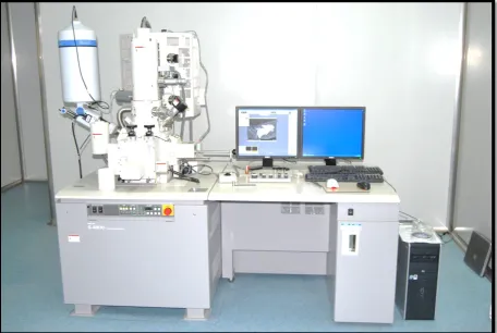
Scanning Electron Microscopic analysis is used in this study to obtain surface characteristics of dentin erosion and smear layer presence in root canal wall.
6 AIM:
The aim of the study was to compare and evaluate the effectiveness of three chelating solutions namely 1% PHTYIC ACID, 18% ETIDRONIC ACID and 17% EDTA on smear layer removal and dentin erosion at different time intervals after 3% NaOCl irrigation for 5min under Scanning Electron Microscopy.
OBJECTIVES:
To evaluate the effectiveness of 1% PHYTIC ACID on smear layer removal and dentin erosion at 5 min, 3 min and 1 min.
To evaluate the effectiveness of 18% ETIDRONIC ACID on smear layer removal and dentin erosion at 5 min, 3 min and 1 min.
7
SMEAR LAYER
Shahravan et al (2007) 66 did a systematic review to determine whether smear layer removal reduces leakage of obturated human teeth in vitro.It was concluded that smear layer removal helps in achieving the fluid-tight seal of the root canal system whereas other factors such as the sealer or the obturation technique, did not produce significant effects.
Pintor et al (2016)52 reviewed whether the smear layer removal procedure influences the outcome of root canal treatment. They concluded that the smear layer removal for root canal treatment of primary teeth with initial clinical signs and symptoms or necrotic status of pulp, could improve the treatment outcome, although further Randomized Control Trial should be performed to achieve evidence.
Likhitkar et al (2016)36 assessed the effect of the presence/absence of a smear layer
on the micro leakage of root canal filled teeth. Elimination of the smear layer enhanced the resistance to micro leakage;
ROTARY INSTRUMENTATION:
Khademi et al. (2006)8 determined to find the minimum instrumentation size required for the effective penetration of irrigants and elimination of debris and smear layer from the apical third of the root canals. They concluded that minimum instrumentation size needed for penetration of irrigants to the apical third of the root canal is a #30 file.
8
coronal and middle thirds, but in the apical third, they were unable to produce a canal surface free from debris and smear layer. However, ProTaper instrumented canals in the apical region showed smaller amounts of debris and smear layer.
Wadhwani et al (2011)82 evaluated the ability of 19% EDTA gel and 17% ethylene
diamine tetra acetic acid (EDTA) solution to remove debris, and smear layer produced during root canal istrumentation with two NiTi files systems, Mtwo and Protaper. They concluded that when used with EDTA gel and EDTA solution both the NiTi instruments produced a similar dentin surface on root canal wall.
Reddy et al (2014) 56 evaluated the amount of smear layer and debris removal on canal walls following the using of rotary ProTaper NiTi files compared with manual Nickel Titanium (NiTi) files using a Scanning Electron Microscope in two individual groups. They concluded that both systems of Rotary ProTaperNiTi and manual NiTi files used did not create completely clean root canals. Manual NiTi files produced significantly less smear layer and debris. Manual instruments were more time consuming when compared to rotary instruments.
Suparna et al (2015) 73 compared the cleaning efficacy of two different rotary file systems- WaveOne and ProTaper NEXT, using a Scanning Electron Microscope. They concluded that both the rotary systems ProTaper NEXT and WaveOne resulted in cleaner canals after instrumentation. However, the apical thirds of the root canals demonstrated more residual debris scores when compared to the middle and coronal thirds
Zarei et al (2016) 89 compared the influence of root canal taper (30/0.02 and 30/0.4) on
9
that greater smear layer was detected in the apical portion of each group. No statistical difference was found between canals with different tapers.
Kiran et al (2016) 32 evaluated the amount of smear layer and debris on the canal walls prepared with a combination of hand and rotary ProTaper technique using NaOCl and ethylene diamine tetra acetic acid (EDTA) alternately as root canal irrigants using scanning electron microscope (SEM).They concluded that none of the instrumentation techniques could completely eliminate the smear layer and debris from the root canal walls. Instrumentation of the canals with hand files after automated rotary preparation could result in cleaner canal walls. Alternative irrigation with NaOCl and EDTA is ineffective in the apical third.
IRRIGATION:
Kalyoncuoğlu and Demiryürek EÖ (2013) 28 evaluated the efficacy of smear layer
removal from teeth following root canals using lasers (Er:YAG and Nd:YAG), NaOCl, 17% EDTA, and MTAD by scanning electron microscopy (SEM). They concluded that although improvement was observed in removal of the smear layer using alternative materials and techniques, application of a combination of EDTA and NaOCl remains an effective technique.
10
Schmidt et al.(2015) 63 evaluated the efficacy of passive ultrasonic irrigation (PUI) with 17% EDTA and 1% NaOCl solutions on smear layer removal. They concluded that when compared with conventional irrigation, PUI did not show higher efficacy in smear layer removal
ROOT CANAL IRRIGANTS:
SODIUM HYPOCHLORITE:
Berber et al (2006) 8 evaluated the efficacy of 0.5%, 2.5% and 5.25% sodium hypochlorite (NaOCl) as intracanal irrigants against Enterococcus faecalis within root canals and dentinal tubule associated with hand and rotary instrumentation techniques. They found that 5.25% NaOCl was shown to be the most effective irrigant solution tested, when dentinal tubules were analysed at all depths and thirds of the root canals and for all techniques used, followed by 2.5% NaOCl. No differences among concentrations in cleaning the canals were found.
Zhang et al (2010) 93 studied the impact on the elastic modulus and flexure strength of standardized human root dentin bars of different irrigation sequences of EDTA (17%; 3 minutes) and NaOCl (2.5% w/v; total exposure time, 24 minutes) .They found that deleterious effects attributed to the use of NaOCl on dentin are time- dependent and concentration-dependent and they are not associated with the demineralization caused by the use of EDTA as the final active irrigant.
Marion et al (2012) 40 evaluated the effectiveness of various concentrations of sodium
11
of root canals is the 2.5% sodium hypochlorite concentration, due to its less cytotoxic properties.
Zargar et al (2015) 90 investigated the antibacterial efficacy in the presence and absence of smear layer (SL) by three root canal irrigants. The 2.61% solution of NaOCl was significantly more effective than 0.2% CHX and 0.2% CHX was more efficient than 1% PI for decreasing fungal and microbial infection of dentinal tubules. The presence of smear layer decreased the efficacy of antimicrobial irrigants.
EDTA:
Scelza et al (2004) 62 evaluated smear layer removal from root canal dentin by 17%
EDTA, EDTA-T, and 10% citric acid after final irrigation for 3, 10, and 15 min. They concluded that these 3 irrigants were effective at the shortest time tested and with an increase in time, they did not demonstrate an improved effect
Teixeira et al. (2005) 76 verified under the scanning electron microscope (SEM), the influence of irrigation time with sodium hypochlorite (NaOCl) and ethylenediaminetetraacetic acid (EDTA) on intracanal smear layer removal. They concluded that canal irrigation with EDTA and NaOCl were equally effective in removing the smear layer from the canal walls of straight roots for 1, 3 and 5 min.
Crumpton et al.(2005) 13 quantified the volume of 17% ethylene diamine tetra-acetic
12
Sayin et al (2007) 61evaluated the effect of single and combined use of ethylenediamine tetra acetic acid (EDTA), ethylene glycol bis [b-aminoethylether] N,N,N=,N=-tetra acetic acid (EGTA), EDTA plus Cetavlon (EDTAC), tetracycline-HCl, and NaOCl on the micro hardness of root canal dentin. They concluded that the use of EDTA alone or prior to NaOCl resulted in the maximum decrease in dentin micro hardness. The softening effect of subsequent NaOCl treatment was both region and material dependent.
Dotto et al (2007) 16 compared the efficacy of 17% EDTA solution and 24% ethylene diamine tetra acetic acid (EDTA) gel in cleaning dentine walls after root canal instrumentation. They concluded that both 24% EDTA gel and 17% EDTA solution used in association with 1% sodium hypochlorite were more effective in removing the smear layer compared with sodium hypochlorite alone and that there was no statistical difference between EDTA gel and EDTA solution in smear layer removal.
Khedmat S and Shokouhinejad N (2008) 31 compared the efficacy of SmearClear,
17% EDTA, and 10% citric acid in smear layer removal. They concluded that especially in the apical third, the application of 1 mL of SmearClear, 17% EDTA, and 10% citric acid for 1 minute followed by 3 mL of 5.25%NaOCl was not sufficient to remove the smear layer completely. When compared with EDTA alone, the addition of surfactants to EDTA in SmearClear did not result in better smear layer removal.
Mello et al. (2008) 41 analysed the influence of different volumes (5 mL, 10 mL, 15
13
Saito et al (2008) 59 evaluated smear layer removal from root canals after rotary instrumentation with irrigation times of 1 minute or less with 1 mL of 17% ethylene diamine tetra-acetic acid (EDTA). They found that the 1-minute EDTA irrigation group had significantly greater smear layer removal than the 30-second or 15-second groups. Chen et al (2011) 10 evaluated the effect of paste and liquid type EDTA during rotary root-canal instrumentation using an incremental crown-down technique on root-canal debris removal. They concluded that the use of paste/gel-type chelators during rotary nickel titanium instrumentation in the coronal and middle parts of the root canal resulted in improved cleanliness. They recommend using liquid EDTA during root-canal preparation as a final flushing solution because it provides a better smear layer-free condition before 3-dimensional root-canal obturation.
Zaparolli et al (2012) 88 evaluated the effect of irrigation regimens on dentin micro hardness at the furcation area of mandibular molars, using sodium hypochlorite and ethylene diamine tetra acetic acid (EDTA), individually and in alternation. They concluded that the 17% EDTA solution, either alone or in combination with 1% NaOCl reduced significantly dentin micro hardness at the furcation area of mandibular molars. Wu et al. (2012) 85 compared the efficacy on smear layer removal of 4 decalcifying agents: 20% citric acid, BioPure MTAD, 17% ethylene diamine tetra acetic acid (EDTA), and SmearClear. They concluded that the 4 decalcifying agents especially in the apical third could not completely remove the smear layer. The efficacy of 17% EDTA was better than that of MTAD and SmearClear.
14
17% EDTA and 2.5% NaOCl could remove the smear layer with no significant alteration in dentinal structure. Partial removal of smear layer was observed at 3 and 5 min of application, and negligible removal of smear layer at 1 min was achieved. Ashraf et al (2014) 5 evaluated the ability of 17% ethylene diamine tetra acetic acid (EDTA), 18% etidronate and Er: YAG on effective removal of the Smear layer. They concluded that EDTA was more effective in removing Smear layer compared to Er: YAG and etidronate.
Vlad et al (2016) 81 measured the cleaning efficiency of irrigating solutions on smear layer removal from the root canal dentin walls. Ethylene diamino tetraacetic acid (EDTA) 17%, citric acid (CA) 10% and sodium hypochlorite (NaOCl) 2.5 % solutions were tested as final irrigating solutions. They reported that at apical level, final irrigation of the root canal with 10% CA is more efficient than 17% EDTA in smear layer removal, which represents the most important area for disinfection. The chelating agents used, especially EDTA, showed high decalcifying effect, therefore the risk of dentin erosion should be taken into consideration.
PHYTIC ACID:
Nassar et al. (2013) 44 evaluated the effect of phytic acid (IP6), when used as etchant,
on resin–dentin bond strength, on smear layer removal, and the viability of pulpal cells. It was concluded that etching of dentin with IP6 enhanced the bond strength of etch-and-rinse adhesive to dentin, efficiently removed the smear layer, and had minimal effects on pulpal cells.
15
activity of osteoblast-like cells (MC3T3-E1). They concluded that IP6 shows the potential to be an effective and biocompatible chelating agent.
Nikhil et al. (2016) 46evaluated the effect of phytic acid, ethylene diamine tetra acetic acid (EDTA), and chitosan solutions on the micro hardness of human radicular dentin. They found that all tested chelating solutions reduced micro hardness of the radicular dentin layer at all the levels. However at the apical level, microhardness reduction was least. Phytic acid caused least microhardness reduction, while EDTA caused more reduction in dentin micro hardness than chitosan.
Kong et al (2016) 33 compared the etching effect of phytic acid (IP6) with phosphoric
acid (PA) and ethylene diamine tetra acetic acid (EDTA) on resin–dentin bond strength, the protecting effect against collagen degradation and nanoleakage formation along resin–dentin interfaces. They concluded that phytic acid (IP6) effectively removed the smear layer and provides high bond strength values on etched dentin, and causing minimal nanoleakage and slight collagen degradation
ETIDRONIC ACID:
Arias-Moliz et al.(2002) 4 evaluated the antimicrobial activity on Enterococcus faecalis growing in biofilms and a dentinal tubule infection model of 9% etidronic acid (HEBP) /2.5% sodium hypochlorite (NaOCl) irrigant solution. They concluded that in biofilms and inside dentinal tubules, HEBP did not interfere with the ability of NaOCl to kill E. faecalis.
Paque et al. (2012) 49 investigated short-term compatibility of etidronate with sodium
hypochlorite-16
compatible chelator – Etidronate can reduce but not completely prevent hard-tissue debris accumulation during rotary root canal instrumentation
Tartari et al. (2013) 74 investigated the effect of sodium hypochlorite (NaOCl), ethylene diamine tetra acetic (EDTA), etidronic (HEBP), and citric acid (CA) on root dentin micro-hardness. They concluded that except saline, all tested irrigation regimens reduced the micro-hardness of human root dentin. Despite being structurally different the root thirds behaved similarly, when subjected to the same irrigation regimen.
Tartari et al (2013) 75 evaluated the effects of sodium hypochlorite (NaOCl), ethylene diamine tetra acetic (EDTA), citric acid (CA), and etidronic (HEBP) on root dentin roughness. They concluded that only the irrigation regimens that used chelating agents altered the roughness of root dentin.
Silva e Souza et al (2014) 67 evaluated the influence of sodium hypochlorite associated with EDTA and etidronate on apical root transportation. They concluded that increased apical transportation in the canals of extracted teeth was seen with the use of NaOCl associated with etidronate
Kuruvilla, et al (2015) 35 evaluated and compared the efficacy of 17% EDTA, 7% maleic acid and 18% etidronic acid in smear layer removal using scanning electron microscopic image analysis. They showed that all the three experimental irrigants removed the smear layer from different tooth levels (coronal, middle, and apical). Etidronic acid was found to have smear layer removal efficacy as equal to that of EDTA and maleic acid in coronal and middle third. But in the apical third it showed less smear layer removal when compared with maleic acid.
17
absorption spectrophotometer. They concluded that SmearClear was the most effective agent for the removal of calcium ions from the root canal. A less aggressive calcium complexing agent such as HEBP could be administered during the whole course of root canal preparation to prevent erosive dentinal change
Arias-Moliz et al. (2016) 3 studied the influence of dentin powder on the concentration,
pH, and antimicrobial activity of sodium hypochlorite (NaOCl) alone and combined with etidronic acid (HEBP). They concluded that the presence of dentin powder significantly decreased the available chlorine and antimicrobial activity of 1% NaOCl/HEBP irrigating solutions, 1% NaOCl and 2.5% NaOCl. The antimicrobial activity of 2.5% NaOCl/HEBP after a 3-minute contact time against E. faecalis biofilms was not affected by the dentin powder.
Morago et al (2016)43 evaluated the influence of the antimicrobial activity of a 2.5% sodium hypochlorite (NaOCl) / 9% etidronic acid (HEBP) irrigating solution against bacteria growing inside dentin tubules of the smear layer. They concluded that the presence of the smear layer reduced the antimicrobial activity of 2.5% NaOCl, wheras the smear layer doesnot reduce the antimicrobial activity of the combination of 2.5% NaOCl / 9% HEBP.
DENTINAL EROSION:
Niu et al. (2002)47 examined dentinal erosion caused by final irrigation with EDTA and
NaOCl. They concluded that final irrigation with 6% NaOCl accelerates dentinal erosion following treatment with 15% EDTA.
18
concentration of calcium ions removal followed by 10% citric acid; Both were the most efficient solutions for removal of smear layer.
Zhang et al (2010)94 evaluated the effects of different NaOCl concentrations and contact times with and without the adjunctive use of EDTA on removal of the organic phase from mineralized dentin, and the effect of NaOCl concentrations on canal wall erosion after the use of EDTA as the final active irrigant. They concluded that the superficial destructive effect of NaOCl is present irrespective of whether EDTA is subsequently employed as the final active irrigant and it is irreversible.
Mai et al (2010)39 studied the use of ethylene diamine tetra acetic acid (EDTA) as a final irrigant in causing canal wall erosion only after prolonged use of 5.25% sodium hypochlorite (NaOCl) as the initial irrigant. They concluded that the apparent aggressiveness of EDTA in causing canal wall erosion is attributed to the prolonged use of NaOCl. The associated decline in dentine flexural strength when thin pulp chamber dentine is immersed in NaOCl for lengthy periods during canal instrumentation has potential clinical relevance. This may render root-treated teeth more prone to vertical fracture.
Mahajan et al (2010)38 evaluated and compared the ability of a mixture of tetracycline isomer, citric acid and detergent (MTAD) and ethylene diamine tetra-acetic acid (EDTA) on smear layer removal and their effects on peritubular and intertubular dentinal structures by scanning electron microscopic (SEM) examination. They concluded that smear layer was removed efficiently by both EDTA and MTAD whereas EDTA shows marked dentinal erosion.
19
NaOCl used as a final irrigant solution after demineralization agents causes marked erosion of root canal dentin.
Cruz-Filho et al (2011)14 evaluated the effect of different chelating solutions (15% EDTA, 10% citric acid, 5% malic acid, 5% acetic acid, apple vinegar, 10% sodium citrate, and control) on the micro hardness of the most superficial dentin layer from the root canal lumen. They concluded that except for sodium citrate, all tested chelating solutions reduced micro hardness of the most superficial root canal dentin layer; EDTA and citric acid were the most efficient.
20
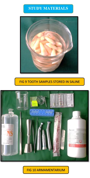
ARMAMENTARIUM: (FIG 10)
One hundred extracted caries free and fracture free, human single rooted maxillary central incisor teeth.
Diamond Disc (MDT Micro Diamond Technologies Ltd)
Micro motor straight hand piece (NSK, Nakanishi Inc., Japan) Airotor hand piece (NSK, Nakanishi Inc., Japan)
Stainless steel K files (No.10 size) (Mani Inc., Japan)
Endo gauge (Dentsply Maillefer, Switzerland)
Endodontic rotary hand piece (Anthogyr, Dentsply, France).
NiTi Rotary files (ProTaper NEXT X1-X5, Dentsply Maillefer). Endo scale (Dentsply Mallifer, Switzerland)
Tweezer (GDC, India).
5ml syringe (Romsons, India). 29 Gauge needle
Beakers
Chisel and mallet
EQUIPMENTS:
21
MATERIALS:
0.9% Normal saline
3% Sodium Hypochlorite (NaOCl) solution (Septodont, France) EDTA solution (Canal Pro, Coltene)
[image:41.595.113.571.354.719.2]Phytic Acid, freshly prepared (TCI CHEMICALS, JAPAN) Etidronic Acid, freshly prepared (TCI CHEMICALS, JAPAN) Paper points (Dentsply Maillefer)
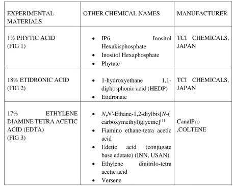
TABLE 1: EXPERIMENTAL MATERIALS USED IN STUDY
EXPERIMENTAL MATERIALS
OTHER CHEMICAL NAMES MANUFACTURER
1% PHYTIC ACID (FIG 1)
IP6, Inositol
Hexakisphosphate
Inositol Hexaphosphate
Phytate
TCI CHEMICALS, JAPAN
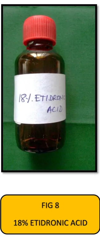
18% ETIDRONIC ACID (FIG 2)
1-hydroxyethane 1,1-diphosphonic acid (HEDP)
Etidronate
TCI CHEMICALS, JAPAN
17% ETHYLENE
DIAMINE TETRA ACETIC ACID (EDTA)
(FIG 3)
N,N'-Ethane-1,2-diylbis[N-( carboxymethyl)glycine][1]
Fiamino ethane-tetra acetic acid
Edetic acid (conjugate base edetate) (INN, USAN)
Ethylene dinitrilo-tetra acetic acid
Versene
22
METHODOLOGY:
SAMPLE SELECTION:
One hundred extracted caries-free and visually assessed fracture-free, human single rooted maxillary incisor teeth with mature apices were selected for the study. Remnants of soft tissue debris, calculus and tissue deposit were mechanically removed from tooth surface with ultrasonic scaler. The radiographs were taken to confirm that each tooth had a single straight canal without curvature and resorption. The teeth were stored in 0.9% normal saline solution until use. (FIG 9)
SAMPLE PREPARATION:
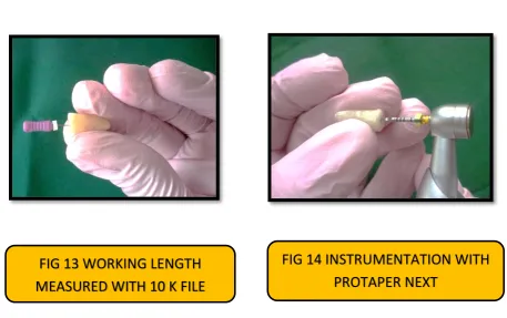
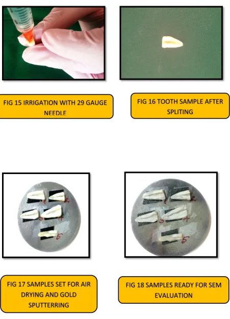
The tooth samples were decoronated with a diamond disc (FIG 11) and straight hand piece at the cemento-enamel junction, measuring root samples of 15 mm (FIG 12) in length. The patency of the canal was checked with a No. 10 K file beyond apical foramen (FIG 13). The teeth were grooved on the buccal and lingual surfaces with a diamond disc. They were split longitudinally with chisel and mallet before instrumentation to avoid creating artificial debris, the disc was not allowed to penetrate the canal space
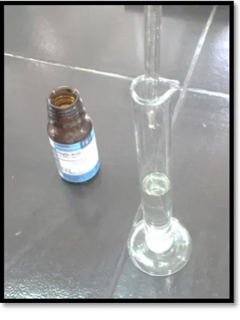
PREPARATION OF IRRIGATING SOLUTION: (FIG 4, 5, 6)
1 % Phytic acid is prepared by adding 1ml of Phytic acid in 100ml of water for injection (FIG 7). 18% Etidronic acid is prepared by adding 18ml of Etidronic acid in 100ml of water for injection (FIG 8).
ROOT CANAL INSTRUMENTATION:
23
[image:43.595.104.526.257.695.2]instrumented in a total time of 4 min each in a crown down manner with Protaper NEXT rotary files upto X5 (ISO size 50) using a 64:1 reduction hand piece (FIG 14) . The irrigation was carried out using 5ml syringe of 29 gauge needle with 14mm length (FIG 15). Samples were irrigated with 3 ml of 3% NaOCl for 5min followed by saline irrigation between every instrument change. The tooth samples were randomly distributed into ten groups of 10 teeth each.
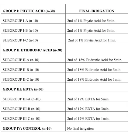
TABLE 2: IRRIGATION PROTOCOL USED IN THE STUDY:
GROUP I: PHYTIC ACID (n-30) FINAL IRRIGATION
SUBGROUP I-A (n-10) 2ml of 1% Phytic Acid for 5min. SUBGROUP I-B (n-10) 2ml of 1% Phytic Acid for 3min. SUBGROUP I-C (n-10) 2ml of 1% Phytic Acid for 1min. GROUP II:ETIDRONIC ACID (n-30)
SUBGROUP II-A (n-10) 2ml of 18% Etidronic Acid for 5min. SUBGROUP II-B (n-10) 2ml of 18% Etidronic Acid for 3min. SUBGROUP II-C (n-10) 2ml of 18% Etidronic Acid for 1min. GROUP III: EDTA (n-30)
SUBGROUP III-A (n-10) 2ml of 17% EDTA for 5min. SUBGROUP III-B (n-10) 2ml of 17% EDTA for 3min. SUBGROUP III-C (n-10) 2ml of 17% EDTA for 1min. GROUP IV: CONTROL (n-10)
No final irrigation
24
SPECIMEN PREPARATION AND SEM EVALUATION:
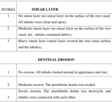
[image:44.595.122.496.349.690.2]The roots were then split longitudinally into two halves with a chisel and mallet. The half with the most visible canal surface of the apex (FIG 16) was used for scanning electron microscopic evaluation. The specimens were air dried, gold sputtered, (FIG 18) and SEM micrographs were obtained at 5000X magnification of the coronal, middle and apical areas of each root canal. The amount of smear layer and degree of dentinal erosion was evaluated using a three step scale given by Torabinajed et al 2003. 78
TABLE: 3 Score Rating system developed by Torabinejad et al.: 78
SCORES SMEAR LAYER
1 No smear layer (no smear layer on the surface of the root canal: All tubules were clean and open).
2 Moderate smear layer (no smear layer on the surface of the root canal, but tubules contained debris).
3 Heavy smear layer (smear layer covered the root canal surface and the tubules).
DENTINAL EROSION
1 No erosion. All tubules looked normal in appearance and size.
2 Moderate erosion. The peritubular dentin was eroded.
3
25
STATISTICAL ANALYSIS:
26
100 SINGLE ROOTED MAXILLARY CENTRAL INCISORS
ROOT SPECIMEN OF 15 mm WORKING LENGTH MEASURED
GROUP I
1 % PHTYIC ACID
GROUP II
18 % ETIDRONIC
ACID
GROUP III
17% EDTA
GROUP IV
CONTROL
(NO FINAL IRRIGATION) DECORONATION OF CROWN USING DIAMOND DISC
INSTRUMENTATION DONE WITH PROTAPER NEXT ROTARY FILES UPTO X5 (ISO SIZE -50)
THE CANALS WERE IRRIGATED WITH SALINE AND 3ml OF 3% NAOCL FOR 5MIN
27
SUBDIVIDED INTO 3 SUBGROUPS BASED ON DURATION OF FINAL IRRIGATION
SUBGROUP I-A (5MIN)
SUBGROUP I-B (3MIN)
SUBGROUP I-C (1MIN)
SUBGROUP II-A (5MIN)
SUBGROUP II-B (3MIN)
SUBGROUP II-C (1MIN)
SUBGROUP III-A (5MIN)
SUBGROUP III-B (3MIN)
SUBGROUP III-C (1MIN)
SAMPLES WERE VIEWED UNDER SCANNING ELECTRON MICROSCOPE (5000X) AT CORONAL, MIDDLE, AND APICAL REGIONS
SAMPLES WERE SPLIT LONGITUDINALLY USING CHISEL AND MALLET
SAMPLES WITH MOST VISIBLE CANAL SURFACE WERE TAKEN
SAMPLES WERE AIR DRIED AND GOLD SPUTTERED
IMAGES WERE EVALUATED FOR SMEAR LAYER AND DENTIN EROSION
[image:48.595.107.240.88.413.2]
FIG 2 ETIDRONIC ACID
[image:48.595.384.527.93.409.2][image:49.595.60.560.73.747.2]
[image:49.595.231.400.465.690.2]
FIG 4 WATER FOR INJECTION (WFI)
FIG 5 WFI ADDED TO TEST TUBE
FIG 8
[image:50.595.331.494.240.638.2]18% ETIDRONIC ACID
FIG 7
1% PHYTIC ACID
FIG 9 TOOTH SAMPLES STORED IN SALINE
STUDY MATERIALS
[image:52.595.67.525.450.737.2]
FIG 14 INSTRUMENTATION WITH
PROTAPER NEXT
FIG 13 WORKING LENGTH
MEASURED WITH 10 K FILE
[image:53.595.71.523.132.758.2]
FIG 15 IRRIGATION WITH 29 GAUGE
NEEDLE
FIG 16 TOOTH SAMPLE AFTER
SPLITING
FIG 17 SAMPLES SET FOR AIR
DRYING AND GOLD
SPUTTERRING
[image:53.595.71.520.144.389.2]28
SCANNING ELECTRON MICROSCOPIC IMAGES OF GROUP I:
PHYTIC ACID (FIG 20)
CERVICAL MIDDLE APICAL
SUBGROUP I-A (5 MIN)
SUBGROUP I-B (3 MIN)
SUBGROUP I-C (1 MIN)
29
SCANNING ELECTRON MICROSCOPIC IMAGES OF GROUP II:
ETIDRONIC ACID (FIG 21)
CERVICAL MIDDLE APICAL
SUBGROUP II-A (5 MIN)
SUBGROUP II-B (3 MIN)
SUBGROUP II-C (1 MIN)
30
SCANNING ELECTRON MICROSCOPIC IMAGES OF GROUP III:
EDTA (FIG 22)
CERVICAL MIDDLE APICAL
SUBGROUP III-A (5 MIN)
SUBGROUP III-B (3 MIN)
SUBGROUP III-C (1 MIN)
31
SCANNING ELECTRON MICROSCOPIC IMAGES OF GROUP IV:
CONTROL (FIG 23)
CERVICAL MIDDLE APICAL
SUBGROUP IV (5 MIN)
SMEAR PLUGS DENTIN EROSION
32
Table no: 5 SMEAR LAYER SCORES OF ALL GROUPS:
SAMPLES (5 MIN) (3 MIN) (1 MIN)
CORONAL MIDDLE APICAL CORONAL MIDDLE APICAL CORONAL MIDDLE APICAL
GROUP I-PHYTIC ACID
1 1 1 2 1 1 2 1 1 2
2 1 1 2 1 1 2 1 1 2
3 1 1 2 1 1 2 1 1 2
4 1 1 2 1 1 2 1 1 2
5 1 1 2 1 1 2 1 1 2
6 1 1 2 1 1 2 1 1 2
7 1 1 2 1 1 2 1 1 2
8 1 1 2 1 1 2 1 1 2
9 1 1 2 1 1 2 1 1 2
10 1 1 2 1 1 2 1 1 2
GROUP II-ETIDRONIC ACID
1 1 1 2 1 1 2 2 2 3
2 1 1 2 1 1 2 2 2 2
3 1 1 2 1 1 2 2 2 3
4 1 1 2 1 1 2 2 2 2
5 1 1 2 1 1 2 2 2 2
6 1 1 2 1 1 2 2 2 3
7 1 1 2 1 1 2 2 2 2
8 1 1 2 1 1 2 2 2 2
9 1 1 2 1 1 2 2 2 2
10 1 1 2 1 1 2 2 2 3
GROUP III-EDTA
1 1 1 2 1 1 2 1 1 2
2 1 1 2 1 1 2 1 1 2
3 1 1 2 1 1 2 1 1 2
4 1 1 2 1 1 2 1 1 2
5 1 1 2 1 1 2 1 1 2
6 1 1 2 1 1 2 1 1 2
7 1 1 2 1 1 2 1 1 2
8 1 1 2 1 1 2 1 1 2
9 1 1 2 1 1 2 1 1 2
10 1 1 2 1 1 2 1 1 2
GROUP IV- CONTROL(5MIN)
1 3 3 3
2 3 3 3
3 3 3 3
4 3 3 3
5 3 3 3
6 3 3 3
7 3 3 3
8 3 3 3
9 3 3 3
33
Table no: 6 DENTIN EROSION SCORES OF ALL GROUPS:
SAMPLES (5 MIN) (3 MIN) (1 MIN)
CORONAL MIDDLE APICAL CORONAL MIDDLE APICAL CORONAL MIDDLE APICAL
GROUP I-PHYTIC ACID
1 2 2 1 1 1 1 1 1 1
2 2 1 1 2 1 1 2 1 1
3 2 2 1 2 2 1 1 1 1
4 2 2 1 1 1 1 2 2 1
5 2 2 1 1 1 1 1 1 1
6 2 2 1 2 1 1 1 1 1
7 2 2 1 1 2 1 1 2 1
8 2 2 1 1 1 1 2 1 1
9 2 2 1 2 1 1 1 1 1
10 2 1 1 1 2 1 1 1 1
GROUP II-ETIDRONIC ACID
1 3 2 1 3 2 1 2 2 1
2 3 2 1 2 2 1 2 2 1
3 3 2 2 3 2 1 2 2 1
4 3 3 1 3 2 1 2 2 1
5 3 2 1 3 2 1 2 2 1
6 3 2 1 3 3 1 2 2 1
7 2 2 2 3 2 1 2 2 1
8 3 3 1 2 2 1 2 2 1
9 2 2 2 3 2 2 2 2 1
10 3 2 1 3 2 1 2 2 1
GROUP III-EDTA
1 3 3 1 3 3 1 3 2 1
2 3 3 2 3 2 1 3 2 1
3 3 3 1 3 3 2 3 2 1
4 3 3 1 3 2 1 3 2 1
5 3 3 2 3 3 1 3 3 1
6 3 3 1 3 3 2 3 2 1
7 3 3 1 3 2 1 3 2 1
8 3 3 2 3 3 1 3 3 1
9 3 3 1 3 3 1 3 2 1
10 3 3 1 3 2 1 3 2 1
34
Table: 6 DESCRIPTIVE TABLE SHOWING MEAN, MEDIAN AND STANDARD DEVIATIONS OF SMEAR LAYER AMONG GROUPS
Groups 5MIN 3MIN 1MIN
Cervical Middle Apical Cervical Middle Apical Cervical Middle Apical PHYTIC
ACID (N-10) Group I
Mean 1.0000 1.0000 2.0000 1.0000 1.0000 2.0000 1.0000 1.0000 2.0000 Std.
Deviation
.00000 .00000 .00000 .00000 .00000 .00000 .00000 .42164 .00000
Median 1.0000 1.0000 2.0000 1.0000 1.0000 2.0000 1.0000 1.0000 2.0000 HEDP
(N-10) Group II
Mean 1.0000 1.0000 2.0000 1.0000 1.0000 2.0000 2.0000 2.0000 2.4000 Std.
Deviation
.00000 .00000 .00000 .00000 .00000 .00000 .00000 .00000 .51640 Median 1.0000 1.0000 2.0000 1.0000 1.0000 2.0000 2.0000 2.0000 2.0000 EDTA
(N-10)
Group III
Mean 1.0000 1.0000 2.0000 1.0000 1.0000 2.0000 1.0000 1.0000 2.0000 Std.
Deviation
.00000 .00000 .00000 .00000 .00000 .00000 .00000 .00000 .00000
Median 1.0000 1.0000 2.0000 1.0000 1.0000 2.0000 1.0000 1.0000 2.0000 Control
(N-10) Group IV
Mean 3.0000 3.0000 3.0000 Std.
Deviation
.00000 .00000 .00000
35
Table 7: ANALYSIS OF SMEAR LAYER VALUES AMONG DIFFERENT GROUPS USING KRUSKAL WALLIS TEST
Statistical analysis
5min 3min 1min
Cervical Middle Apical Cervical Middle Apical Cervical Middle Apical
df 3 3 3 2 2 2 2 2 2
Asymp. Sig.
.000 .000 .000 1.000 1.000 1.000 .000 .000 .012
TABLE: 8 INDIVIDUAL COMPARISONS OF SMEAR LAYER VALUES USING MANN WHITNEY U TEST BETWEEN THE GROUPS
GROUPS
5MIN 1MIN
Cervical Middle Apical Cervical Middle Apical GROUP I VS II 1.000 1.000 1.000 .000 .000 .029
GROUP I VS III 1.000 1.000 1.000 1.000 1.000 1.000 GROUP I VS IV .000 .000 .000
GROUP II VS III 1.000 1.000 1.000 .000 .000 .029
GROUP II VS IV .000 .000 .000
36
TABLE 9: DESCRIPTIVE TABLE SHOWING MEAN, MEDIAN AND STANDARD DEVIATIONS OF DENTIN EROSION AMONG GROUPS
GROUPS 5MIN 3MIN 1MIN
Cervical Middle Apical Cervical Middle Apical Cervical Middle Apical PHYTIC
ACID (N-10) Group I
Mean 2.0000 1.8000 1.0000 1.4000 1.3000 1.0000 1.3000 1.2000 1.0000 Std.
Deviation
.00000 .42164 .00000 .51640 .48305 .00000 .48305 .42164 .00000
Median 2.0000 2.0000 1.0000 1.0000 1.0000 1.0000 1.0000 1.0000 1.0000 HEDP
(N-10) Group II
Mean 2.7000 2.2000 1.3000 3.0000 2.1000 1.1000 2.0000 2.0000 1.0000 Std.
Deviation
.48305 .42164 .48305 .00000 .31623 .31623 .00000 .00000 .00000
Median 3.0000 2.0000 1.0000 3.0000 2.0000 1.0000 2.0000 2.0000 1.0000 EDTA
(N-10) Group III
Mean 3.0000 3.0000 1.3000 3.0000 2.6000 1.2000 3.0000 2.2000 1.0000 Std.
Deviation
.00000 .00000 .48305 .00000 .51640 .42164 .00000 .42164 .00000
37
TABLE: 10 ANALYSIS OF DENTIN EROSION VALUES AMONG DIFFERENT GROUPS USING KRUSKAL WALLIS TEST
Statistical analysis
5min 3min 1min
Cervical Middle Apical Cervical Middle Apical Cervical Middle Apical
df 2 2 2 2 2 2 2 2 2
Asymp. Sig.
[image:65.842.137.792.354.514.2].000 .000 .163 .000 .000 .342 .000 .000 1.000
Table: 11 INDIVIDUAL COMPARISONS OF DENTIN EROSION VALUES USING MANN WHITNEY U TEST BETWEEN THE GROUPS
GROUPS STATISTICS 5MIN 3MIN 1MIN
Cervical Middle Apical Cervical Middle Apical Cervical Middle Apical Group I vs II Asymp. Sig.
(2-tailed)
.001 .051 .067 .000 .001 .317 .001 .000 1.000
Group I VS III Asymp. Sig. (2-tailed)
.000 .000 .067 .000 .000 .146 .000 .000 1.000
Group II VS III Asymp. Sig. (2-tailed)
38
TABLE 12: DESCRIPTIVE TABLE SHOWING MEAN, MEDIAN AND STANDARD DEVIATIONS OF SMEAR LAYER AND DENTIN EROSION OF PHYTIC ACID AT ALL THIRDS
GROUPS
SMEAR LAYER DENTIN EROSION
5MIN 3MIN 1MIN 5MIN 3MIN 1MIN
CERVICAL (N-10) Group 1
Mean 1.0000 1.0000 1.0000 2.0000 1.4000 1.3000
Std. Deviation .00000 .00000 .00000 .00000 .51640 .48305
Median 1.0000 1.0000 1.0000 2.0000 1.0000 1.0000 MIDDLE
(N-10) Group 2
Mean 1.0000 1.0000 1.2000 1.8000 1.3000 1.2000
Std. Deviation .00000 .00000 .42164 .42164 .48305 .42164
Median 1.0000 1.0000 1.0000 2.0000 1.0000 1.0000 APICAL
(N-10) Group 3
Mean 2.0000 2.0000 2.0000 1.0000 1.0000 1.0000
Std. Deviation .00000 .00000 .00000 .00000 .00000 .00000
39
TABLE: 13 ANALYSIS OF SMEAR LAYER AND DENTIN EROSION VALUES OF PHYTIC ACID AT ALL THIRDS USING KRUSKAL WALLIS TEST
STATISTICAL ANALYSIS
SMEAR LAYER DENTIN EROSION
5MIN 3MIN 1MIN 5MIN 3MIN 1MIN
df 2 2 2 2 2 2
[image:67.842.140.728.342.491.2]Asymp. Sig. .000 .000 .000 .000 .096 .197
TABLE: 14 INDIVIDUAL COMPARISONS OF SMEAR LAYER AND DENTIN EROSION VALUES OF PHYTIC ACID AT ALL THIRDS USING MANN WHITNEY U TEST
GROUPS STATISTICS SMEAR LAYER DENTIN EROSION
5MIN 3MIN 1MIN 5MIN 3MIN 1MIN
GROUP 1 VS 2 Asymp. Sig. (2-tailed)
1.000 1.000 .146 .146 .648 .615
GROUP 1 VS 3 Asymp. Sig.
(2-tailed)
.000 .000 .000 .000 .029 .067
GROUP 2 VS 3 Asymp. Sig.
(2-tailed)
40
TABLE 15: DESCRIPTIVE TABLE SHOWING MEAN, MEDIAN AND STANDARD DEVIATIONS OF SMEAR LAYER AND DENTIN EROSION OF PHYTIC ACID AT 5, 3 & 1 MIN
GROUPS SMEAR LAYER DENTIN EROSION
cervical middle apical cervical middle apical
5MIN (N-10) Group 1
Mean 1.0000 1.0000 2.0000 2.0000 1.8000 1.0000 Std.
Deviation
.00000 .00000 .00000 .00000 .42164 .00000
Median 1.0000 1.0000 2.0000 2.0000 2.0000 1.0000
3MIN (N-10) Group 2
Mean 1.0000 1.0000 2.0000 1.4000 1.3000 1.0000 Std.
Deviation
.00000 .00000 .00000 .51640 .48305 .00000
Median 1.0000 1.0000 2.0000 1.0000 1.0000 1.0000
1MIN (N-10) Group 3
Mean 1.0000 1.2000 2.0000 1.3000 1.2000 1.0000 Std.
Deviation
.00000 .42164 .00000 .48305 .42164 .00000
41
TABLE 16: ANALYSIS OF SMEAR LAYER AND DENTIN EROSION VALUES OF PHYTIC ACID AT 5, 3, & 1 MIN USING KRUSKALWALLIS TEST
Statistics SMEAR LAYER DENTIN EROSION
Cervical Middle Apical Cervical Middle Apical
df 2 2 2 2 2 2
Asymp. Sig. 1.000 .126 1.000 .004 .017 1.000
TABLE 17: PAIRWISE COMPARISONS OF SMEAR LAYERAND DENTIN EROSION VALUES OF PHYTIC ACID AT 5, 3 & 1 MIN USING MANN WHITNEY U TEST
GROUPS STATISTICS SMEAR LAYER DENTIN EROSION
Cervical Middle Apical Cervical Middle Apical GROUP 1 VS 2 Asymp. Sig.
(2-tailed)
1.000 1.000 1.000 .004 .028 1.000
GROUP 1 VS 3 Asymp. Sig. (2-tailed)
1.000 .146 1.000 .001 .009 1.000
GROUP 2 VS 3 Asymp. Sig. (2-tailed)
[image:69.842.136.720.343.490.2]42
TABLE 18: DESCRIPTIVE TABLE SHOWING MEAN, MEDIAN AND STANDARD DEVIATIONS OF SMEAR LAYER AND DENTIN EROSION VALUES OF ETIDRONIC ACID AT ALL THIRDS
GROUPS
SMEAR LAYER DENTIN EROSION
5MIN 3MIN 1MIN 5MIN 3MIN 1MIN
CERVICAL (N-10) Group 1
Mean 1.0000 1.0000 2.0000 2.7000 3.0000 2.0000 Std. Deviation .00000 .00000 .00000 .48305 .00000 .00000
Median 1.0000 1.0000 2.0000 3.0000 3.0000 2.0000 MIDDLE
(N-10) Group 2
Mean 1.0000 1.0000 2.0000 2.2000 2.1000 2.0000 Std. Deviation .00000 .00000 .00000 .42164 .31623 .00000
Median 1.0000 1.0000 2.0000 2.0000