0022-538X/06/$08.00⫹0 doi:10.1128/JVI.01049-06
Copyright © 2006, American Society for Microbiology. All Rights Reserved.
Human ESCRT-II Complex and Its Role in Human Immunodeficiency
Virus Type 1 Release†
Charles Langelier,
1Uta K. von Schwedler,
1Robert D. Fisher,
1Ivana De Domenico,
2Paul L. White,
1Christopher P. Hill,
1Jerry Kaplan,
2Diane Ward,
2and Wesley I. Sundquist
1*
Departments of Biochemistry1and Pathology,2University of Utah, Salt Lake City, Utah 84112
Received 22 May 2006/Accepted 8 July 2006
The budding of many enveloped RNA viruses, including human immunodeficiency virus type 1 (HIV-1), requires some of the same cellular machinery as vesicle formation at the multivesicular body (MVB). In
Saccharomyces cerevisiae, the ESCRT-II complex performs a central role in MVB protein sorting and vesicle formation, as it is recruited by the upstream ESCRT-I complex and nucleates assembly of the downstream ESCRT-III complex. Here, we report that the three subunits of human ESCRT-II, EAP20, EAP30, and EAP45, have a number of properties in common with their yeast orthologs. Specifically, EAP45 bound ubiquitin via its N-terminal GRAM-like ubiquitin-binding in EAP45 (GLUE) domain, both EAP45 and EAP30 bound the C-terminal domain of TSG101/ESCRT-I, and EAP20 bound the N-terminal half of CHMP6/ESCRT-III. Consistent with its expected role in MVB vesicle formation, (i) human ESCRT-II localized to endosomal membranes in a VPS4-dependent fashion and (ii) depletion of EAP20/ESCRT-II and CHMP6/ESCRT-III inhibited lysosomal targeting and downregulation of the epidermal growth factor receptor, albeit to a lesser extent than depletion of TSG101/ESCRT-I. Nevertheless, HIV-1 release and infectivity were not reduced by efficient small interfering RNA depletion of EAP20/ESCRT-II or CHMP6/ESCRT-III. These observations indicate that there are probably multiple pathways for protein sorting/MVB vesicle formation in human cells and that HIV-1 does not utilize an ESCRT-II-dependent pathway to leave the cell.
Enveloped RNA viruses like human immunodeficiency virus (HIV) acquire lipid bilayers and exit infected cells by budding through limiting membranes. The process of HIV budding shares a number of similarities with the cellular process of vesicle formation at the multivesicular body (MVB) (reviewed in references 2, 14, 25, 26, 33, 40, and 51). MVB vesicles and HIV virions both bud away from the cytoplasm (58) and share the following mechanistic similarities: (i) a requirement for ubiquitin (Ub) in formation and cargo incorporation (reviewed in references 40 and 81), (ii) recruitment and utilization of the cellular ESCRT-I complex (6, 15, 19, 22, 47, 48, 52, 70, 79), and (iii) a requirement for ESCRT-III, LIP5, and VPS4 ATPase activities (22, 46, 69, 83, 85). In spite of these similarities, however, MVB biogenesis and viral budding ultimately create different vesicles, and it is therefore of interest to determine the precise relationship between the two processes.
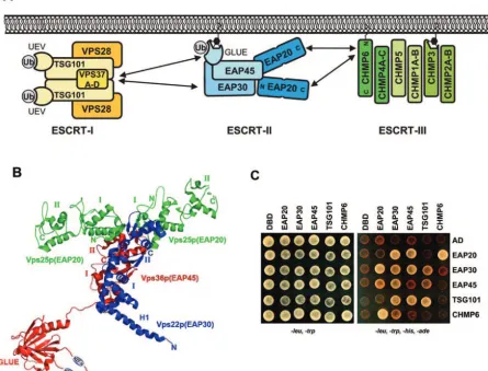
Genetic screens inSaccharomyces cerevisiaedefined the ba-sic machinery of MVB veba-sicle formation and identified 17 soluble yeast proteins that are essential for MVB biogenesis (the “class E” proteins) (40, 59). Complementary biochemical analyses revealed that MVB vesicle formation proceeds through an ordered pathway in which a series of soluble class E complexes, termed ESCRT-I (39), ESCRT-II (4), and ESCRT-III (3), are sequentially recruited to endosomal membranes, where they function in vesicle formation (Fig. 1A). Although the processes of vesicle formation and cargo incorporation are not yet understood
in mechanistic detail, ESCRT-I and -II appear to function pri-marily as adaptors that recognize protein cargoes and help recruit ESCRT-III, which in turn appears to function more directly in protein sorting and vesicle formation.
As the central complex in the yeast ESCRT pathway, ESCRT-II bridges ESCRT-I and ESCRT-III and also interacts with ubiquitylated cargoes. As shown in Fig. 1B, crystal struc-tures of the yeast ESCRT-II core (30, 74) have revealed that the yeast ESCRT-II complex contains two copies of Vps25p (EAP20) that bind asymmetrically to single copies of Vps22p (EAP30) and Vps36p (EAP45) (human protein names are provided in parentheses for reference). The two Vps25p (EAP20) proteins project away from the Vps22p-Vps36p dimer, so that the overall complex assumes a trilobed “Y” shape. The three ESCRT-II subunits are related structurally, as each contains two copies of the winged-helix (WH) protein motif. Two of the proteins also contain N-terminal extensions: Vps22p (EAP30) has an extended helix (Fig. 1B), and Vps36p (EAP45) contains a linker and an N-terminal GRAM-like ubiquitin-binding in EAP45 (GLUE) domain (not present in the core structure). Structural and biochemical analyses of the Vps36p GLUE domain have shown that it is a split PH domain that binds phosphatidylinositol phosphates, particularly PI(3)P (67, 73). An extended insert within the GLUE domain contains two Npl4 zinc fingers (NZF), which are small globular zinc binding modules that mediate protein-protein interactions (49). The first, NZF-N, binds Vps28p (ESCRT-I) and helps recruit ESCRT-II to the endosomal membrane (73), whereas the second, NZF-C, binds ubiquitin and is required for efficient sorting of ubiquitylated cargoes (1).
The human and yeast ESCRT-II complexes are likely to be related structurally, because mammalian ESCRT-II can be
iso-* Corresponding author. Mailing address: Department of Biochem-istry, 15 N. Medical Drive East, Room 4100, University of Utah School of Medicine, Salt Lake City, UT 84112-5650. Phone: (801) 585-5402. Fax: (801) 581-7959. E-mail: [email protected].
† Supplemental material for this article may be found at http://jvi .asm.org.
9465
on November 8, 2019 by guest
http://jvi.asm.org/
lated as a stable, soluble complex composed of three proteins that share 19 to 36% pairwise identity with theirS. cerevisiae
homologs (excluding the N-terminal region of Vps36p/EAP45) (37, 61). We have therefore used the crystal structure of theS. cerevisiaeESCRT-II core as a working model for the human ESCRT-II complex (Fig. 1B) (see Materials and Methods). Mammalian ESCRT-II functions are less well studied than those of the yeast complex, but the mammalian EAP20 and EAP45 proteins do colocalize with ESCRT-III components and with ubiquitylated proteins on endosomal membranes (67, 88). Like their yeast counterparts, EAP20 can bind the ESCRT-III protein, CHMP6 (46, 83, 88), and the N-terminal
[image:2.585.70.515.79.418.2]GLUE domain of murine EAP45 was recently shown to bind ubiquitin (67). These observations all support the idea that the mammalian andS. cerevisiae ESCRT-II complexes are func-tional homologs. Nevertheless, there must also be important differences between human and yeast ESCRT-II proteins. For example, the mammalian complex lacks both NZF domains and therefore must interact differently with both ESCRT-I and ubiquitin. Moreover, ESCRT-II and CHMP6 overexpression does not inhibit HIV type 1 (HIV-1) budding (46), and it was recently reported that ESCRT-II is not required for lysosomal degradation of major histocompatibility complex I (MHC-I) or epidermal growth factor receptor (EGFR), indicating that the
FIG. 1. ESCRT-II structure and interactions. (A) Schematic model summarizing the protein interactions of the human ESCRT-II complex (see the text for details). Note that the stoichiometries and compositions of the ESCRT-I and ESCRT-III complexes have not yet been defined unambiguously. The complexes are therefore simply shown with all possible subunits. (B) Three-dimensional structure of the yeast ESCRT-II complex. The structure of the ESCRT-II core was determined by X-ray crystallography (30, 74). The N-terminal region of Vps36 (EAP45) (shown schematically) has not yet been determined but contains two NZF domains (1) embedded within a GLUE domain (67). The structural elements/domains correspond approximately to the following residues (human homologs are in parentheses): for Vps36p (EAP45), GLUE domain, 1 to 285 (1 to 135); NZF-1, 177 to 205; NZF-2, 242 to 259 (neither NZF motif is present in EAP45); linker domain, 286 to 404 (136 to 238); WH-1, 405 to 491 (239 to 316); and WH-2, 492 to 566 (317 to 386); for Vps22p (EAP30), helix 1 (H1), 1 to 51 (1 to 55); WH-1, 90 to 165 (95 to 176); and WH-2, 166 to 233 (177 to 258); for Vps25p (EAP20), WH-1, 19 to 127 (22 to 103), and WH-2, 128 to 200 (104 to 176). (C) Summary of the positive yeast two-hybrid interactions between ESCRT-II proteins and all known human class E proteins. The right array shows doubly transformed yeast replica plated on minus Leu, minus Trp, minus His, minus Ade selection media, where successful growth represents a positive protein interaction. The left array shows replica-plated yeast on minus Leu, minus Trp media (a control for equivalent transformation and yeast growth). The indicated constructs were fused to DBDs (rows) or ADs (columns). Unfused DBD and AD constructs are shown as negative controls. Note that the interaction between DBD-EAP30 and AD-CHMP6 was judged to be negative because it was very weak and was not detected in the reciprocal direction. Analogous data were summarized previously in reference 83.
on November 8, 2019 by guest
http://jvi.asm.org/
complex is not absolutely required for membrane protein sort-ing or MVB vesicle formation (11). Indeed, several groups have proposed that ESCRT-II complexes in other species may perform functions that appear to be unrelated to MVB protein sorting. These include a role for mammalian ESCRT-II in transcriptional elongation (37, 61, 65) and a role for the Schizo-saccharomyces pombe ESCRT-II protein EAP30 in meiotic spindle pole body amplification (35). The relationship between MVB protein sorting and these other putative ESCRT-II ac-tivities, if any, is not yet clear.
During HIV-1 assembly, human class E proteins are re-cruited by two short sequence motifs (termed “late domains”) within the C-terminal p6 region of the Gag polyprotein (re-viewed in references 15, 19, 22, 47, 48, 52, 70, and 79). The first p6gaglate domain is a PTAP tetrapeptide motif that binds and
recruits TSG101 (tumorsusceptibilitygene 101; yeast Vps23p). TSG101 is the central protein of the human ESCRT-I com-plex, and there is good evidence that the entire ESCRT-I complex functions directly in HIV-1 release (15, 19, 22, 24, 48). The second late domain within p6gagis a YPX
nL motif (where
Xn can vary in identity and length) that binds and recruits
ALIX/AIP1 (yeast Bro1p) (69, 80). ALIX functions late in MVB biogenesis (56) and binds both ESCRT-I (83) and ESCRT-III (41) proteins but is not a permanent subunit of any of the three discrete ESCRT complexes. Like TSG101, ALIX functions directly in virus release, although disruptions of the PTAP-TSG101 interaction generally inhibit HIV-1 budding to a greater degree than do disruptions of the ALIX-YPXnL
interaction (16, 46). As noted above, there are also indications that HIV-1 budding requires the activities of the late-acting ESCRT-III, LIP5, and VPS4 class E proteins (22, 46, 69, 83, 85). Specifically, HIV-1 release is dominantly inhibited by overexpression of VPS4 ATPase mutants and ESCRT-III pro-teins with large N- or C-terminal extensions (22, 46, 69, 83). HIV-1 release is also impaired by small interfering RNA (siRNA) depletion of the VPS4 binding protein LIP5 (85). It is important to note, however, that these experiments do not definitively establish a direct role for these proteins in HIV-1 budding, because the inhibitory effects could also arise from sequestration of the entire ESCRT machinery at aberrant en-dosomal compartments (called “class E” compartments).
In summary, HIV-1 uses the ESCRT-I complex to bud from cells and also requires the activities of downstream ESCRT-III and VPS4-LIP5 complexes. InS. cerevisiae, ESCRT-II physi-cally bridges the ESCRT-I and ESCRT-III complexes and is essential for MVB protein sorting and vesicle formation (4). The mammalian ESCRT-II function appears to be more com-plex, however, and we therefore undertook this study with the goals of characterizing the biochemical properties of human ESCRT-II and testing its role in HIV release.
MATERIALS AND METHODS
ESCRT-II homology modeling.A working model for the human ESCRT-II core was created as follows. Protein Data Bank (PDB) coordinates from a yeast ESCRT-II crystal structure (PDB accession no. 1U5T) were utilized to create a consensus template using Swiss-PdbViewer (http://www.expasy.org/spdbv/). The human and yeast ESCRT-II sequences were then aligned within Swiss-PdbViewer, the alignment was used to thread the human sequence onto the yeast ESCRT-II template, and the model was energy minimized within Swiss-PdbViewer.
Plasmids.Genes for human EAP20, EAP30, EAP45, and CHMP6 used in yeast two-hybrid and biochemical experiments were amplified from expressed sequence tag clones as described previously (83). A complete list of all constructs used in this study is provided in Table S1 in the supplemental material. Genes and fragments for yeast two-hybrid experiments were cloned into pGADT7
(activation domain) and pGBKT7 (binding domain) vectors (Clontech) using 5⬘
NdeI and 3⬘BamHI or BglII cloning sites. Recombinant proteins used in
bio-chemical experiments were expressed as glutathioneS-transferase (GST)
C-terminal fusions from a modified pGEX vector (GE Biosciences). This vector (pGEX2T-TEV) contained a tobacco etch virus (TEV) protease cleavage site
and 5⬘NdeI and 3⬘BamHI or BglII cloning sites following the GST gene.
Expression constructs for deletion mutants were created by amplifying and sub-cloning the desired fragments or by introducing a stop codon using Quickchange mutagenesis (Stratagene). For mammalian expression, ESCRT genes were
cloned as EcoRI-BamHI fragments into pcDNA3.1(⫺)MycHis in frame with the
C-terminal Myc tag, and with a Kozak sequence, ACC, inserted between the EcoRI site and the ATG start codon.
Preparation of recombinant EAP20.GST-EAP20 was expressed inEscherichia
coliBL21(DE3) transformed with WISP05-68 (see Table S1 in the supplemental
material) by induction in mid-log phase (0.4 mM IPTG [isopropyl--D
-thiogalacto-pyranoside]) and allowing the protein to accumulate for 4 hours at 23°C. The cells were lysed by lysozyme treatment (2.5 mg/ml) in 50 mM Tris, pH 8.0, 300 mM NaCl, 10% glycerol, 1 mM dithiothreitol (DTT), followed by sonication and
centrifugation (at 27,000⫻gfor 45 min) to remove cell debris. Soluble
GST-EAP20 was bound to a glutathione-Sepharose affinity matrix (GSTPrep FF; GE Healthcare), washed, and eluted with a step gradient of 20 mM glutathione. Protein fractions were dialyzed into 25 mM Tris (pH 8.0), 150 mM NaCl, and 1 mM DTT), and the fusion protein was cleaved during dialysis with 1 mg TEV protease per 100 mg protein (24 h; 23°C). The TEV cleavage reaction mixture was adjusted to 25 mM Tris (pH 8.0), 300 mM NaCl, 10% glycerol, 1 mM DTT, and free EAP20 was separated from GST and GST-EAP20 by size exclusion chromatography (Superdex 200; GE Healthcare). This purification method
typ-ically yielded⬃1.5 mg of pure protein per liter of starting culture.
Preparation of recombinant CHMP6 proteins.GST-CHMP6 expression, affinity purification, and TEV cleavage were carried out as described for GST-EAP20. Following TEV cleavage, free GST was removed by glutathione-Sepharose affinity chromatography. The solution was then diluted six-fold to reduce the salt in the buffer to 50 mM NaCl, and the CHMP6 protein was purified to homogeneity by anion-exchange chromatography on immobilized Q-Sepharose (GE Biosciences).
CHMP6 eluted at⬃400 mM NaCl from a linear gradient of 50 mM to 1 M NaCl in
25 mM Tris (pH 8.0), 10% glycerol, 1 mM DTT.
GST pull-down assays.GST and GST-CHMP6 fusion proteins were expressed inE. coliBL21(DE3) and induced at mid-log phase with 0.4 mM IPTG. Ten-milliliter cultures of cells expressing GST-CHMP6 were pelleted; resuspended in 2 ml of 300 mM NaCl, 0.1% NP-40, 10% glycerol, 1 mM DTT, 50 mM Tris (pH 8.0); and lysed by lysozyme treatment and sonication. Soluble GST-CHMP6 was
incubated with 100l glutathione-Sepharose resin. Approximately 30M
puri-fied recombinant EAP20 was added to 2M (equivalent) of immobilized
GST-CHMP6 and incubated for 30 min at 4°C in resuspension buffer. Unbound EAP20 was removed by washing with lysis buffer, and bound proteins were released from the resin by boiling it in sodium dodecyl sulfate-polyacrylamide gel electrophoresis (SDS-PAGE) loading buffer and were analyzed on Coomassie-stained 4 to 20% SDS-PAGE gradient gels.
Yeast two-hybrid binding assays.Directed yeast two-hybrid assays were per-formed using the Matchmaker GAL4 Yeast Two Hybrid 3 system (Clontech).
Briefly,Saccharomyces cerevisiaeAH-109 was cotransformed with pGADT7 or
pGBKT7 cloning vector (Clontech) containing the inserts of interest. The trans-formed yeast colonies were grown for 3 days at 30°C on yeast extract-peptone-dextrose plates with minus Leu, minus Trp selection. Ten to 100 colonies were pooled, resuspended in a liquid culture of Sabourand dextrose broth (minus Leu, minus Trp), selected on Sabourand dextrose broth (minus Leu, minus Trp, minus Ade, minus His) plates, and allowed to grow for 3 days.
Antibody production.Antibodies against the purified recombinant proteins EAP20 (UT461 and -462) and CHMP6 (UT463 and -464) were raised in rabbits by Covance Inc., following their 87-day protocol (http://abservices.crpinc.com/ab _sampleProtocols.aspx). UT461 and UT463 were affinity purified with the re-spective proteins, as described previously (83).
Biosensor binding experiments.EAP45-ubiquitin biosensor binding experi-ments were performed at 20°C using a BIACORE 2000 (Biacore AB, Uppsala, Sweden) equipped with a research grade CM4 sensor chip. Approximately 5,000 response units (RU) anti-GST antibody was immobilized on all four flow cells
using amine-coupling chemistry (36). GST-EAP451–229, GST-EAP451–139, or
recombinant GST alone from solubleE. colilysates was diluted in running buffer
on November 8, 2019 by guest
http://jvi.asm.org/
(20 mM Tris, pH 8.0, 100 mM NaCl, 1 mM DTT, 0.2 mg/ml bovine serum albumin [BSA], and 0.005% P20) and captured on the antibody surfaces at
densities of⬃0.5 kRU. Pure GST alone was also captured as a blank reference.
Ubiquitin was purified (84), diluted in running buffer, and injected in duplicate
or triplicate over the four flow cells at concentrations of 0 to 1,250M.
Re-sponses were collected at a rate of 2 Hz during the 30-s association and disso-ciation phases. Binding isotherms from the equilibrium phases were fitted to simple 1:1 binding models to determine the dissociation constant and statistical fitting errors (see Fig. 4) (53). To obtain more accurate estimates for the
disso-ciation constant and standard errors, the interaction between GST-EAP451–139
and ubiquitin was measured in six independent experiments, yielding values of
410⫾90M. Biosensor binding experiments for EAP20 and GST-CHMP6 were
performed as described for the EAP45-ubiquitin interaction, except that a BIA-CORE 3000 instrument was used and binding was examined in a buffer contain-ing 50 mM Tris, pH 8.0, 300 mM NaCl, 10% glycerol, 1 mM DTT, 0.2 mg/ml BSA, and 0.005% P20. Anti-GST antibody was immobilized over all four flow
cells (⬃13,000 RU/cell), and CHMP6, CHMP61–100, and CHMP6101–200were
captured to densities of⬃2,700 RU. Purified EAP20 was diluted in running buffer
and injected in triplicate at 0.03 to 560M concentrations.
Immunofluorescence.COS-7 cells grown on collagen-coated glass coverslips in
12-well plates were transfected with 0.5g of pEGFP-VPS4A-KQ DNA and, in
some experiments, 0.5g pcDNA3.1-EAP20-myc DNA or
pcDNA3.1-EAP30-myc DNA, using 2l Lipofectamine 2000 (Invitrogen) following the
manufac-turer’s instructions. The cells were fixed 24 h posttransfection in 4% paraform-aldehyde-phosphate-buffered saline (PBS), blocked in 10% fetal calf serum (FCS) in PBS containing 0.1% Triton X-100, and incubated at room temperature with murine monoclonal anti-Myc (9E10; Covance Inc.) to detect transfected Myc-tagged ESCRT-II proteins or with EAP20 UT461 (1:1,000) or anti-CHMP6 UT463 (1:1,000) antibody to detect endogenous ESCRT proteins. Sec-ondary antibodies were anti-mouse Alexa 594- or anti-rabbit Alexa
594-conju-gated antibodies (Molecular Probes; 1:1,000). Images of 0.5-m-thick Z sections
were collected as single wavelengths on an Olympus FV300 confocal fluores-cence microscope, using Fluoview 2.0.39 software.
Viral infectivity assays.EAP20 and CHMP6 were silenced by transfecting 5l
of 20M annealed siRNAs, together with 1g of pSL1180 (carrier DNA) and
9l Lipofectamine 2000, into 293T cells in six-well plates (22). The sense strands
of the siRNAs were as follows (lowercase letters denote RNA, and uppercase letters denote DNA nucleotides [nt]): EAP20 (start site, nt 218), cgauccagauug uauuagaTT; CHMP6-1 (start site, nt 257), ccaugguucagaguauugaTT; and CHMP6-2 (start site, nt 284), agaucgaaaugaaagugauTT. TSG101 and INV (TSG101) siRNA constructs have been described previously (22). The levels of protein depletion were estimated by quantifying the intensities of bands in Western blots using NIH ImageJ software.
Viral-particle production was initiated during a second siRNA transfection
(t⫽24 h) by replacing the 1-g carrier DNA either with an HIV-1 vector system
or with an infectious R9 HIV-1 NL4-3 plasmid. Virus was harvested 24 h after the second transfection, and the titers were measured using single-cycle MAGIC
assays (70). The HIV-1 vector system contained 0.38g pCMV⌬R8.2 (54), 0.12
g pRSV-Rev (18), 0.12g pMD.G (57), and 0.38g pWPTS-nlsLacZ (all
generous gifts from D. Trono, Swiss Federal Institute of Technology).
HIV-packagedlacZvectors were harvested 48 h later and titered on HeLaM cells as
described previously (85).
Western blotting.Viral supernatants and cellular proteins from transfected 293T cells were prepared using standard procedures (82). For Western blots, virions or cytoplasmic proteins were separated by 12% SDS-PAGE, transferred to nitrocellulose (TSG101) or polyvinylidene difluoride (all others) membranes in Tris-glycine-10% MetOH buffer, blocked in 1% (TSG101) or 5% milk, and incubated with antibodies diluted in Tris-buffered saline–0.1% Tween 20 buffer (anti-TSG101), or in 1% BSA or 5% nonfat milk in Tris-buffered saline–0.1% Tween 20. Proteins were detected by enhanced chemiluminescence (Pierce) or by imaging Alexa 680-nm (Molecular Probes; 1:10,000) or IRDye 800-nm (Rock-land; 1:20,000) secondary antibody on an Odyssey scanner (Li-Cor Inc.). The primary antibodies used were rabbit anti-HIV CA (UT415 made against purified NL4-3 CA protein and affinity-purified; 1:15,000); rabbit anti-HIV MA from Didier Trono, Swiss Institute of Technology (1:25,000); rabbit anti-EAP20 UT461 (1:1,000); rabbit anti-CHMP6 UT463 or UT464 (1:1,000); and mouse anti-TSG101 (GeneTex; 1:1,000).
EGFR degradation assays.293T or HeLa cells were plated on duplicate six-well plates in Dulbecco’s modified Eagle’s medium-10% fetal bovine serum, and transfected with either oligofectamine or Lipofectamine 2000 and specific
siRNAs (see above). During the second siRNA transfection (t⫽24 h), 1g R9
HIV plasmid was added to one set of cells (to control for knockdown efficiency
and viral production), together with the second aliquot of 5l siRNA. In this
case, virus and cells were harvested 48 h later (t⫽72 h) and analyzed by Western
blotting and MAGIC infectivity assays. The other set of cells, used in EGFR assays, was transfected a second time with siRNA-oligofectamine alone.
Twenty-four hours after the second transfection (t⫽48 h), the cells were reseeded on
polylysine (Sigma)-coated coverslips and the next day were incubated in
serum-free medium for 18 h. The cells were then incubated in the presence of 1g/ml
human EGF (Molecular Probes/Invitrogen) for 1 h (HeLa) or 2 h (293T cells); fixed in 3.7% formaldehyde-PBS, pH 7.2, at room temperature for 20 min; and incubated with mouse anti-human EGFR (1:100; Neomarkers) in 0.1% saponin-1% bovine serum albumin-PBS, pH 7.2, at room temperature for 60 min. The cells were washed extensively and incubated in Alexa 488-conjugated goat anti-mouse immunoglobulin G (1:750) (Molecular Probes/Invitrogen). The cells were im-aged using an epifluorescence microscope with MagnaFire software or an Olym-pus FV300 confocal fluorescence microscope with Fluoview 2.0.3.9 software
using 0.5-m sections. The fluorescence intensities were quantified using NIH
ImageJ software.
EGF-lysosome colocalization assays.HeLa cells were plated on duplicate six-well plates in Dulbecco’s modified Eagle’s medium-10% fetal bovine serum
and transfected twice with either oligofectamine or Lipofectamine 2000 (t⫽0
andt⫽24 h) with the siRNA oligonucleotides listed above and with EAP20-2
(start site, nt 477; gcacaaggccgagaucaucTT). Att⫽48 h, the cells were reseeded
on polylysine (Sigma)-coated coverslips, and att⫽72 h, the cells were switched
to serum-free media for 18 more hours while being incubated with 0.1 mg/ml Alexa 488-conjugated dextran (molecular weight, 10,000). The cells were then washed extensively in growth medium followed by a 4-h chase in dextran-free/
serum-free medium, placed at 0°C, incubated with 1g/ml EGF-Alexa Fluor
555-streptavidin complex (Molecular Probes/Invitrogen) for 60 min, and washed extensively in growth medium. Finally, the cells were incubated for 30 min at 37°C to allow time for EGF internalization and trafficking. Fluorescent images were captured with an Olympus FV300 confocal fluorescence microscope with
Fluoview 2.0.3.9 software using 0.5-m sections and analyzed using NIH ImageJ
software. Three to seven fields of cells were analyzed for each sample, and the EGF-lysosome localization was scored manually, with a positive score given to any cell showing significant colocalization of the EGF and dextran markers.
KK3-mediated MHC-1 downregulation.EAP20 and CHMP6 were silenced as
described above with the addition of either 2g of pTRACER-K3 or pTRACER
empty control plasmid transfected at 24 and 48 h. Thirty-six hours after the
second transfection, 5⫻105
cells were incubated in suspension with anti-human
HLA-ABC antibody (1g/l; eBioscience) for 1 h at 4°C in a solution of 10%
FCS in PBS. The cells were washed once with 1 ml of PBS and then incubated
with phycoerythrin-labeled goat anti-mouse immunoglobulin (1g/l; BD
Bio-sciences) in 10% FCS-PBS, followed by an additional wash with PBS. The cells were pelleted by centrifugation and resuspended in 1 ml PBS, and surface HLA-ABC levels were analyzed using the FACScan system (BD Biosciences).
RESULTS
Protein interactions within the ESCRT-II complex.To sur-vey the protein interactions of human ESCRT-II, we tested for yeast two-hybrid interactions between each of the three human ESCRT-II proteins and all known human class E proteins (83). As shown in Fig. 1C, the following positive interactions were observed: EAP20-EAP30, EAP20-EAP45, EAP20-CHMP6, EAP30-EAP30, EAP30-EAP45, EAP30-TSG101, and EAP45-TSG101 (and EAP45-TSG101-EAP45-TSG101). All of these interactions were positive in both directions, and none of the ESCRT-II proteins interacted with control DNA binding domain (DBD) or acti-vation domain (AD) constructs (Fig. 1C) or with any other class E proteins (data not shown). As noted above, ESCRT-II contains two copies of EAP20 and single copies of EAP30 and EAP45. Hence, all of the expected heteromeric interactions within the human ESCRT-II complex were observed, because EAP20, EAP30, and EAP45 all interacted with one another. The absence of EAP20 and EAP45 homomeric interactions can also be rationalized, because there is only a single copy of EAP45 in the complex and the two copies of EAP20 within ESCRT-II do not contact one another directly (30, 74). The observed homomeric EAP30-EAP30 interaction was
on November 8, 2019 by guest
http://jvi.asm.org/
pected, however, and may have arisen because EAP30 was overexpressed in the absence of its other ESCRT-II binding partners. Alternatively, the interaction may reflect a natural tendency for ESCRT-II oligomerization (either directly or via bridging yeast proteins). In summary, all observed heteromeric intra-ESCRT-II interactions were consistent with structural models for the yeast and human ESCRT-II complexes, but the homomeric EAP30 interaction was not readily explained by the structure of isolated ESCRT-II.
ESCRT-I–ESCRT-II interactions.Two ESCRT-II proteins, EAP30 and EAP45, also interacted with TSG101, the central component of the ESCRT-I complex. Further mapping studies revealed that a fragment corresponding to the C-terminal re-gion of TSG101 (termed the steadiness box [20, 42, 73]), was necessary and sufficient for both ESCRT-II protein interac-tions (Fig. 2A). Conversely, full-length TSG101 interacted with EAP301–173and EAP45114–229, but not with fragments outside
of these regions (Fig. 2B). Thus, the TSG101 interaction site
on EAP45 maps to the linker region between the GLUE do-main and the first WH repeat. The TSG101 interaction site on EAP30 spans both the long N-terminal helical extension and the WH repeat. TSG101 also interacted, albeit less robustly, with an EAP30 fragment missing the first 40 amino acids that form the segment of the N-terminal helix that extends beyond the globular body of EAP30. Hence, the full EAP30 N-termi-nal helical extension is not strictly required for TSG101 bind-ing, although this helix may contribute to the interaction, par-ticularly as the full helix is predicted to extend to EAP30 residue 55.
As noted above, interactions between ESCRT-I and ESCRT-II must differ in the human and yeast systems, because the GLUE domain of human EAP45/ESCRT-II lacks the N-terminal NZF motif that forms the ESCRT-II interface in the yeast Vps28p/ESCRT-I–Vps36p/ESCRT-II complex (73). Nevertheless, we observed two-hybrid interactions for both EAP45/ESCRT-II and EAP30/ESCRT-II with human TSG101/ ESCRT-I, indicating that the human ESCRT-I and ESCRT-II complexes can still interact, albeit via different ESCRT-I binding partners. Furthermore, the overall orientation of the ESCRT-I– ESCRT-II interaction may also be conserved, because the C-terminal region of TSG101 forms an extended interface with VPS28 (9, 42, 48, 73) and the TSG101 binding site on EAP45 lies immediately downstream (or at the very C-terminal end) of the GLUE domain. Thus, interactions between ESCRT-I and ESCRT-II appear to be retained in higher eukaryotes, al-though more detailed analyses of binding sites and energetics must await the production of pure recombinant human ESCRT complexes.
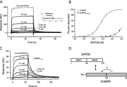
ESCRT-II–ESCRT-III interactions.In yeast, ESCRT-II re-cruits the downstream ESCRT-III complex through direct in-teractions between the ESCRT-II protein Vps25p (EAP20) and the ESCRT-III component Vps20p (CHMP6) (human homologs are in parentheses) (3, 74). Similarly, the human EAP20 and CHMP6 proteins also interacted in our two-hybrid survey of human class E proteins (Fig. 1C and 3A). Yeast two-hybrid mapping studies revealed that this interaction was mediated by the N-terminal half of CHMP6 binding to the C-terminal half of EAP20 (Fig. 3A), consistent with earlier reports (74, 88). The two C-terminal domains of EAP20 are exposed at the ends of the Y-shaped ESCRT-II complex, where they would be ideally positioned to nucleate ESCRT-III deposition through bivalent binding interactions (Fig. 1B).
GST pull-down and biosensor binding experiments with pu-rified recombinant EAP20 and CHMP6 proteins demonstrated that the EAP20-CHMP6 interaction was direct, and these ex-periments again confirmed that EAP20 bound to the N-termi-nal half of CHMP6 (Fig. 3B and 4). As shown in Fig. 4A and B, the equilibrium binding isotherm for EAP20 binding to the full-length CHMP6 proteins fitted a simple 1:1 binding model with a dissociation constant of 5.3 ⫾ 0.2 M. Interestingly, EAP20 bound even more tightly to the N-terminal half of CHMP6 alone (residues 1 to 100) (equilibrium dissociation constant [KD]⫽601⫾9 nM), and the increased affinity
[image:5.585.45.284.78.431.2]pri-marily reflected a significantly lower rate of EAP20 dissocia-tion from the truncated CHMP6 fragment. This observadissocia-tion demonstrates that C-terminal sequences in CHMP6 reduce the EAP20 binding affinity, providing direct physical evidence for the proposal that isolated full-length ESCRT-III proteins can
FIG. 2. Yeast two-hybrid mapping of the interaction sites between TSG101 and the EAP30 and EAP45 proteins. (A) Fragments of TSG101 binding to full-length EAP30 (left) and EAP45 (right). Selec-tion media in the right and left arrays of each pair are the same as in Fig. 1C. (B) Full-length TSG101 binding to fragments of EAP45 (left) and EAP30 (right). (C) Schematic summary of fragment maps and interaction sites of TSG101, EAP30, and EAP45.
on November 8, 2019 by guest
http://jvi.asm.org/
adopt autoinhibited “foldback” conformations (45). In this re-gard, it is interesting that several different CHMP proteins can be posttranslationally modified and that the unmodified CHMP proteins preferentially localize to the cytoplasm (68, 83). The posttranslational modifications might therefore desta-bilize autoinhibitory conformations, thereby enhancing the
protein-protein interactions that help dictate ESCRT-III pro-tein localization.
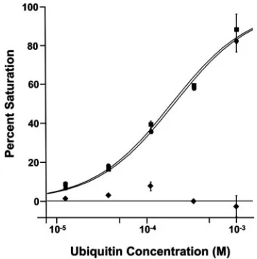
[image:6.585.49.544.71.212.2]EAP45-ubiquitin interactions.The yeast ESCRT-II complex helps sort ubiquitylated protein cargoes and binds ubiquitin through the second NZF motif (NZF-C) within the N-terminal GLUE domain of Vps36p (1). Mammalian EAP45 proteins
FIG. 3. Interactions between EAP20 and CHMP6. (A) Yeast two-hybrid mapping of the interaction sites between EAP20 and CHMP6. (B) GST pull-down experiment showing a direct interaction between EAP20 and the N-terminal half of CHMP6. The proteins present in each incubation mixture are given above lanes 2 to 5. Pure recombinant EAP20 is shown in lane 1 for reference. MW, molecular weight.
FIG. 4. Biosensor quantification of EAP20 binding to full-length and truncated CHMP6 proteins. (A) Sensorgrams showing different concen-trations of EAP20 binding to full-length, immobilized GST-CHMP6. The inset shows EAP20 binding to a control GST surface. (B) Binding isotherms for EAP20 binding to full-length GST-CHMP6 (KD⫽5.3⫾0.2M) and GST-CHMP100–201(KD⬎1 mM). Dissociation constants and errors were derived by fitting simple 1:1 models to the equilibrium binding data (panel A and data not shown). (C) Sensorgrams showing different concentrations of EAP20 binding to GST-CHMP61–100. The association and dissociation phases were globally fitted to simple 1:1 interaction models. The light-gray lines show global fits with the following parameters: association rate constantka⫽(1.46⫾0.04)⫻105M⫺1s⫺1, dissociation rate constantkd⫽0.088⫾0.003 s⫺
1, andK
D⫽601⫾9 nM. (D) Schematic summary of the interaction sites between EAP20 and CHMP6. The arrow with the question mark illustrates a possible “autoinhibitory” interaction between the N- and C-terminal regions of CHMP6.
on November 8, 2019 by guest
http://jvi.asm.org/
[image:6.585.82.499.361.647.2]lack NZF motifs, however, and it has therefore been unclear whether they could still interact with ubiquitylated protein cargoes. This puzzle has recently been resolved by Slagsvold and colleagues, who showed that a minimal N-terminal frag-ment of murine EAP45 spanning residues 1 to 139 binds di-rectly to ubiquitin (KD ⫽460 M) and also binds
phospho-inositides (67).
We independently tested for ubiquitin binding activity in the human EAP45 protein using the entire region of human EAP45 that is missing from the crystal structure of yeast Vps36p (EAP451–229). Biosensor binding analyses revealed
that ubiquitin bound specifically to GST-EAP451–229, with an
estimated dissociation constant of 210⫾10M in the exper-iment shown in Fig. 5. EAP451–229binding was strongly
inhib-ited by the Ub I44A mutation, but not by a F4A mutation, demonstrating that the exposed hydrophobic Ub I44 surface forms at least part of the EAP45 binding site. This same bind-ing surface is also recognized by nearly all of the other ubiq-uitin binding proteins characterized to date (29). Ubiqubiq-uitin also bound with a similar affinity to a minimal EAP451–139
construct (KD ⫽ 410 ⫾ 90 M; n ⫽ 6), indicating that the
ubiquitin binding site is located within the N-terminal GLUE domain. These data are all in excellent agreement with recent analyses of ubiquitin binding to the murine EAP45 GLUE domain (67).
EAP20 and CHMP6 accumulate on aberrant endosomal class E compartments in the absence of VPS4 ATPase activity.
Under steady-state conditions, all three ESCRT complexes are predominantly cytoplasmic but assemble transiently on the en-dosomal membrane to participate in vesicle formation. The assembled ESCRT complexes are then recycled off the endo-somal membrane by the action of the VPS4 AAA ATPases (5). Inhibition of human and yeast VPS4 ATPase activity therefore traps class E proteins, including the VPS4 proteins themselves, on aberrant endosomal class E compartments (3–5, 8, 21, 83, 89).
To test whether human ESCRT-II associated with endosomal membranes in a VPS4-dependent fashion, we examined the localization of two different ESCRT-II components, EAP20 and EAP30, in the presence of a mutant VPS4A protein (green fluorescent protein [GFP]-VPS4AK173Q) previously shown to
block ATP binding and to dominantly inhibit VPS4 activity in cells (8). As shown in Fig. 6A and B, antibodies against EAP20 detected endogenous EAP20 in COS-7 cells, albeit weakly. These antibodies also cross-reacted with a second protein of
⬃36 kDa (data not shown). We therefore also examined the localization of exogenous, Myc-tagged EAP20 to take advan-tage of the enhanced immunofluorescence signal of the tagged, overexpressed protein (Fig. 6C). In both cases, EAP20 local-ized to the enlarged endosomes induced by GFP-VPS4AK173Q
overexpression. In the absence of GFP-VPS4AK173Q, both
en-dogenous and overexpressed EAP20 were distributed through-out the cytoplasm, as has been seen for other ESCRT com-plexes (Fig. 6B and D). To confirm that these results accurately represented the distribution of the entire ESCRT-II complex, the localization of a second ESCRT-II component, EAP30, was also examined. Once again, the overexpressed, Myc-tagged EAP30 protein was seen throughout the cell, but it became trapped on the class E compartments induced by dominant-neg-ative GFP-VPS4AK173Q (compare Fig. 6E and F). Hence,
ESCRT-II was recruited to class E compartments, where it be-came trapped in the absence of VPS4 ATPase activity. Localiza-tion of the endogenous ESCRT-III protein, CHMP6, was also examined, and this protein was also strongly recruited to aberrant class E compartments (Fig. 6G and H and data not shown). Similar results were obtained using the other dominant-negative ATPase constructs, GFP-VPS4AE228Q,
DsRed-VPS4BK180Q, and DsRed-VPS4BE235Q(22, 83), and with
hu-man osteosarcoma cells (data not shown). These data are all consistent with the idea that human EAP20/ESCRT-II, EAP30/ESCRT-II, and CHMP6/ESCRT-III function in vesicle formation and cargo sorting at the MVB.
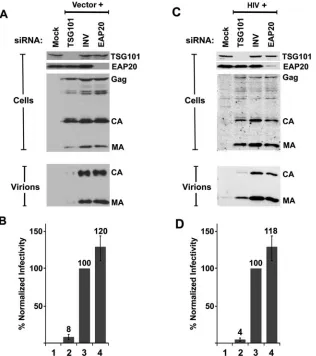
HIV-1 particles are released efficiently from cells depleted of EAP20. To test whether ESCRT-II is required for efficient HIV-1 budding, we measured the levels of HIV-1 vector and virus production following siRNA depletion of EAP20 or TSG101 (positive control). As shown in Fig. 7A and C, both EAP20 and TSG101 were successfully depleted to nearly un-detectable levels, as determined by quantification of Western blots of cellular extracts (Fig. 7A and C, “Cells,” top two rows). Importantly, intracellular Gag expression levels were not sig-nificantly altered by depletion of either protein (“Cells,” bot-tom rows). As expected, the release of virions was dramatically reduced upon TSG101 depletion (Fig. 7A and C, “Virions,” lanes “TSG101”), and the infectious titers were reduced⬃ 20-fold (Fig. 7B and D). Surprisingly, however, depletion of EAP20 did not significantly affect viral release as measured in Western blot assays (Fig. 7A and C, “Virions”, lanes “EAP20”), and actually increased vector titers slightly at the 24-h time point (Fig. 7B and D, bars 4), although at a later time point (48 h), HIV-1 titers were reduced twofold (data not shown). We therefore conclude that efficient depletion of EAP20 does not significantly inhibit the release or infectivity of HIV-1.
HIV-1 particles are also efficiently released from cells de-pleted of CHMP6.The efficient release of HIV-1 from
EAP20-FIG. 5. Biosensor binding analyses of EAP451–229-ubiquitin inter-actions. Binding isotherms for wild-type Ub (circles) (KD⫽210⫾10 M), F4A Ub (squares) (KD⫽170⫾10M), and I44A Ub (dia-monds) (KD⬎1 mM) binding to immobilized GST-EAP451–229. The error bars represent standard deviations.
on November 8, 2019 by guest
http://jvi.asm.org/
[image:7.585.70.255.68.254.2]depleted cells was unexpected, and the implications of this observation were examined further by testing the requirement for CHMP6/ESCRT-III in virus release. CHMP6 was selected for study because it binds EAP20, forms the only known con-nection between ESCRT-II and ESCRT-III, is the only myris-toylated CHMP protein (3, 88), and appears to initiate ESCRT-III assembly in yeast (3).
The effects of CHMP6 depletion on the release and infec-tivity of HIV-1 vectors and virus are shown in Fig. 8. As expected, virus release and infectivity were again substantially reduced upon siRNA depletion of TSG101 (Fig. 8A to D, lanes “TSG101” and bars 2), whereas treatment with an inverted control siRNA had no significant effect on virus release (de-fined as 100% infectivity; lanes “INV” and bars 3). Two dif-ferent siRNAs were used to knock down CHMP6 (designated CHMP6-1 and CHMP6-2), and both siRNAs decreased
CHMP6 levels very significantly (Fig. 8A and C, “Cells,” sec-ond row) without affecting Gag protein expression or process-ing (third row). As with EAP20 depletion, CHMP6 depletion again failed to reduce HIV-1 release or infectivity substantially (Fig. 8A and 8C, “Virions,” and B and D). Although there was some variability in multiple repetitions of this experiment, the average viral titers nevertheless fell within 40% of control levels following treatment with both CHMP6-1 and CHMP6-2 (Fig. 8B and D). At a later time point (48 h), viral infectivities were reduced four- to fivefold, possibly owing to secondary effects on general protein trafficking. We therefore conclude that normal cellular levels of CHMP6 are not required for efficient HIV-1 release or infectivity.
EAP20 and CHMP6 are required for efficient downregula-tion of the EGF receptor.The EGFR is normally present on the cell surface, where it can bind EGF and transmit growth
FIG. 6. Localization of ESCRT-II and ESCRT-III proteins by immunofluorescence. COS7 cells expressing fluorescent, exogenous, dominant-negative GFP-VPS4AK173Q(denoted GFP-VPS4A-KQ) were cotransfected and/or costained to detect the following proteins: (A) endogenous (endog) EAP20 protein (antibody UT461), (C) exogenous EAP20-Myc (anti-Myc antibody), (E) exogenous EAP30-myc (anti-Myc), and (G) en-dogenous CHMP6 (UT463). Column 1 (from left), ESCRT proteins alone (red); column 2, GFP-VPS4A-KQ proteins alone (green); column 3, overlaid (merged) images (colocalization on class E compartments is in yellow); column 4, differential interference contrast (Dic) images. Panels B, D, F, and H show the protein distributions in the absence of dominant-negative VPS4. Scale bars, 20m. Note that our two EAP20 antibodies also stained COS7 and human osteosarcoma cell centrosomes and spindles (Fig. 6B), and one of our two CHMP6 antibodies, UT 464, exhibited a punctate nuclear staining pattern in addition to staining class E membranes (not shown).
on November 8, 2019 by guest
http://jvi.asm.org/
signals. Following high levels of EGF stimulation, the receptor is internalized, trafficked through the MVB pathway, and de-graded in the lysosome, thereby attenuating the growth signal (32, 43). The lack of a requirement for EAP20 and CHMP6 in HIV-1 budding was surprising, and we therefore tested whether depletion of these proteins altered EGFR downregu-lation, as would be expected for proteins that function in MVB vesicle formation and protein sorting. These experiments also served as controls for our ability to deplete TSG101, EAP20, and CHMP6 to functionally significant levels.
[image:9.585.137.453.69.425.2]As shown in Fig. 9, depletion of TSG101/ESCRT-I, EAP20/ ESCRT-II, and CHMP6/ESCRT-III significantly reduced the efficiency of EGFR degradation in response to EGF stimula-tion, although the block was not complete and was somewhat greater for TSG101/ESCRT-I than for EAP20/ESCRT-II and CHMP6/ESCRT-III. Total cellular EGFR protein levels and localization were assayed by immunofluorescence, using an anti-EGFR antibody. In the absence of EGF stimulation, EGFR was located almost exclusively on the surfaces of 293T cells (Fig. 9A, column 1 [from left] and inset). Following EGF
FIG. 7. siRNA depletion of EAP20 does not diminish HIV-1 vector release or transduction efficiency. (A) Western blot analysis of 293T cell extracts showing the efficiency of siRNA depletion of TSG101 (positive control, lane TSG101 in top gel) or EAP20 (lane EAP20 in second gel from top) and the effects on Gag protein expression (third gel from top) and virus-like particle release (virions, bottom gel) from a cotransfected HIV-1 vector system. Lanes Mock and INV are negative controls showing cells that were mock transfected without the HIV-1 vector or cotransfected with an inverted siRNA against TSG101 (INV), respectively. Cellular Gag, CA, and MA levels were monitored to ensure equal transfection and protein expression levels. Extracellular CA and MA levels reflect the relative efficiencies of virus-like particle release. The TSG101 signal intensity for TSG101-depleted cells was 1% of that of the control, and the EAP20 signal intensity for EAP20-depleted cells was 2% of that of the control. Quantification of at least three repetitions of this experiment showed the following levels of MA and CA release: TSG101 siRNA, 9%⫾2% of the control; inverted siRNA, 100% (defined as the control); EAP20, 108%⫾ 12%. The data were collected 48 h after the second siRNA transfection. (B) HIV-1 viral-vector titers produced by cells depleted of EAP20 or TSG101 (positive control). Vector transduction levels were normalized to the negative control (INV) and averaged from six independent experiments. Control vector titers were 2.5⫻105to 3.2⫻105/ml. The data were collected 48 h after the second siRNA transfection. (C) Western blot analysis showing the efficiency and effects of siRNA-mediated silencing of EAP20 in analogous experiments with wild-type (wt) HIV-1NL4-3virus. The experiment was similar to that shown in panels A and B, except that wt HIV-1NL4-3virus was utilized. Quantification of at least three repetitions of this experiment showed the following levels of MA and CA release: TSG101 siRNA, 12%⫾9% of the control; inverted siRNA, 100% (defined as the control); EAP20, 75%⫾4%. The data were collected 26 h after the second siRNA transfection. (D) HIV-1 titers produced by cells depleted of EAP20 or TSG101 (positive control). Control viral titers were 0.9⫻106to 4⫻106/ml of supernatant. The values represent the average and standard error of eight independent experiments. The data were collected 24 to 26 h after the second siRNA transfection.
on November 8, 2019 by guest
http://jvi.asm.org/
stimulation for 2 h,⬎90% of the EGFR was internalized and degraded (column 2). Importantly, EGFR degradation was significantly reduced by treatment with siRNAs against TSG101 (column 4), EAP20 (column 5), and CHMP6 (column
[image:10.585.124.464.69.503.2]6), but not by treatment with a control siRNA (column 3). Depletion of the three ESCRT components did not affect receptor internalization but rather induced intracellular accu-mulation of internalized receptors, consistent with a defect in
FIG. 8. HIV release and infectivity are minimally affected by CHMP6 depletion. (A) Western blot analysis of 293T cell extracts showing the efficiency of siRNA depletion of TSG101 (positive control; lane TSG101 in the top gel) or CHMP6 (lanes CHMP6-1 and -2 in the second gel from the top) and the effects on Gag protein expression (third gel from the top) and virus-like particle release (virions; bottom gel) from a cotransfected HIV-1 vector system. CHMP6 was depleted using two different siRNAs (denoted CHMP6-1 and CHMP6-2). Lanes Mock and INV are negative controls showing cells that were mock transfected without the HIV-1 vector or cotransfected with an inverted siRNA against TSG101 (INV), respectively. Cellular Gag, CA, and MA levels were monitored to ensure equal transfection and protein expression levels. The extracellular CA and MA levels reflect the relative efficiencies of virus-like particle release. The TSG101 signal intensity for TSG101-depleted cells was 2% of that of the control, and the CHMP6 signal intensities for CHMP-6-depleted cells were 12% (CHMP6-1) and 3% (CHMP6-2) relative to the control. Quantification of at least three repetitions of this experiment showed the following levels of MA and CA release: inverted siRNA, 100% (defined as the control); CHMP6-1, 133%⫾14%; and CHMP6-2, 105%⫾35%. The data were collected 48 h after the second siRNA transfection. (B) HIV-1 viral-vector titers produced by cells depleted of CHMP6 or TSG101 (positive control). Vector transduction levels were normalized to the negative control (INV) and averaged from four independent experiments. The data were collected 48 h after the second siRNA transfection. (C) Western blot analysis showing the efficiency and effects of siRNA-mediated silencing of the CHMP6 protein. The experiment was similar to that shown in panel A except that wild-type HIV-1NL4-3virus was used. Quantification of at least three repetitions of this experiment showed the following levels of MA and CA release: inverted siRNA, 100% (defined as the control); CHMP6-1, 109%⫾22%; and CHMP6-2, 94%⫾38%. The data were collected 26 h after the second siRNA transfection. (D) HIV-1 titers produced by cells depleted of CHMP6 or TSG101 (positive control). Viral transduction levels were normalized to the negative control (INV) and averaged from eight independent experiments. The data were collected 26 h after the second siRNA transfection. The error bars represent standard deviations.
on November 8, 2019 by guest
http://jvi.asm.org/
MVB trafficking. We note that treatment with siRNAs against TSG101 resulted in the accumulation of EGFR in larger class E compartments (17) than were seen in cells treated with siRNAs against EAP20 or CHMP6. The EGFR trafficking defects seen in the absence of ESCRT activities were similar in 293T and HeLa cells, as shown quantitatively in Fig. 9B and C. The efficiencies of protein depletion and virus release were monitored in parallel experiments that confirmed that the tar-geted proteins were depleted as expected and that HIV-1 re-lease was significantly reduced by TSG101 depletion, but not by depletion of EAP20 or CHMP6 (data not shown).
Bowers et al. recently reported that siRNA depletion of EAP20 did not measurably inhibit the lysosomal degradation of MHC-I and EGF-EGFR complexes (11). This differs from
[image:11.585.49.539.79.456.2]our conclusion that EAP20 depletion reduced the efficiency of EGFR degradation, and we therefore investigated the issue further by testing the effects of depleting TSG101/ESCRT-I, EAP20/ESCRT-II, and CHMP6/ESCRT-III on (i) MHC-I degradation and (ii) movement of fluorescent EGF to the lysosome. In the former experiments, we found that depletion of TSG101/ESCRT-I reduced the stimulation of MHC-I re-ceptor degradation by the Kaposi’s sarcoma-associated herpes-virus ubiquitin ligase KK3 (13, 28), whereas depletion of EAP20/ESCRT-II and CHMP6/ESCRT-III did not (data not shown). These results are in excellent agreement with those of Bowers et al. (11), and we therefore conclude that efficient MHC-I downregulation requires TSG101/ESCRT-I but not EAP20/ESCRT-II or CHMP6/ESCRT-III. Thus, the ESCRT
FIG. 9. EGF receptor downregulation is inhibited by depletion of TSG101, EAP20, and CHMP6. (A) 293T cells treated with siRNA were serum starved and then incubated in the presence or absence of EGF. Fixed and permeabilized cells were visualized by differential interference contrast (Nomarski) microscopy (top row) or by immunofluorescence to determine EGFR levels and localization (bottom row). Stacked whole-cell images are shown in all cases, with a central confocal slice (inset) also shown in the first image in the bottom row to demonstrate that the EGFR is located on the cell surface prior to EGF stimulation. (B) Quantification of the total levels of EGFR in 293T cells under the different conditions described for panel A. Each bar in the histogram represents the average fluorescence of individual cells as measured in three different images containing two to five cells per image. The error bars show the standard errors of the mean. The data were collected 91 h after the second siRNA transfection. (C) Quantification of total levels of EGFR in HeLa cells under the conditions described for panel A. The data were collected 92 h after the second siRNA transfection.
on November 8, 2019 by guest
http://jvi.asm.org/
requirements for MHC-I downregulation are similar to the requirements for HIV-1 budding.
We examined the trafficking of EGF to the lysosome in cells depleted of TSG101/ESCRT-I, EAP20/ESCRT-II, or CHMP6/ ESCRT-III. HeLa cells depleted of TSG101/ESCRT-I, EAP20/ESCRT-II, or CHMP6/ESCRT-III were incubated with the fluid phase marker Alexa 488-dextran for 18 h, fol-lowed by a 4-h chase in dextran-free medium, which allows the fluid phase marker to accumulate in lysosomes (12, 78). As shown above, the unstimulated EGFR is predominantly local-ized to the plasma membrane. Incubation of cells with EGF-Alexa Fluor 555 streptavidin induced the internalization of the EGFR, and after 30 min, fluorescent EGF colocalized with lysosomes in 41% of the cells examined (Fig. 10, “INV,”)
(86/208 cells). In contrast, cells depleted of TSG101/ESCRT-I showed no colocalization of EGF with lysosomes (“TSG101”) (0/29 cells tested). Cells depleted of either EAP20/ESCRT-II or CHMP6/ESCRT-III showed intermediate phenotypes, with 7 to 10% of the cells showing EGF staining at the lysosome. To control for possible differences between different siRNA oli-gonucleotides, we also tested one of the siRNA oligonucleo-tides used to deplete EAP20 in the study by Bowers et al. This oligonucleotide (EAP20-2) also efficiently depleted EAP20 levels (Fig. 10C) and again gave an intermediate EGF local-ization phenotype (Fig. 10A and B, “EAP20-2”) (19/258 cells positive for EAP20-2 versus 10/140 cells for EAP20-1 and 22/213 cells for CHMP6-2).
[image:12.585.53.534.78.477.2]As will be reported elsewhere, we have also now found that
FIG. 10. Colocalization of EGF and lysosomal fluid phase markers upon depletion of TSG101, EAP20, and CHMP6. (A) HeLa cells were treated with the designated siRNA, Alexa 488-dextran (green), and EGF-Alexa Fluor 555 streptavidin (red) as described in Materials and Methods. (B) Quantification of the fractions of cells showing some colocalization of Alexa 488-dextran and EGF-Alexa Fluor 555 streptavidin under the different conditions described for panel A. The quantification of EGF (red) coincident with lysosome dextran (green) was performed in single-plane overlay analyses using the Olympus Fluoview software. Cells were scored as positive if the overlap coefficient (R) was greater than 0.5. The error bars represent standard deviations. (C) Western blot analysis showing the efficiency of siRNA-mediated depletion of TSG101, EAP20, and CHMP6.
on November 8, 2019 by guest
http://jvi.asm.org/
degradation of ferroportin, a cell surface receptor that is down-regulated by hepcidin binding (55), shows a strong dependence on TSG101, EAP20, and CHMP6 (I. De Domenico, D. M. Ward, C. Langelier, M. B. Vaughn, E. Nemeth, W. I. Sundquist, T. Ganz, G. Musci, and J. Kaplan, unpublished data). We there-fore conclude that lysosomal targeting of different cell surface receptors can differ in EAP20 and CHMP6 requirements. Some receptors, such as MHC-I, exhibit little or no require-ment for these proteins, while other receptors, such as ferro-portin, exhibit strong requirements, and still others, such as EGFR, exhibit intermediate phenotypes. These experiments indicate that there are probably multiple pathways for sorting membrane proteins into the MVB vesicles of mammalian cells.
DISCUSSION
Not surprisingly, our studies have revealed that the human ESCRT-II complex has a number of features in common with its better-characterized yeast counterpart. The similarities in-clude interactions with ESCRT-I, ESCRT-III, and ubiquitin; transient association with endosomal membranes; and func-tional participation in receptor downregulation. These obser-vations are all consistent with the idea that ESCRT-II can play a central role in the human ESCRT pathway, interacting with the upstream ESCRT-I and downstream ESCRT-III com-plexes and with ubiquitylated cargos as they are sorted into MVB vesicles. Nevertheless, depletion of EAP20/ESCRT-II and CHMP6/ESCRT-III did not significantly reduce HIV-1 release from cultured 293T or HeLa cells, indicating that only a subset of the mammalian ESCRT machinery is required for efficient virus release from these cell types.
Role of EAP20/ESCRT-II in HIV-1 release.We considered several possible explanations for the observation that EAP20/ ESCRT-II depletion does not inhibit HIV-1 release. As with all siRNA depletion experiments, it is possible that even the very low levels of EAP20 remaining after depletion were still sufficient to function in HIV-1 release (and MHC-I downregu-lation). While we cannot rule out this possible explanation, it seems unlikely because EAP20 was efficiently depleted in our experiments (Fig. 7 and 10), and this depletion measurably inhibited EGFR degradation and lysosomal targeting (Fig. 9 and 10). Furthermore, EAP20 depletion actually produced a slight, but reproducible, increase in HIV-1 infectivity (Fig. 7), which could be explained if the endogenous ESCRT-II com-plex binds and competes for other factors essential for virus budding (e.g., ESCRT-I). It is also conceivable that ESCRT-II may remain active even without EAP20 subunits. This expla-nation again seems unlikely, because the two copies of EAP20 comprise half of the ESCRT-II subunits and form the only known link between ESCRT-II and ESCRT-III. Furthermore, ESCRT-II activity is blocked by deletion of EAP20/Vps25p in both S. cerevisiae and Drosophila (4, 76, 77). We therefore conclude that the most likely explanation for our data is that ESCRT-II is not required for efficient HIV-1 release from human cells, either because the complex does not normally participate in virus budding or because the viral budding ma-chinery still functions efficiently in its absence.
Our results also imply that if HIV-1 utilizes the late-acting ESCRT-III and VPS4 complexes to bud from cells, then there must be other proteins or complexes, in addition to ESCRT-II,
that can bridge ESCRT-I and ESCRT-III. In principle, ALIX/ AIP1 could be one such bridging protein, as it can bind directly to both TSG101 (83) and to the CHMP4 proteins (38, 41, 69, 83), although simultaneous depletion of ALIX and EAP20 did not synergistically inhibit EGFR degradation (11). Similarly, interactions between the yeast ESCRT-I components, Vps37p and Vps28p, and the ESCRT-III component, Vps20p (CHMP6), have been reported (10), although we did not ob-serve analogous ESCRT-I–ESCRT-III interactions between the isolated human proteins in two-hybrid experiments. Fi-nally, the deubiquitylating enzyme, AMSH, binds both the upstream HRS-STAM complex (which binds to TSG101) (71) and the ESCRT-III components CHMP3 and CHMP4 (23). In principle, these (and other) bridging interactions could help to recruit ESCRT-III to sites of MVB vesicle formation and virus budding even in the absence of ESCRT-II.
Role of ESCRT-III in HIV-1 release.Models for the roles of ESCRT-III (and VPS4) in virus budding must take into ac-count the observation that HIV-1 release is not significantly inhibited by depletion of endogenous CHMP5 and CHMP6 (reference 85 and this work), yet virus release is very potently inhibited by overexpression of dominant-negative CHMP5 or CHMP6 protein (46, 69, 70). siRNA depletion of these indi-vidual CHMP proteins therefore imposes a less severe (or less general) block in the ESCRT pathway than the dominant-negative ESCRT-III and VPS4 constructs. A likely explanation for this difference is that dominant-negative CHMP (and VPS4) constructs function by inducing the formation of aber-rant endosomal compartments (class E compartments) that not only inhibit the functions of the modified CHMP proteins themselves, but also sequester other ESCRT complexes and possibly many other MVB proteins required for HIV-1 bud-ding. Thus, we cannot rule out the possibility that wild-type CHMP and VPS4 proteins do not normally function in HIV-1 release but rather affect virus budding only when present in dominant-negative forms. However, we favor a model in which at least a subset of the ESCRT-III and VPS4 proteins do actually participate directly in virus release but where deple-tion of individual CHMP5 or CHMP6 proteins does not inhibit virus budding owing to (i) functional redundancy within the MVB pathway arising from the presence of 10 different CHMP proteins, (ii) the possibility that CHMP5 (and possibly other CHMP proteins) may function at a step following MVB vesicle formation (7, 66), and/or (iii) the presence of multiple distinct ESCRT pathways in human cells. It therefore continues to be important to determine precisely which subset of different ESCRT proteins is utilized directly by HIV-1 and other vi-ruses.
Complexity of MVB vesicle formation and protein sorting in higher eukaryotes. Genetic studies have identified the basic machinery required for MVB vesicle formation and protein sorting inS. cerevisiae. Importantly, deletion of each of the six known yeast ESCRT-I and ESCRT-II subunits ultimately leads to a strong block in protein sorting and MVB vesicle formation (the class E phenotype) (40). While it seems almost certain that the basic mechanism of MVB biogenesis is highly con-served, because human cells have at least one ortholog of every known yeast class E protein (33, 51), our studies add to the growing number of indications that MVB protein-sorting/ves-icle formation pathways are more complex in higher
on November 8, 2019 by guest
http://jvi.asm.org/
karyotes than in yeast. One level of increased complexity is suggested by the fact that unique yeast class E proteins often have multiple orthologs in mammalian cells. Examples include the 4 different human homologs of the single yeast Vps37p protein (6, 19, 70), the 10 different human homologs of the six yeast ESCRT-III proteins (51), and the 2 (or more) different human homologs of yeast Vps4p (60). Thus, unique proteins or complexes that are essential for MVB formation in yeast may play redundant or overlapping roles in higher eukaryotes. For example, simultaneous depletion of both VPS37B and VPS37C inhibits PTAP-dependent retrovirus budding to a greater ex-tent than depletion of either single VPS37 paralog alone (48). A second level of complexity is suggested by the fact that even unique ESCRT-I or ESCRT-II subunits do not appear to be required for metazoan MVB vesicle formation in all con-texts. For example, a knockout of the Drosophila Vps28/ ESCRT-I protein has only modest effects on MVB morphology and no measurable effect on the downregulation of several different cell surface receptors in vivo, even though there is only a singlevps28gene inDrosophila(63). Similarly, agonist-induced lysosomal degradation of the delta opioid receptor is inhibited by dominant-negative VPS4B mutants and by deple-tion of HRS, but not by depledeple-tion of TSG101/ESCRT-I (31). Hence, an intact ESCRT-I complex is apparently not required for downregulation of all cell surface receptors via the MVB pathway (27, 31). Similarly, EAP20/ESCRT-II is not required for MHC-I receptor downregulation induced by Kaposi’s sar-coma-associated herpesvirus KK3 (reference 11 and data not shown).
Retroviruses also differ in their requirements for ESCRT-I. HIV-1, which binds directly to TSG101, requires all of the known ESCRT-I components for efficient release (19, 22, 48), whereas equine infectious anemia virus and Moloney murine leukemia virus, which bud primarily via ALIX and the Nedd4 E3 ligases, respectively, exhibit little or no requirement for TSG101/ESCRT-I (22, 48, 62, 64, 72). Nevertheless, all retro-viruses are inhibited by dominant-negative ESCRT-III and VPS4 proteins, suggesting a common use of these downstream factors (22, 46, 69, 83). This model is consistent with the idea that ESCRT-I and ESCRT-II may function primarily as adap-tor complexes that help recruit cargoes into the pathway, whereas the ESCRT-III/VPS4 machinery functions more di-rectly in vesicle formation. Indeed, the genomes of Plasmo-dium falciparumand Toxoplasma gondiireportedly lack ES-CRT-I and ESES-CRT-II entirely but do encode ESES-CRT-III and VPS4 proteins and require VPS4 to create multivesicular bod-ies (87). Finally, we note that even the ESCRT-III/VPS4 ma-chinery appears to be dispensable in some cases, as the mela-nosomal protein Pmel17 is sorted into MVB vesicles via an ESCRT-independent pathway that is also insensitive to inhibi-tion by dominant-negative forms of VPS4 (75). The apparent diversity of MVB sorting pathways raises the intriguing possi-bility that viral systems may also have evolved to utilize such “noncanonical” MVB pathways to escape the cell (34).
Roles for mammalian ESCRT-I and ESCRT-II in receptor downregulation. Although ESCRT-I and ESCRT-II are not absolutely required for the efficient downregulation of all re-ceptors or for the release of all retroviruses, both complexes clearly play very important roles in downregulating certain cell surface receptors. Indeed, a number of recent studies indicate
that both ESCRT-I and ESCRT-II exhibit growth/tumor sup-pressor activities by virtue of their roles in cell surface receptor downregulation. For example, all three known ESCRT-I com-ponents (TSG101, VPS28, and VPS37) have now been impli-cated in cell growth and tumor suppression. TSG101 was ini-tially identified in a genetic screen for genes with tumor suppressor activity, and depletion of TSG101 induces NIH 3T3 cell overproliferation in culture and metastatic tumor forma-tion in nude mice (44). Similarly, deleforma-tion oferupted, the Dro-sophilaTSG101 ortholog, inhibits Notch receptor degradation and activates the JAK-STAT (and possibly other) signaling pathway and thereby induces tissue overproliferation (50). Likewise, depletion of one of the four mammalian VPS37 proteins (6, 19, 70), VPS37A/HCRP-1, enhances the growth of cultured hepatocellular carcinoma BEL-7404 cells and elevates their invasive ability (86). Interestingly, VPS37A/HCRP-1 is also downregulated in some hepatocellular carcinomas. Fi-nally, we have observed that depletion of VPS28 can relieve contact-inhibited growth in cultured 293T cells (J. Garrus, per-sonal communication). ESCRT-II also appears to play an im-portant role in growth receptor downregulation, as deletion of the Drosophila vps25 gene leads to tissue overproliferation through aberrant stabilization of the Notch and DPP (and possibly other) receptors (76, 77). Similarly, we observed that depletion of EAP20 (human VPS25) reduces the degradation rates of EGFR (Fig. 9 and 10), ferroportin (De Domenico, et al., unpublished), and presumably other cell surface receptors following ligand stimulation. Thus, just as the proteasome functions to degrade soluble proteins, the mammalian ESCRT pathway, including ESCRT-II, can play a critical role in medi-ating both homeostatic and regulated lysosomal degradation of integral membrane proteins.
ACKNOWLEDGMENTS
This work was supported by NIH funding to C.P.H. (GM066521), J.K. (HL29622 and DK070947), and W.I.S. (AI51174).
We thank Chris Rodesch, David Myszka, and Rebecca Rich for assistance with immunofluorescence and biosensor experiments.
REFERENCES
1.Alam, S. L., J. Sun, M. Payne, B. D. Welch, B. K. Blake, D. R. Davis, H. H. Meyer, S. D. Emr, and W. I. Sundquist.2004. Ubiquitin interactions of NZF
zinc fingers. EMBO J.23:1411–1421.
2.Babst, M.2005. A protein’s final ESCRT. Traffic6:2–9.
3.Babst, M., D. Katzmann, E. Estepa-Sabal, T. Meerloo, and S. Emr.2002. Escrt-III. An endosome-associated heterooligomeric protein complex
re-quired for mvb sorting. Dev. Cell3:271–282.
4.Babst, M., D. Katzmann, W. Snyder, B. Wendland, and S. Emr.2002. Endosome-associated complex, ESCRT-II, recruits transport machinery for
protein sorting at the multivesicular body. Dev. Cell3:283–289.
5.Babst, M., B. Wendland, E. J. Estepa, and S. D. Emr.1998. The Vps4p AAA ATPase regulates membrane association of a Vps protein complex required
for normal endosome function. EMBO J.17:2982–2993.
6.Bache, K. G., T. Slagsvold, A. Cabezas, K. R. Rosendal, C. Raiborg, and H. Stenmark.2004. The growth-regulatory protein HCRP1/hVps37A is a sub-unit of mammalian ESCRT-I and mediates receptor downregulation. Mol.
Biol. Cell15:4337–4346.
7.Bache, K. G., S. Stuffers, L. Malerod, T. Slagsvold, C. Raiborg, D. Lechar-deur, S. Walchli, G. L. Lukacs, A. Brech, and H. Stenmark.2006. The ESCRT-III subunit hVps24 is required for degradation but not silencing of
the epidermal growth factor receptor. Mol. Biol. Cell17:2513–2523.
8.Bishop, N., and P. Woodman.2000. ATPase-defective mammalian VPS4 localizes to aberrant endosomes and impairs cholesterol trafficking. Mol.
Biol. Cell11:227–239.
9.Bishop, N., and P. Woodman.2001. TSG101/mammalian VPS23 and mam-malian VPS28 interact directly and are recruited to VPS4-induced
endo-somes. J. Biol. Chem.276:11735–11742.
10.Bowers, K., J. Lottridge, S. B. Helliwell, L. M. Goldthwaite, J. P. Luzio, and