0022-538X/06/$08.00⫹0 doi:10.1128/JVI.01016-06
Copyright © 2006, American Society for Microbiology. All Rights Reserved.
Human Cytomegalovirus (HCMV) Infection of Endothelial Cells
Promotes Naı¨ve Monocyte Extravasation and Transfer of
Productive Virus To Enhance Hematogenous
Dissemination of HCMV
䌤
Gretchen L. Bentz,
1Marta Jarquin-Pardo,
1Gary Chan,
1M. Shane Smith,
1Christian Sinzger,
2and Andrew D. Yurochko
1*
Department of Microbiology and Immunology, Center for Molecular and Tumor Virology, Feist-Weiller Cancer Center, Louisiana State University Health Sciences Center, Shreveport, Louisiana 71130-3932,1and Institute for
Medical Virology, University of Tu¨bingen, 72076 Tu¨bingen, Germany2
Received 17 May 2006/Accepted 7 September 2006
Human cytomegalovirus (HCMV) pathogenesis is dependent on the hematogenous spread of the virus to host tissue. While data suggest that infected monocytes are required for viral dissemination from the blood to the host organs, infected endothelial cells are also thought to contribute to this key step in viral pathogenesis. We show here that HCMV infection of endothelial cells increased the recruitment and transendothelial migration of monocytes. Infection of endothelial cells promoted the increased surface expression of cell adhesion molecules (intercellular cell adhesion molecule 1, vascular cell adhesion molecule 1, E-selectin, and platelet endothelial cell adhesion molecule 1), which were necessary for the recruitment of naı¨ve monocytes to the apical surface of the endothelium and for the migration of these monocytes through the endothelial cell layer. As a mechanism to account for the increased monocyte migration, we showed that HCMV infection of endothelial cells increased the permeability of the endothelium. The cellular changes contributing to the increased permeability and increased naı¨ve monocyte transendothelial migration include the disruption of actin stress fiber formation and the decreased expression of lateral junction proteins (occludin and vascular endothelial cadherin). Finally, we showed that the migrating monocytes were productively infected with the virus, documenting that the virus was transferred to the migrating monocyte during passage through the lateral junctions. Together, our results provide evidence for an active role of the infected endothelium in HCMV dissemination and pathogenesis.
Human cytomegalovirus (HCMV) is a betaherpesvirus that establishes a life-long persistent infection (10). In immuno-compromised individuals, such as AIDS patients, neonates, and transplant recipients, HCMV infection is associated with significant morbidity and mortality (13, 38, 56, 87). In immu-nocompetent hosts, HCMV infection is generally asymptom-atic, although it can cause mononucleosis (40) and is associated with chronic inflammatory diseases, such as the cardiovascular diseases atherosclerosis and coronary restenosis (1, 19, 35, 46, 51, 57, 59, 86, 90, 107).
HCMV pathogenesis is a direct result of viral spread to host organs and the subsequent infection of those organ systems (6, 44, 54, 83). Systemic spread occurs during both asymptomatic and symptomatic infections (93) and is required for HCMV persistence in the host (83). During primary infection, the virus spreads from the initial site of infection to the peripheral blood and then to host organ tissue (6, 54, 83). In healthy hosts, infection of these organ systems allows for the establishment of the viral persistence needed for viral survival in the infected host and in the general population. In contrast, in
immuno-compromised hosts, because of the absence of a functional immune response, this same strategy of viral spread would lead to the overt organ disease seen in these individuals (83).
The mechanisms of HCMV dissemination remain unclear; however, cells of the myeloid lineage, specifically monocytes, are thought to have a central role in this process for multiple reasons (4, 22, 30, 34, 43, 44, 55, 79, 83, 89). First, monocytes containing viral DNA are found in patients with a cell-associ-ated viremia (79, 83). Second, HCMV-infected macrophages, the differentiated counterparts of monocytes, are detected in organs of symptomatic and asymptomatic hosts (29, 54, 64, 72, 83, 93). Third, animal studies with the related betaherpesvi-ruses murine CMV and rat CMV showed that infected mono-cytes were associated with the systemic spread of the virus (4, 55, 69, 95). These results suggest that infected monocytes serve as a key cell type responsible for viral spread from the blood during a primary infection. A caveat to monocyte participation in viral dissemination are results suggesting that monocytes are only abortively infected (28, 30, 31, 41, 79, 83, 91). Neverthe-less, monocytes appear to be the cell type that is “at the right place at the right time” for the hematogenous spread of HCMV following primary infection. Our recent studies pro-vide a possible answer to this conundrum and support the participation of infected monocytes in viral dissemination (84, 85). We showed that HCMV-infected peripheral blood mono-cytes, which at the time of infection were nonpermissive for
* Corresponding author. Mailing address: Department of Microbi-ology and ImmunMicrobi-ology, Louisiana State University Health Sciences Center, 1501 Kings Highway, Shreveport, Louisiana 71130-3932. Phone: (318) 675-8332. Fax: (318) 675-5764. E-mail: ayuroc@lsuhsc .edu.
䌤Published ahead of print on 20 September 2006.
11539
on November 8, 2019 by guest
http://jvi.asm.org/
viral replication, drove their differentiation into macrophages that became permissive for replication of the original input virus (84). Furthermore, we showed that HCMV infection of monocytes resulted in cellular activation, the induction of cel-lular motility, and monocyte migration through the endothe-lium (84, 85). These studies suggest that primary infection activates monocytes, promoting their migration into the sur-rounding tissue and their differentiation into permissive mac-rophages, which in turn allows for the “viral seeding” of organ systems.
In order for monocytes to carry HCMV from the blood to the tissue, they must pass through an endothelial cell barrier, which separates the blood from the surrounding tissue. The appropriate regulation of endothelial cells is required for the maintenance of existing blood vessels, the formation of new blood vessels (12), and the control of leukocyte migration into and out of the bloodstream (60). Leukocyte transendothelial migration begins with the adhesion of leukocytes to the surface of the endothelium (58, 88). Following adhesion to the endo-thelium, leukocytes move to the junctions between adjacent endothelial cells and migrate through these lateral junctions to enter the surrounding tissues. Thus, endothelial cells are the gatekeepers that control the trafficking of peripheral blood leukocytes.
Endothelial cells are a natural site of HCMV infection in vivo following a primary infection (22, 43, 62, 82, 99) and are a possible viral reservoir (21), suggesting a role for HCMV-infected endothelial cells in viral spread and persistence. HCMV infection of endothelial cells is also associated with atherosclerosis and transplant vascular sclerosis in HCMV-infected patients (reviewed in reference 25), pointing to the possible role infected endothelial cells may play in HCMV-mediated diseases. The specific mechanisms by which infected endothelial cells contribute to viral pathogenesis remain un-clear. We hypothesized that infected endothelial cells recruit naı¨ve monocytes to the infected endothelium, where the mono-cytes become activated and driven to migrate through endo-thelial cell junctions into the surrounding tissue. This strategy would allow for the spread of the virus from the blood into the surrounding tissue, but to be effective, the infected endothe-lium must pass the virus to naı¨ve monocytes. The transfer of the virus between different cell types is supported by studies showing that endothelial cell-monocyte interactions allow for the spread of the virus from one cell type to another (9, 32, 33, 96). Here we describe a potential mechanism for the role infected endothelial cells play in viral pathogenesis. We show that HCMV infection of endothelial cells promoted naı¨ve monocyte adhesion to and migration through the endothelium. Infected endothelial cells exhibited increased cell adhesion molecule expression, increased permeability, decreased actin stress fiber formation, altered expression of tight and adherens junction proteins, and the ability to transfer the virus to the migrating monocytes. These results, along with our previous studies (84), suggest that HCMV utilizes the following two-pronged approach to promote the spread of the virus within the host: (i) HCMV infects monocytes and promotes their migration through the naı¨ve endothelium (84, 85), and (ii) HCMV infects endothelial cells, which then recruit naı¨ve monocytes and promote their migration through the infected endothelium and the transfer of infectious virus from the
in-fected endothelial cell to the migrating monocyte. Biologically, the outcome of this strategy would be the effective and efficient hematogenous spread of the virus. In addition to this strategy serving as a mechanism for viral spread from the blood into the surrounding tissue, it could also promote the vascular diseases associated with HCMV infection.
MATERIALS AND METHODS
HCMV growth and purification.HCMV (Towne/E strain; passage 41 to 44) was cultured in human embryonic lung fibroblasts and purified on a 0.5 M sucrose gradient (103–105). An endothelial cell-tropic strain of HCMV, labeled with green fluorescent protein (GFP) (TB40-UL32-HCMV/E), was constructed similarly to the GFP-labeled fibroblast tropic strain of TB40 (70). TB40-UL32-HCMV/E was grown in a similar manner as the Towne/E strain of HCMV. Endothelial cells were infected at a multiplicity of infection (MOI) of 5 or 20 or mock infected with medium alone. This endothelial cell-tropic strain of HCMV also efficiently infects monocytes (81), as does our Towne/E strain (84, 85, 103). We found that 100% of the endothelial cells and 100% of monocytes were infected with TB40-UL32-HCMV/E and Towne/E when we used an MOI of 5 or an MOI of 20.
Endothelial cell culture.Human microvascular endothelial cells (HMECs)
were cultured at 37°C with 5% CO2in endothelial growth medium (Clonetics,
San Diego, CA) comprised of endothelial cell basal medium supplemented with
3 mg/ml bovine brain extract, 10g/ml human epidermal growth factor, 1 mg/ml
hydrocortisone, 10% fetal bovine serum (FBS), and 0.5 ml gentamicin sulfate– amphotericin B-1000 (all from Clonetics). Cells were harvested by removing medium, washing with Versene (Gibco Life Technologies, Rockville, MD), and incubating cells in 2.5 mg/ml of trypsin (Cellgro; Mediatech, Herndon, VA) for 10 minutes. Cells were collected in endothelial growth medium and pelleted by centrifugation. Medium was removed, and cells were resuspended in fresh medium.
Human umbilical vein endothelial cells (HUVECs) and immortalized human endothelial cells (IHECs) were also used in our experiments. These endothelial cells were cultured similarly to the HMECs as described above with the following exceptions: HUVECs were cultured in endothelial cell basal medium-2
supple-mented with 10g/ml human epidermal growth factor, 10% FBS, vascular
endothelial growth factor, human fibroblast growth factor-basic (with hepa-rin), R3-insulin-like growth factor 1, ascorbic acid, heparin, hydrocortisone, and GA-1000 (all components from Clonetics); IHECs were cultured in RPMI (Cellgro; Mediatech) supplemented with 10% heat-inactivated FBS (Gibco Life Technologies).
Monocyte isolation. Human peripheral blood monocytes were isolated by double density gradient purification (103, 106). Briefly, whole blood was taken by venipuncture from donors and centrifuged through a Ficoll Histopaque 1077 (Sigma, St. Louis, MO) gradient. The mononuclear cells were collected, and the platelets were removed by washing the mononuclear cells with ice-cold saline. Monocytes were then purified by centrifugation through a Percoll (Pharmacia, Piscataway, NJ) gradient. Isolated monocytes were labeled with CellTracker-Green 5-chloromethylfluorescein diacetate (CMFDA; Molecular Probes, Eu-gene, OR) according to the manufacturer’s protocol, and the cells were washed, pelleted by centrifugation, and resuspended in RPMI (Cellgro) supplemented with 10% human serum (Gemini Bio-products, Woodland, CA). The CellTracker-Green CMFDA did not affect the infection of monocytes or alter monocyte adhesion and motility (data not shown). University Institutional Review Board and Health Insurance Portability and Accountability Act guidelines were fol-lowed for all experiments involving human subjects.
Antibodies.A rabbit anti-occludin antibody was obtained from ZYMED Lab-oratories Inc. (San Francisco, CA). A mouse anti-intercellular cell adhesion molecule-1 (ICAM-1) monoclonal antibody, rabbit anti-vascular cell adhesion molecule-1 (VCAM-1) polyclonal antibody, goat anti-E-selectin polyclonal an-tibody, goat anti-platelet endothelial cell adhesion molecule-1 (PECAM-1) poly-clonal antibody, and goat anti-actin polypoly-clonal antibody were obtained from Santa Cruz Biotechnology, Inc. (Santa Cruz, CA). A mouse anti-vascular endo-thelial-cadherin (VE-cadherin) monoclonal antibody was obtained from Immu-notech (Marseille, France). For use as secondary antibodies, horseradish perox-idase (HRP)-conjugated donkey anti-goat and goat anti-rabbit polyclonal antibodies were obtained from Santa Cruz Biotechnology, Inc., while an HRP-conjugated sheep anti-mouse polyclonal antibody was obtained from Amersham Biosciences (Piscataway, NJ). For immunofluorescence microscopy, a fluorescein isothiocyanate (FITC)-conjugated goat anti-rabbit polyclonal antibody was ob-tained from Santa Cruz, and a FITC-conjugated donkey anti-mouse polyclonal
on November 8, 2019 by guest
http://jvi.asm.org/
antibody was obtained from Jackson ImmunoResearch Laboratories Inc. (West Grove, PA). Neutralizing monoclonal antibodies against ICAM-1, VCAM-1, E-selectin, and an immunoglobulin G1 (IgG1) isotype control antibody were obtained from R&D Systems (Minneapolis, MN). A blocking monoclonal anti-body to VE-cadherin (cl75) and a monoclonal antianti-body against HCMV IE1-72 were obtained from BD Transduction Laboratories (San Jose, CA).
Transendothelial migration assay.Transendothelial migration assays were performed as previously described (84, 85) using HMECs, HUVECs, or IHECs cultured in 24-well plates containing cell culture inserts (BD Falcon, Bedford,
MA) with 8-m pores (Fig. 1). A total of 3.5⫻104
endothelial cells were
cultured on the inserts and grown to confluence at 37°C in 5% CO2. Once
confluent, endothelial cells were mock infected, infected with HCMV (MOI, 5 or
20), or stimulated with lipopolysaccharide (LPS; 10g/ml; Sigma) or phorbitol
myristate acetate (PMA; 10 ng/ml; Sigma) (61, 63, 65, 92, 101). At 24 h
postin-fection (hpi), endothelial cells were washed, and 2.5⫻104
mock-infected
mono-cytes were added to each transwell and incubated at 37°C with 5% CO2. At 24 h
after the addition of monocytes to the transwells, three random fields of view (FOV) of monocytes from the top of the insert were counted by fluorescence microscopy to determine the percentage of monocytes undergoing migration on the endothelial cell layer versus the percentage of monocytes stationary on the endothelial cell layer (67). At 96 h after the addition of monocytes, the number of monocytes that migrated completely through the endothelial cell layer and into the bottom well was determined by fluorescence microscopy for 10 random FOV. Additionally, the percentage of monocytes that completely migrated through the endothelial cell layer, compared to the total number of monocytes added to the transwell, was determined by removing and counting monocytes from the bottom of the well (85). For all transendothelial migration assays,
results are plotted as mean⫾standard deviation (SD), and the statistical
sig-nificance between experimental means (Pvalue) was determined using Student’s
ttest.
In experiments where blocking antibodies were used, endothelial cells were in-fected for 24 h, washed, and incubated with blocking antibodies to ICAM-1 (R&D
Systems; 20g/ml), VCAM-1 (R&D Systems; 30g/ml), E-selectin (R&D Systems;
50g/ml), VE-cadherin (BD Pharmingen; 50g/ml), or IgG1 (R&D Systems; 50
g/ml) for 1 h at 37°C. Following washing, naı¨ve, labeled monocytes were added to
the wells and transendothelial migration assays were carried out as described above.
Plaque assay. Plaque assays were performed as previously described (84). HMECs were grown to confluence on tissue culture inserts and infected with HCMV (MOI, 20). At 24 hpi, the wells were washed. At daily intervals from 24 hpi to 120 hpi, medium from the inner chamber of the inserts and medium from the outer chamber of the well were collected, serially diluted, and added to HEL fibroblasts. Fibroblasts were overlaid with 50% methyl cellulose 4000 (Sigma)–
50% 2⫻minimum essential medium (Cellgro) supplemented with 10% FBS. As
a positive control, dilutions of titered virus were added to fibroblasts.
Adhesion molecule and junctional protein expression assay.Surface expres-sion of the adheexpres-sion molecules ICAM-1, VCAM-1, E-selectin, and PECAM-1 was determined using a cell-based enzyme-linked immunosorbent assay (ELISA) (78). HMECs were grown to confluence on 24-well plates (BD Falcon) and mock
infected, infected with HCMV (MOI, 20), or stimulated with LPS (10g/ml) or
PMA (10 ng/ml) for the indicated times (61, 63, 65, 92, 101). Cells were washed with 1:1 phosphate-buffered saline–Hanks balanced salt solution (PBS-HBSS; Cellgro) and stained with the appropriate primary antibodies (1:500 in PBS-HBSS with 5% FBS). Cells were washed, stained with an HRP-conjugated secondary antibody (1:2,000 in PBS-HBSS with 5% FBS), and washed again. A
colorimetric reaction was then performed using 3,3⬘,5,5⬘-tetramethylbenzidine
(Sigma). The color reaction was stopped using 2 N H2SO4(Fisher), and the
absorbance was measured at 450 nm. Results are plotted as means⫾SD, and the
statistical significance between experimental means (Pvalue) was determined
using Student’sttest.
Permeability assays.Using the protocol of Rotrosen et al. (68), endothelial cell permeability was examined after HCMV infection. HMECs were grown to confluence on tissue culture inserts (BD Falcon) in 24-well plates. A stable trypan blue (36 mg; Cellgro) and bovine serum albumin (800 mg; Sigma) com-plex was prepared in 100 ml of HBSS. The permeability of the endothelial cell monolayer was examined at various times following mock infection, infection
with HCMV (MOI, 20), or cytochalasin D (1M; Sigma) treatment by testing
the diffusion of the trypan blue-bovine serum albumin (TBA) complex. To perform the assay, the tissue culture inserts were removed from the 24-well plates along with the medium from the inner chamber. In a new 24-well plate, 0.5 ml of the TBA complex in HBSS was placed in the inner chamber of the tissue culture inserts, while 1.5 ml of HBSS was placed in the outer well surrounding
the insert. The wells were incubated at 37°C for 30 min. A 40-l sample was
collected from the outer chamber and added to 960l of water, and the
absor-bance of the collected sample was examined at 590 nm. Results are plotted as
means⫾SD, and the statistical significance between experimental means (P
value) was determined using Student’sttest.
Electrical resistance.Transmembrane electrical resistance (TER) was mea-sured using a Millicell-ERS meter (Millipore Corp., Bedford, MA) to examine the permeability of the endothelial cell monolayer (24). HMECs were grown to confluence on tissue culture inserts in 24-well plates, and the permeability of the endothelial cell monolayer was examined at various times following mock
infec-tion, HCMV infection (MOI, 20), or cytochalasin D (1M; Sigma) treatment.
The resistance across an empty transwell was also measured to determine the
background resistance. Results are plotted as means⫾SD, and the statistical
significance between experimental means (Pvalue) was determined using
Stu-dent’sttest.
In some experiments, blocking antibodies were used. For these experiments, endothelial cells were treated with blocking antibodies to VE-cadherin (BD
Pharmingen; 50g/ml) or IgG1 (R&D Systems; 50g/ml) and mock infected or
infected with HCMV, and then the permeability of the monolayer was examined at various times.
Western blot analysis.HMECs were grown to confluence on 100-mm dishes and mock infected or infected with HCMV (MOI, 20) for various lengths of time. Cells were harvested in whole-cell extract buffer containing 5 mM HEPES (pH 7.9), 250 mM KCl, 0.1 mM EDTA, 0.1 mM EGTA, 10% glycerol, 0.1% NP-40,
1 mM dithiothreitol, 10M trichostatin A (Calbiochem, San Diego, CA), 1⫻
protease inhibitor cocktail (Sigma), 1⫻phosphatase inhibitor cocktail 1 (Sigma),
and 1⫻phosphatase inhibitor cocktail II (Sigma). Samples were incubated for 30
min on ice and centrifuged. Supernatants were collected and stored at⫺80°C.
The DC protein assay (Bio-Rad, Hercules, CA) was used for protein quantifi-cation of the samples. Prior to sodium dodecyl sulfate-polyacrylamide gel elec-trophoresis (SDS-PAGE) analysis, samples were added to Laemmli sample
buffer (Bio-Rad) and boiled. Equal protein amounts (10g protein/lane) were
added to each lane. Sample were separated by SDS-PAGE and transferred to polyvinylidene difluoride membranes (Bio-Rad). Blots were blocked in a 5% milk–Tris-buffered saline–Tween 20 solution (0.005 M Tris, 0.15 M NaCl, and 0.1% Tween 20). Following incubation with a primary antibody, the blots were washed, incubated with an HRP-conjugated secondary antibody, and washed again. Protein was detected by using Enhanced Chemiluminescence Plus (ECL; Amersham) following the manufacturer’s protocol. Relative levels of occludin and VE-cadherin were determined by densitometry and were expressed in arbi-trary units as the protein/actin ratio.
[image:3.585.53.274.69.162.2]Immunofluorescence. HMECs were grown to confluence on fibronectin-coated coverslips and mock infected or infected with HCMV (MOI, 20). At 96 hpi, the cells were fixed in 3% paraformaldehyde (Sigma) and permeabilized in
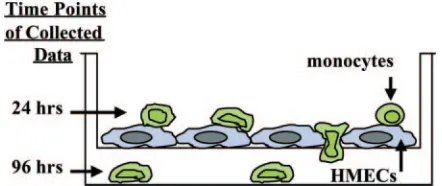
FIG. 1. In vitro model for studying the effects of viral infection on monocyte diapedesis. HMECS were grown to confluence on polysty-rene transwell plates and infected with HCMV (MOI, 20), mock in-fected, or treated with PMA (10 ng/ml) or LPS (10g/ml). After 24 h, the endothelial cells were washed and monocytes labeled with a green cell tracker dye were added to the wells. At 24 h after the addition of monocytes, three random FOV of monocytes on the HMECS in the top of the insert were counted to determine the percentage of cells adhering to the endothelial cell layer and beginning to undergo dia-pedesis versus the percentage of cells rounded up and stationary on the endothelial cell layer. At 96 h after the addition of monocytes, the number of monocytes that migrated completely through the endothe-lial cell layer was determined for 10 random FOV in the bottom chamber. The percentage of added monocytes that migrated through the wells was also determined at 96 h after the addition of monocytes to the transwells.
on November 8, 2019 by guest
http://jvi.asm.org/
a PBS solution containing 0.2% Triton X-100 (Sigma) and 0.05 M sucrose (Sigma). Cells were blocked with 10% FBS in PBS prior to incubation with primary antibodies against occludin or VE-cadherin for 1 h. Cells were washed, incubated with FITC-conjugated anti-rabbit or anti-mouse secondary antibodies for 1 h, and examined by fluorescence microscopy. The Slowfade antifade kit
containing 4⬘,6-diamidino-2-phenylindole (DAPI; Molecular Probes) was used to
mount the slides according to the manufacturer’s protocol.
Phalloidin staining.HMECs were grown on fibronectin-coated coverslips and mock infected or infected with either Towne/E HCMV (MOI, 20) or TB40-UL32-GFP HCMV (MOI, 20). At 24 hpi, the cells were fixed in 3% paraform-aldehyde (Sigma) and permeabilized in a PBS solution containing 0.2% Triton X-100 (Sigma) and 0.05 M sucrose (Sigma). Cells were stained with Alexa Fluor 546 (Molecular Probes) for 1 hour. When TB40-UL32-GFP HCMV was used to infect cells, the cells were also stained with a FITC-conjugated GFP anti-body (Molecular Probes). The Slowfade antifade kit containing DAPI (Molec-ular Probes) was used to mount the slides, and F-actin staining was examined by fluorescence microscopy.
Viral transfer studies.Transendothelial migration assays were performed as described above with the exception that HMECs were infected with either Towne/E HCMV (MOI, 20) or TB40-UL32-GFP HCMV (MOI, 20), and fi-bronectin-coated coverslips were placed in the bottom well to collect monocytes that migrated through the endothelial cell layer and into the bottom well. At 96 h after adding the naı¨ve monocytes to the inserts, cells were fixed and permeabil-ized as described above. When the Towne/E strain of HCMV was used to infect endothelial cells, we stained for the presence of the tegument protein pp65 in the monocytes. When TB40-UL32-GFP HCMV was used to infect endothelial cells, monocytes were stained with FITC-conjugated anti-GFP antibody (Molecular Probes). The Slowfade antifade kit containing DAPI (Molecular Probes) was used to mount the slides, and the presence of the major tegument protein or the capsid-associated protein was examined by fluorescence microscopy.
To more thoroughly investigate viral transfer, we examined the migrating monocytes for the presence of viral DNA (see below) and performed long-term tissue culture of monocytes that migrated through the HCMV-infected or mock-infected HMEC monolayer into the bottom of the well to determine if virus was released from the monocytes/macrophages. Four days after monocytes were added to the inserts, the inserts were removed and the cells in the bottom of the well were washed. Medium was replaced and changed every 3 to 5 days. At weekly intervals, medium was harvested. To test for the release of infectious virus, the medium was added to HEL fibroblasts. At 12 h after the harvested medium was added to the fibroblasts, the cells were fixed and IE1-72 protein expression was examined by immunohistochemistry (Histomouse SP Broad Spectrum; Zymed), according to the manufacturer’s protocol.
Southern blotting.Transendothelial migration assays were performed with TB40-UL32-GFP HCMV as described above. Monocytes that migrated through the endothelial cell layer into the bottom of the well were washed, and total
cellular DNA was harvested from the cells using DNA lysis buffer (1⫻
Tris-EDTA with 0.5% SDS). As a control, DNA was also harvested from mock-infected and HCMV-mock-infected (MOI, 20) monocytes. Using equal concentrations of DNA, PCR amplification of the genomic sequence of IE1-72 from exon 2 to exon 4 was performed as previously described (16). The amplification product of 587 bp was confirmed by Southern blot analysis. The PCR products were trans-ferred to a nylon membrane (Roche, Mannheim, Germany) and probed with a radioactive probe specific for the genomic sequence in exon 3 of IE1-72. The probe was generated by end labeling an oligonucleotide specific for exon 3 of
IE1-72 (5⬘ACGACGTTCCTGCAGACTA) with [32P]ATP (ICN Biomedicals,
Inc., Irvine, CA). Hybridization with the probe was performed overnight at 42°C.
The membrane was washed twice (2⫻SSC [1⫻SSC is 0.15 M NaCl plus 0.015
M sodium citrate] and 0.1% SDS), and the membrane was visualized by auto-radiography.
RESULTS
HCMV infection of endothelial cells promotes naı¨ve mono-cyte adhesion and diapedesis.Because we hypothesized that HCMV infection of endothelial cells recruits naı¨ve monocytes to the endothelial cell surface and promotes monocyte transen-dothelial migration, we first examined the potential of HCMV-infected or mock-HCMV-infected endothelial cells to regulate naı¨ve monocyte transendothelial migration. A modified Boyden chamber consisting of endothelial cells grown to confluence on
tissue culture inserts was used to examine transendothelial migration (Fig. 1). Endothelial cells were infected for 24 h, and prior to the addition of uninfected human peripheral blood monocytes to the tissue culture inserts, all inserts were washed extensively to remove extracellular virus. Plaque assays were performed to verify that no extracellular virus remained in the inserts (data not shown). As previously described (84, 85), 24 h after monocytes were added to the tissue culture inserts the percentage of monocytes undergoing migration on the endo-thelial cell layer versus the percentage of monocytes stationary on the endothelial cell layer was determined (Fig. 2A). Using the classification of Ronald et al. (67), which we employed in our previous study on HCMV-induced changes in monocyte function (84), we defined spherical or rounded monocytes as stationary. Monocytes were defined as moving if they exhibited the following characteristics: lamellipodia formation, spread-ing out on top of the endothelial cell layer, migratspread-ing through the endothelial junctions, or located on the transwell mem-brane beneath the endothelial cell layer. We found that HCMV infection of endothelial cells resulted in a significant (P ⬍ 0.01) increase in the percentage of naı¨ve monocytes migrating on the infected endothelial cell layer (70%) (Fig. 2A) compared to the percentage of monocytes migrating on the mock-infected endothelial cell layer (50%) (Fig. 2A). Nu-merically, this increased percentage of naı¨ve monocyte migra-tion translated to more than a twofold increase in the number of naı¨ve monocytes undergoing diapedesis on the infected en-dothelium compared to the mock-infected enen-dothelium. There was an increase in the ratio of moving to rounded naı¨ve mono-cytes from 1:1 on the mock-infected endothelium to 2:1 on the HCMV-infected endothelium. As positive controls, endothe-lial cells were stimulated with PMA or LPS, which have pre-viously been shown to activate endothelial cells at the concen-trations used (61, 63, 65, 92, 101). An increase in naı¨ve monocyte adhesion to the PMA-treated or LPS-treated endo-thelium was observed, similar to that of the HCMV-infected endothelium. We previously observed that infected monocytes migrated through the uninfected endothelium (84). Therefore, we used monocytes that were HCMV infected and added to a monolayer of uninfected endothelial cells as an additional pos-itive control in these experiments, and we found that infected monocyte migration on the naı¨ve endothelium occurred at rates similar to that seen with naı¨ve monocytes on infected endothelial cells (data not shown), suggesting that the increase in monocyte migration occurred regardless of the cell type infected. This common outcome supports our hypothesis that both cell types play a critical role in the hematogenous dissem-ination of HCMV.
Our results with the HMEC cell line suggested that HCMV infection of dermal microvascular endothelial cells promotes monocyte migration. Endothelial cells from diverse vascular beds have different responses, such as altered viral replication and cell adhesion molecule expression, following HCMV in-fection (17, 21, 45, 48, 52, 76, 80). To examine if HCMV infection of endothelial cells from different origins promoted monocyte migration, we performed migration assays on two additional endothelial cell lines, IHECs and HUVECs, which are microvascular endothelial cells of brain origin and macro-vascular endothelial cells of venous origin, respectively. We found that HCMV infection of IHECs and HUVECs
on November 8, 2019 by guest
http://jvi.asm.org/
strated a similar increase in naı¨ve monocyte adhesion to the infected endothelium (data not shown). Thus, the ability of the infected endothelium to recruit naı¨ve monocytes was not de-pendent on the origin of the endothelial cells.
HCMV infection of endothelial cells results in increased monocyte transendothelial migration.Following adhesion to the surface of the endothelium, monocytes locate the lateral junctions in the endothelial cell layer and migrate through these junctions into the surrounding tissue (60, 66, 74). We next examined the bottoms of the wells containing the tissue culture inserts at 96 h post-addition of monocytes to the in-serts, a time previously used in our system (84, 85), to deter-mine the average number of monocytes per FOV that under-went complete transendothelial migration (Fig. 2B). At 96 h after the addition of monocytes to the inserts, there was a significant (P⬍0.01) increase in the average number of mono-cytes per FOV that migrated through the infected endothelial cell layer (average of 35 monocytes per FOV) compared to mock-infected endothelial cells (average of 10 monocytes per FOV). Control PMA- or LPS-treated endothelial cells showed increased monocyte migration equivalent to that observed in HCMV-infected endothelial cells.
To rule out the possibility that only a small subset of naı¨ve monocytes underwent transendothelial migration following ex-posure to infected or activated endothelial cells, the percent-age of monocytes that migrated through the endothelium was determined. There was greater than a threefold increase in total naı¨ve monocyte transendothelial migration when endo-thelial cells were infected with HCMV (Fig. 2C). A similar increase in the percentage of monocytes that migrated through the endothelium was observed with PMA- or LPS-treated en-dothelial cells. Together, these data suggest that the virus ac-tivates endothelial cells and promotes naı¨ve monocyte transen-dothelial migration.
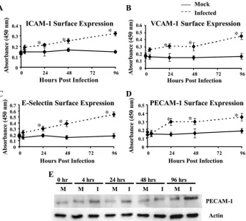
[image:5.585.65.259.79.458.2]HCMV infection of endothelial cells increases adhesion molecule surface expression.The increased monocyte transen-dothelial migration suggested that HCMV-induced cellular changes in endothelial cells promoted adhesion and diapede-sis, so we next wanted to examine the expression of the adhe-sion receptors ICAM-1 (Fig. 3A), VCAM-1 (Fig. 3B), E-selec-tin (Fig. 3C), and PECAM-1 (Fig. 3D), which are needed to promote firm adhesion of leukocytes to the endothelium (50, 67). Previous work showed that HCMV infection of macrovas-cular HUVECs increased the surface expression of the cell adhesion molecules ICAM-1, VCAM-1, and E-selectin (17, 45, 48, 52, 76). In contrast, another study showed that HCMV infection of HUVECs increased VCAM-1 expression and had no change in E-selectin expression, while HCMV infection of human intestinal microvascular endothelial cells decreased VCAM-1 expression and increased E-selectin expression (80). Because these previous findings demonstrated that HCMV-altered adhesion molecule expression differs between macro-vascular and micromacro-vascular endothelial cells and between dif-ferent studies, we examined the surface expression of adhesion receptors on endothelial cells in our system (HMECs) using a cell-based ELISA (78). In these experiments, cells were grown to confluence and mock infected or HCMV infected (MOI, 20) for the indicated lengths of time. When the HMECs were mock infected, there was no significant change in the expres-sion of any of the adheexpres-sion molecules tested during the course
FIG. 2. HCMV infection of endothelial cells promotes naı¨ve mono-cyte recruitment and transendothelial migration. (A) HCMV infection of endothelial cells resulted in increased naı¨ve monocyte recruitment. HMECs were mock infected, HCMV infected (MOI, 20), PMA treated (10 ng/ml), or LPS treated (10g/ml) for 24 h. Human mono-cytes were isolated, labeled with a green cell tracker dye, and added to each well. At 24 h after the addition of monocytes, the percentage of monocytes undergoing diapedesis versus the percentage of monocytes rounded up and stationary on the endothelial cell layer was deter-mined. Results are plotted as means⫾SD of three random fields of view. Magnification,⫻200. (B) HCMV infection of endothelial cells resulted in increased naı¨ve monocyte transendothelial migration. At 96 h after the addition of monocytes, the number of monocytes that migrated completely through the endothelial cell layer was deter-mined. Results are plotted as means⫾SD of 10 random fields of view. Magnification,⫻200. (C) HCMV infection of endothelial cells resulted in increased total naı¨ve monocyte transendothelial migra-tion. At 96 h after the addition of monocytes to the transwell inserts, monocytes that had migrated through both the endothelial cell layer and the insert and had adhered to the bottom of the well were collected and counted. The number of monocytes that mi-grated through the endothelium and the transwell versus the total number of monocytes added to the wells was determined. Results are plotted as means⫾SD of the percentage of added monocytes that underwent transendothelial migration. Results are an average of four independent experiments from separate human blood do-nors. HCMV-infected, LPS-treated, and PMA-treated groups were significantly different (P⬍0.01) than the mock-infected group.
on November 8, 2019 by guest
http://jvi.asm.org/
of the experiment (Fig. 3A to C, mock). Our findings showed that HCMV infection of HMECs increased the surface expres-sion of ICAM-1 (Fig. 3A), VCAM-1 (Fig. 3B), and E-selectin (Fig. 3C). Similar increases in ICAM-1, VCAM-1, and E-se-lectin surface expression were observed when the HMECs were treated with LPS or PMA (data not shown), suggesting that HCMV activates endothelial cells to the same extent as other known stimuli. The increased surface expression of these cell adhesion molecules coincides with the increased naı¨ve monocyte adhesion to the infected endothelium, suggesting a mechanism by which HCMV infection of endothelial cells stimulates naı¨ve monocyte recruitment to the apical surface of the endothelium.
We next examined HCMV-induced alterations in endothe-lial cell PECAM-1 expression. PECAM-1 is a cell adhesion molecule important in leukocyte recruitment (7, 53, 74), cell adhesion (5), endothelial cell permeability (49), and angio-genesis (2, 11). In addition, it has been shown that blocking PECAM-1 function inhibits transendothelial migration (74). We found that HCMV-infected HMECs exhibited increased PECAM-1 surface expression (Fig. 3D), which was similar to the increased PECAM-1 surface expression observed when the
endothelial cells were activated with LPS or PMA (data not shown). In addition, total PECAM-1 protein levels increased following HCMV infection (Fig. 3E). The initial increase in total PECAM-1 protein levels coincided with the increased PECAM-1 surface expression through 96 hpi. These data show that HCMV infection of endothelial cells increases the surface expression of PECAM-1 along with ICAM-1, VCAM-1, and E-selectin and that this increase in adhesion receptor expres-sion could enhance naı¨ve monocyte recruitment to the infected endothelium.
[image:6.585.122.467.68.379.2]HCMV-induced increased cell adhesion molecule expres-sion is functional.While the data from Fig. 3 show that E-selectin, ICAM-1, VCAM-1, and PECAM-1 expression levels are upregulated following infection, these results do not as-cribe a function to this increased expression. To determine if these changes in adhesion receptor expression correlated with increased naı¨ve monocyte attachment to the infected endothe-lium, we next examined whether HCMV-induced increased ICAM-1, VCAM-1, and/or E-selectin surface expression was required for HCMV-induced naı¨ve monocyte recruitment and transendothelial migration. PECAM-1-specific neutralizing antibodies are not commercially available, so the role of
FIG. 3. HCMV infection of endothelial cells resulted in the upregulation of cell adhesion molecule expression. (A to D) HCMV infection of endothelial cells promoted increased surface expression of cell adhesion molecules. Cell-based ELISAs were performed as previously described (78) to examine the surface expression of ICAM-1 (A), VCAM-1 (B), E-selectin (C), and PECAM-1 (D) at the indicated times after infection. The straight line corresponds to mock-infected cells, while the dashed line corresponds to HCMV-infected cells (MOI, 20). Results are plotted as means⫾SD of three independent experiments performed in triplicate. HCMV-infected HMECs showed a significant increase (P⬍0.01) in surface expression of ICAM-1, VCAM-1, E-selectin, and PECAM-1 by 24 hpi compared to mock-infected HMECs. (E) HCMV infection of endothelial cells promoted increased PECAM-1 protein expression. Western blot analyses of PECAM-1 and actin were performed using equal protein loading of mock-infected and HCMV-infected (MOI, 20) HMEC lysates harvested at the indicated times after infection.
on November 8, 2019 by guest
http://jvi.asm.org/
HCMV-induced increased PECAM-1 expression in naı¨ve monocyte recruitment could not be examined at this time. Transendothelial migration assays were performed, and mock-infected and HCMV-mock-infected endothelial cells were treated with blocking antibodies to ICAM-1, VCAM-1, and/or E-se-lectin prior to the addition of monocytes. At 24 h after naı¨ve monocytes were added to the inserts, monocyte adhesion to and motility on the apical surface of the endothelial cells was examined (Fig. 4A). Similar percentages (50%) of naı¨ve mono-cyte adhesion to and motility on the endothelial cell surface (or a ratio of 1:1 when comparing the number of moving naı¨ve monocytes to those with a rounded phenotype) were obtained when the endothelial cells were mock infected or mock in-fected and pretreated with antibodies to ICAM-1, VCAM-1, and/or E-selectin or IgG1, the isotype control antibody (Fig. 4A). We found that monocyte adhesion to and motility on the infected HMECs returned to mock levels, which was signifi-cantly lower than their infected, untreated counterparts (P⬍
0.05), when the endothelial cells were pretreated with antibod-ies to ICAM-1, VCAM-1, and/or E-selectin but not when the HMECs were treated with the isotype control antibody (Fig. 4A). The percentage of monocytes migrating on HMECs treated with the blocking antibodies remained at approxi-mately 50%, similar to that seen in mock-infected cells, sug-gesting that the HCMV-induced increase in the surface expres-sion of ICAM-1, VCAM-1, and E-selectin promoted naı¨ve monocyte recruitment to the infected endothelium.
[image:7.585.112.470.76.321.2]Each step in leukocyte extravasation is in turn required for the subsequent step(s) (75). Therefore, using the blocking an-tibodies, we examined if inhibition of monocyte recruitment abrogated HCMV-induced naı¨ve monocyte transendothelial migration (Fig. 4B). We observed that pretreatment of in-fected endothelial cells with blocking antibodies prior to the addition of naı¨ve monocytes significantly inhibited (P⬍0.05) the average number of monocytes per FOV that migrated through the endothelial cell layer into the bottom of the well. When HMECs were mock infected and pretreated with anti-bodies to ICAM-1, VCAM-1, and/or E-selectin or the isotype control, an average of 10 naı¨ve monocytes were observed per FOV in the bottom well of the insert, similar to that observed when the endothelial cells were mock infected and not pre-treated with any antibodies. When HMECs were infected and pretreated with the ICAM-1, VCAM-1, and/or E-selectin blocking antibodies, the average number of monocytes that underwent transendothelial migration into the bottom well returned to that observed when the endothelial cells were mock infected, 10 monocytes per FOV. Pretreatment of in-fected endothelial cells with the isotype control did not signif-icantly alter naı¨ve monocyte transendothelial migration, sug-gesting that HCMV-induced naı¨ve monocyte recruitment, due to the increased surface expression of ICAM-1, VCAM-1, and E-selection, was required for the increased monocyte migra-tion through the infected endothelium.
FIG. 4. HCMV-induced cell adhesion molecule surface expression is functional. (A) The increased surface expression of ICAM-1, VCAM-1, and E-selectin following HCMV infection is necessary for the recruitment of naı¨ve monocytes. HMECs were mock infected or HCMV infected (MOI, 20) for 24 h. Cells were washed and incubated for 1 h at 37°C with medium containing blocking antibodies (ICAM-1, 20g/ml; VCAM-1, 30g/ml; E-selectin, 50g/ml; all three antibodies; or the IgG1 isotype control, 50g/ml), after which the cells were washed again and isolated labeled human monocytes were added to each well. At 24 h after the addition of monocytes, the percentage of monocytes undergoing diapedesis versus the percentage of monocytes rounded up and stationary on the endothelial cell layer was determined. Results are plotted as means⫾SD of three random fields of view. Magnification,⫻200. (B) Blocking the adhesion of monocytes to the HCMV-infected endothelial cells inhibited HCMV-induced naı¨ve monocyte transendothelial migration. At 96 h after the addition of monocytes, the number of monocytes that migrated completely through the endothelial cell layer was determined. Results are plotted as means⫾SD of 10 random fields of view. Magnification,⫻200.
on November 8, 2019 by guest
http://jvi.asm.org/
HCMV infection increases endothelial cell permeability.
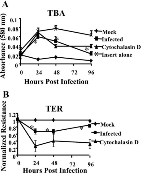
Following naı¨ve monocyte adhesion to the surface of the in-fected endothelium, the monocytes must migrate through the lateral junctions of the endothelial cell layer. We observed increased naı¨ve monocyte transendothelial migration following infection of the endothelium. To investigate changes in endo-thelial cell function that could contribute to increased naı¨ve monocyte transendothelial migration, we first investigated whether HCMV altered the permeability of the infected en-dothelium, because increased permeability can lead to leuko-cyte transendothelial migration (77). HMECs were grown to confluence on tissue culture inserts, and the permeability of the endothelium was examined by measuring the diffusion of a TBA complex across the endothelial cell layer and by measur-ing the TER of the endothelial cell monolayer.
Using TBA diffusion, we detected an increase in endothelial cell permeability beginning at 24 hpi (Fig. 5A). The increase in the permeability of the infected endothelium remained through 96 hpi. As a positive control, endothelial cells were treated with cytochalasin D, an inhibitor that induces actin
depolymerization and blocks actin-dependent cellular pro-cesses (15).
TER was used to confirm that endothelial cell permeability was enhanced following infection. We found a significant in-crease in HCMV-infected endothelial cell permeability com-pared to mock-infected cells (Fig. 5B). As a control, endothe-lial cells were treated with cytochalasin D. Similar results were seen with cytochalasin D-treated cells in both the TER assay and the TBA assay. These assays showed that endothelial cell permeability increased significantly (P ⬍ 0.01) following HCMV infection.
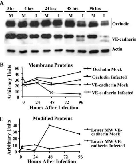
[image:8.585.49.279.69.353.2]HCMV infection of endothelial cells results in reduced tight and adherens junction protein expression.The increase in the permeability of the infected endothelium suggested that endo-thelial cells were pulling apart following HCMV infection and that their lateral junctions were altered. Therefore, we next investigated if HCMV altered tight and adherens junctions in infected endothelial cells. Lateral junctions, consisting of tight and adherens junctions, are essential for connecting adjacent cells and for controlling the movement of cells and molecules between the blood and the surrounding tissue (14, 20). Tight junctions are located just below the apical surface of the cells, and they regulate the movement of integral membrane pro-teins between the apical and the basolateral surfaces of the cell. In addition, tight junctions regulate the passage of mole-cules and cells from the blood into the surrounding tissue. One transmembrane protein involved in tight junction formation is occludin, which binds to other occludin molecules on neigh-boring endothelial cells and is linked to intracellular cytoskel-etal proteins (23, 37). Below the tight junctions are the adhe-rens junctions, which regulate cell migration from the blood into the tissue and provide a strong mechanical attachment between cells. Adherens junctions consist of the transmem-brane protein VE-cadherin, a calcium-dependent cell adhesion molecule which is linked to actin and myosin filaments (102). To examine the protein levels of occludin and VE-cadherin at various times following HCMV infection, we performed Western blot analyses (Fig. 6A) and normalized occludin and VE-cadherin protein levels to actin protein levels by densitom-etry to quantify changes in the expression of the lateral junc-tion proteins (Fig. 6B and C). The time course used allowed us to examine early times, 4 and 24 hpi, and later times, 48 and 96 hpi. We observed decreased occludin (both the 65- and 72-kDa forms) (Fig. 6A and B) and VE-cadherin (130 kDa) (Fig. 6A and B) protein levels in HCMV-infected endothelial cells com-pared to mock-infected endothelial cells at 48 hpi, which con-tinued through 96 hpi. Mock-infected endothelial cells exhib-ited relatively steady levels of occludin and VE-cadherin expression over time. A faster-migrating form of VE-cadherin (100 kDa) was detected in infected HMECs at 48 and 96 hpi (Fig. 6A and C). VE-cadherin is known to be processed and degraded following endocytosis (100). During this process, the cytoplasmic tail of the protein is cleaved, resulting in a 100-kDa protein (100). We observed the presence of a 100-100-kDa form of VE-cadherin in infected cells at 48 and 96 hpi, sug-gesting that VE-cadherin is being internalized and degraded following HCMV infection. Cell-based ELISAs, examining to-tal protein levels, were also used to examine the expression of these junctional proteins. The results were similar to the West-ern blot analyses (data not shown). The decrease in the protein
FIG. 5. HCMV infection of endothelial cells resulted in increased endothelial cell permeability. (A) Diffusion of a TBA complex (68) across a confluent monolayer of mock-infected, HCMV-infected (MOI, 20), or cytochalasin D-treated (1M) HMECS or across the insert alone was determined at the indicated times. Results are plotted as means⫾SD of three independent experiments performed in trip-licate. (B) TER of a confluent monolayer of mock-infected, HCMV-infected (MOI, 20), or cytochalasin D-treated (1 M) HMECS or across the insert alone was determined at various times after treat-ment. Results are plotted as means⫾SD of three independent exper-iments done in triplicate. HCMV-infected and cytochalasin D-treated HMECs showed a significant increase (P⬍0.01) in permeability com-pared to mock-infected HMECs.
on November 8, 2019 by guest
http://jvi.asm.org/
expression of these critical junction components suggests that endothelial cells are pulling apart from each other, thereby aiding monocyte diapedesis.
HCMV infection of endothelial cells results in the internal-ization of junctional proteins. Lateral surface expression of VE-cadherin and E-cadherin, a separate member of the cad-herin superfamily, has been reported to be regulated through endosomal recycling from the membrane to the endosomal pathway and back to the cell membrane (3, 7, 26). A contrast-ing report suggested that the endosomal pathway regulated VE-cadherin by targeting it for degradation rather than recy-cling it back out to the membrane (100). These studies suggest that the normal maintenance of endothelial cell VE-cadherin, and perhaps junctional proteins in general, is regulated by an endosomal recycling pathway, but that following inflammation and/or vascular damage, the junctional proteins are degraded rather than being recycled to the cell surface (8). A virus-stimulated degradation of these proteins would account for the decreased occludin and VE-cadherin protein levels observed in our Western blot analyses.
To examine the possibility that the junctional proteins were in fact being internalized and/or degraded following HCMV
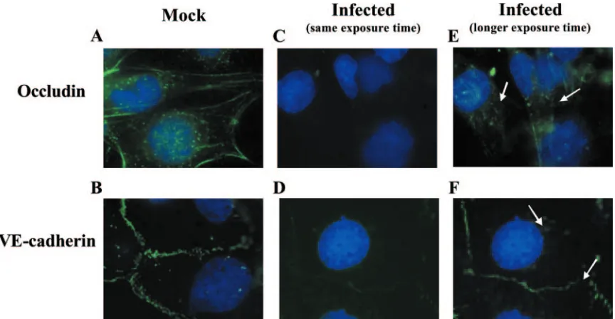
infection, occludin and VE-cadherin expression levels were examined by immunofluorescence microscopy. No changes in the localization of occludin and VE-cadherin were observed at early times following infection (data not shown). Decreased protein expression, however, was detected by 96 hpi. In mock-infected HMECs, occludin (Fig. 7A) and VE-cadherin (Fig. 7B) expression was primarily observed at the periphery of the cells where the cell-to-cell contacts occur. Using the same exposure time to examine the expression of these proteins in cells infected for 96 h, we found that infected HMECs ex-pressed lower levels of occludin (Fig. 7C) and VE-cadherin (Fig. 7D), supporting our Western blot analyses. When the exposure time for the captured images was lengthened, occlu-din (Fig. 7E) and VE-cadherin (Fig. 7F) expression could be detected. Less protein was observed at the periphery of the cell, while increased internalization of both proteins was seen. The increased internalization and decreased amounts of VE-cadherin are supported by our Western blotting data showing that a 100-kDa intermediate fragment of VE-cadherin, which is reported to accumulate in the cell until it is degraded in the lysosome (100), is present in infected HMECs. These data suggested that enhanced internalization and degradation of occludin and VE-cadherin may be a mechanism to account for the decreased expression of these proteins that we observed in infected endothelial cells.
HCMV-induced lateral junction degradation promotes in-creased endothelial cell permeability and naı¨ve monocyte transendothelial migration.Because lateral junctions are es-sential for controlling the permeability of the endothelium and transendothelial migration (14, 20), we proposed that HCMV-induced internalization and degradation of occludin and VE-cadherin aided HCMV-induced permeability of the endothe-lium. Our results, however, did not directly test this possibility. Thus, we next asked whether, in our system, the inhibition of the binding of lateral junction proteins would promote naı¨ve monocyte transendothelial migration. To test this possibility, we utilized a monoclonal VE-cadherin-specific antibody previ-ously shown to disrupt the homotypic binding of VE-cadherin, resulting in increased permeability of the endothelial cell monolayer (39, 94). A similar reagent targeting occludin is not available at this time. Therefore, only the role of the loss of VE-cadherin binding could be examined in endothelial cell permeability and naı¨ve monocyte transendothelial migration.
TER was used to determine if the loss of VE-cadherin-based cell-cell adhesion was sufficient to increase the permeability of the endothelial cell monolayer (Fig. 8A). We found that treat-ment of endothelial cells with the VE-cadherin-specific anti-body significantly (P⬍0.01) increased the permeability of the monolayer regardless of whether the cells were infected or not, while treatment with the isotype control antibody did not alter the permeability (Fig. 8A), suggesting that the loss of the lateral junction proteins, specifically VE-cadherin, is critical for maintaining the integrity of the endothelium. In addition, the inhibition of VE-cadherbased cell-cell adhesion in-creased endothelial cell permeability similar to that seen when cells were treated with cytochalasin D. These findings suggest that HCMV infection of endothelial cells results in the in-creased permeability of the endothelium due to dein-creased expression of the lateral junction proteins.
[image:9.585.44.282.69.356.2]Next, transendothelial migration assays were performed to
FIG. 6. HCMV infection of endothelial cells resulted in decreased junctional protein expression. (A) Western blot analyses of actin, VE-cadherin, and occludin protein levels were performed using equal protein loading of mock-infected and HCMV-infected (MOI, 20) HMEC lysates harvested at the indicated times after infection. (B and C) Bands were analyzed by densitometry to determine relative levels of occludin (both the 65- and 72-kDa forms) and VE-cadherin (130 kDa) (B) and the alternative forms of VE-cadherin (lower molecular mass, ⬃100 kDa; represented as lower MW VE-cadherin) (C) and are ex-pressed in arbitrary units as a ratio of x/actin. A representative exper-iment from three independent experexper-iments is shown.
on November 8, 2019 by guest
http://jvi.asm.org/
examine if the loss of VE-cadherin-based cell-cell adhesion aided naı¨ve monocyte transendothelial migration (Fig. 8B). We found that treatment of endothelial cells with the VE-cadherin-specific antibody significantly (P ⬍ 0.01) increased the average number of monocytes per FOV that underwent transendothelial migration in mock-infected and HCMV-in-fected endothelial cells. Treatment of the endothelial cells with the isotype control antibody did not alter HCMV-induced naı¨ve monocyte transendothelial migration. Together these data identify that the HCMV-induced degradation of the lat-eral junction proteins, VE-cadherin and possibly occludin, pro-motes virus-induced naı¨ve monocyte transendothelial migra-tion via the loss of cell-cell adhesion and the increased permeability of the endothelium.
It should be noted from these results that the disruption of the lateral junctions by the antibody against VE-cadherin re-sults in a greater loss of junctional integrity, as measured by the permeability of the endothelium, than infection alone; an in-crease in permeability similar to that seen with cytochalasin D was observed. Although our findings define the de facto im-portance of junctional proteins in the integrity of the endothe-lium and in the regulation of monocyte trafficking, the results should not be interpreted to mean that only the loss of junc-tional integrity is required to promote monocyte extravasation following HCMV infection. Based on our results from Fig. 4, both the increased adhesion to the endothelium and the de-creased barrier function of the lateral junctions contribute to the increased trafficking of monocytes following infection of the endothelium.
HCMV infection of endothelial cells disrupts stress fiber formation.Our results documenting that HCMV infection of endothelial cells promoted the increased permeability of the
[image:10.585.77.509.68.292.2]endothelium and the degradation of the lateral junction pro-teins pointed to a possible mechanism for why infection of the endothelium resulted in enhanced naı¨ve monocyte migration. From a temporal standpoint, however, there appear to be two distinct events occurring in infected endothelial cells. We pro-pose that HCMV infection promotes a two-step change in vascular permeability. One step, as described above (Fig. 6 and 7), occurs around 48 hpi; HCMV induces the internalization and degradation of the lateral junction proteins and the in-creased permeability of the endothelium. Because peak mono-cyte migration occurs in this time frame, these changes likely contribute to the enhanced monocyte migration. Furthermore, these changes would maintain the increased permeability we observed at these times. Because our results showed that changes in endothelial cell permeability occurred prior to 48 hpi, an additional mechanism, the first step in our proposed two-step process, must be responsible for the changes that occur earlier following infection. It is reported that enhanced endothelial cell permeability can be accompanied by reorgani-zation of the actin cytoskeleton and the loosening of intercel-lular junctions (18) and that HCMV infection of fibroblasts results in the disruption of actin stress fiber formation to allow for the nuclear translocation of the virus (97). We suggest that HCMV infection of endothelial cells alters actin stress fiber formation at early times after infection to allow the virus to get to the nucleus but, as a result, this change alters the perme-ability of the endothelium. Therefore, we propose the follow-ing two-step process for how HCMV alters the permeability of the endothelium: first, HCMV infection decreases actin poly-merization in endothelial cells, resulting in an initial increase in permeability of the endothelium, and second, HCMV infec-tion promotes the degradainfec-tion of the lateral juncinfec-tion proteins,
FIG. 7. HCMV infection of endothelial cells promoted junctional protein internalization. HMECs were grown to confluence on fibronectin-coated coverslips and mock infected or HCMV infected (MOI, 20) for 96 h. The cells were then fixed, stained with the appropriate primary and secondary antibodies and DAPI, and examined by immunofluorescence microscopy. Magnification,⫻1,000. (A and B) Expression of occludin (A) and VE-cadherin (B) was examined in mock-infected cells. (C and D) Using the same exposure time, expression of occludin (C) and VE-cadherin (D) was examined in HCMV-infected cells. (E and F) A longer exposure time was used to examine the expression of occludin (E) and VE-cadherin (F) in HCMV-infected cells. A representative experiment from three independent experiments is shown.
on November 8, 2019 by guest
http://jvi.asm.org/
further increasing the virus-induced increase in endothelial cell permeability.
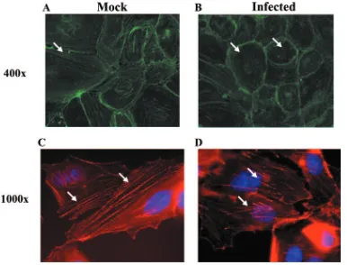
To test our proposed idea that changes occur in the actin cytoskeleton shortly after infection of endothelial cells, we next examined if HCMV infection of endothelial cells disrupted actin stress fiber formation. We performed phalloidin stains to examine actin polymerization in mock-infected and HCMV-infected endothelial cells (Fig. 9). We found that at 24 hpi there was a decrease in actin stress fiber formation in infected HMECs compared to mock-infected HMECs. At both⫻400 (Fig. 9A versus B) and⫻1,000 (Fig. 9C versus D) magnifica-tion, infected endothelial cells showed less stress fiber forma-tion than their uninfected counterparts. In addiforma-tion, the actin stress fibers present were shorter and more branched than the stress fibers in mock-infected cells. Because actin depolymer-ization and/or hyperpolymerdepolymer-ization have been shown to in-crease the permeability of the endothelium (98), we suggest that our data verify that the HCMV-induced changes in the actin cytoskeleton result in the increased permeability ob-served in the endothelium early after infection.
HCMV can be transferred from the infected endothelial cell to a monocyte undergoing transendothelial migration.For en-dothelial cells to be important in the hematogenous
dissemi-nation of HCMV within the host, the infected endothelial cell must be capable of transferring the virus to the migrating monocyte. Previous work showed the bidirectional transmis-sion of HCMV between endothelial cells and monocytes (27, 96). Specifically, monocytes were infected following adhesion to the infected endothelial cells (27, 96), probably due to a series of transitory microfusion events between the monocytic cell membrane and the endothelial cell membrane (27). These results suggested that the mechanism we proposed for the role endothelial cells play in viral spread was possible. Neverthe-less, in our system, it remained unclear if the naı¨ve monocyte migrating through the infected endothelial cell layer was ca-pable of acquiring HCMV from the infected endothelium. To test this possibility, we performed transendothelial migration assays using two different strains of HCMV, Towne/E and an endothelial cell-tropic, GFP-labeled HCMV (TB40-UL32-HCMV/E). HMECs were infected for 24 h, at which time the cells were washed to remove extracellular virus. Medium from this final wash was examined for virus presence by plaque assay or by immunofluorescence microscopy. No free virus was de-tected, and all of the labeled viral particles were found in the cytoplasm of the endothelial cells (data not shown). Naı¨ve monocytes were added to the wells, and 96 h after the addition
FIG. 8. HCMV-induced lateral junction disruption promotes increased endothelial cell permeability and naı¨ve monocyte transendothelial migration. (A) TER of a confluent monolayer of endothelial cells treated with a VE-cadherin-specific blocking antibody (cl75; 25g/ml) or the isotype control antibody (50g/ml) and either mock infected, HCMV infected (MOI, 20), or cytochalasin D treated (1M) was determined at various times after treatment. The resistance across the empty insert was also determined. Results are plotted as means⫾SD of three independent experiments performed in triplicate. HMECs that were infected or infected and treated with the isotype control, with the VE-cadherin-specific blocking antibody, or the IgG1 isotype control and mock infected or HCMV infected or treated with cytochalasin D showed a significant increase (P ⬍ 0.01) in permeability compared to mock-infected HMECs. (B) Blocking VE-cadherin-based cell adhesion promotes naı¨ve monocyte transendothelial migration. HMECs were mock infected or HCMV infected (MOI, 20) for 24 h. Cells were washed, and medium containing the VE-cadherin-specific blocking antibody (25g/ml) or the IgG1 isotype control (50g/ml) was added to each well. Isolated and labeled human monocytes were added to each well. At 96 h after the addition of monocytes, the number of monocytes that migrated completely through the endothelial cell layer was determined. Results are plotted as means⫾SD of 10 random fields of view. Magnification,⫻200.
on November 8, 2019 by guest
http://jvi.asm.org/
of monocytes, monocytes that migrated through the endothe-lial cell layer were collected and examined for the presence of the virus, viral proteins, and viral DNA. First, we examined monocytes for the presence of the major tegument protein pp65 by confocal microscopy, which translocates to the nucleus shortly following infection (36, 71). We observed punctuate nuclear pp65 staining in⬃10% of the monocytes that migrated through the infected endothelium (Fig. 10B), while monocytes that migrated through the mock-infected endothelium did not stain positive for HCMV pp65 (Fig. 10A). Because pp65 is synthesized late after infection and we did not observe the pro-duction of pp65 in HCMV-infected endothelial cells at the times used in these experiments, the presence of pp65 in the migrated monocytes likely represents the tegument of the virus transferred to the migrating monocyte. To determine if only the tegument protein was transferred from the infected HMECs to the migrating monocytes, we also examined migrat-ing monocytes for the presence of TB40-UL32-GFP HCMV (Fig. 10C and D). Around 10% of the monocytes, which mi-grated through the HCMV-infected endothelium, were found to contain the GFP-labeled HCMV capsid (Fig. 10D), while monocytes that migrated through the mock-infected endothe-lial cells did not acquire the GFP-labeled viral capsid (Fig. 10C). To rule out the possibility that only viral proteins were transferred between the cells and not viral DNA, the migrating monocytes were assayed for the presence of HCMV genomic
[image:12.585.97.483.67.361.2]DNA. By Southern blot analysis, we found that only the mono-cytes that migrated through the HCMV-infected HMECs or monocytes that were directly infected with HCMV contained HCMV genomic DNA (Fig. 10E). An analysis of HCMV genomic DNA in the different populations of monocytes found that the amount of viral genomic DNA in the monocytes that migrated through the HCMV-infected HMECs was roughly 1/10 of the amount of viral genomic DNA present in the monocytes that were directly infected with HCMV. Finally, the migrating monocytes were examined for productive infection and the release of infectious virus. We previously reported that HCMV infection of monocytes induced monocyte-to-phage differentiation and that these differentiated macro-phages were productive for HCMV replication starting at 3 to 4 weeks postinfection (84). We found that monocytes that migrated through HCMV-infected HMECs had a more differ-entiated phenotype than monocytes that migrated through mock-infected HMECs (data not shown), consistent with our earlier study (84). In addition, when the supernatants were harvested from monocyte cultures 2 to 6 weeks post-migration through the HCMV-infected or mock-infected HMECs and assayed for the release of infectious virus, we found that only monocytes that migrated through HCMV-infected HMECs released infectious virus (Table 1). In these experiments, the peak virus titer from these cells was 30 PFU/500 monocytes,
FIG. 9. HCMV infection of endothelial cells disrupts actin stress fiber formation. HMECs were grown on fibronectin-coated coverslips and mock infected or HCMV infected (MOI, 20) for 24 h. The cells were then fixed, stained with phalloidin conjugated to Alexa Fluor 488 (A and B) or 546 (C and D) and DAPI (C and D), and examined by immunofluorescence microscopy. Stress fiber formation in mock-infected and HCMV-infected HMECs was examined at⫻400 (A) and⫻1,000 (B) magnification. Representative images of two different experiments from a
total of three independent experiments are shown.
on November 8, 2019 by guest
http://jvi.asm.org/
similar to the rate of productive HCMV infection that we previously observed in monocytes (84).
The above experiments showed that viral proteins and viral DNA are transferred from the infected endothelial cells to the
[image:13.585.101.491.69.389.2]migrating monocytes, which differentiate into macrophages and release infectious virus 4 to 6 weeks after migrating through the infected endothelial cells. We observed that⬃10% of the migrating monocytes acquired HCMV proteins and DNA during the process of migration, and it is unclear why only a percentage of cells acquired the virus. One possible explanation is biological timing, in that for viral transfer to occur, the monocyte must traverse the endothelial cell lateral junction at approximately the same time an infectious particle is localized near the lateral junction. Thus, we propose that although viral transfer is directed by the viral manipulation of the host cell, its probability of occurring is dependent on the timing of two independent biological variables: (i) viral traf-ficking within the infected endothelial cells, and (ii) monocyte transendothelial migration. In order for viral transfer to occur, both variables must spatially and temporally converge. We are currently attempting to test these possibilities.
FIG. 10. HCMV can be transferred from the infected endothelial cell to a monocyte undergoing transendothelial migration. HMECs were grown to confluence in tissue culture inserts and mock infected or HCMV infected (Towne/E or TB40-UL32-HCMV/E; MOI, 20) for 24 h. Isolated peripheral blood monocytes were then added to the wells. At 96 h after the addition of monocytes, cells that migrated through the insert into the bottom of the well were collected on the fibronectin-coated coverslips in the bottom of the well and fixed and permeabilized. The cells were then stained with TO-PRO3-iodide (A and B) or DAPI (C and D) and an anti-pp65 antibody or an anti-GFP antibody and examined by immunofluorescence microscopy. (A and B) Confocal microscopy was performed to examine the transfer of pp65 from the infected endothelial cells to the migrating naı¨ve monocytes. Only monocytes that migrated through the HCMV-infected endothelial cells stained positively for pp65. Magnification,⫻1,000. (C and D) Bright-field and immunofluorescence microscopy was performed to examine the transfer of HCMV from the infected endothelial cells to migrating naı¨ve monocytes. Only monocytes that migrated through the HCMV-infected endothelial cells stained positively for TB40-UL32-GFP HCMV. Magnification,⫻1,000. (E) A PCR/Southern blot analysis was utilized to confirm the presence of HCMV genomic DNA in various populations of monocytes. Lane 1, monocytes that migrated through mock-infected HMECs; lane 2, monocytes that migrated through TB40-UL32-GFP HCMV-infected HMECs; lane 3, mock-infected monocytes; lane 4, HCMV-infected monocytes; lane 5, water control. Only monocytes that migrated through HCMV-infected HMECs or monocytes that were directly infected with HCMV were positive for HCMV genomic DNA, suggesting that viral DNA can be transferred from the infected endothelial cell to the migrating, naı¨ve monocyte.
TABLE 1. Monocytes that migrate through HCMV-infected HMECs release infectious HCMVa
Cell type through which monocytes migrated
Presence (⫹) or absence (⫺)
of PFU at wk:
2 3 4 5 6
Mock-infected HMECs ⫺ ⫺ ⫺ ⫺ ⫺
HCMV-infected HMECs ⫺ ⫺ ⫹ ⫹ ⫹
aFor four different donors, monocytes that migrated through mock-infected or
HCMV-infected HMECs were cultured. Cellular supernatants were harvested at weekly intervals and assayed for release of infectious virus. The peak virus titer was 30 PFU/500 monocytes by 6 weeks after the monocytes migrated through the HMECs.