0022-538X/06/$08.00⫹0 doi:10.1128/JVI.80.7.3515–3522.2006
Copyright © 2006, American Society for Microbiology. All Rights Reserved.
Tumor Necrosis Factor Alpha Enhances Influenza A Virus-Induced
Expression of Antiviral Cytokines by Activating
RIG-I Gene Expression
Sampsa Matikainen,
1* Jukka Sire
´n,
1Jorma Tissari,
2Ville Veckman,
1Jaana Pirhonen,
1Martina Severa,
3,4Qiang Sun,
5Rongtuan Lin,
5Seppo Meri,
2Gilles Uze
´,
4John Hiscott,
5and Ilkka Julkunen
1Department of Viral Diseases and Immunology, National Public Health Institute, Helsinki, Finland1; Haartman Institute,
Department of Bacteriology and Immunology, University of Helsinki, Helsinki, Finland2; Department of Infectious, Parasitic,
and Immunomediated Diseases, Istituto Superiore di Sanita`, Rome, Italy3; Centre National de la Recherche Scientifique,
Institute of Molecular Genetics, Montpellier, France4; and McGill University, Montreal, Canada5
Received 1 June 2005/Accepted 8 November 2005
Epithelial cells of the lung are the primary targets for respiratory viruses. Virus-carried single-stranded RNA (ssRNA) can activate Toll-like receptors (TLRs) 7 and 8, whereas dsRNA is bound by TLR3 and a cytoplasmic RNA helicase, retinoic acid-inducible protein I (RIG-I). This recognition leads to the activation of host cell cytokine gene expression. Here we have studied the regulation of influenza A and Sendai virus-induced alpha interferon (IFN-␣), IFN-, interleukin-28 (IL-28), and IL-29 gene expression in human lung A549 epithelial cells. Sendai virus infection readily activated the expression of the IFN-␣, IFN-, IL-28, and IL-29 genes, whereas influenza A virus-induced activation of these genes was mainly dependent on pretreatment of A549 cells with IFN-␣or tumor necrosis factor alpha (TNF-␣). IFN-␣and TNF-␣induced the expression of the RIG-I, TLR3, MyD88, TRIF, and IRF7 genes, whereas no detectable TLR7 and TLR8 was seen in A549 cells. TNF-␣also strongly enhanced IKKmRNA and protein expression. Ectopic expression of a constitu-tively active form of RIG-I (⌬RIG-I) or IKK, but not that of TLR3, enhanced the expression of the IFN-, IL-28, and IL-29 genes. Furthermore, a dominant-negative form of RIG-I inhibited influenza A virus-induced IFN-promoter activity in TNF-␣-pretreated cells. In conclusion, IFN-␣and TNF-␣enhanced the expression of the components of TLR and RIG-I signaling pathways, but RIG-I was identified as the central regulator of influenza A virus-induced expression of antiviral cytokines in human lung epithelial cells.
Influenza A viruses are negative-strand RNA viruses that are capable of infecting a variety of avian and mammalian species. In humans, influenza A viruses cause widespread ep-idemics. The primary targets of influenza virus, parainfluenza virus, and other respiratory viral pathogens are the epithelial cells of the upper respiratory tract. Influenza viruses can also infect dendritic cells (DCs) and macrophages, which elicit a strong cytokine production response to the infection. At early phases of infection, influenza A virus-infected macrophages produce alpha/beta interferon (IFN-␣/) and tumor necrosis factor alpha (TNF-␣), which are the key cytokines regulating innate immune responses. IFN-␣/and TNF-␣directly inhibit viral replication and activate NK cells, DCs, and macrophages. However, influenza A virus nonstructural protein 1 (NS1) (3) has been shown to interfere with host cell IFN production (12, 20). Recently, two novel IFN-␣/-related cytokines, interleu-kin-28A/B (IL-28A/B; also called IFN-2/3) and IL-29
(IFN-1), were described (18, 41). Like that of IFN-␣/, IL-28 and IL-29 gene expression is activated during viral infections, and the corresponding proteins have antiviral activity. The IL-28 and IL-29 receptor is a heterodimeric class II cytokine receptor consisting of IL-28R␣and IL-10R. IL-28 and IL-29 activate
the Jak-STAT signaling pathway (18, 41). Thus, IL-28 and IL-29 may contribute to the activation of innate immunity by a mechanism similar to but independent from that of IFN-␣/.
In viral infections, innate immune responses are initiated when viruses or their genetic material is recognized by cellular pattern recognition receptors (PRRs), including Toll-like re-ceptors (TLRs) (16, 45). Recently, it was shown that single-stranded RNA (ssRNA) from influenza A virus is recognized by TLR7 and TLR8 (6, 14, 27), leading to the production of IFN-␣/. Viral dsRNA is formed during the replication cycle of many RNA viruses and is recognized by TLR3. TLR3 acti-vation leads to the production of IFN-␣/and other cytokines (2). Although TLRs play important roles in the establishment of the antiviral response, accumulating evidence suggests that other PRRs are involved in the activation of the IFN response in viral infections. Recently, a cytoplasmic RNA helicase, ret-inoic acid-inducible protein I (RIG-I), was reported to bind viral dsRNA and activate IFN-gene expression (48).
Signal transduction via TLRs requires a conserved Toll/IL-1 receptor domain, which recruits adapter molecules to the re-ceptor complex (1). Most TLRs utilize a common adapter molecule, MyD88. TLR3, however, signals independently of MyD88 via an adapter called Toll/IL-1 receptor domain-con-taining adapter inducing IFN-(TRIF) (1). TRIF is known to associate with IB kinaseε(IKKε) and TANK-binding kinase 1 (TBK1), which are the virus-activated kinases that regulate the phosphorylation and activation of IRF3 and subsequent
* Corresponding author. Present address: Unit of Excellence in Im-munotoxicology, Finnish Institute of Occupational Health (FIOH), Topeliuksenkatu 41b, 00250 Helsinki, Finland. Phone: 358-30-4742592. Fax: 358-30-4742116. E-mail: sampsa.matikainen@ttl.fi.
3515
on November 8, 2019 by guest
http://jvi.asm.org/
in human lung epithelial cells.
MATERIALS AND METHODS
Cell culture and viruses.A549 human lung carcinoma cells (ATCC CCL185) were maintained in Eagle’s minimal essential medium supplemented with 0.6 g/ml penicillin, 60g/ml streptomycin, 2 mM L-glutamine, and 10% heat-inactivated fetal calf serum (Integro, Zaandaam, The Netherlands). Influenza A/Beijing/353/89 (H3N2) virus, wild-type (wt) influenza A/Udorn/72 (H3N2) virus, influenza A/Udorn/72 CPSF mutant virus, which has a mutation in the NS1A gene that prevents the interaction of the NS1A protein with the 30-kDa subunit of the cleavage and polyadenylation specificity factor (CPSF) (31), and Sendai (strain Cantell) virus were grown in 11-day-old embryonated eggs as previously described (34). The infectivity titers of the stock viruses in A549 cells were 2⫻107PFU/ml for the wt Beijing and Udorn viruses, 1⫻107PFU/ml for the Udorn CPSF mutant virus, and 4⫻107
PFU/ml for Sendai virus. In all experiments, 2 PFU/cell of influenza A or Sendai virus was used. With these virus doses, all cells were infected (data not shown).
Cytokines.Human leukocyte IFN-␣was provided by the Finnish Red Cross Blood Transfusion Service (Helsinki, Finland) and was used at 100 IU/ml. TNF-␣was purchased from R&D Biosystems (Abingdon, United Kingdom) and used at 10 ng/ml.
Transfections.The IKKεexpression vector has been described previously (40). RIG-IC and⌬RIG-I plasmids were prepared as described previously (48). Ex-pression constructs were transfected into A549 cells, using FuGENE 6 transfec-tion reagent (Roche Molecular Biochemicals) according to the manufacturer’s instructions. For luciferase assays, the cells were transfected with the indicated expression plasmids, aRenillaluciferase expression plasmid, and a firefly lucif-erase reporter under the control of the IFN-promoter. The firefly andRenilla
luciferase activities were measured using a dual-luciferase reporter assay system (Promega) and a Victor multilabel reader (Wallac).
Real-time PCR.Real-time PCR quantification of different type I IFNs and IL-29 was done as previously described (5).
RNA isolation and Northern blot analysis.Total cellular RNA was isolated with an RNeasy kit (QIAGEN, Valencia, CA) according to the manufacturer’s instructions. Samples containing equal amounts of RNA (10g) were size fractionated in 1% formaldehyde-agarose gels, transferred to nylon membranes (Hybond; Amersham, Buckinghamshire, United Kingdom), and hybridized with specific probes. IFN-␣(36), IFN-(35), MyD88 (37), TLR3, TLR7, TLR8 (29), TRIF, IKKε, TBK1, IRF7, IL-28, and IL-29 (42) probes have been described previously. The probe for RIG-I was cloned from total cellular RNA obtained from Sendai virus-infected macrophages by reverse transcription-PCR (RT-PCR) using oligonucleotides TGTTTCCAGGGATCCCAGCAATGA and AC TTCACATGGATCCCCCAGTCATGGC. Ethidium bromide staining of rRNA bands was used to ensure equal RNA loading. The probes were labeled with [␣-32
P]dATP (3,000 Ci/mmol; Amersham), using a random-primed DNA label-ing kit (Boehrlabel-inger, Mannheim, Germany). The membranes were hybridized (Ultrahyb; Ambion, Austin, TX), washed with 1⫻SSC (0.15 M NaCl plus 0.015 M sodium citrate)–0.1% sodium dodecyl sulfate (SDS), and exposed to Kodak AR X-Omat films at⫺70°C using intensifying screens.
Oligonucleotide DNA precipitation.To analyze the activation and DNA bind-ing of IRF3 and IRF7 in A549 cells, the cells were left unstimulated or stimulated with IFN-␣or TNF-␣followed by infection with influenza A virus for 6 h. The
intervals with Escherichia coli-expressed glutathione S-transferase–⌬RIG-I (amino acids 1 to 284) (50 mg/immunization). Horseradish peroxidase-conju-gated anti-guinea pig (P0141; Dako A/S, Glostrup, Denmark), anti-goat (P0449; Dako), and anti-rabbit (P0448; Dako) immunoglobulins were used as secondary Abs. The protein bands were visualized on Hyper-Max film using the ECL chemiluminescence system (Amersham).
RESULTS
Kinetics of IFN-␣, IFN-, IL-28, and IL-29 gene expression in virus-infected lung epithelial A549 cells. Epithelial cells, DCs, and macrophages in the lungs are the targets for respi-ratory viruses. We have previously shown that influenza A and Sendai viruses differ in the ability to induce cytokine expression in human macrophages (28, 33, 34). For this report, we studied the expression of the IFN-␣, IFN-, IL-28, and IL-29 genes in A549 lung epithelial cells in response to influenza A and Sen-dai virus infections. To study the kinetics of type I IFN and IL-29 gene expression, total cellular RNA was isolated at dif-ferent time points after infection, and the expression of the IFN-␣1, IFN-␣2, IFN-, and IL-29 genes was studied by real-time RT-PCR. Influenza A virus was able to induce weak IFN-␣1 and IFN-␣2 responses and somewhat more expression of IFN- and IL-29 mRNAs. IFN- mRNA expression was initially seen 3 h after infection, whereas the other IFN types and IL-29 mRNA started to accumulate 6 h after infection (Fig. 1A). Sendai virus, in contrast, induced IFN-and IL-29 mRNA expression very rapidly (at 3 h), and 50- to 80-fold higher mRNA expression levels were seen in Sendai virus-infected cells than in influenza A virus-virus-infected cells. Sendai virus infection had little effect on IFN-␣ mRNA expression (Fig. 1A). To confirm the results obtained by RT-PCR, we performed Northern blot analyses. In influenza A virus-in-fected A549 cells, weak IFN-␣, IFN-, IL-28, and IL-29 mRNA expression was seen 12 and 24 h after infection (Fig. 1B). In Sendai virus-infected cells, the expression of these genes was rapid, high levels of IFN-␣, IFN-, IL-28, and IL-29 mRNAs were detected 6 h after infection, and the genes per-sisted in an up-regulated state for up to 24 h (Fig. 1B). As a whole, Sendai virus induced much higher expression levels of the IFN-, IL-28, and IL-29 genes in A549 cells than did influenza A virus.
IFN-␣and TNF-␣pretreatment enhances influenza A virus-induced IFN-, IL-28, and IL-29 gene expression in A549 cells.
It is well established that IFN-␣ genes are regulated by a
on November 8, 2019 by guest
positive feedback mechanism by IFN-␣and IFN-during virus infection (23, 46). We have previously shown that IFN-␣ en-hances influenza A virus-induced expression of antiviral cyto-kines in human primary macrophages (42). Like IFN-␣/, TNF-␣ is produced at early times of infection and has an important role in enhancing innate immune responses. To study whether IFN-␣ and TNF-␣ would positively regulate virus-induced IFN-, IL-28, and IL-29 gene expression in lung
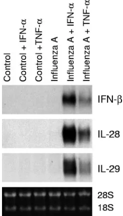
epithelial cells, A549 cells were pretreated with IFN-␣ and TNF-␣for 24 h, followed by infection with influenza A virus for 6 h. IFN-␣and TNF-␣clearly enhanced influenza A virus-induced expression of the IFN-, IL-28, and IL-29 genes in A549 cells (Fig. 2).
Influenza A NS1A CPSF mutant virus is a more potent activator of IL-28 and IL-29 gene expression than wild-type virus. It has been shown that the influenza A virus NS1A protein interferes with the host cell IFN response by inhibiting the posttranscriptional processing of cellular mRNAs (20). To address the role of NS1A in influenza A virus-induced IFN-␣, IFN-, IL-28, and IL-29 responses, wt and CPSF-binding-de-fective NS1A mutant Udorn viruses were used (31). A549 cells were infected with wt or CPSF mutant Udorn virus for 6 h, total cellular RNA was isolated, and IFN-␣, IFN-, IL-28, and IL-29 mRNA expression was analyzed by Northern blotting (Fig. 3). Strongly enhanced IFN-␣, IFN-, IL-28, and IL-29 mRNA expression was seen in IFN-␣- and TNF-␣-pretreated cells that were infected with wild-type or CPSF mutant virus. In addition, the CPSF mutant virus was a more potent activator of IFN-␣, IFN-, IL-28, and IL-29 mRNA expression than the wild-type virus (Fig. 3).
[image:3.585.61.266.60.508.2]IFN-␣ and TNF-␣ activate RIG-I and TLR3 mRNA and protein expression in A549 cells. RIG-I, TLR3, TLR7, and TLR8 are PRRs that detect viral RNAs. TLR7 and TLR8 are involved in ssRNA recognition, whereas RIG-I and TLR3 me-diate dsRNA-induced IFN gene expression (2, 6, 14, 27, 48). Since IFN-␣and TNF-␣pretreatment enhanced virus-induced IFN-␣/, IL-28, and IL-29 gene expression, we studied whether these cytokines would stimulate RIG-I or TLR expression in A549 cells. In line with previous studies (29, 42, 47, 48), IFN-␣ activated RIG-I and TLR3 gene expression in A549 cells
[image:3.585.359.482.71.286.2]FIG. 1. Kinetics of IFN-␣, IFN-, IL-28, and IL-29 gene expression in virus-infected lung epithelial cells. A549 cells were infected with influenza A H3N2 (A/Beijing/353/89) or Sendai (strain Cantell) virus (multiplicity of infection, 2). At the times indicated, the cells were collected and total cellular RNA was isolated. (A) Quantitative RT-PCR analysis was carried out for virus-induced expression of the IFN-␣1 and IFN-␣2 subtypes, IFN-, and IL-29 (IFN-1). All quan-tification data are presented as ratios to the glyceraldehyde-3-phos-phate dehydrogenase level. (B) RNA samples (10g/lane) were sub-jected to Northern blot analysis with IFN-␣2, IFN-, IL-28, and IL-29 probes. Ethidium bromide staining of rRNA bands was used to control for equal RNA loading.
FIG. 2. IFN-␣ and TNF-␣ enhance influenza A virus-induced IFN-, IL-28, and IL-29 gene expression. A549 cells were left un-treated or preun-treated with IFN-␣(100 IU/ml) or TNF-␣(10 ng/ml) for 24 h, followed by infection with influenza A virus (A/Beijing/353/89; multiplicity of infection, 2) for 6 h. The cells were collected, total cellular RNA was prepared, and IFN-, IL-28, and IL-29 mRNA expression was studied by Northern blotting.
on November 8, 2019 by guest
http://jvi.asm.org/
(Fig. 4A). Surprisingly, TNF-␣ also clearly enhanced RIG-I and TLR3 mRNA synthesis, although with delayed kinetics compared to that of IFN-␣(Fig. 4A). Furthermore, IFN-␣and TNF-␣ had an additive effect on RIG-I and TLR3 mRNA expression. Similarly, both IFN-␣and TNF-␣induced RIG-I and TLR3 protein expression in A549 cells (Fig. 4B and C). In line with our previous observations (47), no detectable expres-sion of TLR7 or TLR8 mRNA was seen in untreated or cyto-kine-stimulated A549 cells (data not shown).
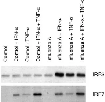
IFN-␣and TNF-␣enhance the expression of genes involved in the TLR/RIG-I signaling pathway.The recognition of vi-ruses by PRRs activates antiviral genes via recruitment of adapter proteins and protein kinases to the receptor complex. Most TLRs utilize a common adapter, MyD88, whereas TLR3 also signals via TRIF (1). TRIF associates with kinases IKKε and TBK1, which phosphorylate the transcription factors IRF3 and IRF7 (10, 40). To further study putative positive feedback mechanisms in IFN-␣- and TNF-␣-pretreated cells, we carried out Northern blot analyses of cytokine-stimulated cells (Fig. 5A). IFN-␣and TNF-␣enhanced MyD88 mRNA synthesis 3 h and 6 h after cytokine stimulation, respectively. IFN-␣- and
TNF-␣-induced TRIF expression was seen already 3 h after the cytokine treatment. IFN-␣had no effect on IKKεand TBK1 mRNA expression, whereas TNF-␣ strongly activated both IKKεmRNA and protein expression in A549 cells (Fig. 5A and B). TNF-␣also up-regulated TBK1 mRNA expression. IRF3 was expressed at a basal level in A549 cells, and its expression was not affected by cytokine stimulation (Fig. 5B). In IFN-␣
[image:4.585.309.505.63.568.2]-FIG. 4. IFN-␣ and TNF-␣ activate RIG-I and TLR3 mRNA and protein expression in A549 lung epithelial cells. (A) A549 cells left un-treated or stimulated with IFN-␣(100 IU/ml), TNF-␣(10 ng/ml), or their combination for the times indicated. Total cellular RNA was prepared, and RNA samples (10g/lane) were subjected to Northern blot analysis with RIG-I and TLR3 probes. Ethidium bromide staining of rRNA bands was used to control for equal RNA loading. (B) A549 cells left untreated or stimulated with IFN-␣(100 IU/ml), TNF-␣(10 ng/ml), or their combination for 24 h. RIG-I protein expression was studied by Western blotting. To control for equal loading, the membranes were stained with anti-STAT5 Ab. (C) The cells were left untreated or treated with IFN-␣(100 IU/ml), TNF-␣(10 ng/ml), or their combination for 24 h. After treatment, the cells were collected and prepared for immunoprecipitation with anti-TLR3 Ab. After immunoprecipitation, the samples were run in 10% SDS-PAGE gels under reducing conditions. To control for equal loading, the membranes were stained with Ponceau S and then immunoblotted with anti-TLR3 Ab.
FIG. 3. Expression of IFN-␣, IFN-, IL-28, and IL-29 mRNAs in wild-type and CPSF-binding-defective NS1A mutant Udorn virus-in-fected cells. A549 cells were left untreated or pretreated with IFN-␣(100 IU/ml) or TNF-␣(10 ng/ml) for 24 h, followed by infection with wild-type or CPSF mutant influenza A/Udorn/72 virus for 6 h. IFN-␣, IFN-, IL-28, and IL-29 gene expression was studied by Northern blotting.
on November 8, 2019 by guest
[image:4.585.55.270.71.330.2]and TNF-␣-stimulated cells, IRF7 mRNA and protein expres-sion was clearly activated (Fig. 5A and B). In concluexpres-sion, we observed that IFN-␣and TNF-␣enhanced the expression of the signaling components known to be involved in the activa-tion of IFN-␣/gene expression.
IFN-␣or TNF-␣pretreatment is essential for enhanced influ-enza A virus-induced activation of IRF3. IFN-␣ and TNF-␣ enhanced IRF7 expression, whereas IRF3 expression was not affected by these cytokines. The effects of IFN-␣and TNF-␣on influenza A virus-induced activation of IRF3 and IRF7 were further studied with the oligonucleotide immunoprecipitation method. Low basal DNA binding of IRF3 to the IFN- pro-moter PRDI-PRDIII region was seen in untreated A549 cells. Influenza A virus infection itself did not clearly enhance IRF3 binding to the IFN-promoter PRDI-PRDIII region, whereas a strong enhancement in IRF3 binding was seen in influenza A virus-infected cells that had been pretreated with IFN-␣ or TNF-␣(Fig. 6). IRF7 binding appeared to be associated with the overall expression of IRF7 (compare Fig. 6 to Fig. 5), and no further enhancement of its binding to the IFN-␣14 PRD-like element was seen in influenza A virus-infected cell extracts (Fig. 6).
IKK and a constitutively active form of RIG-I induce IFN-, IL-28, and IL-29 gene expression. It has previously been shown that ectopic expression of IKKε(40) and a con-stitutively active form of RIG-I,⌬RIG-I, that contains the two caspase recruitment domains induces IFN- expression (48). To study whether overexpression of IKKε or⌬RIG-I would result in the activation of IL-28 and IL-29 gene expression, we transfected A549 cells with IKKεand⌬RIG-I expression plas-mids. As shown in Fig. 7, overexpression of IKKεor⌬RIG-I activated the endogenous IFN-, IL-28, and IL-29 genes in A549 cells.
[image:5.585.332.512.74.249.2]RIG-I is involved in influenza A virus-induced expression of IFN-. Gene targeting studies have shown that TLR3 is not
FIG. 5. IFN-␣and TNF-␣induce the expression of genes involved in the TLR/RIG-I signaling pathway. (A) A549 cells left unstimulated or stimulated with IFN-␣(100 IU/ml) and/or TNF-␣(10 ng/ml) for the times indicated. Total cellular RNA was prepared, and RNA samples (10g/lane) were subjected to Northern blot analysis with MyD88, TRIF, IKKε, TBK1, and IRF7 probes. (B) A549 cells were stimulated with IFN-␣, TNF-␣, or their combination for the times indicated. IKKε, IRF3, and IRF7 protein expression was studied by Western blotting. To control for equal loading, the membranes were stained anti-STAT5 Ab.
FIG. 6. IFN-␣and TNF-␣enhance influenza A virus-induced IRF3 and IRF7 DNA binding. A549 cells were left untreated or pretreated with IFN-␣, TNF-␣, or their combination for 24 h, after which the cells were infected with influenza A virus (A/Beijing/353/89). After 6 h of infection, nuclear extracts were prepared and precipitated with oligonucleotides containing the promoter elements of the IFN-␣14 or IFN-gene. IRF3 and IRF7 binding was visualized by Western blotting.
on November 8, 2019 by guest
http://jvi.asm.org/
[image:5.585.72.255.93.616.2]required for IFN-␣/production in virus-infected cells (6, 7, 9, 27). Similarly, in our experiments, transfection of TLR3 into A549 cells did not enhance the expression of antiviral cytokines in response to influenza A virus infection (data not shown). To study the role of RIG-I in influenza A virus-induced expression of antiviral cytokines, we used a dominant-negative form of RIG-I, RIG-IC. To enhance the expression of endogenous RIG-I, the cells were treated with TNF-␣, followed by transfection with the RIG-IC expression plasmid and infection with the CPSF mutant influenza A virus (Fig. 8). IFN-mRNA synthesis was measured using a dual-luciferase assay. RIG-IC expression clearly reduced
virus-induced IFN-reporter activity in TNF-␣-pretreated A549 cells. These results suggest that RIG-I is involved in influenza A virus-induced expression of IFN-.
DISCUSSION
We have previously shown that A549 lung epithelial cells are highly susceptible to influenza A virus infection and that low levels of IFN-␣/ are secreted from virus-infected cells (35). Recently, two IFN-␣/-related cytokines, IL-28A/B and IL-29, were described (18, 41). We have previously shown that IFN-␣ enhances TLR- and virus-induced expression of the IL-28 and IL-29 genes in human primary macrophages (42). In the present study, we analyzed the regulation of IFN-␣, IFN-, IL-28, and IL-29 gene expression in human lung epithelial cells in response to influenza A and Sendai virus infection. Sendai virus infection in A549 cells resulted in rapid and efficient expression of the IFN-␣, IFN-, IL-28, and IL-29 genes. In contrast, influenza A virus-stimulated A549 cells expressed these genes at high levels only when the cells were pretreated with IFN-␣or TNF-␣. This up-regulation correlated with en-hanced expression of RIG-I and TLR3. Similarly, the expres-sion of MyD88, TRIF, IKKε, TBK1, and IRF7, signaling com-ponents involved in the activation of IFN gene expression, was up-regulated in response to TNF-␣stimulation. It is well es-tablished that most IFN-␣ genes are regulated by a positive feedback mechanism by IFN-␣/ during virus infection (23, 46). Our finding that TNF-␣ can also prime cells for high IFN-␣/production is a novel one and suggests an important role for TNF-␣in enhancing innate immune responses during viral infections.
[image:6.585.316.531.68.198.2]TLRs play a crucial role in the mammalian host defense against microbes. TLRs recognize conserved structural com-ponents of microbes and activate intracellular signaling path-ways leading to the production of cytokines (1). TLRs that have been associated with virus-induced cytokine production include TLR4, which recognizes the fusion protein of respira-tory syncytial virus (22), and TLR2, which induces chemokine
FIG. 7. Activation of endogenous IFN-, IL-28, and IL-29 gene expression by ectopic expression of IKKεand a constitutively active form of RIG-I. A549 cells were mock transfected or transfected with an IKKεexpression vector (A) or⌬RIG-I expression vector (B). After 24 h of transfection, the cells were collected, and total cellular RNA was prepared. Endogenous IFN-, IL-28, and IL-29 mRNA expression was studied by Northern blotting.
Renilla luciferase constructs, with or without a RIG-IC expression plasmid. After 24 h, cells were infected with influenza A/Udorn CPSF mutant virus for 18 h. Firefly andRenilla luciferase activities were measured, and firefly/Renillaluciferase ratios (FF/Ren rlu) were cal-culated.
on November 8, 2019 by guest
production in response to measles virus infection (3). TLR2 and TLR9 are involved in the activation of the innate immune response against herpes simplex virus (19, 21, 26). TLR3 is a receptor for dsRNA, whereas TLR7 and TLR8, which are preferentially expressed in macrophages or DCs, are activated by ssRNA originating from influenza A virus or other RNA viruses (2, 6, 14, 27). Our results show that in human lung epithelial cells, TLR7 and TLR8 are not the crucial compo-nents in virus-induced IFN-, IL-28, and IL-29 gene expres-sion, since A549 cells appear to be devoid of TLR7 and TLR8 expression (47; data not shown) and yet virus-induced cytokine gene expression occurred at a high level. In addition to TLRs, it was recently shown that the RNA helicase RIG-I is a cyto-plasmic receptor for dsRNA (48). In our experimental setting, IFN-␣and TNF-␣induced high expression levels of RIG-I and TLR3 in A549 cells, which correlated with an enhanced re-sponsiveness of the cells to influenza A virus infection. There-fore, RIG-I or TLR3 could be involved in the activation of IFN-␣/, IL-28, and IL-29 genes in response to influenza A virus infection. However, gene targeting studies have shown that TLR3 is not required for IFN-␣/ production in virus-infected cells (6, 7, 9, 27). In accordance with these studies, transfection of TLR3 into A549 cells did not enhance the expression of antiviral cytokines in response to influenza A virus infection (data not shown). In our transfection experi-ments, a constitutively active form of RIG-I (⌬RIG-I) induced IFN-, IL-28, and IL-29 gene expression. In addition, a dom-inant-negative form of RIG-I (RIG-IC) effectively inhibited influenza A virus-induced IFN-promoter activity in TNF-␣ -pretreated A549 cells. These results show that TNF-␣-induced RIG-I, not TLR3, mediates influenza A virus-induced IFN- gene expression in human lung epithelial cells. Previously, it has been shown that RIG-I is involved in Newcastle disease virus and hepatitis C virus recognition (4, 11, 44). Our results suggest that RIG-I is involved in influenza A virus recognition. The binding of microbial components to TLRs activates target genes via adapter proteins and protein kinases that are associated with or recruited to the receptor complex (1). Most TLRs utilize a common adapter, MyD88, whereas TLR3 and TLR4 also signal independently of MyD88 via TRIF. TRIF associates with IKKε and TBK1, which phosphorylate the transcription factors IRF3 and IRF7. Gene targeting studies have shown that IKKε and TBK1 are essential for TLR- and virus-induced activation of IFN-␣/genes (10, 15, 32, 40). Here we show that both IFN-␣ and TNF-␣up-regulate MyD88 and TRIF expression. In contrast to IFN-␣, TNF-␣also up-regulated TBK1 and especially IKKε expression in epithelial cells. Furthermore, in transfection exper-iments, enhanced IKKεexpression resulted in the activation of IFN-, IL-28, and IL-29 gene expression. Previously, it has been shown that IFN-promoter activity is induced by ectopic expres-sion of IKKε(10, 40). Our results show that IFN- as well as IL-28 and IL-29 gene expression can be activated via an IKKε -dependent pathway.
The molecular mechanisms of IFN-␣/gene regulation have been extensively studied. Gene disruption studies have shown the critical roles of IRF3 and IRF7 in the activation of IFN-␣/ genes (38). IRF3 is mainly responsible for the initial activation of IFN- gene expression, whereas enhanced expression of IRF7 leads to the production of the majority of IFN-␣subtypes (24, 46). Our results show that like that of IFN-␣genes,
effi-cient expression of the IL-28 and IL-29 genes during influenza A virus infection is also dependent on IFN-␣or TNF-␣ prim-ing. IRF3 activation and enhanced binding to the IFN- pro-moter PRDI-III element were seen only in influenza A virus-infected cells that had been primed with IFN-␣or TNF-␣. In contrast to that of IRF3, the DNA binding of IRF7 appeared to be dependent on IFN-␣- and TNF-␣-induced IRF7 expres-sion, suggesting that the IRF7 protein itself has some intrinsic DNA binding activity. Similar to that of IRF3, the transcrip-tional activity of IRF7 does, however, require activation by virus infection (17, 39, 43).
Many viruses have evolved mechanisms to interfere with the activation of host antiviral responses. Sendai virus C proteins target STAT proteins for degradation, leading to defective IFN signaling (13), whereas the influenza A virus NS1A protein has been shown to interfere with the production of IFN-␣/(12, 20). Initially, we observed that influenza A virus-infected A549 cells express the IFN-␣, IFN-, IL-28, and IL-29 genes at low levels (Fig. 1). However, after IFN-␣or TNF-␣priming, A549 cells expressed these genes very well. To address the role of NS1A in influenza A virus-induced cytokine gene expression, we used an NS1A mutant in which the NS1A protein is defec-tive in its ability to bind to CPSF and interfere with host cell mRNA processing (31). In these experiments, we observed that in spite of a functionally defective NS1A protein, influenza A virus-induced IFN-␣, IFN-, IL-28, and IL-29 gene expres-sion was greatly dependent on IFN-␣or TNF-␣priming (Fig. 4). Our results strongly suggest that IFN-␣and/or TNF-␣ prim-ing, followed by enhanced expression of PRRs and their sig-naling molecules, is a crucial factor in influenza A virus-in-duced expression of antiviral cytokine genes. This does not, however, rule out the role of the wild-type NS1A protein as an antagonist of IFN-␣/synthesis (8) or as a destabilizer of IFN mRNAs (30), since in our experiments we also observed higher cytokine mRNA levels in influenza A CPSF mutant virus-infected cells.
In conclusion, our results show that IFN-␣ and/or TNF-␣ priming of human lung epithelial cells is an essential factor for influenza A virus-induced production of antiviral cytokines. IL-28 and IL-29 were found to be produced by epithelial cells at high levels, which may have an important role in activating the innate immune response and in restricting the spread of influenza A virus infection. Our results also suggest that RIG-I, but not TLR3, is involved in influenza A virus recognition in epithelial cells.
ACKNOWLEDGMENTS
We thank Robert Krug for kindly providing us with the wt and CPSF mutant Udorn influenza viruses. We thank Mari Aaltonen and Hanna Valtonen for expert technical assistance.
This study was supported by grants from the Medical Research Council of the Academy of Finland, the Sigrid Juselius Foundation, and the Finnish Cancer Foundation.
REFERENCES
1.Akira, S.2003. Toll-like receptor signaling. J. Biol. Chem.278:38105–38108. 2.Alexopoulou, L., A. C. Holt, R. Medzhitov, and R. A. Flavell.2001. Recog-nition of double-stranded RNA and activation of NF-kappaB by Toll-like receptor 3. Nature413:732–738.
3.Bieback, K., E. Lien, I. M. Klagge, E. Avota, J. Schneider-Schaulies, W. P. Duprex, H. Wagner, C. J. Kirschning, V. Ter Meulen, and S. Schneider-Schaulies.2002. Hemagglutinin protein of wild-type measles virus activates Toll-like receptor 2 signaling. J. Virol.76:8729–8736.
on November 8, 2019 by guest
http://jvi.asm.org/
10.Fitzgerald, K. A., S. M. McWhirter, K. L. Faia, D. C. Rowe, E. Latz, D. T. Golenbock, A. J. Coyle, S. M. Liao, and T. Maniatis.2003. IKKεand TBK1 are essential components of the IRF3 signaling pathway. Nat. Immunol.
4:491–496.
11.Foy, E., K. Li, R. Sumpter, Jr., Y. M. Loo, C. L. Johnson, C. Wang, P. M. Fish, M. Yoneyama, T. Fujita, S. M. Lemon, and M. Gale, Jr.2005. Control of antiviral defenses through hepatitis C virus disruption of retinoic acid-inducible gene-I signaling. Proc. Natl. Acad. Sci. USA102:2986–2991. 12.Garcia-Sastre, A.2004. Identification and characterization of viral
antago-nists of type I interferon in negative-strand RNA viruses. Curr. Top. Micro-biol. Immunol.283:249–280.
13.Garcin, D., J. B. Marq, L. Strahle, P. le Mercier, and D. Kolakofsky.2002. All four Sendai virus C proteins bind Stat1, but only the larger forms also induce its mono-ubiquitination and degradation. Virology295:256–265. 14.Heil, F., H. Hemmi, H. Hochrein, F. Ampenberger, C. Kirschning, S. Akira,
G. Lipford, H. Wagner, and S. Bauer.2004. Species-specific recognition of single-stranded RNA via Toll-like receptor 7 and 8. Science303:1526–1529. 15.Hemmi, H., O. Takeuchi, S. Sato, M. Yamamoto, T. Kaisho, H. Sanjo, T. Kawai, K. Hoshino, K. Takeda, and S. Akira.2004. The roles of two IkappaB kinase-related kinases in lipopolysaccharide and double stranded RNA sig-naling and viral infection. J. Exp. Med.199:1641–1650.
16.Hiscott, J.2004. Another detour on the Toll road to the interferon antiviral response. Nat. Struct. Mol. Biol.11:1028–1030.
17.Iwamura, T., M. Yoneyama, K. Yamaguchi, W. Suhara, W. Mori, K. Shiota, Y. Okabe, H. Namiki, and T. Fujita.2001. Induction of IRF-3/-7 kinase and NF-kappaB in response to double-stranded RNA and virus infection: com-mon and unique pathways. Genes Cells6:375–388.
18.Kotenko, S. V., G. Gallagher, V. V. Baurin, A. Lewis-Antes, M. Shen, N. K. Shah, J. A. Langer, F. Sheikh, H. Dickensheets, and R. P. Donnelly.2003. IFN-lambdas mediate antiviral protection through a distinct class II cytokine receptor complex. Nat. Immunol.4:69–77.
19.Krug, A., G. D. Luker, W. Barchet, D. A. Leib, S. Akira, and M. Colonna.
2004. Herpes simplex virus type 1 activates murine natural interferon-pro-ducing cells through Toll-like receptor 9. Blood103:1433–1437.
20.Krug, R. M., W. Yuan, D. L. Noah, and A. G. Latham.2003. Intracellular warfare between human influenza viruses and human cells: the roles of the viral NS1 protein. Virology309:181–189.
21.Kurt-Jones, E. A., M. Chan, S. Zhou, J. Wang, G. Reed, R. Bronson, M. M. Arnold, D. M. Knipe, and R. W. Finberg.2004. Herpes simplex virus 1 interaction with Toll-like receptor 2 contributes to lethal encephalitis. Proc. Natl. Acad. Sci. USA101:1315–1320.
22.Kurt-Jones, E. A., L. Popova, L. Kwinn, L. M. Haynes, L. P. Jones, R. A. Tripp, E. E. Walsh, M. W. Freeman, D. T. Golenbock, L. J. Anderson, and R. W. Finberg.2000. Pattern recognition receptors TLR4 and CD14 mediate response to respiratory syncytial virus. Nat. Immunol.1:398–401. 23.Levy, D. E., I. Marie, and A. Prakash.2003. Ringing the interferon alarm:
differential regulation of gene expression at the interface between innate and adaptive immunity. Curr. Opin. Immunol.15:52–58.
24.Levy, D. E., I. Marie, E. Smith, and A. Prakash.2002. Enhancement and diversification of IFN induction by IRF-7-mediated positive feedback. J. In-terferon Cytokine Res.22:87–93.
25.Lin, R., P. Genin, Y. Mamane, and J. Hiscott.2000. Selective DNA binding and association with the CREB binding protein coactivator contribute to differential activation of␣/interferon genes by interferon regulatory factors 3 and 7. Mol. Cell. Biol.20:6342–6353.
26.Lund, J., A. Sato, S. Akira, R. Medzhitov, and A. Iwasaki.2003. Toll-like receptor 9-mediated recognition of herpes simplex virus-2 by plasmacytoid dendritic cells. J. Exp. Med.198:513–520.
33.Pirhonen, J., S. Matikainen, and I. Julkunen.2002. Regulation of virus-induced IL-12 and IL-23 expression in human macrophages. J. Immunol.
169:5673–5678.
34.Pirhonen, J., T. Sareneva, M. Kurimoto, I. Julkunen, and S. Matikainen.
1999. Virus infection activates IL-1and IL-18 production in human mac-rophages by a caspase-1-dependent pathway. J. Immunol.162:7322–7329. 35.Ronni, T., S. Matikainen, T. Sareneva, K. Melen, J. Pirhonen, P. Keskinen,
and I. Julkunen.1997. Regulation of IFN-␣/, MxA, 2⬘,5⬘-oligoadenylate synthetase, and HLA gene expression in influenza A-infected human lung epithelial cells. J. Immunol.158:2363–2374.
36.Ronni, T., T. Sareneva, J. Pirhonen, and I. Julkunen.1995. Activation of IFN-␣, IFN-␥, MxA, and IFN regulatory factor 1 genes in influenza A virus-infected human peripheral blood mononuclear cells. J. Immunol.154:
2764–2774.
37.Sareneva, T., I. Julkunen, and S. Matikainen.2000. IFN-␣and IL-12 induce IL-18 receptor gene expression in human NK and T cells. J. Immunol.
165:1933–1938.
38.Sato, M., H. Suemori, N. Hata, M. Asagiri, K. Ogasawara, K. Nakao, T. Nakaya, M. Katsuki, S. Noguchi, N. Tanaka, and T. Taniguchi.2000. Dis-tinct and essential roles of transcription factors IRF-3 and IRF-7 in response to viruses for IFN-␣/gene induction. Immunity13:539–548.
39.Servant, M. J., B. ten Oever, C. LePage, L. Conti, S. Gessani, I. Julkunen, R. Lin, and J. Hiscott.2001. Identification of distinct signaling pathways leading to the phosphorylation of interferon regulatory factor 3. J. Biol. Chem.
276:355–363.
40.Sharma, S., B. R. tenOever, N. Grandvaux, G. P. Zhou, R. Lin, and J. Hiscott.2003. Triggering the interferon antiviral response through an IKK-related pathway. Science300:1148–1151.
41.Sheppard, P., W. Kindsvogel, W. Xu, K. Henderson, S. Schlutsmeyer, T. E. Whitmore, R. Kuestner, U. Garrigues, C. Birks, J. Roraback, C. Ostrander, D. Dong, J. Shin, S. Presnell, B. Fox, B. Haldeman, E. Cooper, D. Taft, T. Gilbert, F. J. Grant, M. Tackett, W. Krivan, G. McKnight, C. Clegg, D. Foster, and K. M. Klucher.2003. IL-28, IL-29 and their class II cytokine receptor IL-28R. Nat. Immunol.4:63–68.
42.Siren, J., J. Pirhonen, I. Julkunen, and S. Matikainen.2005. IFN-␣regulates TLR-dependent gene expression of IFN-␣, IFN-, IL-28, and IL-29. J. Im-munol.174:1932–1937.
43.Smith, E. J., I. Marie, A. Prakash, A. Garcia-Sastre, and D. E. Levy.2001. IRF3 and IRF7 phosphorylation in virus-infected cells does not require double-stranded RNA-dependent protein kinase R or Ikappa B kinase but is blocked by vaccinia virus E3L protein. J. Biol. Chem.276:8951–8957. 44.Sumpter, R., Jr., Y. M. Loo, E. Foy, K. Li, M. Yoneyama, T. Fujita, S. M.
Lemon, and M. Gale, Jr.2005. Regulating intracellular antiviral defense and permissiveness to hepatitis C virus RNA replication through a cellular RNA helicase, RIG-I. J. Virol.79:2689–2699.
45.Takeda, K., T. Kaisho, and S. Akira.2003. Toll-like receptors. Annu. Rev. Immunol.21:335–376.
46.Taniguchi, T., and A. Takaoka.2002. The interferon-alpha/beta system in antiviral responses: a multimodal machinery of gene regulation by the IRF family of transcription factors. Curr. Opin. Immunol.14:111–116. 47.Tissari, J., J. Siren, S. Meri, I. Julkunen, and S. Matikainen.2005. IFN-␣
enhances TLR3-mediated antiviral cytokine expression in human endothe-lial and epitheendothe-lial cells by up-regulating TLR3 expression. J. Immunol.174:
4289–4294.
48.Yoneyama, M., M. Kikuchi, T. Natsukawa, N. Shinobu, T. Imaizumi, M. Miyagishi, K. Taira, S. Akira, and T. Fujita.2004. The RNA helicase RIG-I has an essential function in double-stranded RNA-induced innate antiviral responses. Nat. Immunol.5:730–737.