THE ROLE OF COLLAGEN COMPOSITION IN THE AETIO-PATHOGENESIS OF INGUINAL HERNIA
Dr. SIVA PRASANNA K Dissertation submitted to
The Tamil Nadu Dr. M.G.R Medical university, Chennai In partial fulfillment of the requirements for the degree of
Master of Surgery in General Surgery
Under the guidance of
Professor. Dr. VIMAL KUMAR GOVINDAN Department of General Surgery
ENDORSEMENT BY THE HOD, DEAN / HEAD OF THE
INSTITUTION
This is to certify that this dissertation entitled “THE ROLE OF
COLLAGEN COMPOSITION IN THE AETIO-PATHOGENESIS OF INGUINAL HERNIA” is a record of bonafide research work done by Dr. Siva Prasanna K, under the guidance of Dr. Vimal Kumar Govindan, in the Department of General Surgery, PSG Institute of Medical Sciences and Research,
Coimbatore – 641004.
Dr. Premkumar .S Dr. S. Ramalingam
HOD & Professor of Surgery Dean,
PSG IMS&R, PSG IMS&R,
CERTIFICATE
This is to certify that this dissertation entitled “THE ROLE OF
COLLAGEN COMPOSITION IN THE AETIO-PATHOGENESIS OF INGUINAL HERNIA” is a record of bonafide research work done by Dr. Siva Prasanna K, under Our guidance in the Department of General Surgery, PSG Institute of Medical Sciences and Research, Coimbatore – 641004.
Dr. Vimal Kumar Govindan Dr. S. Shanthakumari,
Guide Co guide
Professor of Surgery, Professor of Pathology,
PSG IMS&R, PSG IMS&R,
DECLARATION
I, Dr. Siva Prasanna .K, solemnly declare that this dissertation “THE
ROLE OF COLLAGEN COMPOSITION IN THE
AETIO-PATHOGENESIS OF INGUINAL HERNIA” is a bonafide record of work done by me in the Department of General Surgery, PSG Institute of Medical Sciences &
Research, Coimbatore, under the guidance of Dr.Vimal Kumar Govindan,
Professor of Surgery. This dissertation is submitted to The Tamilnadu Dr.M.G.R.
Medical University, Chennai, in partial fulfillment of the University regulations for
the award of MS Degree (General Surgery) Branch-I, Examination to be held in
April 2018.
CERTIFICATE – II
This is to certify that this dissertation work titled “THE ROLE OF
COLLAGEN COMPOSITION IN THE AETIO-PATHOGENESIS OF INGUINAL HERNIA” of the candidate Dr. Siva Prasanna .K, with Registration Number 221511502 for the award of Master of Surgery in the branch of General Surgery. I personally verified the urkund.com website for the purpose of
plagiarism Check. I found that the uploaded thesis file contains from introduction
to conclusion pages and result shows 7% of plagiarism in the dissertation.
ACKNOWLEDGEMENT
I would like to convey my utmost gratitude to my patients for they are
always an unparalleled source of knowledge.
I am thankful to the Institute and to our Principal for permitting me to
conduct this study. I am forever grateful to the faculty of the Department of
General Surgery and Pathology for actively taking part in the study and providing
samples.
I sincerely thank the technicians from the Department of Pathology who
have done a great job staining the slides.
My thanks are also to my colleagues, Interns and Staff Nurses for the
considerable help extended to me.
Finally, I would like to thank my family for their support, encouragement
CONTENTS
S NO. TOPIC PAGE NO.
1 INTRODUCTION 1
2 AIMS AND OBJECTIVES 3
3 REVIEW OF LITERATURE 4
4 MATERIALS AND METHODS 55
5 OBSERVATION AND RESULTS 64
6 DISCUSSION 76
5 SUMMARY AND CONCLUSION 81
6 BIBLIOGRAPHY
INTRODUCTION
Over the years mesh repair has evolved as the gold standard for the
treatment of groin hernias. The concept of mesh repair was introduced some 50
years ago. Lichtenstein and his colleagues popularized it in the last few decades.
An abdominal wall hernia is regarded as a localized weakness in the
background of mechanical problems. It has to be repaired through technical means.
Despite a legion of therapeutic modifications and refinements, recurrence of
hernias still appears to be a challenge in 10–15% of cases. Explanations for the
cause of recurrence are propounded in a more elementary way. It is presumed that
a debacle in the form of recurrence mainly depends on the quality of the repair.
Regarding sequence curves after hernia repair, the conglomerate incidences for
recurrences of inguinal hernias show a linear rise over years. Considering the
frame of these outcome curves of patients with hernia recurrence, explanations
pointing solely to failed technique are found wanting.
Alternatively, biological reasons should be discussed as a cause of flawed
wound healing with impaired scarring process. Lately, molecular-biological
awareness provides escalating evidence of elemental biochemical variations in
with a flawed wound healing are prevalent insufficient scar formation as the
underlying disease is assumed to be the end result of every surgical repair.
The question of whether the patient or the surgeon is liable for a recurrence
is of basic pertinence not only in hernia repair but also in other therapeutic
endeavors, such as in oncological surgery. While viewing recurrence as a sequel of
defective biology and collagens, one should not trivialize technical debacles
directly leading to a poor outcome.1,2However, considering the prevailing rates of
operations for recurrent hernia of 10–15% worldwide despite all therapeutic
breakthroughs, a more fundamental pathology for hernia occurrence and
AIM OF THE STUDY
To compare the composition of collagen in the tissues – Skin and Transversalis
fascia from the abdominal wall of patients with inguinal hernia and age matched
controls with no hernia.
To compare the Collagen I/Collagen III ratio in transversalis fascia and skin in
REVIEW OF LITERATURE
HISTORY OF INGUINAL HERNIA SURGERY
Hernia (from Greek hernios – meaning to bud or offshoot) is a well known
and a widely dealt with surgical disease throughout history.7It is one of the diseases
that haunted humanity from the very beginning. Ancient surgeons treated hernias
based on their size and the threat they posed to the patient. Although the natural
course of the disease is insidious it eventually reaches the size that severely impairs
the patient ability to perform daily activities (Fig1). This is why surgeons and
physicians alike were trying to find the solution for this highly disturbing and if left
[image:12.612.216.399.428.672.2]untreated deadly condition.
Fig 1: Frank Lamb, a slave in North Carolina with a 69 year history of groin swelling.
There are five eras described for the evolution of Hernia surgery.7
The first era dates back to Ancient Egypt where the first documented
description of a hernia was discovered in the Papyrus of Ebers. A cough impulse
was first described in this manuscript. Galen of Greece also contributed a lot to
understanding hernia after this period. Hippocratic corpus states that hernia was the
result of drinking water from large rivers.
The second era began during the renaissance in Europe. Anatomy as a
science flourished during this era and surgeons had a deeper insight into the subject
of hernia. The mainstay of treatment remained the use of various types of inguinal
belts that were supposedly designed to maintain the hernia sac inside the body
cavity. To successfully apply the herniary belt the hernia was first manually
reduced and then, the custom-made herniary belt was applied. The use of hernia
belts was widespread and even today it can be found in some regions of the world.
The wide popularity of the belts was maintained because the surgical option for the
cure of inguinal hernia was extremely dangerous and unfortunately not very
convincing.
The third era is termed ‘Era of Hernia repair under tension’. One of the first
attempts to solve inguinal hernia by the means of surgical knife came from the
idea of wide excision of the sac with the ensheathed skin and all its contents,
securing the neck with a significant suture (so-called golden stitch).This technique
did not enjoy widespread acceptance, especially among patients because it resulted
in castration and sometimes in the permanent stoma from the cut intestinal loop.
The risk of death from bleeding and peritonitis was also an important limiting factor of this primitive technique. This is why many ‘barber-surgeons’ of that time
suggested that the Fallopio operation should be considered only for marked
hernias, which could not be held even with the strongest and sturdiest bands at
their right place.8,9Another attempt worth mentioning was by the London surgeon
Claudius Amyand who in 1735 operated on an 11-year-old boy. The patient
suffered from a right inguinal hernia complicated by a fecal fistula. The operation
performed by Amyand is important for two reasons. First, it is the earliest
description of a hernia containing a vermiform appendix (known even today as Amyand’s hernia). And second, it is the earliest documented appendicectomy in
the history of surgery. The child died due to sepsis a few days later.
The age of asepsis brought about by Joseph Lister marked the beginning of
this era. The three cardinal features of hernia surgery performed during this period
are
1. Strict adherence to aseptic techniques.
3. Narrowing the internal inguinal ring.
The results of surgeries performed during this era were disappointing. There
was a high recurrence rate and significant mortality. The introduction of antisepsis,
asepsis, anesthesia and anatomical clarity allowed for safer procedures. Among
techniques that received some fame among renaissance surgeons, we should
mention the techniques of William Wood, Vinzenz Czerny and James Heaton.
Heaton was performing injection of the mixture of white oak and morphine into the
hernia sac to obtain its fibrosis.10Czerny was performing the high ligation of the
hernia sac and complete closure of the internal inguinal ring with sutures.11In the Wood’s method, the surgeon was supposed to double ligate hernial sac to perform
a natural ‘plug’ and use it to close internal inguinal ring. Although these techniques
looked appealing at first, virtually all patients experienced hernia recurrence. All
these procedures had an unacceptable mortality rate of over 7%.11
The fourth era began with Eduardo Bassini introducing the technique of
strengthening the posterior wall of the inguinal canal. This became the fourth and a
vital component of hernia surgeries performed thereafter. Bassini suffered a
bayonet wound to his right groin that resulted in a caecal fistula. He remained a
patient at Pavia for more than 6 months. His Caecostomy healed but during this
the problem was due to the distorted anatomy and physiology rather than the
technique.
Bassini had presented his first results in Padua, Italy during the surgical
congress in Genoa and after a few years in Germany to gain a wider
audience12.The significance of Bassini repair lies in the fact that most of the
procedures that were conceived later were modifications of this hallmark
procedure.
The Halsted’s modification left the spermatic cord in the subcutaneous plane
so that the surgeon can perform repair of the posterior wall using deep transfixation
sutures. This resulted in troublesome urinary fistulae.
The Polish technique propounded by Slawinski of Warsaw consisted of
dissecting, ligating and cutting only the neck of the sac. The divided sac is left
insitu. This procedure was carried out on a cardinal who would later become Pope
Pio IX.
Canadian surgeon E.Shouldice advocated strengthening the posterior wall using four layers of fasciae and aponeurosis. This was considered the ‘last big step’
in a barrage of Bassini repair modifications. In his world-renowned clinic, he
were ambulant in 3 days and recurrence rate 3% as against patients in other centers
who were advised strict bed rest for 3 weeks and had a dismal recurrence rate of
20%. His patients include several prominent monarchs and presidents.
Next began the era of tensionless hernia surgery. The tension in the suture
line was reduced initially by incisions over the rectus sheath. Norman Tanner
brought about this method as a tension reducing approach to conventional surgeries performed then. His ‘slide operation’ was meant for both inguinal and femoral
hernias8. The Bassini operation was later denounced in many centers and remained
only in academic interest.
There were more than 70 modifications for the Bassini repair. This very
plethora of modifications is by itself a hostile piece of evidence against the Bassini
repair. In anatomy, the function of muscles is motion, locomotion and stabilization
and not a buffer for a weakened posterior wall.
Reasons for the failure of Bassini repair are many. Uniting muscle with
fascia is against the rules of anatomy. This can take place only when the areolar
tissue in the region has been meticulously removed and tight tying of sutures thus
effecting a fibrin reaction and connective tissue formation. The surface area of
The Bassini operation fails13if applied to cases not demanding a reinforcing
procedure like cases of indirect hernia where the muscles are undamaged by
distension and the rings sound. In the direct hernias where it is required, the
conjoined tendon may be weak or have anomalous insertions into the rectus sheath.
This makes repair in the medial portion (the area most prone to recurrence)
demanding. Even when used efficiently the muscle does not function properly as it
is pulled away from the normal line of direction of its fibers. Slack sutures are
ineffective and tight sutures cause pressure necrosis of the vital lower part of the
conjoined tendon.
The era of plastics was ushered into herniology and foreign materials were
introduced as means of reducing tension between tissues.
The terms ‘onlay’ and ‘sublay’ were in reference to the position of mesh in relation to the posterior wall ofthe inguinal canal. The ‘onlay’ technique consisted
of placing the mesh superficial to the posterior wall and ’sublay’ position requires
Fig 2: Onlay [A] and Sublay [B] positions of mesh
Irving Lichtenstein published his paper on 1000 surgeries performed using14
Marlex mesh. Marlex is a high-density polyethylene which is loosely woven to
form a mesh which is quite pliable. It is well tolerated by the tissues even in the
presence of infection.
The Marlex mesh currently available is made of knitted polypropylene fiber,
which is more pliable and has a higher melting point [335 F]. This permits
sterilization by autoclaving. The original Marlex mesh had to be boiled in water,
cold-sterilized, or gas sterilized for reuse [melting point 270F]. It stimulates
fibroblastic proliferation.
Lichtenstein himself said to incise a strong posterior layer and, then, to
prosthetic mesh, one which serves only to strengthen such a floor, is harmless and
should reduce the incidence of recurrences.15
More recently in 1999, Arthur Gilbert described a technique that allows placing a mesh both in ‘onlay’ and ‘sublay’ position. His Prolene Hernia System
consisted on introducing a complex mesh build from two meshes of distinctive
shapes connected with a small tube. This allowed reinforcing the posterior wall of
the inguinal canal both from pre-peritoneal site and from the ‘onlay’ position.16,17
PREPERITONEAL REPAIRS:
Rene Stoppa in1969 developed a technique of Giant prosthetic
reinforcement of the visceral sac. This technique was supposed to be applied to
large, complicated and bilateral inguinal hernias and consisted on implanting a
large polyester mesh in pre-peritoneal connective tissue between the peritoneum
and fascia transversalis. The incision of choice for preperitoneal access was a low
midline incision and the mesh need not be fixed with sutures due to its size and
intraabdominal pressure maintaining it in situ.18Another preperitoneal hernia repair
was described in 1976 by Lloyd Nyhus from Chicago. Unlike in the Stoppa
method, the incision was made above the inguinal ligament. A similar incision was
used also for a pre-peritoneal placement of a sutureless mesh by Robert Kugel
LAPAROSCOPIC REPAIR:
With the advent of computer chip technology, laparoscopic picturing and
treatment of inguinal hernia got introduced in the surgical field.Ralph Ger was the
first in 1982 to report a transabdominal closure of an inguinal hernia defect during
a laparoscopy for other reasons. Some years later, in 1989, the gynecologist
S.Bogojavalensky showed a video demonstrating the laparoscopic intraabdominal
incision of the peritoneal hernia sac, subsequently closing the visible muscular
defect with a rolled-up piece of polypropylene mesh.
The early 1990’s saw a rapid rise in the number of publications, confirming
the feasibility of laparoscopic hernia repair. Although the first interventions were
limited to a plug and patch repair, later transabdominal approaches opted for the
fixation of a large preperitoneal mesh, either sutured or stapled to the posterior
muscular wall. A first attempt was made by applying a synthetic mesh to the
peritoneal wall and got the name IPOM (IntraPeritoneal Onlay Mesh).
Another approach involved making an intraperitoneal U-type incision in the
peritoneal wall and introducing the mesh in a preperitoneal position. It became
known asthe TAPP technique (TransAbdominal PrePeritoneal approach).Soon
Mac Kernan and Laws in 1993.It became known as the TEP technique (Total
ExtraPeritoneal approach). Even a special balloon dissector was introduced to
facilitate this extraperitoneal approach.
ANATOMY OF THE ABDOMINAL WALL
The abdominal wall is made up of muscles and fascia which cover the
anterolateral area between the xiphoid process and a line that goes through the iliac
[image:22.612.90.526.326.670.2]crests, femoral arches, and pubis.
Skin (Integument)
The epidermis has great ability to regenerate because its nutrition comes via
diffusion from the underlying vascular planes. Arterial vessels form a subdermal
plexus from which some branches leave to enter the subcutaneous tissue. In the
subdermal plexus, arteriovenous anastomoses exist; some of these are glomus
under the control of the autonomous nervous system. The area least irrigated is the
midline of the abdomen as the plexus comes from the back to the front area. The
subdermal lymphatics are anastomosed at all levels, so a free exchange is produced
between regions.
Subcutaneous Tissue (Adipose Tissue or Hypodermis)
This consists of areolar tissue and/or unilocular white adipose tissue according to the constitution of the individual and the subject’s nutritional status
and according to factors of hereditary.
The mobilization and deposition of lipids are influenced by nerve factors
(nor adrenaline, which activates the lipase) and hormonal factors (insulin, thyroid
hormones, glucocorticoids, and pituitary hormones).
In the subcutaneous tissue, the blood vessels are the vessels from perforating
Musculoaponeurotic Plane
This is composed of the Transversus abdominis, Internal oblique muscle,
External oblique muscle and Rectus abdominis. Transverse and oblique muscles
go forward internally and externally forming the rectus sheath and the white line.
Blood supply of these muscles depends on the epigastric vessels, which come up
from the external iliac vessels to get to the internal mammary artery and vein, in
the thorax. Epigastric vessels are included in the rectus muscle inside the sheath. 20-25
Extraperitoneal or Subperitoneal Space
This is placed between the inner surface of the abdominal wall, roofed by its
fascia, and the parietal peritoneum. It encloses vessels, nerves, organs, and extra-
peritoneal adipose tissue, in a variable arrangement according to regions and
subjects.
We can identify the following regions or extra-peritoneal spaces26-31:
Lateroperitoneal spaces: at the level of the iliac fossa internal to the external
iliac vessels, gonadal, and nerve genitocrural.
Preperitoneal spaces: at the round ligament and lower and include prevesical
Pelvic subperitoneal space: comprised of a visceral mediastinum,
laterovisceral spaces.
Retroperitoneal spaces
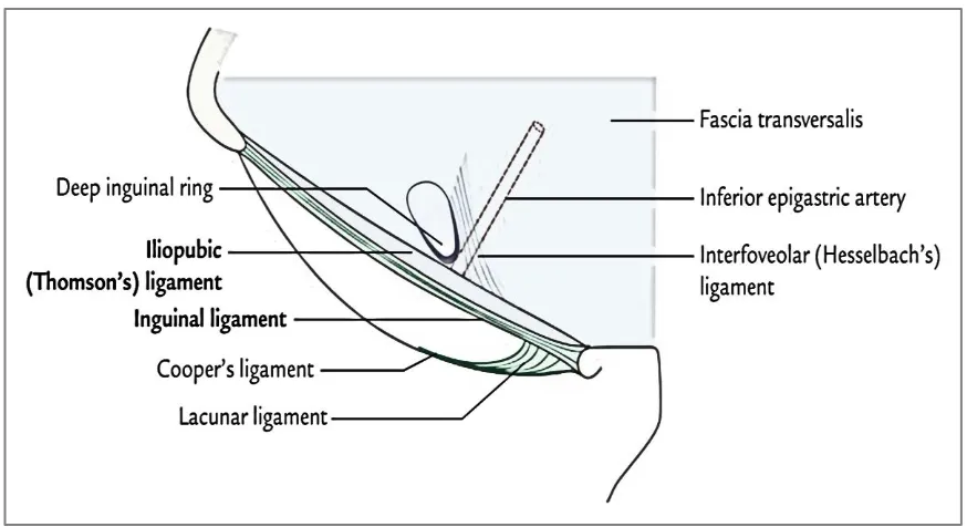
Transversalis fascia is a thin layer of fascia lining the transversus abdominis muscle. It lies between the transversus abdominis and the
extraperitoneal fascia.It is imperative to understand that the transversalis fascia, the
diaphragmatic fascia, the iliacus fascia, and the pelvic fascia form one continuous
lining to the abdominal and pelvic cavities. They are named according to the
structures they are associated with. The femoral sheath in the lower limbs is
formed from this fascia, in addition to the iliacus fascia
Borders and attachments of transversalis fascia:
Posterior aspect: In the posterior aspect, the transversalis fascia is lost in the fat covering the posterior surface of the kidneys.
Inferior aspect: Inferiorly, the transversalis fascia is attached to the iliac crest in its entire length and posterior margin of the inguinal ligament.
Medial to femoral vessels: In this region, the fascia is thin and attached to the pubis and pectineal line. It descends in front of the femoral vessels to
Beneath the inguinal ligament: In this region, it is strengthened by a band of fibrous tissue that loosely connects to the inguinal ligament and forms the
[image:26.612.193.439.182.376.2]iliopubic tract.
Fig 4a: Fascia transversalis
[image:26.612.187.429.441.684.2]Fig 4c: Relation of the fascia to the structures in the Inguinal Canal
The iliopubic tract32 (Thomson ligament) is a band in the posterior inguinal region distinct from the inguinal ligament and is sometimes known as the deep
crural arch. The advent of laparoscopic approaches to herniorrhaphy has recently
heightened awareness of the importance of the iliopubic tract. The histological
composition of the iliopubic tract is strikingly different from the inguinal ligament,
Fig 5: Iliopubic tract
Opening in the transversalis fascia (Deep inguinal ring):
The transversalis fascia is called the Achilles heel of the abdomen. It has an
oval opening near the midpoint of the inguinal ligament Fig 4c. This opening is
known as the deep inguinal ring. It transmits the spermatic cord (in males) or round
ligament of the uterus (in females).
The opening in the fascia is not visible externally. It is very hard to locate
during dissection of a cadaver. The reason is that transversalis fascia is prolonged
on the structures passing through the opening as the internal spermatic fascia.
Peritoneum–This holds the intraperitoneal organs and is divided into areas and regions that are useful in surgical exploration and intraperitoneal pathways and
structures ideal for transperitoneal/ extraperitoneal approach.
[image:29.612.106.510.229.565.2]Areas of Weakness the in the inguinal region:
The area of inguinal weakness is oval-shaped and is limited superiorly by
the conjoined tendon (inferior border of the abdominal internal and transverses
oblique muscle) and inferiorly by the inguinal ligament.
The Hesselbach’s triangle is an anatomic landmark and is bounded by the rectus muscle medially, the inguinal ligament inferiorly, and the inferior epigastric
vessels laterally. Indirect inguinal hernias are produced by the area of weakness
created by the deep inguinal orifice, in such a way that the hernia sac is directed
into the interior of the spermatic cord towards the testicle. Direct inguinal hernias
are produced by the area of weakness localized medial to the deep inguinal orifice
BIOLOGY OF COLLAGEN
Collagen is the ubiquitous protein of mammals. The term collagen is derived
from the Greek word – ‘Kolla’ meaning ‘glue’ and ‘gen’ meaning ‘to produce’.
This is related to the practice of the ancient Greeks who used crude collagen as
glue. They obtained this viscous substance by boiling the hides and tendons of
horses. Such glue was used to hold together and preserve manuscripts like the
Dead sea scrolls.33
In Chinese medicine, it is stated for millennia to consume animal cartilage to
treat joint disorders.
Within the body of both vertebrates and invertebrates, collagen is the
predominant structural biopolymer of extracellular connective tissue matrix. It
contributes to 25-35% of the protein content of the whole body. It is produced by
fibroblasts and is extracted into the extracellular space. The arrangement of
collagen determines the mechanical response to stress. The tensile strength of the
tissues depends on the degree of mineralization of the predominant collagen that
makes up the tissue. This can result in rigid (bone) to compliant (tendons) tissues.34
Collagen occurs in many places throughout the body. Over 90% of the
So far, 29 types of collagen have been identified and described. They can be
divided into several groups according to the structure they form:
Fibrillar (Type I, II, III, V, XI)
Non-fibrillar
o FACIT (Fibril Associated Collagens with Interrupted Triple Helices) (Type IX, XII, XIV, XVI, XIX)
o MACIT (Membrane Associated Collagens with Interrupted Triple Helices) (Type XIII, XVII)
o Multiplexin (Many Triple Helix domains with breaks) (Type XV, XVIII)
o Short chain (Type VIII, X)
o Basement membrane (Type IV)
o Other (Type VI, VII)
Type I: Skin, tendon, vascular ligature, connective tissue, bone (main
component of the organic part of bone)
Type II: Cartilage (main collagenous component of cartilage)
Type III: Reticulate (the main component of reticular fibers), commonly
found alongside type I.
Type IV: Forms basal lamina, the epithelium-secreted layer of the basement
Type V: Cell surfaces, hair and placenta
Type VI: Collagen VI is a form of collagen primarily associated with the
extracellular matrix of skeletal muscle.
Type VII: Forms anchoring fibrils between the basal and reticular laminae of
the basement membrane
Type VIII: Endothelial cells of cornea, keratinocytes, mast cells,
microvascular endothelial cells and some tumor cells, sclera, skin and
glomerulus (Mesangial cells)
Type IX: Collagen component of Hyaline cartilage
Type X: Collagen expressed by chondrocytes during enchondral ossification
Type XI: Part of the inner ear and the nucleus pulposus, which is the of the
vertebrae.
Type XII: Type XII collagen is found in association with type I collagen, an
association that modifies the interactions between collagen I fibrils and the
surrounding matrix.
Type XIII: cell adhesion-associated function in a wide array of cell-matrix
junctions – Intercalated discs of heart, Talin, Vinculin.
Type XIV: embryonic heart, notably within the cardiac interstitium of the
Type XV: Expressed in fetal kidney and Lung tissue (especially
microvasculature). Also found in the interstitium of adult kidneys.
Type XVI: Found in keratinocytes, and in smooth muscle and amnion
Type XVII: It is a transmembrane protein that forms a significant part of
hemidesmosomes. It binds keratinocytes to the underlying dermo-epidermal
junction and its defect is implicated in epidermolysis bullosa
Type XVIII: Component of basement membrane. It possesses molecular
properties of both collagen and proteoglycan. Chemical cleavage of its C
terminal end yields endostatin that has marked anti angiogenic properties
Type XIX: It is also called cuticle collagen. It forms polymers with fibril
forming collagen I and maintains the integrity of extracellular matrix
Type XX: It is very prevalent in the Corneal epithelium, embryonal skin,
notochord, neural retina.
Type XXI: It is almost exclusively localized to tissue containing Collagen I
and forms a part of the extracellular matrix.
Type XXII: It strengthens skeletal muscles and stabilizes myotendinous
junctions.
Type XXIII: It was discovered from rat prostate carcinoma and its presence
in the developing epidermis and other epithelia such as those in tongue, git,
pulmonary parenchyma also in the brain, renal tissue and the cornea.
Type XXIV: It is a marker of osteoblastic differentiation. It is predominantly
a skeletal collagen and is expressed in embryonal trabecular bone and
periosteum.
Type XXV: It is expressed in the cell membrane of brain parenchyma and
plays a role in the progression of Alzheimer amyloid plaques.
Type XXVI: It is made of a cysteine-rich Emilin core and two protein
stretches. It is investigated as a cause of aspirin-induced asthma.
Type XXVII: It plays a major role in the calcification of cartilage and
furtherance of cartilage to bone.
Type XXVIII: It forms a part of von Willebrand protein
Type XXIX: It is a newly discovered epidermal collagen and being studied
as a cause of atopic dermatitis.37
Thus the collagen superfamily comprises 29 members in vertebrates
numbered with Roman numerals (I–XXVIII). The novel skin is collagen called
collagen XXIX, but the COL29A1 gene was demonstrated to be akin to the COL6A5 gene, and the α1(XXIX) chain corresponds to the α5(VI) chain.38
collagen XII). The heterogeneity of the collagen family is further increased by the existence of several α chains, multitudes of molecular isoforms and the presence of
supramolecular structures for a single collagen type, and the use of many
promoters and alternative splicing.
SUPRAMOLECULAR ASSEMBLY OF COLLAGEN:
When viewed through electron microscopy after rotary shadowing, collagen
molecules are seen as rods varying in length from almost 75 nm for collagen XII to
425 nm for collagen VII.39 The molecular structure of collagen XV removed from
tissues haveunusual shapes, many molecules being found in a pretzel,
knot,figure-of-eight, configuration.40,41Presence of non-collagenous domains causes kinks that
are viewed in electron microscopy of nonfibrillar collagen preparations. Electron
microscopy can also characterize Non-collagenous domains after rotary
shadowing.42
FIBRIL-FORMING COLLAGENS
Collagens can be divided into subfamilies based on their supramolecular
assemblies: fibrils, beaded filaments, anchoring fibrils, and networks as shown in
(Fig. 7). Heterotypic collagen fibrils are the ones that are made of several collagen
types. In cartilage collagen fibrils are composed of collagens II, XI, and IX or of
cornea.44Besides collagen fibrils can be thought of as macromolecular alloys made
of collagenous and non-collagenous proteins or proteoglycans. Inarguably, small
leucine-rich proteoglycans regulate the formation of fibrils, as do collagens V and
XIV45, and could also influence collagen cross-linking.46For the process of
fibrillogenesis of collagens I and II, collagens V and XI could act as nucleators and
fibronectin and integrins as organizers.47
Collagen fibrillogenesis has been extensively studied in tendons, although
the site of the initial steps of fibrillogenesis is not clearly defined so far. They may
take place in extracellularly where fibril intermediates are clustered and mature
fibrils grow through a fusion process of intermediates48or they may occur
intracellularly, in 28-nm-diameter Golgi-to-membrane carriers with fibrils that are
Collagen fibrils exhibit a banding pattern with a periodicity (D) of 64–67
nm50 and usually collagen molecules are D-staggered within the fibrils. Depending
on the tissue collagen fibrils range in diameter from nearly 15 nm up to 500 nm or
more.51,52The microfibrillar structure of collagen I fibrils has been investigated in
situ by X-ray diffraction of rat tail tendons.53
Collagen I forms five molecules of supertwisted fibrils that interdigitate with
adjacent microfibrils, leading to the quasi-hexagonal packing of collagen
molecules.53In contrast, cartilage fibrils are composed of a nexus of four
microfibrils (two of collagen II and two of collagen XI) wreathed by a ring of ten
microfibrils, In cross-section, each microfibril contains five collagen molecules.54
FIBRIL-ASSOCIATED COLLAGENS
The FACITs do not form fibrils by themselves, but they are associated with
the surface of collagen fibrils. Collagen IX is covalently linked to the exterior of
cartilage collagen fibrils mostly made of collagen I.55 Collagens XII and XIV are
associated to Collagen I-containing fibrils. Collagen XV is associated with
basement membrane fibrils and forms a bridge linking large, banded fibrils, likely
NETWORK-FORMING COLLAGENS
Collagen IV forms a network in which four molecules assemble to form
tetramers via 7S domain, and two molecules assemble to form dimers via their
carboxy-terminal NC1 domain. Because the NC1 domains are trimeric, the NC1
dimer is a hexamer. The three-dimensional structure of the hexameric form of the
NC1 domain that plays a major role in collagen IV assembly and in the
stabilization of the collagen.57
Collagens VIII and X form similar hexagonal networks in Descemet’s
membrane of cornea and in hypertrophic cartilage, respectively. Collagen VI forms
beaded filaments and collagen VII assembles into anchoring fibrils connecting the
epidermis to the dermis.58
Some collagens participate in distinct molecular assemblies in different
tissues. Collagen XVI is a component of microfibrils containing fibrillin-1 in skin,
whereas it is incorporated into thin, weakly banded fibrils containing collagens II
and XI in cartilage.59,60 Supramolecular assemblies of many collagens are capable
of interaction as shown by the anchoring fibrils that are tightly attached to striated
COLLAGEN BIOSYNTHESIS
Collagen biosynthesis has been studied in depth for fibril-forming collagens
that are formed as procollagen molecules composed of an amino-terminal
propeptide followed by a small, nonhelical, N-telopeptide, a central triple helix, a
C-telopeptide and a carboxy-terminal propeptide. Individual proα chains are
subject to many posttranslational modifications (hydroxylation of proline and
lysine residues, glycosylation of lysine and hydroxylysine residues, sulfation of
tyrosine residues,62that end with the formation of the triple helix. In the
endoplasmic reticulum, the heat shock protein 47 (HSP47) binds to procollagen. It
is a specific molecular chaperone of procollagen.63More than 20 such HSP47
molecules have to bind per triple helix for the stabilization of procollagen at body
temperature.64 It has been recently suggested that intracellular Secreted Protein
Acidic and Rich in Cysteine (SPARC) may be a collagen chaperone because it
binds to the triple-helical domain of procollagens and its absence leads to flaws in
collagen deposition in tissues.65
Propeptides of procollagens are cleaved during the maturation process.66 The
N-propeptide is cleaved by proteinases belonging to the Disintegrin And
Metalloproteinase with Thrombospondin motifs (ADAMTS) family, except the N-propeptide of the proα1(V) chain that is cleaved by the procollagen C-proteinase
carboxy-terminal propeptide of procollagens, except the carboxy-terminal propeptide of the proα1(V) chain, that is processed by furin. The telopeptides
contain the sites where cross-linking occurs. This process is initiated by the
oxidative deamination of lysyl and hydroxylysyl residues catalyzed by the
enzymes of the lysyl oxidase family.68
COVALENT CROSS-LINKING OF COLLAGENS
Collagen is thought to be an elastic protein with a resilience of
approximately 90%. Collagen fibrils are thus able to deform and form back and
their mechanical properties can be studied by force spectroscopy.69
Cross-linking is tissue-specific and not collagen-specific. Reducible,
bifunctional, cross-links (aldimines and keto-imines) are formed in newly
synthesized collagens, and they automatically mature into nonreducible
trifunctional cross-links, pyridinoline and deoxypyridinoline in bone and cartilage,
pyrrole cross-links in bone, and histidinohydroxylysinonorleucine in skin73 (Fig.
3). Cross-link maturation provides added resistance to shear stress.
Collagens are long-lived proteins that are altered by glycation.71-75 Glycation
expands with age and several advanced glycation endproducts contribute to the
from ribose), and glucosepane (a nonfluorescent product formed from glucose)
have been identified in collagens. Glucosepane, the most abundant cross-link in
senescent skin collagen, is able to cross-link one in five collagen molecules in the
skin of the elderly.76
COLLAGEN DEGRADATION
Matrix metalloproteinases (MMPs) are zinc-dependent endopeptidases
belonging to the metzincin superfamily. They partake in physiological
(development and tissue repair) and pathological (tumorigenesis and metastasis)
processes. Fibril-forming collagens I, II, and III are cleaved by MMP-1 (interstitial
collagenase), MMP-8 (neutrophil collagenase), MMP-13 (collagenase 3). Collagen
II is a preferential substrate of MMP-13, whereas collagens I and III are
preferentially cleaved by MMP-1 and MMP-8.77-79Another group of enzymes,
collectively called sheddases80releases the ectodomain of membrane collagens as
soluble forms.
COLLAGEN AND DISEASE
More than one thousand mutations have been identified in 12 out of more
than 20 types of collagen. These mutations can lead to various diseases at the tissue
Osteogenesis imperfecta – Caused by a mutation in Type 1 collagen,
autosomal dominant disorder, results in brittle bones and irregular connective
tissue, some cases can be mild while others can be lethal. Mild cases have lowered
levels of collagen while severe cases have structural defects in collagen.82
Chondrodysplasias – Skeletal disorder believed to be caused by a mutation
in Type 2 collagen; further research is being conducted to confirm this.83
Ehlers-Danlos syndrome – Six different types of this disorder, which lead to
deformities in connective tissue, are known. Some types can be lethal, leading to
the rupture of arteries. Each syndrome is caused by a different mutation, for
example, type four of this disorder is caused by a mutation in collagen type 3.84
HERNIA AND COLLAGEN BIOLOGY:
All techniques employed for hernia repair have to depend on the formation
of adequate scar tissue. The scarring process is a form of flawed healing replacing
physiological tissues by fibrotic tissues that are rich in fibroblasts and collagens. It
forms from a complex network with interactions of many mediators of wound
healing and an intensive cross talk between cells, in particular macrophages.
Because of the long half-life of the collagens when compared to other growth
have been observed, such as in the expression of the matrix metalloproteinase 2
(MMP-2).85-90
Therefore, it is not amazing that a decreased collagen type I/III ratio could
be verified in adult patients with groin hernia (and in the scar of patients with
recurrent hernia.91,92 Collagen type I is the hallmark for mature scars or fascial
tissue whereas the collagen type III represents the mechanically unstable, sparsely
cross-linked collagen formed during the early periods of wound healing.
In patients with recurrent hernias, there seems to be an impaired maturing of
their scar tissue, which is not able to close the hernial gap or fix the mesh in place
for long. As a result, a recurrence may develop either through a scar or at the
border of a synthetic mesh through its defective and flawed fixation.93
Interestingly, an altered function could be detected even in vitro in macrophages’
free cultures of fibroblasts from patients with recurrent hernia, indicating a built-in
Fig 8: Cross polarization microscopical (CPM) and immunohistochemical features of
human fascial tissue. A- Tissue with normal Collagen I/III ratio. B – Scar tissue with
reduced Collagen I/III ratio. C,D – Positive cytoplasmic expression of MMP-2. Cells
marked with arrows
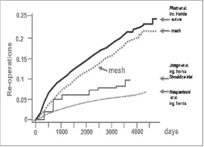
The low quality of the scar of patients with recurrences explain the outcome
curves [Fig 9]. exogenous factors such as smoking could be identified as major risk
factors. Likewise, it clarifies the high frequency of incisional hernia in patients
with abdominal aortic aneurysm and their demonstrated defect of the collagen
metabolism. It explains the frequent development of recurrences, if not the entire
Fig 9: Cumulative incidences of recurrences after incisional and inguinal hernia repair95,96
Patients with hernia and in particular those with an incisional hernia are
likely predisposed for recurrent hernia formation. Unfortunately, until now we do
not have any predicting markers to identify those with an impaired wound healing
and scar formation. The most substantial factor still is a patient's history of hernia
THE BLADE, BIOLOGY OR BOTH:
While recurrent and incisional hernias following suture repair are most
likely caused by a faulty biology, the recurrence subsequent to mesh repair may be
regarded as a technical fault. In reflection of the tensile strength of existing mesh
materials; it is the extent of overlap, which defines whether and when a recurrence
may appear. Almost all recurrences manifest at the border of a dislocated,
shrunken or undersized mesh, almost never through a mesh itself. Thus, it really
should be possible to prevent recurrences by mesh repair.
It is the possibility of an insufficient scar formation, that requires a
supplementary reinforcement with non-absorbable alloplastic nets as flat meshes
with an extensive overlap. Taking into consideration all patients with primary
hernia, the experiences of the past decades with suture repairs indicate that we
should expect 15–20% to develop a recurrent hernia. It will depend on the
long-term biocompatibility of mesh materials whether it is justified to apply a mesh
repair to all of the patients or to restrict it to selected patients at risk. Future
perspective may provide further possibilities to improve scar quality itself, e.g., by
The correlation between connective tissue and hernia began with a
significant discovery in 1964. Zolton T. Wirtschafter and J. Peter Bentley97 did a study providing a lathyritic diet to young and old Long-Evans rats. The ingestion
of seeds of Lathyrus odoratus caused generalized connective tissue disorder
especially osteolathyrism in mammals. The authors extracted collagen from the
Skin and peritoneum of Lathyrus fed rats and concluded that the rates of
occurrence of hernia was greater in this group compared to controls. Abnormal
fibrinogenesis and defective collagen formation were postulated to cause
diminished tensile strength of fascia
In a study by MA Ajabnoor et al98 Fibroblastic cell cultures were derived from the hernial sac and the surrounding muscles especially internal oblique of 130
Saudi patients with different types of hernias, and from 21 control subjects. The
tissues were subject to explantation and subcultures. Three variables were studied
1. Cell proliferation 2. Rates of incorporation of 14C proline 3.Rates of collagenase
activity. The rates of cell proliferation were studied for 39 days. Results implied
decreased rates of proliferation of cells derived from patients compared to controls.
In vitro studies of the rates of incorporation of 14C proline into the muscle biopsies
revealed decreased rates of label incorporation in the samples derived from patients
compared to controls. However, no differences were detected between rates of
controls. These findings suggested that collagen synthesis is probably defective in
the patients with hernia.
Similarly, in an article next year (1993), it wasdetermined99 if alterations in
fibrillar collagen synthesis were associated with the development of inguinal
hernias. This study was undertaken to study collagen synthesis in patients with
inguinal hernia in the absence of any other connective tissue disease.
Trypsin-chymotrypsin-resistant type I and III collagens were isolated and analyzed. The
study concluded that a constitutive and systemic increase in type III collagen
synthesis might result in reduced collagen fibril assembly in the abdominal wall,
eventually leading to the development of herniation. Although it is not yet clear
what genetic factors are responsible for the elevation in type III collagen synthesis
in patients with hernias. This study was a first attempt to define individuals with an
abnormality in collagen production that may be specifically related to herniation.
JM Bellon et al100 aimed to examine the transversalis fascia of patients with direct and indirect hernia in an effort to identify the differences between each type
of hernia. They analyzed the ultrastructure of the fascia surrounding the hernial
lesions, the proline and lysine hydroxylation in the tissue, the type I–type III
collagen ratio and the presence of metalloproteinases. They did not detect
abundant electron-dense particles. No differences in proline hydroxylation were
observed between each type of hernia. A small decrease in lysine hydroxylation
was detected in patients with direct hernia. As opposed to our study this one
evaluated collagen I/III ratio based on Enzyme-linked immunosorbent assays
(ELISAs). This showed no statistically significant differences in the type I–type III
collagen absorbance ratios. Immunohistochemistry revealed no differences in the
expression of matrix metalloproteinase-1. FT from patients presenting direct hernia
showed a very strong staining vs. metalloproteinase-2 when compared with that
observed in indirect hernia.
The relation between Collagen I and III is regulated by several collagenases,
mainly matrix metalloproteinases-1 and -13101 whereas fibronectin plays a key role
in the adherence of cells within the extracellular matrix. The aim of this study was
to investigate whether an alteration in type I and type III collagen synthesis,
amounts of MMP-1 and MMP-13 and the expression of fibronectin were
associated with the development of inguinal hernia. They analyzed the hernial sac
of patients with indirect and direct inguinal hernias and peritoneum in controls by
immunohistochemistry and Western blot analysis. The results showed that the ratio
of I/III collagen was markedly decreased in patients with either indirect or direct
hernias as compared with controls with a concomitant increase in type III collagen
of the controls, but the positive reactions of MMP-1 were found in the surface of
the subserosa of the hernial sac in patients with indirect or direct hernia without
any difference compared to controls. In regard to the known alterations of the
collagen metabolism in fascia and skin of hernia patients the changed collagen I/III
ratio with its increase of type III collagen in hernial sacs support the presence of a
systemic disturbance of collagen metabolism. The absence of changes in the
expression of collagenases (MMP-1, MMP-13) and the constant levels of
fibronectin underline the central role of collagen synthesis for the development of
indirect or direct hernias.
Alain Pans et al102suggested that a defect in collagen fiber structure may play a role in inguinal hernia formation. They performed a biochemical
investigation of the collagen in the transversalis fascia and rectus sheath. The
samples were collected from 40 adult patients with uni- or bilateral hernias and
from 20 control subjects without hernia (autopsies and organ donors). A constant
area of tissue was taken by using a calibrator. The wet and dry weights per 100
mm2 were determined and the total collagen concentration as well as its sequential
extractability in NaCl, acetic acid, and pepsin was measured. The ratios of type
I/III collagen were assessed by polyacrylamide gel electrophoresis.The significant
hernias. This connective tissue pathology is expressed only in the inguinal region,
since they observed no major difference between the rectus sheaths of controls and
those of patients. The qualitative study I/III collagen ratio showed no difference
between the fascia groups.
The study by Pablo Bórquez M103 is solely dedicated to the quality of skin in patients with and without hernia. Skin from the surgical wound was obtained
from 23 patients with and 23 patients without inguinal hernia. Patients without
hernia had compact collagen tracts homogenously distributed towards the deep
dermis. In contrast, patients with hernia had zones in the dermis with thinner and
disaggregated collagen tracts. Connective tissue had a lax aspect in these patients.
Collagen fiber density was 52% lower in patients with hernia, compared to subjects
without hernia. No differences in elastic fiber density or distribution was observed
between groups. Thus collagen fiber quality was distorted in the skin of patients
with hernia.
Rodrigues Jr AJet al104demonstrated structural and quantitative age-related changes of the elastic fibers in transversalis fascia, which may play a role in
inguinal hernia formation. Transversalis fascia fragments were acquired during
surgical intervention and underwent histological quantitative analysis of collagen
by colorimetry and analysis of elastic fibers by histomorphometry. They
elastic fibers in transversalis fascia from patients with direct inguinal hernia
compared to indirect inguinal hernia patients. The transversalis fascia from direct
inguinal hernia patients exhibited structural changes of the mature elastic fibers,
which are accountable for elasticity, and lower density of oxytalan elastic fibers,
which are responsible for resistance. These variations encouraged loss of resiliency
of the transversalis fascia.
Raphael Rosch105analyzed type I and type III procollagen messenger ribonucleic acid (mRNA) and MMP-1 and MMP-13 mRNA in fibroblasts from the
skin of patients with and without hernia (controls) by reverse transcription
polymerase chain reaction (RT-PCR) and Northern Blot. The results indicated that
the ratio of type I to type III procollagen mRNA was decreased in patients with
primary hernia, showing significant differences as compared to controls (p = 0.01).
This decrease like the previous study was mainly due to the increase of type III
procollagen mRNA. Furthermore, RT-PCR analysis revealed that the expression of
MMP-1 mRNA in patients with primary hernia is equivalent to that of controls. In
addition, MMP-13 mRNA is expressed neither in patients with primary hernia nor
When studying recurrent inguinal hernia Zheng H106extracted RNA from skin fibroblasts of three groups (control group I = healthy skin; control group II =
plain skin scar; recurrent inguinal hernia group = skin of recurrent inguinal hernias;
each n = 5). Reverse transcription-polymerase chain reaction (RT-PCR) and
Northern blot analysis were used to investigate the expression of procollagen type
I/- III. Both ratios of procollagen types I to III mRNAs and collagen types I to III
were decreased in the recurrent hernia group compared to those of both control
groups. Significant differences were caused by the increase of both procollagen
type III mRNA and collagen type III protein synthesis. The data of this study
strongly suggest recurrent inguinal hernias to be a disease of the collagen matrix.
With regards to prosthesis, Karsten Junge107studied Seventy-eight prostheses (Prolene, Atrium, Marlex, Vypro, Mersilene, Gore-Tex) implanted for
inguinal and incisional hernia repair that was explanted because of recurrence,
chronic pain or infection. The mean implantation period was 17.9±11.2 (range 0.5–
48) months. Collagen formation was investigated quantitatively (collagen–protein
ratio) and qualitatively (collagen type I/III ratio). Results were related to clinical
data that included gender, age, implantation period, indication for
implantation/explantation, type and location of the prosthesis. Samples explanted
for recurring hernias exhibited a significantly decreased ratio compared to samples
independent effects of age, gender, indication for implantation of prostheses,
location and implantation period on collagen type I/III ratio.
This group also studied gentamicin-supplemented polyvinylidenfluoride
mesh materials. Gentamicin mesh108 induced a significantly decreased expression
of MMP-8 and MMP-13 at the interface after implantation compared to the other
groups. The quality of collagen formation conveyed by the collagen type I/III ratio
showed significantly higher ratios around the Gentamicin mesh 21 and 90 days
after implantation. A 5.3-fold expression of type I alpha 1 collagen mRNA was
found.
ALM Meyer et al109did a Qualitative and Quantitative analysis of collagen types in inguinal hernia patients. The inclusion criteria were similar to this study
but the controls were 24 corpses which had passed away less than 8 hours.
Collagen I was visualized as thicker fibers that were strongly birefringent while
Collagen III was thin, sparse fibers that were stained with Picosirius stain.
A statistically meaningful difference (P = 0.788) was not found in the
amount of collagen type I in the fascia of the patients and of the controls. On the
other hand, a greater amount of collagen type III was found in the patients, which
Fig 10: Collagen I and III – Difference between cases and controls
The compared the amount of collagen type III superior to 5% - in the sample
field to the Nyhus classification for inguinal hernia. The patients classified as
Nyhus IIIa had a greater amount of collagen type III with a statistical meaningful
The second part of this study uses skin as a marker of collagen composition
of hernia defects. A similar study is by Peeters E.110Collagenorganization was examined in Haematoxylin-Eosin sections of anterior rectus sheath fascia, and
collagen type I/III ratio. The study concluded that in both skin and abdominal wall
fascia of hernia patients, collagen type I/III ratio was lower compared to control
patients, with more pronounced abnormalities in incisional and recurrent inguinal
hernia patients. Importantly, collagen type I/III ratio in skin was representative for
that in abdominal wall fascia.
A more recent study is that of Lazarenko111 of Russia. The trial included 141 patients for the period 2012-2015. Group I of patients without ventral hernias
was divided into subgroup AI - primary operation and BI. Group II consisted
patients with ventral hernias. There were significant differences between collagen
type I/III ratio in skin and aponeurosis. In patients with ventral hernias collagen
type I/III ratio in skin is 2.54 times lower than in patients without hernias.
Significant correlation of collagen types in skin and aponeurosis allows predicting
the risk of postoperative ventral hernias on basis of skin fragment.
In a study very similar to ours Koruth S112included a total of 90 patients, of which 45 patients underwent mesh repair for the various hernias and 45 patients
recurrent and primary inguinal hernias are not just caused because of a primary
defect but an acquired disorder with respect to collagen distribution.
One another recent study carried out in a similar fashion as ours was that of
Gonçalves O.113Samples of the transverse fascia and of the anterior sheath of the rectus abdominis muscle were collected from 40 men aged between 20 and 60
years with type II and IIIA Nyhus inguinal hernia and from 10 fresh male cadavers
(controls) without hernia in the same age range. The staining technique was
immunohistochemistry for collagen I, collagen III and elastic fibers; quantification
of fibrillar components was performed with an image analysis processing software.
But no statistically significant differences were found in the amount of elastic
fibers, collagen I and collagen III, and the ratio of collagen I / III among cases and
controls.
The importance of collagen in herniology is stressed in the Danish paper by
Burcharth J and Rosenberg J.114 Patient groups with reduced type-I/III collagen ratio and consequently increased risk of herniation include patients with Ehlers-Danlos, Marfan’s syndrome, osteogenesis imperfecta, cutis laxa, and patients with
abdominal aortic aneurysms, colonic diverticula or stress urinary incontinence. The
future perspective may be individualization of the operative technique for patients
Franz MG115 while studying the biology of hernia formation reinforced that all abdominal wall hernias occur when tissue structure and function are lost at the
load-bearing muscle, tendon, and fascial layer. The basic biologic mechanisms are
primary fascial pathology or surgical wound failure. In both cases, cellular and
extracellular molecular matrix defects occur. Primary abdominal wall hernias are
associated with extracellular matrix diseases. Incisional hernias and recurrent
inguinal hernias involve a combination of technical and biologic curbs.
Oğuzkurt P116
investigated and compared the distribution and intensity of
staining of extracellular matrix proteins--laminin, fibronectin, and types 1 and 4
collagen--in various congenital inguinoscrotal abnormalities and the peritoneum
through immunohistochemical staining. The sacs associated with undescended
testis (n = 28), hydrocele (n = 29), inguinal hernia (n = 31), and parietal
peritoneum (n = 28) were stained with antibodies for laminin, fibronectin, and
types 1 and 4 collagen. The peritoneum served as the control group. Type 1
collagen was intensely expressed in the sacs obtained from the hydroceles
compared with the other groups and the peritoneum. Expression of type 4 collagen
was significantly increased in the sacs associated with hydrocele and inguinal
hernia compared with the peritoneum.
inguinal hernia patients. This study was similar to the current one except that wide
variety of tissues were used and the staining was different. Twenty patients
operated on for inguinal hernia included in the study (11 direct and 9 indirect).
Nine patients underwent open cholecystectomy served as the control group. A 0.5
x 1 cm. tissue was sampled from skin, rectus sheath, transversalis fascia and
peritoneum in HR group. Skin, rectus sheath and peritoneum samples were taken
from the patients in Control group. The sections of those samples were submitted
to two different staining methods: "Masson's trichrome" for collagen and "van
Gieson" for elastin fibers and graded with light microscopy.
The rectus sheath samples of Control had higher staining scores for both
collagen and elastin fibers in comparison with hernia group. The control group had
a significantly higher score for collagen in peritoneum samples. There were no
statistically significant differences between the patients with direct and indirect
inguinal hernias for collagen or elastin fibers scores in skin, rectus sheath,
transversalis fascia and peritoneum samples.
Taniguchi Set al118 studied the Impact of collagen subtype proportions in peritoneal tissues on inguinal hernia formation in adults and infants.They
correlated the ratios of collagen type I to type III between adults and infants with
and without inguinal hernia, in an attempt to clarify the pathogenesis of this
hernia, and from the normal peritoneum of patients without an inguinal hernia.
After separation by electrophoresis, the collagen bands were quantified and we
compared the ratios of collagen type I to type III between the cases and controls.
The ratio of collagen type I to type III was significantly lower in the adults with an
inguinal hernia than in those without the disorder. Whereas the ratios were similar
in infants with and without an inguinal hernia confirming that a simple herniotomy
is an adequate treatment.
One another study carried out in infants by Hosgor et al119aim to investigate whether an alteration in type I and type III collagen synthesis was associated with
the development of childhood inguinoscrotal pathologies. The expression pattern
of type I and IIIcollagen did not differ among sacs obtained from patients with
inguinal hernia, hydrocele, and undescended testis when compared with that of
controls. However, strong expression of type III collagen was observed in the
hernial sacs of right-sided male inguinal hernia compared with the left side.
Casanova AB120 studied specifically the Collagen in the transversalis fascia of patients with indirect inguinal hernia. Biopsy samples from 26 patients and 26
cadavers were analyzed. The results showed 17.3% less total collagen in patients
with hernias compared with the control group. Type I collagen in patients with
To conclude we would like to quote the comprehensive paper published by
Henriksen NA121 where Fifty-two papers were included. Collagen alteration depended on the type of hernia; there were more pronounced changes in patients
with a direct inguinal hernia than in those with an indirect inguinal hernia. A
constant finding was a significant increase in immature type III collagen relative to
the stronger type I collagen in patients with a hernia. This resulted in thinner
collagen fibers with lessened biomechanical strength. It was also suggested that
these alterations are due to variation in the synthesis, maturation or degradation of
collagen by matrix metalloproteinases.
Robert Bendavid from Israel propounded the Unified theory of hernia formation122whereinhe states that we have progressed from the simple concept of increased intra-abdominal pressure overwhelming a weak abdominal wall to the
complex malady that calls upon several basic sciences to clarify the countless
facets, though one final common pathway, of its pathophysiology. The target organ
of all the known injurious stimuli is the collagen matrix. One day, classification

![Fig 2: Onlay [A] and Sublay [B] positions of mesh](https://thumb-us.123doks.com/thumbv2/123dok_us/170841.46736/19.612.132.505.67.257/fig-onlay-sublay-b-positions-mesh.webp)