JOURNAL OFVIROLOGY, Sept. 2006, p. 9115–9133 Vol. 80, No. 18 0022-538X/06/$08.00⫹0 doi:10.1128/JVI.00909-06
Copyright © 2006, American Society for Microbiology. All Rights Reserved.
Amino Acids in the Basic Domain of Epstein-Barr Virus ZEBRA
Protein Play Distinct Roles in DNA Binding, Activation of
Early Lytic Gene Expression, and Promotion of
Viral DNA Replication
Lee Heston,
1Ayman El-Guindy,
2Jill Countryman,
2Charles Dela Cruz,
1Henri-Jacques Delecluse,
4and George Miller
1,2,3*
Departments of Pediatrics,1Molecular Biophysics and Biochemistry,2and Epidemiology and Public Health,3Yale University School of
Medicine, New Haven, Connecticut 06520, and Department of Tumor Virology, German Cancer Research Center,
Im Neuenheimer Feld 242, Heidelberg, Germany4
Received 3 May 2006/Accepted 30 June 2006
The ZEBRA protein of Epstein-Barr virus (EBV) drives the viral lytic cycle cascade. The capacity of ZEBRA to recognize specific DNA sequences resides in amino acids 178 to 194, a region in which 9 of 17 residues are either lysine or arginine. To define the basic domain residues essential for activity, a series of 46 single-amino-acid-substitution mutants were examined for their ability to bind ZIIIB DNA, a high-affinity ZEBRA binding site, and for their capacity to activate early and late EBV lytic cycle gene expression. DNA binding was obligatory for the protein to activate the lytic cascade. Nineteen mutants that failed to bind DNA were unable to disrupt latency. A single acidic replacement of a basic amino acid destroyed DNA binding and the biologic activity of the protein. Four mutants that bound weakly to DNA were defective at stimulating the expression of Rta, the essential first target of ZEBRA in lytic cycle activation. Four amino acids, R183, A185, C189, and R190, are likely to contact ZIIIB DNA specifically, since alanine or valine substitutions at these positions drastically weakened or eliminated DNA binding. Twenty-three mutants were proficient in binding to ZIIIB DNA. Some DNA binding-proficient mutants were refractory to supershift by BZ-1 monoclonal antibody (epitope amino acids 214 to 230), likely as the result of the increased solubility of the mutants. Mutants competent to bind DNA could be separated into four functional groups: the wild-type group (eight mutants), a group defective at activating Rta (five mutants, all with mutations at the S186 site), a group defective at activating EA-D (three mutants with the R179A, S186T, and K192A mutations), and a group specifically defective at activating late gene expression (seven mutants). Three late mutants, with a Y180A, Y180E, or K188A mutation, were defective at stimulating EBV DNA replication. This catalogue of point mutants reveals that basic domain amino acids play distinct functions in binding to DNA, in activating Rta, in stimulating early lytic gene expression, and in promoting viral DNA replication and viral late gene expression. These results are discussed in relationship to the recently solved crystal structure of ZEBRA bound to an AP-1 site.
ZEBRA (BamHiZ Epstein-Barr virusreplicationactivator), the Epstein-Barr virus (EBV) protein encoded by the viral
BZLF1gene, is necessary and sufficient to initiate the complete
viral lytic cascade (16, 17, 22, 33, 61). Since ZEBRA is an essential contributor to the viral life cycle, it plays an indis-pensable role in viral pathogenesis by permitting the virus to spread between cells and among individuals. ZEBRA is a po-tential target for antiviral and oncolytic strategies. ZEBRA activates viral replication, which ultimately leads to cell death; therefore, overexpression of the protein may be used to de-stroy EBV-containing tumor cells in which the virus was orig-inally latent (23, 35).
ZEBRA protein exerts many functions in the viral life cycle. It is a transcriptional activator of early viral lytic cycle genes (15, 16, 42, 55). One of ZEBRA’s primary targets is the pro-moter of theBRLF1gene, encoding the R transactivator (Rta) (36, 40, 44, 60). Rta and ZEBRA synergistically activate
down-stream genes, such as the BMRF1 gene, encoding the viral DNA polymerase processivity factor, a protein originally de-tected by immunofluorescence as diffuse early antigen (EA-D) (51). At early times in the viral life cycle, ZEBRA also acts as a temporally regulated repressor of the capacity of Rta to activate a late gene,BLRF2, that encodes a tegument protein (19, 54). ZEBRA binds to the origin of lytic DNA replication (oriLyt), where it functions as an essential replication protein (24, 57, 58). ZEBRA interacts with other viral replication pro-teins, including the EBV-encoded helicase-primase complex (BBLF2/3, BBLF4, and BSLF1), the viral DNA polymerase (BALF5), and the DNA polymerase processivity factor (BMRF1) (5, 30, 69).
ZEBRA also impinges on many different cellular pathways. When expressed in the absence of other EBV genes, ZEBRA modulates the cell cycle, promoting G1cell cycle arrest (10, 11,
66). ZEBRA influences the host immune response by inhibit-ing the actions of gamma interferon, NF-B p65, and tumor necrosis factor alpha (46–48). ZEBRA influences signaling pathways by activating c-Jun N-terminal and p38 mitogen-activated protein kinases (1).
ZEBRA is a member of a large family of transcription
fac-* Corresponding author. Mailing address: Department of Pediatrics, Yale University School of Medicine, 4th Floor LSOG 420, New Haven, CT 06520. Phone: (203) 785-4758. Fax: (203) 785-6961. E-mail: George [email protected].
9115
on November 8, 2019 by guest
http://jvi.asm.org/
tors that are conserved fromSaccharomyces cerevisiae to hu-mans (21, 64). Other members of this family include GCN4, the activator of histidine biosynthesis inS. cerevisiae, and c-Fos and c-Jun, mammalian early-response proteins. Members of this family of proteins possess a generally modular structure with an N-terminal transcriptional activation domain, a DNA recognition domain enriched in basic amino acids, and a C-terminal dimerization domain, such as a “leucine zipper” or other hydrophobic coiled-coil structure (25, 38, 41). The DNA recognition region of these “basic zipper” (b-zip) proteins obey the consensus: BB-BN–AA-B-R-BB (where B is a basic amino acid, N is any amino acid, A is alanine, and R is arginine) (34). ZEBRA generally conforms to this organizational plan: the 245-amino-acid (aa) protein contains a transcriptional activa-tion domain (aa 1 to 166), a basic domain (aa 178 to 194), and a dimerization domain (aa 195 to 225) (13). ZEBRA aa 167 to 177 constitute a “regulatory domain” in which phosphorylation of S173 by casein kinase 2 (CK2) modulates DNA binding, transcriptional repression, and lytic viral DNA replication (19, 37). A functionally poorly characterized accessory activation domain is found in the C terminus, aa 230 to 245 (26, 45). Secondary-structure algorithms predict a long continuous al-pha helix to exist between aa 174 and 205; this is flanked by two short alpha helixes [aa 168 to 171 and aa 212 to 221]. Such a long alpha helix encompassing the basic and dimerization do-mains is characteristic of the b-zip proteins whose structures have been solved (20, 32). The recently solved crystal structure of ZEBRA bound to an AP-1 site shows that ZEBRA forms a continuous alpha helix between amino acids 175 and 221 (50). The basic domain of ZEBRA and other b-zip proteins is of obvious interest, since this region of the protein contains amino acid residues which make contact with specific DNA sequences (13, 43, 63). ZEBRA recognizes a heptad sequence with the consensus 5⬘-T(T/G)(T/A)G(T/C)(G/C/A)A-3⬘ (39). These ZEBRA response elements (ZREs) are heterogenous; they vary in their affinities for binding ZEBRA and in their responses to ZEBRA in transcriptional activation assays (39). The binding site for AP-1 factors, such as c-Fos, is included in the ZRE consensus, and ZEBRA is competent to bind an AP-1 site, TGAGTCA (21, 43). However, a chimeric mutant protein in which ZEBRA’s basic domain has been replaced with that of c-Fos can bind to an AP-1 site but cannot activate the EBV lytic cascade (36). This finding indicates that specific recognition of ZEBRA response elements is an essential com-ponent of the capacity of ZEBRA to disrupt latency. Since c-Fos and c-Jun cannot substitute for this function, recognition of AP-1 sites is not sufficient for lytic cycle activation.
The crystal structure of GCN4 and a c-Fos/c-Jun het-erodimer bound to an AP-1 site demonstrated that five amino acids of these b-zip proteins contacted DNA (20, 32). Four of the five contacting amino acids in c-Fos/c-Jun are identical at the colinear amino acids in ZEBRA, namely, at positions N182, A185, C189, and R190. However, S186 in ZEBRA is colinear with an alanine in GCN4 and c-Fos/c-Jun that con-tacted DNA. Mutation of S186 in ZEBRA to alanine in the mutant Z(S186A) to mimic the contacting amino acids in c-Fos/c-Jun eliminated the capacity of ZEBRA to activate the lytic cascade (29). The Z(S186A) mutant preserved the capac-ity of ZEBRA to bind DNA in vitro. However, this mutation altered the affinity of ZEBRA for some ZREs. Z(S186A) was
deficient in binding to TGAGCGA (ZRE-R or ZRE2) and TGAGCCA (ZIIIA), the two ZREs present in Rp, the pro-moter of the BRLF1 gene, and enhanced in binding to an AP-1 site, TGAGTCA (28). A major defect in the Z(S186A) mutant, namely, loss of its capacity to activate transcription of the BRLF1 gene, could be attributed to a decreased affinity of the mutant for the two ZREs, ZRE-R and ZIIIA, located in Rp (28). Overexpression of Rta could rescue the ability of the Z(S186A) mutant to activate the expression of EA-D (3, 28). The importance of position S186 in the ZEBRA basic domain in the biologic properties of the protein became further evi-dent with the demonstration that nearly all amino acid substi-tutions at position S186 abrogated the capacity of ZEBRA to drive the EBV lytic cycle (28). The single exception was Z(S186T), whose mutation diminished, but did not destroy, ZEBRA’s ability to activate the lytic cycle. Z(S186A), however, was the only S186 site mutant that could be reproducibly com-plemented by concomitant overexpression of Rta (28).
The defect responsible for the dramatic phenotype of the Z(S186A) mutant has been investigated intensely. The Z(S186) mutation is located in a consensus site for protein kinase C (PKC). The ZEBRA protein can be phosphorylated in vitro by PKC at S186, and Z(S186) may be phosphorylated in cells in which PKC is activated (6). However, ZEBRA is competent to activate the lytic cycle in cells in which PKC is not activated (18). Moreover, in cells where ZEBRA is fully com-petent to activate the lytic cycle, the in vivo phosphopeptide maps of the wild type and the Z(S186A) mutant are identical; therefore, it is unlikely that lack of phosphorylation by PKC explains the inability of Z(S186A) to disrupt latency (18). In addition to its lower binding affinity than that of the wild type for the two ZREs in Rp, the Z(S186A) mutant is impaired in its ability to bind to methylated DNA (7). A methylated CpG is embedded in the ZRE-R site in Rp. Z(S186A), unlike wild-type ZEBRA, was found to be deficient in binding to methylated DNA in Rp (7). However, defects in interactions of ZEBRA protein with DNA at Rp are unlikely to explain the entire phenotype of the Z(S186A) mutant, since even in the presence of exogenously expressed Rta, the mutant is unable to activate late genes (3).
Observations about the behavior of the Z(S186A) mutant stimulated the current study. These earlier experiments pro-voked a number of questions. Was position S186 unique among residues in the basic domain in its importance for EBV lytic cycle activation? Which residues in the ZEBRA basic domain were needed for binding to DNA? Could DNA binding-defective mutants activate the lytic cycle? The last question was stimulated by earlier experiments showing that three ZEBRA mutants defective at binding DNA were able to autostimulate the promoter of the BZLF1 gene linked to a reporter (27).
To address these questions, we carried out extensive mu-tagenesis of residues in the basic DNA recognition domain of ZEBRA. We investigated whether these mutants affected the capacity of the protein to bind DNA in vitro and to activate early and late lytic viral gene expression in latently infected cells. Not only did we find that the basic domain influences DNA binding, which is obligatory for the disruption of latency, we demonstrated that individual residues in the basic domain
on November 8, 2019 by guest
http://jvi.asm.org/
play very specific roles in the activation of early gene expres-sion and in stimulating lytic viral DNA replication.
MATERIALS AND METHODS
Cell lines. The HKB5 (hybrid kidney B-cell) line is a somatic cell hybrid between the 293 human embryonic kidney cell line and the HH514-16 subclone of the P3J-HRlK line from Burkitt lymphoma (12). HKB5/B5 is a subclone. Raji is an EBV-positive Burkitt lymphoma cell line in which EBV is tightly latent. Upon lytic cycle activation, Raji expresses only early viral lytic genes due to a
deletion of the EBV genome, which encompassesBALF2, the viral gene
encod-ing the sencod-ingle-stranded DNA bindencod-ing protein (68). 293/EBV-ZKO (BZKO) cells are 293 cells that contain an EBV bacmid in which the BZLF1 gene has been inactivated by insertion of a hygromycin resistance cassette (22). HKB5/B5 and Raji were grown in RPMI 1640 medium with 8% fetal bovine serum and anti-biotics (penicillin-streptomycin at 50 units/ml and amphotericin B [Fungizone] at
1g/ml). BZKO cells were maintained in Dulbecco’s modified minimal essential
medium with high glucose (4.5 gm/liter), 10% fetal bovine serum, antibiotics, and
100g/ml hygromycin B (Calbiochem).
Construction of mutations in the BZLF1 gene.The template for mutagenesis was 10 ng to 50 ng of DNA, purified by CsCl centrifugation, of a cytomegalovirus (CMV) vector (pHD 1013) containing wild-type genomic EBV DNA encom-passing the BZLF1 gene (CMV/gZ) (29). Mutations were made using the QuikChange site-directed mutagenesis kit (Stratagene). Mutagenic primers were 20 to 30 bases in length, and the desired mutation was in the center of the primer. Primers, purchased from the oligonucleotide synthesis laboratory of the Yale University Department of Pathology, contained one or more G or C bases at the
5⬘and 3⬘ends. Small amounts of DNA fromEscherichia colicontaining possibly
mutagenizedBZLF1sequences were prepared by the alkaline lysis miniprep
procedure. These DNA samples were first screened by restriction enzyme diges-tion for correct-size inserts; several DNA samples were then screened for the correct mutation by automated sequence analysis at the Yale University Keck Biotechnology Resource Center. The DNA from one correct sample was
ampli-fied inE. coliand purified by CsCl centrifugation. The completeBZLF1insert in
the purified DNA was sequenced.
Transfection of HKB5/B5 cells and preparation of cell extracts for electro-phoretic mobility shift assays (EMSA).Twelve micrograms of purified DNA was
transfected into 6⫻106
HKB5/B5 cells using 72g DMRIE-C reagent (1,2–
dimyristyloxypropyl-3-dimethyl-hydroxy ethyl ammonium bromide and choles-terol), according to the manufacturer’s protocol (Life Technologies). After 24 h of incubation at 37°C, the transfected HKB5/B5 cells were centrifuged at low speed and washed once in phosphate-buffered saline (PBS). Each pellet,
previ-ously flash frozen in dry ice and ethanol, was suspended in 100l whole-cell
extract buffer (20 mM HEPES, pH 7.5, 25% glycerol, 0.42 M NaCl, 1.5 mM
MgClz, 0.2 mM EDTA, 0.5 mM phenylmethylsulfonyl fluoride, 0.5 mM
dithio-threitol [DTT], and aprotinin at 2g/ml). Samples were centrifuged at 90,000
rpm for 15 min at 4°C in a model TLA 100 rotor in a Beckman TLX
ultracen-trifuge. Supernatants were flash frozen and stored at⫺70°C. Protein
concentra-tions were determined by a Bradford assay.
EMSA.The EMSA binding reactions contained 10g protein from HKB5/B5
cell extracts, 10l 2⫻buffer (20 mM HEPES, pH 7.5, 100 mM NaCl, 4 mM
MgCl2, 5M ZnSO4, 1 mM EDTA, 2 mM DTT, 30% glycerol), 500 ng
poly(dI-dC) in 0.5l, duplex oligonucleotide probe at approximately 0.1 ng in 0.5l, and
double-distilled H2O to a 20-l final volume. A probe containing ZEBRA
rec-ognition element ZIIIB (TTAGCAA [underlined]) was made by annealing two
oligonucleotides: (5⬘-GATCGGTACATTAGCAATGCCTG-3⬘and 5⬘-GATCC
AGGCATTGCTAATGTACC-3⬘), which were end labeled with␥-32P by T4
polynucleotide kinase. The 2⫻buffer, water, and protein were mixed first on ice.
The probe and poly(dI-dC) were mixed together and then added to the protein mix. The samples were held at room temperature for 20 min. For supershift
reactions, 1l to 4l of BZ-l monoclonal antibody (MAb) was added to the
reaction mixture and the samples were held another 20 min at room tempera-ture. BZ-l MAb was either purchased from DAKO or received as a gift from Lawrence Young, University of Birmingham, United Kingdom (67). The samples
were loaded onto a 6% polyacrylamide gel made with 0.5⫻TBE
(Tris-borate-EDTA) buffer. The gel was prerun for 0.5 h at 225 V in 0.5⫻TBE buffer before
being loaded. After the samples were loaded, the gel was run at 225 V until a bromphenol blue marker lane reached three-fourths down the gel. The gel was vacuum dried onto Whatman paper at 80°C for 1 to 2 h. The gel was exposed to Kodak XAR-5 film overnight.
Quantitation of DNA binding reactions.The percentage of the probe shifted by the wild-type or a mutant ZEBRA protein was calculated as follows. A sum of the intensities of the ZIIIB probe, the nonspecific band (Ku), and the ZEBRA/ZIIIB
complex, measured by densitometry, was obtained. The ZEBRA/ZIIIB complex
was corrected (Zcorr
) for intensity at the same position when a cell extract transfected with CMV vector alone was incubated with the probe. The
percent-age of the probe shifted was equal toZcorrdivided by the total intensity. The
binding of each mutant was calculated relative to binding by wild-type ZEBRA in the same experiment. The binding of DNA binding-defective mutants was
⬍5% of that of the wild type; that of weakly binding mutants was 5% to 37% of
that of the wild type. The percentages of the probe shifted in DNA
binding-competent mutants were 67% to⬎100% of that of the wild type.
Mapping the epitope of BZ1 MAb by blocking EMSA supershifting. Five peptides, corresponding to ZEBRA amino acids 199 to 213, 214 to 230, 231 to 245, 221 to 236, and 206 to 220, were synthesized by the Keck Foundation
Biotechnology Resource Center. The peptides were diluted to 10 mg/ml in H2O
or 50% dimethyl sulfoxide (DMSO). DNA binding reaction mixtures for EMSA,
containing 10g protein extract from CMV/gZ-transfected HKB5/B5 cells and
the ZIIIB probe, were incubated for 20 min at room temperature. The BZ1 MAb
(3l of BZ-l MAb from DAKO or 1l of BZ-l MAb from L. Young) was mixed
with 1l of peptide or 1l 50% DSMO before being added to the mixture of
protein and probe. The reaction mixtures were held at room temperature for 20 min before they were loaded on a gel that was electrophoresed at 225 V.
Mapping the epitope of BZ1 MAb by ELISA.Six replicate wells of a Nunc
96-well flat-bottom Maxisorp immunoplate were coated with 20g of each
peptide in coating buffer (CB; 0.1 M carbonate buffer, pH 9.5). Six wells were
coated with 100l HKB5/B5 cell extract transfected with CMV/gZ at 5g/well
diluted in CB. Six wells were coated with 100l 50% DMSO diluted 1:50 in CB.
Six wells received 100l CB only. The plate, covered in Saran wrap, was held at
4°C overnight. The wells were washed three times with 1⫻PBS with 0.05%
Tween 20 using a manual plate washer. Three hundred microliters of blocking
buffer (BB; 10% heat-inactivated fetal bovine serum in 1⫻PBS passed through
a 22-m filter) was added to each well. The plates were held at room
tempera-ture for 3 h. The wells were washed three times with PBS-Tween 20. Duplicate
wells for each peptide, cell extract, and blanks received 100l of BZ1 MAb
diluted 1:50 or 1:100 or BB only. The plates were wrapped and held at 4°C overnight. The wells were washed three times with PBS-Tween 20. One hundred
microliters of peroxidase-conjugated AffiniPure F(ab⬘)2fragment Donkey
anti-mouse IgG heavy and light chains (Jackson ImmunoResearch), 3l diluted in 10
l BB, was added to each well. The plate was incubated at room temperature for
20 min. The wells were washed 10 times with PBS-Tween 20. Fifty microliters of the tetramethylbenzidine substrate (BD Pharmingen) was added to each well. A blue color was allowed to develop in immunoreactive wells for 5 min, after which
50l stop solution (2 N sulfuric acid) was added to each well. The plate was read
on an enzyme-linked immunosorbent assay (ELISA) reader (Coulter) at 450 nm.
Transfection and immunoblotting for detection of EBV lytic cycle proteins.
Raji cells (107
) were transfected by electroporation at 0.25 V with 20g
CsCl-purified DNA using a Bio-Rad gene pulser. After 48 h incubation at 37°C, Raji cells were spun in a clinical centrifuge and suspended in sodium dodecyl sulfate
(SDS) sample buffer (0.2 M Tris, pH 6.8, 0.1% SDS, 30% glycerol, 15%
-mer-captoethanol) at 106cells per 20l. BZKO cells, plated at 2⫻105cells per well
of a six-well plate were 80% confluent in 4 to 5 days. Six micrograms of DNA was
transfected into three wells of BZKO cells using 72 g DMRIE-C reagent
(Invitrogen) according to the manufacturer’s protocol.
After 24 h of incubation at 37°C, BZKO cells were removed from the plastic surface by forceful pipetting, pooled, centrifuged, and resuspended in PBS. The cell suspension was divided into five tubes and spun down. Each cell pellet was
flash frozen. To assay for viral proteins, one pellet, containing 2⫻106
cells, was
resuspended in 40l SDS sample buffer.
Samples were sonicated for 30 s and heated to 100°C for 5 min. Forty micro-liters was loaded per lane of a 10% SDS–polyacrylamide gel. After electrophore-sis, the proteins were transferred to a nitrocellulose membrane by being elec-troblotted for 30 min at 15 V using a Bio-Rad Transblot semidry transfer cell. The blots were blocked with 5% nonfat dry milk for 1 h and were incubated for 1 to 2 h with human, rabbit, or murine antibodies to EBV lytic cycle proteins, diluted in 5% nonfat dry milk. The blots were washed twice in Tris saline (TS) (10 mM Tris, pH 7.5, 200 mM NaCl, 5% Tween 20), incubated for 1 to 2 h with secondary antibodies appropriate for the species diluted in 5% nonfat dry milk, and washed twice in TS. To detect immunoreactive bands, blots were incubated
with 1Ci125I-protein A (Amersham) in nonfat dry milk for 1 h and washed
twice. The blots were exposed overnight with intensifying screens to Kodak
XAR-5 film at⫺70°C.
Antibodies for immunoblotting.Anti-Rta is a polyclonal rabbit antiserum raised to the 320-aa N-terminal fragment of Rta generated using the pET expression system (Novagen) (53). Anti-ZEBRA is a polyclonal rabbit antiserum raised to a TrpE-BZLF1 fusion protein (62). BZ1 is a murine monoclonal
VOL. 80, 2006 PHENOTYPES OF EBV ZEBRA BASIC DOMAIN MUTANTS 9117
on November 8, 2019 by guest
http://jvi.asm.org/
antibody to ZEBRA (67). Anti EA-D is the R3.1 mouse monoclonal antibody to BMRF1 (a gift from G. Pearson) (49). Anti-LR2 is a polyclonal rabbit antiserum raised to a TrpE-BLRF2 fusion protein. Anti-FR3 is SJ, a human serum that reacts strongly to BFRF3 and also detects EBNA1 (59).
Assay for solubility of wild-type and mutant ZEBRA proteins.Pellets of transfected BZKO cells were suspended in whole-cell EMSA extract buffer (20
mM HEPES, pH 7.5, 25% glycerol, 1.5 mM MgCl2, 0.2M EDTA, 0.5 mM
phenylmethylsulfonyl fluoride, 0.5 mM DTT, 2g/ml aprotinin) containing
dif-ferent concentrations of NaCl, (0.3 M, 0.4 M, 0.6 M, or 1.0 M NaCl) and were thereafter processed as described for the preparation of cell extracts to be used in EMSA.
One hundred micrograms of protein was mixed with SDS sample buffer and
the volume adjusted to 60l. Before being loaded on an SDS–10% acrylamide
gel, the samples were heated at 100°C for 5 min. After electrophoresis, the proteins were transferred to a nitrocellulose membrane by electroblotting and blocked with 5% nonfat milk for 0.5 h to overnight. To detect immunoreactive bands, the blots were incubated for 1 to 2 h with rabbit antiserum to BZLF1 diluted 1:100 in 5% nonfat milk, then washed twice for 10 to 20 min in TS that
had been incubated with 1Ci125
I-protein A (Amersham) in nonfat milk for 1 h, and washed twice with TS. The blots were exposed overnight with intensifying screens to Kodak XAR-5 film. The solubilities of mutants were compared to that of wild-type ZEBRA protein.
Northern blotting.Samples of 5⫻106
to 8⫻106
BZKO cells were harvested 48 h after transfection. RNA was prepared with a “QIAshredder” and RNeasy minikits (QIAGEN). Twenty micrograms of RNA was electrophoresed through
a 1% agarose–6% formaldehyde gel in 20 mM MOPS (3-[N-morpholino]
pro-panesulfonic acid)–6% formaldehyde buffer. The RNA was transferred to a
Hybond N⫹filter (Amersham). The probe for BRLF1 mRNA was a 680-bp
XbaI/XhoI subfragment of EBV BRLF1 contained in the plasmid pRTS/Rta (53). The probe for BZLF1 mRNA was a 301-nucleotide PCR fragment
encom-passing EBV DNA nucleotides 102927 to 103227. The probe for BMRF1 mRNA was two restriction fragments, 773 bp and 454 bp, excised with EcoRI and XbaI from the plasmid pFLAG-CMV/BMRF1. The control for loading was a probe for the H1 component of RNase P (4). The probes were purified from agarose
with a Gene Clean II kit (Q-Biogene) and were radiolabeled with32
P by the random-primer method (Pharmacia Biotech).
DNA replication assays.BZKO cells (4⫻106
to 8⫻106
) isolated 24 to 72 h after transfection, were used to prepare total cellular DNA. The cells were pelleted and suspended in buffer containing 0.2 M Tris, pH 8.5, 0.1 M EDTA, 1 mg/ml pronase, and 1% SDS and incubated at 60°C for 2 h. The sample was cooled on ice, and a one-fifth volume of 5 M potassium acetate was added. The sample was held on ice for 0.5 h. The sample was centrifuged for 15 min at 8,000 rpm in a model SA600 rotor in a Sorvall RC-SB centrifuge at 4°C. The super-natant was treated with RNase at a final concentration of 1 mg/ml for 15 min at room temperature. Two volumes of cold ethanol were added, and the sample was
held at⫺20°C overnight. The DNA was centrifuged at 10,000 rpm for 20 min at
4C. The DNA pellet was suspended in 500l of TE buffer (0.01 M Tris, pH 8,
0.001 M EDTA). The DNA concentration was determined by calculating the optical density at 260 nM. Ten micrograms of DNA was digested with 40 units BamHI (New England BioLabs) at 37°C for 3 h. The reaction was stopped by incubation at 60°C for 5 min.
After electrophoresis, DNA was denatured with 0.4 M NaOH and transferred to a Zeta probe GT genomic membrane (Bio-Rad) by Southern’s blotting pro-cedure. One probe for Southern blots was made from a subregion of the unique Xho 1.9-kb sequence found adjacent to the terminal repeats in the B95-8 EBV genome (52). A 336-bp subfragment was isolated from pBR/Xho 1.9 by PCR
using the primers 5⬘-CTCACGAGCAGGTGG and 5⬘-CGCAGTCTTAGGTA
TCTGG. The other probe was EBV BamHI W.
[image:4.585.53.533.72.390.2]The PCR product or restriction fragment to be used as a probe was excised from an 0.8% low-melting-point agarose gel and processed with a Gene Clean II
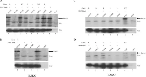
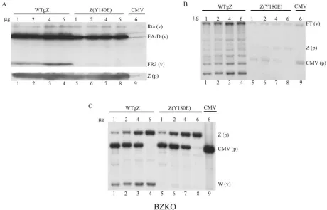
FIG. 1. Representative assays for DNA binding by ZEBRA basic domain point mutants. (A and C) Examples of results of EMSA using extracts of HKB5/B5 cells prepared 24 h after transfection with the individual point mutants. In each EMSA, the mutant was tested for binding to the ZIIIB site without (0) and with (⫹) the addition of 1l of monoclonal antibody BZ1 to ZEBRA. (B and D) Immunoblots of cell extracts used for EMSA probed with BZ1 MAb.
on November 8, 2019 by guest
http://jvi.asm.org/
kit (Q-Biogene). The excised DNA (4l) was labeled with [␣-32P]dCTP using 1
ng of random-primer oligonucleotides (Pharmacia) and 10 units of the Klenow fragment of DNA polymerase (New England Biolabs). The labeled DNA was separated from unincorporated radioactive nucleotides on a G-50 Sephadex spin
column. The probe was hybridized to the Zeta probe membrane at 65°C in 6⫻
SSC (1⫻SSC is 0.15 M NaCl plus 0.015 M sodium citrate), 0.1% Denhardt’s
solution, 0.5% SDS, 100g/ml salmon sperm DNA for at least 16 h. The
membrane was washed and exposed to film.
Quantitation of replication of viral DNA by real-time PCR.Real-time PCR
was performed in 20 mM Tris-HCl (pH 8.4), 50 mM KCl, and 2.5 mM MgCl2
with 2 ng of intracellular DNA as the template, 20 mM of each nucleotide, 50 ng
of each primer, and 1.25 units of PlatinumTaqpolymerase (Invitrogen) containing
SYBR green (0.25⫻; Molecular Probes) and 4 mM MgCl2. Quantitative real-time
PCR was measured with a Smart Cycler (Cepheid). DNA concentration was deter-mined using a standard curve generated from purified plasmid DNA containing Rp,
the promoter of the BRLF1 gene. The primers used were 5⬘-TTAGTTAATGCC
CCAGCCAGA and 5⬘-CTTTAAAAAGGCCGGCTGAC.
RESULTS
Experimental strategy.A series of 46 individual point mu-tations were installed into residues in the basic DNA
recogni-tion domain of ZEBRA. After they were expressed in mam-malian cells, the mutants were compared with wild-type ZEBRA for their capacity to bind DNA in EMSA (Fig. 1). For the purpose of screening for DNA binding activity, the highest-affinity ZRE, ZIIIB, was used as a probe. Specificity of binding was confirmed by supershift analysis with a BZ1 monoclonal antibody. Explanations for failure to supershift some mutants were sought by investigating the solubility of mutants (Fig. 2) and the epitope recognized by BZ1 (Fig. 3). The mutant ZEBRA proteins were analyzed for their ability to activate viral lytic early gene expression in Raji and BZKO cells (Fig. 4 to 6). The ability of Rta to rescue mutants that failed to activate early gene expression was investigated (Fig. 7). The mutants were also tested for their capacity to activate late protein expression (Fig. 6 and 8). Selected late mutants were studied for the capacity to activate lytic viral DNA replication (Fig. 9 and 10).
[image:5.585.51.527.69.424.2]Mutant ZEBRA proteins vary in DNA binding activity. Fig-ure 1 illustrates the results of two representative experiments
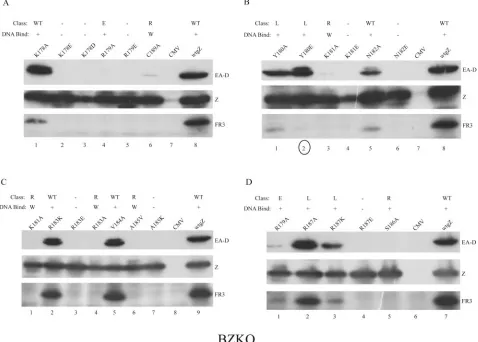
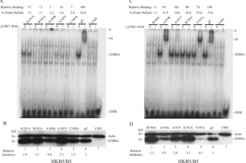
FIG. 2. Effect of ZEBRA protein concentration on supershifting with BZ1 MAb. (A) Comparison of the solubilities of wild-type ZEBRA (wtgZ) and two basic domain point mutants, the K192A protein (early mutant [E]) and the F193A protein (WT), in 0.3 M and 1.0 M NaCl. ZEBRA proteins were expressed in BZKO cells. Cell extracts were prepared 24 h after transfection and analyzed by immunoblotting with polyclonal antibody to ZEBRA. (B) Protein extracts of HKB5 cells transfected with a plasmid vector containing the CMV promoter were mixed in various ratios with protein extracts from cells transfected with CMV/gZ. The extracts were prepared in EMSA buffer containing 0.42 M NaCl 72 h after transfection. The proteins were assessed for their ability to shift a ZIIIB probe in an EMSA assay and to be supershifted by MAb BZ1 (⫹). SS, supershifted complex; NS, nonspecific. (C) Immunoblots of CMV- or gZ-transfected total cell extracts mixed in different proportions were probed with a 1:100 dilution of MAb BZ1.
VOL. 80, 2006 PHENOTYPES OF EBV ZEBRA BASIC DOMAIN MUTANTS 9119
on November 8, 2019 by guest
http://jvi.asm.org/
in which 10 different ZEBRA point mutants were examined for their ability to bind the high-affinity ZIIIB site in EMSA. Inspection of Fig. 1A and C shows that some mutants bound to the ZIIIB site strongly and that others bound weakly or not at all. Four mutants, those with a A191K, A191V, K192A, or F193A mutation, bound ZIIIB from 70% to 103% as actively as the wild type based on the percentage of probe shifted. These mutants are considered to be DNA binding competent (Table 1). The K181A mutant was moderately impaired in DNA binding and about 37% as active as the wild type. The R183A, A185K, A185V, C189A, and R190A mutants were markedly impaired in DNA binding (⬍10% of that of the wild type). In replicate experiments, the R183A, A185V, and C189A mutants showed weak, but detectable, DNA binding activity, whereas the A185K and R190A mutants did not re-producibly bind ZIIIB.
In Fig. 1B and D, the same cell extracts used for EMSA were examined for the levels of the mutant ZEBRA proteins by probing an immunoblot with BZ1 MAb. The amounts of ZEBRA protein present in the extracts varied. However, Fig. 1B and D show that mutant ZEBRA proteins, such as the R183A, A185K, A185V, C189A, and R190A proteins, which were markedly deficient in binding to ZIIIB, were present in the cell extracts used for EMSA at levels equivalent to or greater than that of the wild-type ZEBRA protein. Thus, the DNA binding defect was not due to the underexpression or the low solubility of mutant ZEBRA proteins.
Similar experiments screening for DNA binding to ZIIIB were conducted for all 46 individual point mutant proteins with mutations installed into residues of the basic DNA recognition domain of ZEBRA. When assayed by EMSA, 19 mutants did
not bind (⬍5% of wild-type binding), four mutants bound weakly (5% to 37% of wild-type binding), and 23 mutants were judged to be competent (67% to⬎100% of wild-type binding) to bind a ZIIIB site in vitro (Table 1).
Mutant ZEBRA proteins differ in susceptibility to super-shifting by monoclonal antibody BZ1. In the experiments whose results are illustrated in Fig. 1, a portion of each cell extract was treated with 1 l of BZ1 MAb before EMSA. Wild-type ZEBRA protein was readily recognized by the an-tibody, as evidenced by an antibody-induced supershift of the ZIIIB site. The DNA binding reactions of two mutants, with A191K and F193A mutations, were also supershifted by BZ1 MAb, whereas the ZEBRA/ZIIIB complex of three mutants, those with the K181A, A191V, and K192A mutations, was resistant to antibody supershifts.
The two mutant proteins, the A191K and F193A proteins, whose DNA binding reaction was supershifted by BZ1 MAb, were present at relatively low levels in the cell extracts, whereas cell extracts that contained relatively high levels of mutant ZEBRA proteins were resistant to supershifting by BZ1. This result suggested that supershifting by BZ1 MAb was sensitive to the amount of ZEBRA protein present in the cell extract. Moreover, the A191K and F193A mutants, which could be supershifted, were wild type in their capacity to activate the EBV lytic cycle, whereas two mutants that could not be super-shifted (the K181A and K192A proteins) were defective at activating the lytic cycle (Table 1).
Mutant ZEBRA proteins differ in their solubilities in NaCl.
[image:6.585.64.534.70.294.2]Cell extracts transfected with ZEBRA mutants were conven-tionally prepared in 0.42 M NaCl. The data of Fig. 1C and D showed that in such cell extracts, the abundance of the F193A
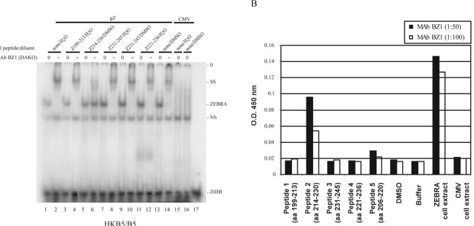
FIG. 3. Mapping the epitope of ZEBRA recognized by the BZ1 monoclonal antibody. (A) Mapping the BZ1 MAb epitope by peptide by inhibition of antibody-induced supershifting. Protein extracts of HKB5/B5 cells prepared 24 h after transfection with CMV/gZ (lanes 1 to 14) or CMV (lanes 15 to 16) were used to shift a ZIIIB probe in an EMSA in the absence (0) and presence (⫹) of BZ1 MAb. The reaction mixtures also contained diluent (water or DMSO) and various ZEBRA peptides or diluent alone (lanes 1 and 2 and 13 and 14). (B) Mapping the BZ1 epitope by ELISA. Individual peptides were coated on plates, and the reactivity of BZ1 MAb was determined by ELISA. An extract of HKB5 cells expressing ZEBRA served as the positive control, and cells transfected with the CMV vector served as the negative control. SS, supershifted complex; NS, nonspecific; O.D., optical density.
on November 8, 2019 by guest
http://jvi.asm.org/
protein, a mutant whose phenotype was similar to that of the wild type, was considerably lower than that of the K192A protein, a mutant that was defective in early gene activation. When total cell extracts prepared in SDS sample buffer were examined by immunoblotting or by immunoprecipitation with BZ1 MAb, the concentration of the K192A and F193A mu-tants was the same (data not shown). This result suggested that ZEBRA mutants differed in solubility in EMSA buffer. The solubilities of the wild-type ZEBRA protein and two mutants, the K192A and F193A proteins, were compared when trans-fected BZKO cells were extracted with EMSA buffer contain-ing 0.3 M or 1.0 M NaCl (Fig. 2A). Wild-type ZEBRA was relatively insoluble in 0.3 M NaCl; solubility was increased 10-fold in 1.0 M NaCl (Fig. 2A). The solubility profile of Z(F193A) was similar to that of the wild-type protein. The solubility of Z(K192A) was maximal in 0.3 M NaCl and did not increase in 1.0 M NaCl. A separate report will describe the solubility characteristics of a large panel of mutant ZEBRA proteins and the correlation of solubility with biologic pheno-type (A. El-Guindy, unpublished data).
The amount of ZEBRA proteins in cell extracts affects the capacity to be supershifted by BZ1 MAb.The foregoing data showed that mutant ZEBRA proteins differed in their solubil-ity in cell extracts; moreover, mutants with high solubilsolubil-ity (e.g., the K181A, A191V, and K192A proteins) were refractory to supershifts, whereas mutants of lower solubility (e.g., the A191K and F193A proteins) reacted with BZ1 MAb in the supershift assay. These results suggested that supershifts were very sensitive to the amount of ZEBRA protein present in the
cell extracts used for EMSA. To examine this hypothesis, ex-tracts from cells transfected with a wild-type BZLF1 gene were mixed in various proportions with extracts of cells transfected with a cytomegalovirus immediate early expression vector lack-ing an insert. These extracts were incubated with the ZIIIB probe in the absence and presence of BZ1 MAb (Fig. 2B). Twenty micrograms of cell extract programmed to express ZEBRA protein bound the ZIIIB duplex oligonucleotide strongly, but the DNA-protein complex could not be super-shifted. At lower ZEBRA concentrations, the bound complex was reduced in intensity by the antibody, but there was no clearly supershifted complex. However, extracts containing the smallest amounts of ZEBRA protein were supershifted effi-ciently. An experiment using serial dilutions of cell extracts showed that the BZ1 MAb could readily detect on an immu-noblot amounts of ZEBRA protein, present in 25g to 100g of cell extract, that could not be detected by EMSA supershift-ing (Fig. 2C). Thus, one factor responsible for the differences in the behaviors of mutants in ZEBRA’s basic domain in the EMSA supershift assay was the solubility of the mutant pro-tein, reflected in the amounts of ZEBRA protein present in the cell extract.
[image:7.585.49.537.72.330.2]Mapping the epitope recognized by the BZ1 MAb.Although variations in the solubility of mutant ZEBRA proteins could account for differences in recognition by the BZ1 MAb in EMSA supershift assays, another hypothesis was that muta-tions in the basic domain led to alteramuta-tions in the conformation of ZEBRA which masked the epitope recognized by the anti-body. Therefore, the epitope of BZ1 MAb was mapped by two
FIG. 4. Testing the capacity of ZEBRA basic domain mutants to activate the expression of the EBV Rta protein. DNA binding-competent (⫹) or -defective (⫺) mutants were transfected into BZKO cells. Cell extracts in SDS sample buffer prepared 48 h after transfection were examined by immunoblotting with polyclonal rabbit antisera directed against the Rta and ZEBRA proteins. WT, wild type; E, early; L; late; R, Rta; Rta (v), Rta expressed from the EBV endogenous to BZKO cells; Z (p), ZEBRA expressed from the transfected plasmid; X (c), cross-reactive cellular protein that was detected by the antibody to Rta.
VOL. 80, 2006 PHENOTYPES OF EBV ZEBRA BASIC DOMAIN MUTANTS 9121
on November 8, 2019 by guest
http://jvi.asm.org/
methods, peptide inhibition of supershifting (Fig. 3A) and ELISA (Fig. 3B). Published data based on immunoprecipita-tion of in vitro-translated subregions of ZEBRA protein indi-cated that the BZ1 epitope was loindi-cated in the C-terminal portion of ZEBRA, containing the dimerization domain (67). To confirm and refine this conclusion, a set of peptides, each 15 to 17 aa in length, encompassing aa 199 to 245 of ZEBRA were preincubated with BZ1 MAb1 before EMSA supershift assays were conducted. A single peptide, Z (aa 214 to 230), inhibited the supershift (Fig. 3A). The same peptide was reac-tive with BZ1 MAb in an ELISA assay (Fig. 3B). The only other peptide that reacted by ELISA (to a level slightly above the background) contained aa 206 to 220; this peptide over-lapped the peptide containing amino acids 214 to 230.
How-ever, peptide Z (aa 206 to 220) did not inhibit the supershift by BZ1 MAb (data not shown).
Mutants defective at activating Rta expression. Three dif-ferent classes of mutants were defective at activating the ex-pression of Rta. First, mutants that were unable to bind DNA did not activate Rta protein (Fig. 4). Second, mutants that bound weakly to DNA, such as the K181A, R183A, and C189A proteins, also weakly activated the expression of Rta protein and Rta mRNA (Fig. 5). Third, with one exception, namely, Z(S186T), proteins with mutations at position S186 were un-able to activate the expression of Rta protein (Fig. 4C and D) or BRLF1 mRNA (Fig. 5 and data not shown). Proteins with substitutions at position S186 that were competent to bind DNA and those that were impaired in binding DNA all failed to activate Rta. All three classes of mutants were equally ex-pressed, as judged by immunoblotting of total cell extracts (Fig. 4).
Mutants that fail to activate EA-D. Among the ZEBRA mutants that were competent to bind DNA were two distinct groups with a defect in activating the expression of early anti-gen (Fig. 6). The first group consisted of all mutants described above, such as those with the K181A, R183A, C189A, A185V, and S186 substitutions, that were impaired in the activation of expression of the BRLF1 mRNA or Rta protein (Fig. 4 and 5). The second group consisted of three mutants, the R179A, S186T, and K192A proteins, which were competent to activate BRLF1 mRNA (Fig. 5) and Rta protein (Fig. 4 and 7) but nonetheless failed to activate EA-D to wild-type levels.
We determined whether concomitant overexpression of EBV Rta protein could rescue the capacity of seven different ZEBRA mutants to activate the expression of early antigen (Fig. 7; Table 2). As reported previously from experiments con-ducted with Raji cells, Z(S186A) could be rescued by the cotrans-fection of Rta in BZKO cells (Fig. 7B) (2, 28). The ability of the mutants Z(K181A) and Z(K192A) to activate the expression of EA-D was also rescued by coexpression of Rta (Fig. 7B). How-ever, the R183A, C189A, and R179A mutants could not be rescued by Rta. Among the mutants that failed to be rescued were two, the R183A and C189A mutants, that were impaired in binding DNA and activated Rta only weakly and one, the R179A mutant, that was competent to activate BRLF1 mRNA (Fig. 5) and Rta protein (Fig. 7). Both the R179A and R183A mutants appeared to suppress the expression of cotransfected Rta (Fig. 7 B and D).
Point mutants that were defective at activating late gene expression.During the survey of mutants for their ability to activate EA-D (Fig. 6), the Y180E mutant was noted to acti-vate the expression of EA-D at wild-type levels but was mark-edly deficient in activation of the small viral capsid antigen (sVCA) protein encoded by the BFRF3 late gene (Fig. 6B, lane 2). The Y180A, R187K, and K188A mutants were also reproducibly weaker than wild-type ZEBRA in activating sVCA though equivalent to wild-type ZEBRA in the activation of Rta and EA-D (Fig. 6 and 8). For comparison, the K88R mutant, with a conservative mutation, activated both early and late proteins at levels comparable to those of the wild type (Fig. 8).
[image:8.585.49.285.75.443.2]Seven mutants with substitutions at positions Y180, R887, K188, F193, and K194 that were specifically defective at acti-vating late genes were encountered (Table 1). Of these, the
FIG. 5. Activation of BRLF1 and BMRF1 mRNA by ZEBRA and eight basic domain point mutants. BZKO cells were transfected with vectors expressing wild-type or mutant ZEBRA proteins. Total RNA was prepared 48 h after transfection. (A) A Northern blot was probed for viral BRLF1 mRNA, for BZLF1 mRNA expressed from the trans-fected plasmids, and for cellular RNase P to control for RNA loading. WT, wild type; E, early; L; late; R, Rta. (B) A Northern blot was probed for BMRF1 and RNase P. RNAs from HH514-16 (Cl16) cells treated with butyrate or left untreated were used as positive and negative controls.
on November 8, 2019 by guest
http://jvi.asm.org/
Y180E and K188A proteins demonstrated the most profound defect; the others could weakly induce BFRF3 protein or BLRF2 protein, the two components of the p21 late antigen complex (59; data not shown). Five of the seven late mutants were deficient at activation of both the BFRF3 and BLRF2 proteins (data not shown). However, R187A could strongly activate BFRF3 (Fig. 6D lane 2) but could only weakly activate BLFR2 (not shown). The R187K mutant weakly activated BFRF3 (Fig. 6D, lane 3) but failed to activate BLFR2 (not shown).
Three late mutants exhibit a specific defect in activating lytic EBV replication.In the experiment illustrated in Fig. 8, one aliquot of BZKO cells was examined for early and late viral protein expression and another aliquot from the same transfection was studied for the abundance of EBV DNA by quantitative PCR. Transfection of wild-type ZEBRA induced a 22-fold increase in viral DNA. The amount of EBV DNA measured after the transfection of three late mutants, the K188A, Y180E, and Y180A proteins, was 11% to 15% of wild-type levels.
The capacity of mutant Z(Y180E) to stimulate lytic viral DNA replication was compared with that of the wild type in a
dose-response experiment (Fig. 9). One to 6g of the wild-type BZLF1 or Z(Y180E) expression vector was transfected into BZKO cells. The Z(Y180E) mutant activated levels of Rta protein comparable to those of the wild type and levels of EA-D protein nearly equivalent to those of the wild type (Fig. 9A) but, unlike wild-type protein, did not activate detectable BFRF3 protein expression. Following transfection of 1 to 6g of wild-type BZLF1, there was stimulation of lytic viral DNA replication, as is evident from the appearance of a ladder of restriction fragments containing various numbers of EBV ge-nome terminal repeats (Fig. 9B). Plasmid expressing wild-type BZLF1 also induced a notable amplification of viral DNA, as indicated by the appearance of a strong signal on a Southern blot probed with EBV BamHI W, the large internal repeat (Fig. 9C). Parallel experiments carried out with the Z(Y180E) plasmid failed to detect lytic viral DNA replication or ampli-fication of the viral genome (Fig. 9B and C, lanes 5 to 8). The Southern blot illustrated in Fig. 9C demonstrated that equiv-alent amounts of plasmid containing the Z(Y180E) mutant or wild-type BZLF1 were transfected.
To determine whether mutants were delayed in the activa-tion of EBV DNA replicaactiva-tion, wild-type ZEBRA and the
mu-FIG. 6. Activation of early and late EBV proteins by ZEBRA and mutants with point mutations in the basic domain. BZKO cells were transfected with vectors expressing the wild-type or mutant ZEBRA proteins. Cell extracts were prepared 48 h after transfection. Immunoblots were sequentially probed with antibodies to EA-D, ZEBRA (Z), or BFRF3 (FR3). Each panel represents the same immunoblot. WT, wild type; E, early; L; late; R, Rta; W, weak. Lane 2 in panel B is circled to indicate a mutant, Y180E, that activates EA-D but not FR3.
VOL. 80, 2006 PHENOTYPES OF EBV ZEBRA BASIC DOMAIN MUTANTS 9123
on November 8, 2019 by guest
http://jvi.asm.org/
[image:9.585.55.533.72.414.2]tant Z(K188A) were compared at 24-h intervals from 48 to 96 h following transfection of 6g of DNA. Wild-type ZEBRA induced the expression of slightly more BFRF3 sVCA at each 24-h interval between 48 and 96 h after the transfection of
BZKO cells. A near-maximal level of lytic EBV replication was observed 72 h after the transfection of wild-type BZLF1. Even at 96 h, the latest time after transfection, Z(K188A) did not detectably induce sVCA (Fig. 10A). A faint signal of lytic viral DNA replication was observed 96 h after the transfection of the Z(K188A) mutant; this signal was less intense than that observed 48 h after the transfection of wild-type ZEBRA (Fig. 10B). Similarly, EBV genome amplification, as detectable with the BamHI W probe, was seen to increase between 48 and 96 h after the transfection of wild-type ZEBRA (Fig. 10C); how-ever, only a slight amplification of EBV BamHI W was seen 96 h after the transfection of the Z(K188A) mutant. Figure 10C demonstrates that the cells received slightly more of the mutant than the wild-type expression vector.
Thus, whether in terms of dose-response results (Fig. 9) or the time course (Fig. 10), the late mutants Z(Y180E) and Z(K188A) were markedly deficient at activating lytic viral DNA replication.
[image:10.585.51.532.66.364.2]Summary of phenotypes of point mutants that altered the charge of residues in the basic domain of ZEBRA protein.The basic DNA recognition domain of ZEBRA consists of 17 amino acids (Fig. 11A). Nine of them are basic, and none of them are acidic. A single acidic substitution at any one of the 9 basic amino acids destroyed DNA binding and the capacity of the mutant ZEBRA protein to activate lytic gene expression. Con-servative substitutions in basic residues in three mutants, the R183K, R187K, and K188R proteins, did not affect DNA binding.
FIG. 7. Effect of the coexpression of EBV Rta on the activation of early antigen by mutants with point mutations in the basic domain of ZEBRA. BZKO cells were transfected with vectors expressing the wild-type (gZ) or mutant ZEBRA proteins. In one set of cells, the empty vector, pRts, was cotransfected; in the other set of cells, the pRts vector expressing Rta (pRta) was cotransfected. Immunoblots of cell extracts prepared 48 h after transfection were probed for Rta, EA-D, and ZEBRA (Z) with monospecific antibodies. X, a cross-reactive protein detected by the antibody to Rta that controls for protein loading; E, early mutant; R, Rta.
FIG. 8. Basic domain point mutants that specifically fail to activate late protein expression. BZKO cells were transfected with vectors expressing the wild-type (wtgZ) or mutant ZEBRA proteins. SDS cell extracts and total cellular DNA were prepared 48 h after transfection. Immunoblots were probed for Rta, EA-D, and FR3 encoded by the EB virus (v) endogenous to BZKO cells and for ZEBRA expressed by the transfected plasmid [Z (p)]. The relative amounts of EBV DNA were determined by quantitative PCR.
on November 8, 2019 by guest
http://jvi.asm.org/
[image:10.585.43.285.494.651.2]Two of these conservative basic-amino-acid-substitution mutants were wild type, but the R187K protein was a late mutant.
Amino acid substitutions that altered the charge were in-stalled at six nonbasic amino acids, Y180, N182, A185, S186, A191, and F193. Both acidic and basic substitutions at posi-tions N182 and S186 destroyed the capacity to bind DNA. A basic substitution in the A185K mutant also destroyed DNA binding. In the Y180E and F193E mutants, an acidic substitu-tion did not impair DNA binding or the activasubstitu-tion of early gene expression, but both mutants were defective in late gene expression. A basic substitution was tolerated in the A191K mutant, which behaved like the wild type. Eleven of 19 mutants that lost the capacity to bind DNA (Table 1) did so as the result of substitution of a negatively charged amino acid for a basic one. Six nonbinding mutants resulted from acidic or basic substitutions at position S186 or N182. The other two nonbind-ing mutants were the A185K and R190A mutants.
Phenotypes of alanine or valine substitutions.Fifteen of the 17 residues in the DNA recognition domain of ZEBRA were changed to alanine. The two remaining residues in this region, which are already alanine, were mutated to valine. The effects of these alterations were markedly different from the effects of introducing a charged substitution (compare Fig. 11A with B). At 12 of 17 positions, DNA binding activity was maintained at wild-type levels in the alanine substitution mutants. Alanine
substitutions were tolerated in DNA binding at six of nine basic amino acids and six of eight nonbasic amino acids. However, at five positions, an alanine or valine substitution weakened or eliminated binding to a ZIIIB site. The R190A mutant lost DNA binding and three mutants, the R183A, A185V, and C189A proteins, were markedly impaired in binding DNA; all four mutants were defective at activating downstream lytic cycle genes. The K181A protein, which was moderately im-paired in binding ZIIIB, failed to activate Rta. Of the 12 alanine or valine substitution mutants with intact competence to bind DNA, 5 were wild type, 1 was defective at activating Rta, 2 were early mutants, and 4 were late mutants.
DISCUSSION
DNA binding by ZEBRA. Considerable new information about the interaction of ZEBRA with DNA was revealed by making a large number of mutations that either altered the charge of amino acids or substituted alanine or valine in the basic domain. Changing any single basic amino acid to an acidic residue, from arginine or lysine to either glutamate or aspartate, abolished DNA binding (Fig. 11A). Therefore, each of the individual basic residues seems to be required to contact DNA possibly via an electrostatic interaction with the nega-tively charged phosphate backbone. Seven of nine basic
resi-FIG. 9. The Z(Y180E) late mutant is defective at activating EBV lytic-DNA replication. From 1 to 6g of vectors expressing either wild-type ZEBRA (WTgZ) or the Z(Y180E) mutant were transfected into BZKO cells. Cells were transfected with an empty vector and the BZLF expression plasmid in different ratios. Cellular protein and DNA extracts were prepared 96 h after transfection. (A) Immunoblots of cell protein extracts were probed for Rta, EA-D, and FR3. (B) A Southern blot of BamHI-digested total cellular DNA was probed for the replication of lytic viral DNA with a subfragment of Xho (52). The probe detects the ladder indicative of lytic-DNA replication, the fused terminal (FT) fragment of the virus (v), transfected BZLF1 expression plasmid [Z (p)], and CMV plasmid (p). (C) A Southern blot of a BamHI digest of total cellular DNA was probed with EBV BamHI W.
VOL. 80, 2006 PHENOTYPES OF EBV ZEBRA BASIC DOMAIN MUTANTS 9125
on November 8, 2019 by guest
http://jvi.asm.org/
[image:11.585.61.521.68.363.2]dues are likely to make essential, but nonspecific, contacts with DNA since at these positions, substitution of alanine did not destroy DNA binding (Fig. 11B). At two positions, N182 and S186, neither basic nor acidic substitutions were tolerated. This result suggests that both N182 and S186 may play some specific role in binding to DNA or may alter the DNA binding behavior of other amino acids. However, alanine substitution at these two positions maintained the protein’s ability to bind a ZIIIB site.
Alanine substitutions at three positions, R183, C189, and R190, markedly reduced or abolished the ability of ZEBRA to bind ZIIIB DNA, as did an alanine-to-valine substitution at position A185 (Fig. 1B and 2A). We hypothesize that these four residues of ZEBRA are likely to be required to make specific contacts with DNA bases. When the amino acids of c-Jun and c-Fos that are known to make specific contacts with DNA in the crystal structure of a heterodimer of c-Jun and c-Fos bound to an AP-1 site are compared with the four amino acids of ZEBRA proteins in which an alanine or valine sub-stitution impairs DNA binding, it can be seen that ZEBRA A185, C189, and R190 are colinear with the amino acids of c-Fos/c-Jun that contact DNA. R183 of ZEBRA is not colinear with any amino acid of c-Fos/c-Jun that was found to contact DNA. However, the amino acid colinear with Z(R183) is also an arginine or lysine in c-Jun/c-Fos.
Our conclusions about the importance of residue C189 in
con-tacting DNA is supported by a recent report showing that the mutant Z(C189S) is impaired in binding the ZIIIB site and is unable to activate the reporter ZIIIB/CAT (9, 56). However, another recent study finds that the Z(C189S) mutant is not sig-nificantly altered for DNA binding activity in vitro or in vivo (65). It is likely that individual residues in the basic domain of ZEBRA play specific roles in contacting individual ZREs. For example, the mutant Z(S186A) is relatively deficient in binding to ZRE-R and ZIIIA, two ZREs in Rp, even though the same mutant is unimpaired in binding to ZIIIB and enhanced in binding to an AP-1 site (28). Testing this prediction will re-quire examination of the crystal structures of individual mutant ZEBRA proteins binding to an array of ZEBRA response elements.
Implications of the recently solved crystal structure of ZEBRA bound to an AP-1 site for our observations of the DNA binding phenotypes of basic domain mutations.The structure of ZEBRA amino acids 175 to 236 bound to 19 base pairs of DNA containing an AP-1 site and flanking sequences from the promoter of the BMLF1 gene was recently published while our manuscript was in the final stages of preparation (50). The truncated ZEBRA protein used to determine this structure contained two mutations, S186A and C189S. An essential fea-ture of the strucfea-ture is that two long alpha helix monomers, one from R179 to M221 and the other from L175 to M221, dimerize and contact both strands of DNA. Two main features
FIG. 10. Time course of activation of EBV lytic replication by wild-type ZEBRA (WTgZ) and the Z(K188A) mutant. (A) Cell extracts prepared 48, 72, and 96 h after transfection with 6g of plasmid DNA were probed for Rta, EA-D, and FR3 expressed from the virus (v) and ZEBRA from the transfected plasmids [Z (p)] by immunoblotting. (B) A Southern blot of a BamHI digest of cellular DNA harvested 48, 72, and 96 h after transfection was probed with a subfragment of Xho 1.9 or EBV BamHI W (C).
on November 8, 2019 by guest
http://jvi.asm.org/
of the contacts of amino acids in the basic domain of ZEBRA with DNA were evident (Fig. 12A): eight of the nine arginines and lysines in the basic domain contacted the phosphate back-bone of the DNA. The ninth basic amino acid, K178, was not shown to contact DNA. Four amino acids, N182, A185, S186A, and R190, contacted bases. The phosphate backbone was con-tacted at seven consecutive positions, from⫹2/⫹1 to⫺5/⫺6 relative to C or G at the center of the AP-1 site, TGACTCA. Thus, ZEBRA forms contacts not only with the AP-1 site itself but with DNA flanking the specific binding site. N182 from each monomer contacted T at position⫺3 on one strand and C at position⫹2 on the other strand. S186A of each monomer
made contacts with T at position ⫹1 of each strand. R190 contacted C or G in the center of the AP-1 site, position 0. A185 contacted both the phosphate backbone and T at posi-tion⫺3.
This structure validates a number of our functional obser-vations about the phenotypes of mutations in the basic domain of ZEBRA. Our conclusions about the importance of contacts of arginines and lysines with the negatively charged phosphate back-bone of the ZIIIB site was evident from the deleterious effects of changing any one of the basic amino acids to a negatively charged one (Fig. 11A). Since the mutations A185V and R190A markedly weakened or destroyed binding to DNA (Fig. 11B), we postu-lated, in agreement with the structure, that these two amino acids contacted bases in the DNA (Fig. 12B). It is notable that A185 and R190, residues that we deem essential, contact DNA bases at positions that are invariant in the ZRE consensus, T at⫺3 and C/G at position 0, respectively.
Our experiments allow the formulation of additional hypoth-eses about the interactions of ZEBRA with the phosphate backbone that were not evident from the structure. The con-tacts of six of nine basic amino acids with the phosphate back-bone are either redundant or specific for the AP-1 site. We found that single alanine substitutions at positions K178, R179, R187, K188, K192, and K194 were tolerated in binding to ZIIIB DNA. However, since the structure shows a series of seven consecutive backbone contacts, it is likely that multiple alanine substitutions in basic amino acids impair DNA binding. We find that the K178D and K178E mutants are unable to bind DNA. Residue K178 would not contact the AP-1 site or any other ZRE itself but may be important in interacting with flanking DNA. R183 in the structure makes contacts with the phosphate backbone between DNA residues 0 and⫹1. These phosphate contacts may be essential for all ZREs since the R183A mutation nearly eliminates binding to ZIIIB, the high-est-affinity site (Fig. 1A).
Our data also indicate similarities and differences from the crystal structure in the putative contacts of individual amino acids of ZEBRA with DNA bases. The role of residues C189 and S186 cannot be definitively determined from the structure since substitution mutants at these positions were used to crys-tallize the protein. The importance of C189 is evident from our results showing that C189A markedly decreased binding to ZIIIB (Fig. 1A). Contacts of S186 with DNA are obviously dependent on the individual ZRE site and its methylation status (7, 8, 28). The contacts of S186A with T at position⫹1 of the AP-1 site in the structure is consistent with earlier observations that this mutant is enhanced in binding to an AP-1 site (29). The S186A and C189S mutations might also change the conformation of the protein. Such conformational alterations might affect not only contacts of the mutated resi-dues with DNA but also the contacts of other resiresi-dues in the basic domain (18).
Our results do not confirm the essential nature of N182. We find that the N182A mutant binds ZIIIB normally (not shown) and is capable of activating early and late lytic gene expression. Yet in the crystal structure with the AP-1 site, N182 was found to contact two bases on each strand of the DNA. This result suggests that the amino acids of ZEBRA that contact DNA will differ, depending on the DNA site studied. The contacts of N182 with the AP-1 site may not be crucial in disrupting
la-TABLE 1. Summary of phenotypes of DNA binding-competent point mutants with mutations in the basic DNA
recognition domain of ZEBRAa
DNA binding phenotype
Amino
acid Mutation
Expression of indicted protein in
BZKO cells Class
Rta EA-D FR3 LR2
Competent K178 A Yes Yes Yes Yes WT
R179 A Yes Weak Weak Weak E
Y180 A Yes Yes Weak Weak L
Y180 E Yes Yes No No L
N182 A Yes Yes Yes Yes WT
R183 K Yes Yes Yes Yes WT
V184 A Yes Yes Yes Yes WT
S186 C No No No No R
S186 G No No No No R
S186 V No No No No R
S186 Y No No No No R
S186 A No No No No R
S186 T Yes Weak Weak Weak E
R187 A Yes Yes Yes Weak L
R187 K Yes Yes Weak Weak L
K188 A Yes Yes No No L
K188 R Yes Yes Yes Yes WT
A191 V Yes Yes Yes Yes WT
A191 K Yes Yes Yes Yes WT
K192 A Yes Weak No No E
F193 E Yes Yes Weak Weak L
F193 A Yes Yes Yes Yes WT
K194 A Yes Yes Weak Weak L
K181 A Weak Weak No No R
R183 A Weak No No No R
A185 V No No No No R
C189 A Weak Weak No No R
Defective K178 D No No No No
K178 E No No No No
R179 E No No No No
K181 E No No No No
N182 E No No No No
N182 K No No No No
R183 E No No No No
A185 K No No No No
S186 D No No No NT
S186 E No No No NT
S186 K No No No NT
S186 R No No No NT
R187 E No No No NT
K188 E No No No NT
R190 A No No No No
R190 E No No No No
K192 E No No No NT
K194 D No No No NT
K194 E No No No NT
a
Forty-six mutants were classified as DNA binding competent or DNA bind-ing defective based on their capacity to bind a ZIIIB site in an electrophoretic mobility shift assay (Fig. 1). Four mutants with weak DNA binding activity (the K181A, R183A, A185V, and C189A mutants) are also listed. The ability of ZEBRA mutants to activate the expression of Rta, EA-D, BFRF3 (FR3), and BRLF2 (LR2) in BZKO cells was determined by immunoblotting with mono-specific antibodies. The DNA binding-competent mutants are classified as wild type (WT), defective in activating Rta (R), defective in EA-D activity (E), or defective in activating late protein (L).
VOL. 80, 2006 PHENOTYPES OF EBV ZEBRA BASIC DOMAIN MUTANTS 9127
on November 8, 2019 by guest
http://jvi.asm.org/
[image:13.585.43.282.99.499.2]tency since ZEBRA/c-Fos chimeras, which bound strongly to both heptamer and octamer AP-1-like sites, did not activate the lytic cycle (36).
Figure 12 compares the contacts between ZEBRA amino acids in the basic domain evident from the crystal sructure and those predicted from our mutagenesis study. There are many consistencies. Differences are likely to result from the DNA
sequences used for the crystal and those used for DNA binding studies with the mutants.
[image:14.585.43.543.81.187.2]Variations in supershifts by monoclonal antibody and the solubility of ZEBRA mutants. We studied the interactions between ZEBRA and DNA using extracts of mammalian cells rather than purified ZEBRA protein, because it was likely that in mammalian cells the protein would be properly folded and
FIG. 11. Summary of phenotypes of point mutants arranged according to the linear amino acid sequence of the DNA binding domain of ZEBRA. Basic amino acids in the DNA binding domain are indicated in bold. (A) Effect of substitutions that alter the charge of the amino acid side chain. (B) Effects of alanine substitution mutants or alanine-to-valine changes. DNA binding was determined by EMSA with a ZIIIB site. Activation of expression of EBV lytic-cycle proteins was determined by immunoblotting of extracts of transfected BZKO cells with specific antibodies (Table 1).⫹, positive;⫺, negative; W, weak. Data are given for 38 mutants. Eight mutants that are listed in Table 1 but not included in this figure include five with substitutions at S186 and three with conservative changes, R183K, R187K, and K188R.
TABLE 2. Examples of ZEBRA mutants that are defective in activating the of expression of EA-Da
Mutation Binding to
ZIIIB
% of WT binding
Activation of:
EA-D rescue
by Rta Possible defect
BRLF1
mRNA Rta
BMRF1
mRNA EA-D
R179A ⫹ 102 ⫹ ⫹ W W ⫺ Synergy with Rta
S186A ⫹ 93.8 ⫺ ⫺ ⫺ ⫺ ⫹ Activation of Rta
K192A ⫹ 88.7 W ⫹/⫺ ⫹ W ⫹ Weak activation of Rta
S186T ⫹ 97.3 ⫹ ⫹ ⫹⫹ W ⫺ Translation or modification of EA-D
C189A W 4.7 W W ⫹ W ⫺ Weak DNA binding
K181A W 37.6 W W W ⫺ ⫹ Weak DNA binding
R183A W 10.8 W W ⫺ ⫺ ⫺ Weak DNA binding
aMutants were tested for binding in vitro to ZIIIB and activating BRLF1 mRNA, Rta protein, BMRF1 mRNA, and EA-D protein in BZKO cells. The capacity of
coexpression of Rta to rescue a mutant’s ability to activate EA-D was also examined in BZKO cells.⫹, positive;⫺, negative; W, weak; WT, wild type. DNA binding
relative to that of the wild type was based on the percentage of probe shifted.
on November 8, 2019 by guest
http://jvi.asm.org/
[image:14.585.105.478.369.667.2]modified. Therefore, it was essential that the identity of the DNA-protein complexes be confirmed by use of specific anti-bodies to ZEBRA. A surprising result (Fig. 1A and C) was that some ZEBRA mutants formed a complex with DNA that could not be supershifted by the BZ1 monoclonal antibody. We initially hypothesized that these mutants had undergone a conformational change that masked the epitope recognized by
[image:15.585.98.487.99.529.2]the BZ1 MAb. Therefore, it became imperative to map this epitope precisely. In previous work, the epitope had been mapped to the carboxy end of the protein downstream of aa 197 (67). Using two different techniques, peptide inhibition of supershifting and ELISA, the epitope recognized by the BZ1 MAb to ZEBRA was mapped to amino acids 214 to 230, a region distant from the sites of the point mutations (aa 178 to
FIG. 12. Comparison of the ZEBRA/AP-1 crystal structure with ZEBRA proteins in the current study with mutations that affect binding to ZIIIB and the biologic phenotype. (A) AP-1 site from the BMLF1 promoter in the crystal. Invariant nucleotides in the ZRE are shown in bold. The numbering system for the ZRE is indicated. Amino acids in the basic domain are shown. Basic amino acids are bold. *, S186A and C189S were used in the crystallized ZEBRA protein. The ZEBRA amino acids that contact the phosphate backbone (⫹) and the positions are indicated. W, water. The amino acids that contact bases are shown. (B) ZIIIB site from the BZLF1 promoter. Amino acids are predicted to contact the phosphate backbone if an acidic substitution destroys binding to ZIIIB. Base contacts are predicted from a loss of or decrease in DNA binding resulting from alanine or valine substitutions. Boxes represent concordance between the ZEBRA crystal structure and mutagenesis. Biologic phenotypes of alanine or valine substitutions mutants are represented as follows: WT, wild type; E, early; L; late; R, Rta; and DB, DNA binding defective. ND, not determined.
VOL. 80, 2006 PHENOTYPES OF EBV ZEBRA BASIC DOMAIN MUTANTS 9129
![FIG. 2. Effect of ZEBRA protein concentration on supershifting with BZ1 MAb. (A) Comparison of the solubilities of wild-type ZEBRA(wtgZ) and two basic domain point mutants, the K192A protein (early mutant [E]) and the F193A protein (WT), in 0.3 M and 1.0 M](https://thumb-us.123doks.com/thumbv2/123dok_us/168906.45791/5.585.51.527.69.424/protein-concentration-supershifting-comparison-solubilities-mutants-protein-protein.webp)