A HUMAN CADAVERIC STUDY ON THE FORMATION AND
BRANCHING PATTERN OF LUMBAR PLEXUS.
DISSERTATION SUBMITTED FOR
BRANCH – XXIII - M.D. DEGREE
(ANATOMY)
MAY 2018
THE TAMILNADU
DR.M.G.R.MEDICAL UNIVERSITY
BONAFIDE CERTIFICATE
This is to certify that the dissertation entitled “A HUMAN CADAVERIC
STUDY ON THE FORMATION AND BRANCHING PATTERN OF LUMBAR
PLEXUS” submitted by Dr.K.PUSHPA, to the Tamil Nadu Dr. M.G.R. Medical
University, Chennai in partial fulfillment of the requirement for the award of M.D
degree Branch– XXIII (ANATOMY) is a bonafide research work carried out by her
under direct supervision & guidance.
Prof.DR.S.MARUTHUPANDIAN,M.S., Dr.VINO VICTOR JESUDAS,M.S ., F.I.C.S.,F.A.I.S.,
DEAN DIRECTOR AND PROFESSOR,
Madurai Medical College & Institute of Anatomy,
Govt.Rajaji Hospital, Madurai Medical College,
CERTIFICATE FROM THE GUIDE
This is to certify that the dissertation “A HUMAN CADAVERIC STUDY ON
THE FORMATION AND BRANCHING PATTERN OF LUMBAR PLEXUS” is
a bonafide record of work done by Dr.K.PUSHPA, under my guidance and
supervision in the Institute of Anatomy, Madurai Medical College, Madurai during the
period of her Post graduate study of M.D. ANATOMY from 2015 – 2018.
Dr.VINO VICTOR JESUDAS.M.S.,
DIRECTOR AND PROFESSOR,
DECLARATION
I, DR.K.PUSHPA declare that, I carried out this work on, “A HUMAN
CADAVERIC STUDY ON THE FORMATION AND BRANCHING PATTERN OF LUMBAR PLEXUS” at the Institute of Anatomy, Madurai Medical College. I also
declare that this bonafide work or a part of this work was not submitted by me or any
others for any award, degree or diploma to any other University, Board, either in India
or abroad.
This is submitted to The Tamilnadu Dr. M. G. R. Medical University, Chennai
in partial fulfillment of the rules and regulations for the M.D. Degree examination in
Anatomy.
Place : MADURAI Dr.K.PUSHPA.
ACKNOWLEDGEMENT
I am grateful to the Dean, Prof.DR.S.MARUTHUPANDIAN,M.S.,F.I.C.S
.,
F.A.I.S.,
Madurai Medical College and Government Rajaji Hospital, Madurai forpermitting me to carry out this study.
I would like to express my deep sense of gratitude and sincere thanks to our
Director Dr.VINO VICTOR JESUDAS M.S., Institute of Anatomy, Madurai
Medical College, for his constant help, guidance and encouragement given to me
throughout this study.
I would like to express my sincere thanks to my guide Prof.Dr.VINO VICTOR
JESUDAS M.S, co-guide Dr.P.G.ANANTHI., for their valuable suggestions and
guidance given to me.
I would like to express my sincere thanks to all my Associate and Assistant
professors for their valuable suggestions and guidance given to me.
I extend my thanks to all staff members, Institute of Anatomy for giving full
cooperation and timely help in carrying out the study.
I extend my thanks to my parents, my family membersfor their esteemed moral
support rendered during the study.
CONTENTS
S.No Title Page. No
1 INTRODUCTION
1
2 AIMS &OBJECTIVES
4
3 REVIEW OF LITERATURE
5
4 MATERIALS AND METHODS
24 5 OBSERVATIONS 37 6 DISCUSSION 51 7 SUMMARY 77 8 CONCLUSION 79 9 BIBILIOGRAPHY
10 ANNEXURE
Master Chart
abbreviations
Ethical Committee Approval Form
1
INTRODUCTION
The lumbar plexus, which is one of the main nervous pathway supplying
the lower extremity is located retroperitoneally within the posterior part of
psoas major muscle. It is formed by the union of ventral rami of upper three
lumbar nerves and a greater part of fourth lumbar nerve along with a
contribution from the twelfth thoracic spinal nerve. Smaller portion of fourth
lumbar nerve joins with fifth lumbar nerve to form lumbosacral trunk22. The
pattern of formation of lumbar plexus is altered, if the plexus is prefixed or post
fixed, that is the fibre contribution is moved cranially or caudally
respectively13.
The mode in which the plexus is arranged varies in different subjects.
It differs from brachial plexus in not forming an interlacement, but several
nerves of distribution arise from one or more of spinal nerves. The first lumbar
nerve, frequently supplemented from a twig from twelfth thoracic nerve splits
into an upper and lower branch, the upper and larger branch divides into
iliohypogastric and ilioinguinal nerve. The lower and smaller branch unites
with a branch of second lumbar to form genitofemoral nerve. The second, third
and fourth nerves divide into dorsal and ventral divisions. The ventral division
of second, third and fourth unites to form obturator nerve and the dorsal
division unite to form femoral nerve. A branch from dorsal division of second
and third nerve join to form lateral femoral cutaneous nerve. The accessory
obturator nerve if present ,is formed by union of ventral division of third and
2
The branches of lumbar plexus may be injured during certain surgical
procedures in the lower abdominal region (inguinal hernia repair, iliac crest
bone grafting, gynaecological procedures through transverse incision).After
such surgery several clinical conditions such as meralgia paresthesia,groin
pain, testicular pain in which lateral femoral cutaneous nerve,ilioinguinal
nerve, and genitofemoral nerve are mostly involved13.
Recently, retroperitoneal endoscopic surgery has been applied to
various spinal disorders. When the psoas major muscle is separated during
retroperitoneal endoscopic surgery, there is potential risk of injury to lumbar
plexus or nerve roots. Due to fast growing technology, awareness of consumer
protection movement and medicolegal issues, it is essential for the medical
personnels, particularly general Surgeons, Traumatologist, and Neurosurgeons
to have a thorough knowledge in normal anatomy and the possible variations,
which will help them to device appropriate treatment plan and to avoid huge
compensation.
The study on formation and branching pattern of lumbar plexus will
help to achieve the above principles in the clinical conditions like
In retroperitoneal endoscopic surgeries, injury to lumbar plexus can be avoided.
1. To diminish the possibility of post operative complication in surgery
like laproscopic herniorraphy.
2. While doing routine herniorraphy, injury to ilioinguinal and genital
branch of genito femoral nerve during skin incision is definitely
3
3. To prevent injury to lateral femoral cutaneous nerve (LFCN) which
causes meralgia paresthesia in case of iliac crest bone graft.
4. Knowledge of dermatomal distribution by nerves of lumbar plexus
helps in diagnosis of symptoms of disc prolapse and lumbar
spondylosis.
5. Femoral nerve injury can be avoided during psoas abscess drainage,
hip joint dislocation and during surgery on pelvic fractures due to road
traffic accidents.
6. Obturator nerve palsy resulting from intra pelvic extrusion of cement
during total hip replacement can be avoided.
7. Knowledge of anatomy of lumbar plexus and its variations is essential
during comprehensive electromyographic evaluation during
radiculopathy and lumbosacral plexopathy.
8. For various approaches to lumbar plexus block –such as psoas
compartmental block, femoral nerve block, and obturator nerve block.
Hence this extensive clinically oriented topic has created much interest
in me to dissect and analyse the complicated lumbar plexus.
4
AIM AND OBJECTIVE
1. To study the formation of lumbar plexus.
2. To observe its branching pattern and its relation with psoas major
muscle.
3. To document any variations in the formation and branching
5
REVIEW OF LITERATURE
Many studies on anatomy of lumbar plexus had been conducted by
many Surgeons, Anatomist, and Anaesthetist. The lumbar plexus has been
dissected and analysed thoroughly for more than hundred years.
At the earliest, first detailed description of the normal anatomy of lumbar
plexus was provided by Cruveilher (1844) – a French Anatomist and
pathologist who had special interest in nervous system.
FORMATION OF LUMBAR PLEXUS:
EISLER19 (1891) described that in normal condition, the lumbar plexus
is formed by whole of first three lumbar nerves and a part of fourth lumbar
nerve. While the fifth lumbar nerve contribute entirely to the formation of
sacral plexus. The fourth lumbar nerve has been described by V.JHERING as
the nervus furcalis as he found that it is divided between the lumbar and sacral
plexus.
C.S.SHERRINGTON (1893) introduced the term prefixed and
postfixed, when the plexus originated rostrally or caudally respectively than
that of the usual pattern.
A.M.PATERSON41 (1893) described that the nerve divides into ventral
and dorsal groups and documented the normal pattern of formation of lumbar
6
BARDEEN and ELTING8 (1901) used the term proximal and distal in
the same way that Sherrington used prefixed and postfixed . They concluded
that sex, race and laterality has no relation with variation in the formation and
branching pattern of lumbar plexus.
SEVEREANO (1904) studied lumbar plexus in 50 cadavers on both
sides and described the formation of lumbar plexus into ordinary (usual
pattern) –formation normalis, prefixed –formation superieur , and postfixed
–formation inferieur.
J.SYMINGTON and E.A.SCHAFER43 (1909) - Quains element of
Anatomy described that the lumbar plexus is formed by ventral primary
division of upper four lumbar nerves and it is placed inside the substance of
psoas major muscle. It lies in front of the transverse process of corresponding
lumbar vertebrae. The plexus is narrow above and is usually connected with
last thoracic nerve by a small twig. The plexus is wider below and is formed by
the sacral plexus by means of a branch passing from fourth lumbar nerve (L4)
to fifth lumbar nerve (L5).
FARNY J, DROLET P, GIRARD M,21 (1994) in Can J Anaesthesia 41
(12) : 1238-9 Anatomy of posterior approach to lumbar plexus nerve says that
four cadavers were dissected and 22 computed tomography files of the
lumbosacral region were studied. Cadaver dissection demonstrated that the
lumbar plexus at the level of L5, is within the substance of psoas major muscle
7
YUSUF IZCL58 et al (2005) studied lumbar plexus in 10 human
anatomical specimen. He dissected lumbar plexus bilaterally and exposed it in
all specimen. He studied the size and course of lumbar nerves in posterior
abdominal wall with reference to bony landmarks. He concluded that the
thinnest nerve is L1 (4.1 mm) and the thickest root is L4(5.5mm). The longest
is iliohypogastric nerve (210mm) and ilioinguinal nerve is the thinnest (1.2mm)
of lumbar plexus.
DENIZ UZMANSEL53 et al ( 2008) found the presence of multiple
variation in the branching pattern of lumbar plexus in the right side of a 35 year
old female cadaver. The variations were, presence of double ilioinguinal nerve,
an accessory branch joined with genital branch of genitofemoral nerve, and
also an accessory lateral femoral cutaneous nerve(LFCN) arising from femoral
nerve.
KUSUM R GANDHI22 et al (2013) conducted a study on 30 formalin
fixed cadavers in PIMS Maharashtra. He described prefixation of lumbar
plexus in one cadaver bilaterally. Measurements were made from nearby bony
points to the point of emergence of branches of lumbar plexus. These accurate
measurements will be essential for the surgeons to prevent nerve injury during
surgical procedures.
DEEPTI ARORA13 et al (2016) described in a study on 30 formalin
fixed cadavers and observed an unusual pattern of lumbar plexus on the left
8
twelfth thoracic nerve. The ventral rami of T12 to L3 formed three loops.
Genitofemoral nerve arose from second loop and lateral femoral cutaneous
nerve arose from third loop. The whole trunk divided into femoral and
obturator nerve.
KOTIAN SR34 et al (2015) done dissection in 25 formalin embalmed
foetal cadavers within a range of 20-37 weeks of gestation . He had reported
that multiple variation in the formation and the branching pattern of lumbar
plexus.
J.JEYARANI 29(2015) stated that in a study of 25 cadavers prefixed
cadavers was found on left side in one cadaver and post fixed on right side of
another cadaver. Variant origin of ilioinguinal and lateral femoral cutaneous
nerve was also observed.
DEEPTI ARORA13 et al ( 2016). Reported presence of psoas minor
muscle on right side of one cadaver and bilaterally in two cadavers. Variable
origin of iliohypogastric nerve was observed in 8.33%, genitofemoral nerve in
13.36%. Absent lateral femoral cutaneous nerve in 16.67%.
BRANCHES OF LUMBAR PLEXUS.
ILIO HYPOGASTRIC NERVE :
Iliohypogastric nerve is formed mainly from the ventral rami of first
lumbar nerve and may also receive fibres from 12th thoracic nerve(T12). The
9
anterior to quadratus lumborum muscle of abdomen. After furnishing a lateral
cutaneous nerve to the skin of the hip it becomes subcutaneous anteriorly and
ends in suprapubic skin of lower part of abdomen above the superficial inguinal
ring.
EISLER19 (1891) states that 12th thoracic nerve gives a branch to first
lumbar nerve which in turn gives ilio hypogastric and ilioinguinal nerve.
BARDEEN C.R8(1906-07) in development and variation of nerves and
the musculature of the inferior extremity and of neighbouring regions of the
trunk of man ,American journal of anatomist 6:259-390, states that
iliohypogastric nerve occasionally is derived from the twelfth thoracic nerve
and it may receive fibres from eleventh thoracic nerve. The iliohypogastric
nerve divides into iliac and hypogastric branches. The iliac nerve varies in size
with lateral cutaneous branch of 12th thoracic nerve or may be absent. The
hypogastric branch is joined with last thoracic nerve.
W.HENRY25 (1976) gives the following variation in the formation of
iliohypogastric nerve. He states that iliohypogastric nerve can arise from T11
or T12 thoracic nerve or may arise from T12 thoracic nerve alone or from
first lumbar nerve ( L1) alone.
RONALD A.BERGMAN 9 Phd., in compendium of human anatomical
variation in Urban and Schwarzenberg , Munich and Baltimore 1988 states that
the iliac branch of iliohypogastric nerve may be absent , or replaced by lateral
10
JACK JOSEPH (1990) in Aids to Anatomy states that both ilioinguinal
and iliohypogastric nerve are derived from first lumbar nerve.
YUSUF IZCL58 (2005) demonstrated in a study of morphological
aspects of lumbar plexus , that according to morphometric analysis of branches
of lumbar plexus, he concluded that iliohypogastric nerve is the longest
measuring 210mm.
KUSUM R.GANDHI22 et.al (2008) observed in a dissection of 30
cadavers, that iliohypogastric and ilioinguinal nerve arose from a common
trunk in 11.66%. In 88.33% they had separate origin. The common trunk after
traversing a distance of 6 cm it divided into iliohypogastric and ilioinguinal
nerve.
DEEPTI ARORA13 et al (2016) performed dissection on 30 formalin
embalmed cadaver and reported that iliohypogastric nerve was not found in 8
specimens. It was absent bilaterally in three cadavers and on the left side in one
cadaver and on right side in another cadaver. The origin of iliohypogastric
nerve was from T12 and L1 in 5 specimen and from L1 alone in 47 specimens.
According to GRAY’S 49Anatomy (2016) it is mentioned that, ilio
hypogastric nerve arises from first lumbar nerve and connects the ilioinguinal
11
ILIOINGUINAL NERVE:
EISLER19 (1891) states that ilioinguinal nerve along with
iliohypogastric nerve arises from a common trunk from the ventral rami of
first lumbar nerve(L1). It may also be derived from ventral rami of twefth
thoracic nerve(T12). It may arise from a loop between first and second lumbar
nerve or even from second lumbar nerve alone.
E.A SCHAFER AND J.SYMINGTON43 (1909) in Quains Anatomy it is
mentioned that ilioinguinal nerve is derived from first lumbar nerve and
sometimes it receives fibres from twelfth thoracic nerve. It appears beneath the
outer border of psoas major muscle just below the iliohypogastric nerves and
runs obliquely downwards and outwards over the quadratus lumborum and
iliacus muscle to the anterior superior iliac spine. After piercing the transverses
abdominis the nerve enters the inguinal canal and emerges out through
superficial inguinal ring. After which it becomes subcutaneous and supplies
skin of mons pubis and adjoining labium majus in females and proximal medial
part of thigh.
GRIFFIN. M24 (1909) in Quains Anatomy illustrates that, the
ilioinguinal nerve may also be absent. In such case, it may be replaced by
genital branch of genitofemoral nerve or rarely by femoral branch of
genitofemoral nerve or sometimes by the lateral femoral cutaneous nerve of
12
DAVIES (1935) - describes that ilioinguinal nerve may also be
regarded as the collateral branch of first lumbar nerve.
According to HENRY HOLLINGSHED25 (1976) the following
variations in formation may occur such as ilioinguinal nerve may arise from
T12 alone or from T12 and L1 spinal nerves.
RONALD BERGMAN9 (1988) done dissection on 200 cadavers and
observed that in 25% of cases ilioinguinal nerve arises from a common
trunk along with iliohypogastric nerve. The nerve was absent in 2.5 % of
cases. Ilio inguinal nerve was formed from one root in 92.5% cases and from
two roots in about 5% of cases. Ilio inguinal nerve arose primarily from L1 in
86% of cases and from two spinal nerves in 14% that is either T12,L1 or
L1,L2 or L2,L3.
DENIZ UZMANSEL53 et al (2008) had demonstrated that the
iliohypogastric and paired ilioinguinal nerves arose from a common trunk
formed by L1 ventral rami.
DEEPTI ARORA 5et al (2014) observed in a study of 60 specimen
ilioinguinal and iliohypogastric nerve arose from T12 and L1. In one case
ilioinguinal nerve was seen arising from L2.
SUSHMA R KOTIAN34 et al (2015) reported in a study of 25 foetuses
within age of 20-37 weeks of gestation observed absence of inguinal nerve in
13
J.JAYARANI 29(2015) performed an anatomical study of lumbar plexus
in 25 cadavers and reported variation in formation in 8%. In 2% the nerve arose
from L1-L2 loop, from L2 alone in 2%, arose from L3 alone in 2% and absent
ilioinguinal nerve on left side in 2% of specimen.
DEEPTI ARORA13 et al (2016) observed that ilioinguinal nerve was
absent in 14.97%. Origin of ilioinguinal nerve varied from T12,L1 in 5
plexus, from L1 in 44 specimens, and from L1,L2 in 1 specimen and L2 alone
in 1 specimen.
GENITOFEMORAL NERVE :
EISLER19 (1891) describes that Genitofemoral nerve is the most
variable branch of lumbar plexus.
QUAINS Anatomy – states that the two divisions of genitofemoral
nerve often arise independently from the lumbar plexus.
E.A.SCHAFER43 and J. SYMINGTON in Quains element of Anatomy
Says that genitofemoral nerve originates chiefly from L2, but receives few
connecting fibres from first and second lumbar nerves, and emerges on the
anterior surface of psoas major muscle and divides into genital and femoral
branches.
RONALD A.BERGMAN,9 PhD,(1998) in compendium of Human
Anatomical variation , has stated that in a study of 200 cadavers , the
14
genital and femoral branches in 20 %. The single trunk may arise from L1, L2
or L2,L3. In case of separate branches it arises from L1, L2 and L2,L3 or L3
alone.
W.HENRY HOLLINGSHED25 (1976) - states that the following
variations can be observed in formation of genito femoral nerve . It may arise
from L1, or L2, L1-L3 or L2, L3.
UZMANSEL53 et al (2008) reported an additional genital branch from
L2 joining the genital ramus of genitofemoral nerve.
KUSUM R.GANDHI22 et al (2013) observed in a study of 30 cadavers
genitofemoral nerve pierced the middle one third of psoas major muscle at the
level of L3-L4 in 81.66%. In 19% it pierced the upper one third of muscle.
Genito femoral nerve emerges as a single stem and then it divides into genital
and femoral branches at a mean distance of 5.5cm above the inguinal ligament.
In 21.7% cases genito femoral nerve bifurcated into genital and femoral
branches in that the femoral branch lies medial to genital branch. In one case
GF nerve arose solely from L2, in rest of cases it arises from L1-L2, in prefixed
cases the fibre contribution shifted cranially.
DEEPTI ARORA5 et al (2014 ) genitofemoral nerve arose from second
loop formed by ventral rami of T12 to L3 in the form of continuous trunk . GF
15
GURBACHAN SINGH23 et al (2015) done a study on variant origin of
genitofemoral nerve and observed that GF nerve originated from normal root
valve of L1,L2 in 58.33% cases. Variant origin from T12,L1 was observed in
3.33% cases, from L1 alone in 1.67 % . Ventral rami of L1,L2,L3 gave origin
to genitofemoral nerve in 3 specimens in male cadaver, the nerve arises from
L2 alone in 4 specimens and from L2,L3 in 2 specimens. Genitofemoral nerve
was absent bilaterally in six cases and unilaterally in one cadaver.
DEEPTI13 et al (2016) reported absent genito femoral nerve in 3
cadavers bilaterally and unilaterally in 1 cadaver . He also describes variant
origin of genito femoral nerve from T12,L1 -2 plexus , L1 alone -1 plexus.
From L1,L2 (normal root value) - 45 plexus. L1,L2,L3 in 2 specimen , from
L2 alone -2 plexus. L2,L3 in 1 plexus.
LATERAL FEMORAL CUTANEOUS NERVE:
SCHAFER and SYMINGTON43 of Quains Anatomy (1909) – describes
that normally lateral cutaneous nerve of thigh arises from L2 and L3 lumbar
nerves . It emerges from lateral border of psoas major muscle and it crosses
iliacus lying beneath the iliac fascia ,passes under the inguinal ligament to enter
the thigh and then it divides into anterior and posterior branches.
GRIFFIN.M (1909) in Quains Anatomy states that in the normal form
lateral cutaneous nerve is derived mainly from second lumbar nerve and it also
16
arises from second lumbar nerve or from first and second lumbar nerve. In the
low form of lumbar plexus the nerve arises from the third lumbar nerve.
W.HENRY HOLLINGSHED25 (1976), States that lateral cutaneous
nerve may present the following variations. In the high form it arises from
T12-L2 , or L1-L2. In the low form it arises from L3-L4 lumbar nerves.
RONALD A.BERGMAN9 Ph.D., (1988) in compendium of human
anatomic variation – Urban and Schwarzenberg ,Munich and Baltimore reports
that this nerve may arise from femoral nerve or as an independent branch of
lumbar plexus from L2,L3. The nerve may be absent or may be replaced by
anterior femoral cutaneous nerve or by ilioinguinal nerve or by a branch of
genito femoral nerve.
HOSPODAR27 PP, et al (1999) states that the course of lateral femoral
cutaneous nerve is highly variable. The nerve lies approximately 1-1.5 cm
medial to anterior superior iliac spine and as far medially as forty six
millimetres . When using ilioinguinal surgical approach ,if the lateral femoral
cutaneous nerve is not encountered immediately adjacent to anterior superior
iliac spine , dissection upto five centimetres medial to anterior superior iliac
spine will be necessary to locate the nerve.
DENIZ UZMANSEL53 et al (2008) reported variation in origin of lateral
femoral cutaneous nerve . In addition to normal LFCN that arises from dorsal
division of L2 and L3 spinal nerve, that passes 0.4 cm medial to anterior
17
LFCN arising 6.5 cm distal and posterolateral to origin of femoral nerve was
observed. It passes 4 cm medial to anterior superior iliac spine and supplies
superoanterior region of thigh.
KUSUM R GANDI22 et al (2013) - observed that LFCN arose from L2
and L3. In 11.6% cases the nerve was bound to femoral nerve and hence called
as anterior femoral cutaneous nerve. Lateral femoral cutaneous nerve passes
through the inguinal ligament in 41 cases (68.3%) and beneath the ligament in
19 (31%) cases .
SUSHMA R KOTIAN34 et al (2015) performed a study on 25 formalin
fixed foetuses and reported anomalous formation of LFCN. The two roots L2
,L3 arises separately from lateral side of psoas major and united to form a
single nerve inside the pelvic cavity.
J.JAYARANI29 (2015) –In an anatomical study on 50 specimen . High
form of LFCN was found in 4 specimens with fibre contribution from L1 L2
Loop and L2. 3 specimens showed low formation of LFCN by the fibre
contribution by L3.
DEEPTI ARORA5 et al (2015) : Morphology of LFCN was studied in
30 cadavers and observed that LFCN arose from L2 and L3 in 46.6% ( 28
plexuses) .
Variant morphology was noted in the other plexus. Root valve of
L1,L2 in 8 plexuses. L1,L2,L3 in 6 plexus and L3 in 1 plexus. LFCN was
18
DEEPTI ARORA13 et al (2016) stated that dissection done in 30
cadavers showed absent lateral femoral cutaneous nerve in 16.67% . LFCN
arises from femoral nerve in 8.33%.variant origin of LFCN in 15 plexuses. Out
of 15 plexus in 8 plexus it originates from L1,L2 whereas in remaining six
plexus the nerve arises from L1,L2,L3.
GRAY’S ANATOMY (2016) states that lateral cutaneous nerve of
thigh arise from dorsal branches of second and third lumbar ventral rami. On
the right side it descends below the caecum and on the left side it passes behind
the descending colon and at avariable distance from anterior superior iliac
spine it passes behind or through the inguinal ligament to enter into thigh.
FEMORAL NERVE:
According to EISLER19(1981) femoral nerve is the largest nerve arising
from lumbar plexus. It arises usually from ventral rami of third and fourth
lumbar nerves but in high form it arises from first and second lumbar nerves
and in low form it originates from fourth and fifth lumbar ventral rami.
BARDEEN C.R AND A.W. ELTING8 (1901) states that femoral nerve
in normal form arises from second lumbar to fourth lumbar nerves. In high
form it arises from twelfth thoracic to fourth lumbar nerve(T12-L4). In low
form it arises from third lumbar to fifth lumbar nerve(L3-L5).
W. HENRY HOLLINSHED25 (1976) observed that femoral nerve
arises from dorsal division of first lumbar to fourth lumbar nerve (L1-L4) or
19
R.J.LAST (2011) in twelfth edition states that posterior division of
second ,third and fourth lumbar nerve join to form femoral nerve. It emerges on
the lateral side of psoas major passes in the gutter between the psoas and
iliacus, supplies the iliacus muscle and passes beneath inguinal ligament lateral
to femoral sheath.
RAJESH. B ASTIK Urvi H6 . dave (2011) observed variant origin of
femoral nerve in 25% of plexus. Variants are abnormal long L2 root in1 plexus,
early division of femoral nerve 3.2 above inguinal ligament in 1 plexus, origin
of lateral cutaneous nerve from femoral nerve in 10 plexus, splitting of femoral
nerve by psoas major in 3 plexus, and splitting of femoral nerve by iliacus
muscle in 2 plexus.
KUSUM R.GANDI22 et al (2013). Observed in one case femoral nerve
had a contribution from L5 along with its normal root valve. The nerve
emerges 6 cm inferior to iliac crest .Sometimes anomalous slip of psoas major
split the femoral nerve.
DEEPTI ARORA et al (2014). Ventral rami from T12 to L3 nerve
form 3 loops in the form of continous trunk .This trunk as a whole divides into
femoral and obturator nerves.
GURBACHAN SINGH GINDHA23 et al (2015) studied 60 lumbar
plexus and reported that 3.33% of FN arose from T12,L1,L2,L3,L4. In 50%
20
L1,L2,L3.In 1.67% plexus originated from L2,L3 and in 35% the nerve arose
from L2,L3,L4.
DEEPTI ARORA13 et al (2016) said that variant origin was seen in
43.32 % .It arises from T12,L1,L2,L3 in 2 plexus. From L1,L2,L3 in 5 plexus.
L1,L2L3,L4 gives origin to femoral nerve in 18 plexus.L2,L3 -1 specimen.
Normal root valve in 34 specimens.
OBTURATOR NERVE:
EISLER19 (1981) noted that the obturator nerve sometimes has an
additional root from the first lumbar nerve in the high form or the fifth lumbar
nerve in the low form of lumbar plexus. The nerve root from second lumbar
spinal nerve is always present.
W.HENRY HOLLINGSHED25 (1976) gives the following variation in
the formation of obturator nerve. In the high form of plexus it arises from first
to fourth lumbar nerves and in low form it arise from third lumbar nerve to fifth
lumbar nerve.
GRAY’S ANATOMY 41st edition - obturator nerve usually arises from
anterior division of second ,third and fourth lumbar ventral rami. The branch
from the third is the largest and that from the second is the smallest. It
descends along the medial border of psoas major muscle and descends along
21
KUSUM R GANDI22 et al (2013) observed that L5 root contributed to
its formation in one case. In 5% of cases it was found at the level of iliac crest.
In 36.6 % case it was found 2-3 cm below iliac crest. In 58.33 % it was located
3-3.5 cm below supracristal plane.
DEEPTI5 et al ( 2014): concludes that in 1.67% cases the obturator
nerve originated from T12,L1,L2,L3.
DEEPTI ARORA13 et al (2016) reported variant origin of obturator
nerve such as from T12,L1,L2,L3 -2 specimens. Arises from L1,L2,L3 in 7
Specimens , from L1,L2,L3,L4 in 16 specimens, from L2,L3 in 3 specimens
and normal root valve (L2,L3,L4) in 32 specimens.
LUMBOSACRAL TRUNK :
BARDEEN C.R8 (1901) states that contribution to lumbosacral trunk
may come from third lumbar to fifth lumbar nerve. Sometimes the branch from
fourth lumbar nerve to lumbosacral plexus is absent. In such cases the fifth
lumbar nerve supplies branches to both lumbar and sacral plexus. Thus
contribution to both plexuses may come from fourth lumbar nerve, the third
and fourth, fourth and fifth or from fifth lumbar nerve alone.
W.HENRY HOLLINGSHED25 (1976), states that the lumbosacral trunk
is formed from the part of ventral branch of fourth lumbar nerve .The other part
form part of obturator nerve. It descends downwards to join the fifth lumbar
22
R.J.LAST (2011) states that lumbosacral trunk is formed from fourth
and fifth lumbar nerves which forms part of sacral plexus.
GRAYS ANATOMY (2016) 41st Edition. – the lumbosacral trunk
comprises part of fourth and all the fifth lumbar ventral rami ,it descends on the
medial margin of psoas major muscle ,over the pelvic brim and anterior to
sacroiliac joint and join the first sacral ramus to form sacral plexus.
ACCESSORY OBTURATOR NERVE.
EISLER19 (1891) in his discussion on lumbosacral plexus of man,
reported the presence of accessory obturator nerve in 8 out of 32 cases.
Presence of accessory obturator nerve has a frequency of 25 %.
BARDEEN8 (1906) found that accessory obturator nerve to be present in 21 of
250 specimens.
DE SOUSA 16(1942) reported 19 % occurrence of accessory obturator
nerve.
KAISER31 (1949) observed 8.3 % frequency of accessory obturator
nerve.
W.HENRY HOLLINGSHED25 (1946) says that it arises from L1-L3,
L2, L2, L3, L4.
R.J LAST (2011) States that it is derived from posterior division of third
and fourth lumbar plexus . It should have been named as accessory femoral
23
KATRITIS E32 et al (1980) observed accessory obturator nerve to arise
from L3,L4- 63.6% .L2,L3,L4 -10.6%. L2,L3 in 7.6 % from L3 in 6.1 %
.Incidence of frequency - 13.2 %.
ROHINI. M et al (2012 ) in a case report had observed an unilateral
variation in a male cadaver in that the nerve after its origin passes superficial
to pectineus and divides into three branches.
ARCHANA B.J3 et al (2016 ) concluded that accessory obturator nerve
was present in 2% males ,2% females. Incidence of accessory obturator nerve
being 8%. Origin from L2,L3 in 2% and from L3,L4 in 6 % cases.
GRAYS ANATOMY (2016) 41st Edition . Accessory obturator nerve is
small and occasionally present. It arises from ventral branches of third and
fourth lumbar ventral rami. The nerve descends along the medial border of
psoas major muscle and divides into three branches, one supplying pectineus
muscle, another supply the hip joint and the third connecting with the anterior
24
MATERIALS AND METHODS
MATERIALS:
This study was conducted in 25 formalin embalmed cadavers. Extensive
dissection of lumbar plexus was conducted in both sides in 17 male and 8
female cadavers.
The cadavers utilised in this study were of donated and unclaimed in
nature. After routine fulfilment of administrative formalities, the cadavers were
received in the Institute of Anatomy, Madurai Medical College. Madurai.
Embalmed cadavers were stored in tanks filled with preservative fluid
(formalin, glycerine and thymol crystals) in correct proportion. These cadavers
were used for dissection purpose for the undergraduate students.
After obtaining permission from our Director and the Head of the
Department, along with the undergraduate students, I started dissecting and
analysing the lumbar plexus, its formation and branching pattern and noted
down the important variations observed. The features of normal and abnormal
pattern were recorded for future tabulation.
25
METHODS OF COLLECTION OF DATA.
• Study design:
Observational study.
• Study place:
Institute of Anatomy,
Madurai Medical college.
Madurai.
• Study duration : 2015- 2017
Sample size :
50 dissections from 25 embalmed cadavers.
Inclusion criteria :
Adult human cadavers belonging to both the sexes - from the
Institute of Anatomy, Madurai Medical college which were of donated
or unclaimed in nature.
Exclusion criteria :
The specimen will be excluded if there is evidence of surgical
26
METHODOLOGY
DISSECTION METHOD:
EXLORATION OF LUMBAR PLEXUS WAS DONE AS GIVEN BELOW.
Exploration of lumbar plexus was done as per Cunninghams manual of
practical Anatomy (volume two). Abdomen was opened by routine method.
Peritoneal cavity was reached and all visceral organs like stomach, liver,
spleen, pancreas, kidney, jejunum, ilium, ascending, transverse, descending,
and sigmoid colon was removed along with mesentry. Aorta and its branches
like coeliac trunk, superior, and inferior mesenteric vessels were excised and
removed.
By exposing the ilio psoas fascia branches like femoral, lateral
cutaneous nerve of thigh, iliohypogastric, ilioinguinal nerves were identified on
the lateral border of psoas major muscle. Obturator nerve was seen medial to
psoas major muscle. Genitofemoral nerve was seen on the anterior surface of
psoas major muscle. Psoas major muscle was removed piece by piece to study
the formation and situation of lumbar plexus. Branches of lumbar plexus were
traced upto iliac crest laterally and inguinal ligament anteriorly.
27
INSTRUMENTS USED
1. Stainless steel students scapel.
2. Stainless steel 14” blade with handle.
3. Stainless steel long and short scissors curved and straight.
4. Stainless steel long and short forceps toothed and non - toothed.
5. Cotton
6. Thin emulsion of yellow colour paint.
28
PARAMETERS TO BE STUDIED:
Formation of lumbar plexus
Relation of branches of lumbar plexus to psoas major muscle.
Formation of iliohypogastric nerve.
Formation of ilioinguinal nerve.
Formation and division of genito femoral nerve into genital and femoral
branches .
Formation of lateral femoral cutaneous nerve .
Formation of femoral nerve.
Formation of obturator nerve.
29
CRITERIA FOR IDENTIFICATION OF LUMBAR PLEXUS FORMATION AND ITS BRANCHES.(GRAY’S ANATOMY).
1. The lumbar plexus is placed in the posterior part of psoas major muscle.
2. The ventral rami of lumbar spinal nerves pass anterior to transverse process
of corresponding lumbar vertebrae.
3. The upper four ventral rami along with a twig from twelfth thoracic ventral
ramus (dorsolumbar nerve) form lumbar plexus.
4. The lower part of fourth lumbar nerve join with fifth lumbar nerve to
form lumbosacral trunk. Hence the fourth lumbar ventral rami is called as
furcal nerve.
5. The nerves of lumbar plexus do not form interlacement as in the brachial
plexus. The nerves of this plexus arise from two or more roots from the
corresponding number of spinal nerves to produce an appearance of series
of loops.
Although the lumbar plexus is a potential site to show variations, the
30
NORMAL ANATOMY OF LUMBAR PLEXUS
[image:40.612.141.505.189.486.2]FORMATION OF LUMBAR PLEXUS.
Fig:1 –Formation of lumbar plexus.
Lumbar plexus is seen in the posterior part of psoas major muscle. The
first lumbar nerve (L1) having been joined by branch from the twelfth thoracic
nerve gives off iliohypogastric and ilioinguinal nerve. It runs laterally in the
posterior abdominal wall. A communicating branch from ventral ramus of L1
runs downwards and join with second lumbar nerve to form genitofemoral
31
The second lumbar nerve contributes to greater part of genito femoral,
lateral femoral cutaneous nerve, femoral nerve and obturator nerve through its
ventral and dorsal divisions.
The second, third and greater part of fourth lumbar nerve divide into
anterior and posterior division .The posterior divisions join together to form
the femoral nerve. The anterior division of ventral rami of L2,L3,L4 unite to
form the obturator nerve. When accessory obturator nerve if present, is formed
by anterior division of L3, L4 ventral rami. Branches from the dorsal
division of ventral rami of L2,L3 form the lateral femoral cutaneous nerve
(LFCN). A small portion of fourth lumbar nerve (L4) unite with fifth lumbar
ventral rami (L5) to form lumbosacral trunk which descends to form the sacral
plexus.
In normal condition, the upper three lumbar spinal nerves enter wholly
into the formation of lumbar plexus and the fifth lumbar nerve enters wholly to
form sacral plexus. Whereas the fourth lumbar nerve takes part partially in the
formation of lumbar and sacral plexus. Because of this forking nature, the
fourth lumbar nerve is called as furcal nerve or nervus furcalis. Sometimes the
third lumbar nerve (L3) or the fifth lumbar nerve (L5) may be forking in nature
and in such occasion the sacral plexus may be called as prefixed or postfixed
32
BRANCHES AND DISTRIBUTION:
The branches of lumbar plexus is divided depending upon its relation
to psoas major muscle into medial, lateral and anterior branches.
Medial branch : Obturator nerve.
Lateral branch : Iliohypogastric nerve, ilioinguinal nerve, lateral femoral
cutaneous nerve ( LFCN) and femoral nerve.
Anterior branch : Genitofemoral nerve.
BRANCHES OF LUMBAR PLEXUS.
ILIOHYPOGASTRIC NERVE
L1
ILIOINGUINAL NERVE
L1
GENITOFEMORAL NERVE
L1 ,L2
LATERAL FEMORAL
CUTANEOUS NERVE
L2,L3
FEMORAL NERVE
L2,L3,L4(dorsal division)
OBTURATOR NERVE
L2,L3,L4(ventral division)
ACCESSORY OBTURATOR
33
ILIOHYPOGASTRIC NERVE :
Iliohypogastric nerve arise from ventral rami of first lumbar nerve (L1).
Sometimes it receives contribution from twelfth thoracic nerve. It emerges on
the lateral border of psoas major muscle. The nerve runs in front of quadratus
lumborum and behind the lower pole of the kidney. It enters the anterolateral
abdominal wall after piercing the anterior abdominal muscles and supplies the
suprapubic region. The lateral cutaneous branch of iliohypogastric nerve
supplies the posterolateral gluteal skin.
ILIO INGUINAL NERVE :
The anterior rami of L1 gives origin to ilioinguinal nerve. This nerve
runs parallel and caudal to iliohypogastric nerve. Hence sometimes called as
collateral branch of iliohypogastric nerve. It enters the inguinal canal after
piercing the internal oblique just above the anterior superior iliac spine. Within
the inguinal canal it runs superficial to spermatic cord in males or round
ligament in females. After emerging through superficial inguinal ring it
supplies the upper part of medial side of thigh , the root of penis and the
proximal part of scrotum in males. In females this nerve supplies the skin of
mons pubis and labium majora .
GENITO FEMORAL NERVE:
Genito femoral nerve is formed within the psoas major muscle.The
nerve penetrates the substance of the psoas major and runs inferiorly along the
anterior aspect of the muscle belly beneath the transversalis fascia and the
34
branch passes through the transverse and spermatic fascia, traverses the internal
inguinal ring and then reaches the spermatic cord. Lying on the dorsal aspect of
the cord, this nerve supplies the cremaster muscle and the skin of the scrotum
and thigh. In females, the genital nerve accompanies the round ligament of the
uterus. The femoral branch travels beneath the inguinal ligament alongside the
external iliac artery. After entering the femoral sheath superficial and lateral to
the femoral artery, the femoral branch exits the sheath and fascia lata to supply
the skin of the proximal anterior thigh.
LATERAL FEMORAL CUTANEOUS NERVE:
Lateral femoral cutaneous nerve is formed by the union of dorsal
division of ventral rami of L2,L3. It emerges on the lateral aspect of psoas
major muscle and runs across iliacus muscle towards the anterior superior iliac
spine. The nerve passes beneath the inguinal ligament and enters into the thigh
about 1-2 cm medial to anterior superior iliac spine. In the thigh this nerve
supplies the antero lateral side of thigh upto the knee.
OBTURATOR NERVE:
Obturator nerve emerges on the medial side of psoas major muscle
near the pelvic brim. It is formed by the union of anterior division of L2,L3,L4.
Emerging from the medial border of the psoas major beneath the common iliac
vessels, this nerve travels along the lateral wall of the lesser pelvis and enters
35
After entering the thigh, it bifurcates into an anterior and posterior
branch. The anterior branch passes anterior to the obturator externus, deep to
the pectineus and adductor longus, and superficial to the adductor brevis.
Muscular branches are supplied to the adductor longus, gracilis, and adductor
brevis. The posterior branch of the obturator nerve exits the anterior aspect of
the obturator externus, travels beneath the adductor brevis anterior to the
adductor magnus, and then gives off muscular and articular branches. The
muscular branches innervate the obturator externus, adductor magnus, and the
adductor brevis.
FEMORAL NERVE :
The femoral nerve is formed by the union of dorsal division of L2,L3,L4
ventral rami. It is the primary nerve innervating the anterior aspect of the thigh
and the largest of the peripheral nerve.It emerges through the psoas major
fibers and passes down between the psoas major and the iliacus, then passes
underneath the inguinal ligament just lateral to the femoral artery as it enters
the thigh. Within the abdomen, the femoral nerve gives off muscular branches
to the iliacus. Peripherally, there are two large anterior cutaneous branches
(intermediate and medial cutaneous nerves). The intermediate cutaneous
branch descends along the anterior thigh to supply the skin and then contributes
to the patellar plexus.The medial cutaneous branch supplies the skin on the
medial side of the thigh. The femoral nerve sends several terminal branches
including the nerve to pectineus, nerve to vastus medialis obliquus, nerve to
36
LUMBOSACRAL TRUNK :
Formed by part of fourth lumbar spinal nerve (L4) and whole of fifth
(L5) lumbar ventral rami. It descends on the medial margin of psoas major
nearer to pelvic brim and anterior to sacroiliac joint and joins with the first
sacral nerve to form the sacral plexus.
ACCESSORY OBTURATOR NERVE :
Accessory obturator nerve may be occasionally present , and if present
it usually arises from third and fourth lumbar ventral rami. The nerve
descends on the medial side of psoas major muscle, crosses the superior pubic
ramus behind the pectineus muscle and divides into two branches. One of the
branch supplies the hip joint and the other branch joins with the anterior branch
of obturator nerve.
With the above criteria, as cited in the standard literature, the formation
and the branches of lumbar plexus are identified and studied in all of the
specimens.
37
OBSERVATION
The formation and branching pattern of lumbar plexus was analysed in
25 cadavers. Both the sides of the cadaver were dissected to observe the
formation and branching pattern of lumbar plexus. Out of 25 cadavers studied,
17 were male and 8 were female cadavers.
Table no 1: Sex distribution
Sex No.of specimen Percentage
Male 17 68
Female 8 32
SITUATION:
In all 50 specimen, lumbar plexus was seen to be situated in the
posterior aspect, within the substance of psoas major muscle , anterior to the
transverse process of lumbar vertebrae, emerging from the intervertebral
foramina below the corresponding vertebrae. The lumbar ventral rami
increases in size from above downwards in all 50 specimens. Psoas minor
Fig :3 picture showing normal branching pattern of lumbar plexus.
38
FORMATION OF LUMBAR PLEXUS.
I have observed normal lumbar plexus pattern – the upper three lumbar
ventral rami and a part of fourth lumbar ventral rami unite together to form
lumbar plexus.
The fourth lumbar nerve divides into upper and lower part and so the
fourth nerve is the furcal nerve (Nervus furcalis). The lower part descends to
unite with the fifth lumbar nerve to form the lumbosacral trunk.
The lumbosacral trunk passes downwards to join with the first sacral
nerve to form sacral plexus. This normal pattern is observed in 49 out of 50
specimens.
In one specimen fibre contribution in the formation of lumbar plexus is
moved cranially (from T12) and hence it is classified as high form or prefixed
in type(fig.22).
BRANCHES OF LUMBAR PLEXUS:
ILIOHYPOGASTRIC NERVE
According to Gray’s Anatomy iliohypogastric nerve is formed normally
by the ventral rami of first lumbar nerve (L1). In the present study of lumbar
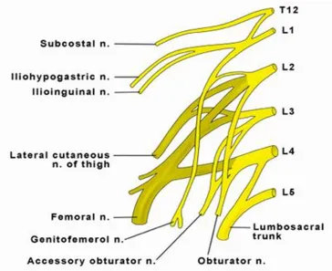
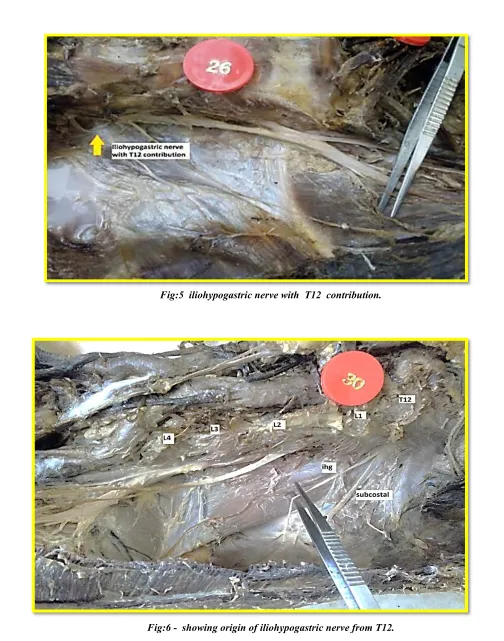
Fig:5 iliohypogastric nerve with T12 contribution.
39
Table : 2- Observation in the formation of ilio hypogastric nerve.
Iliohypogastric nerve was formed from ventral rami of L1 in
29 specimens (14 on right side and 15 on left side) in male cases and 14
specimens (7 on right and 7 on left side) in female cases. The nerve arises from
T12 in 1male case(on right side)(Fig.6) Iliohypogastric nerve was found to be
absent(fig.8) in left side of one male case. T12 contributes to the formation of
iliohypogastric nerve in 5 specimens, of which 1 on right and 2 on left side of
male cadaver, and one on right and one on left side of female cadaver(fig.5,7)
Root value Male cases Female cases
Right % Left % Right % Left %
L1 14 28 15 30 7 14 7 14
L1 with contribution
from T12 1 2 2 4 1 2 1 2
T12 1 2
40
Table : 3- Variation in origin of iliohypogastric nerve.
ARISES FROM NO.OF.PLEXUS PERCENTAGE
L1 43 86 %
L1 with contribution from T12
5 10 %
T12 1 2%
Absent 1 2%
Iliohypogastric nerve was found to arise from ventral rami of L1 in 43
specimens (86%). It arises from ventral rami of L1 along with contribution
from T12 nerve in 5 specimens (10%) Iliohypogastric nerve arises from T12
in 1 plexus (2%). The nerve was found to be absent in 1 plexus (2%).
MODE OF ORIGIN OF ILIOHYPOGASTRIC NERVE.
Normally iliohypogastric and ilioinguinal nerve arises from a single
stem formed by the ventral ramus of first lumbar nerve. In the present study,
Iliohypogastric nerve was found to arise from a single stem in 9
41
Table : 4-Mode of origin.
origin No.of specimen Percentage
Common trunk 9 18%
Separate branch 41 82%
Table :5- Percentage of variant origin.
Origin No.of specimen Percentage
Normal 43 86%
Variant 7 14%
Iliohypogastric nerve is found to have normal origin in 86 % cases and
variant origin in 14 % cases.
FORMATION OF ILIOINGUINAL NERVE .
GRAY’S ANATOMY – states that ilioinguinal nerve arises from
ventral ramus of L1 normally. The following observations were made in the
Fig: 7- showing iliohypogastric nerve with T12 contribution and ilioinguinal nerve with L1,L2 root.
42
Table:6 – Formation of ilioinguinal nerve.
Root value
Male cases Female cases
Right % Left % Right % Left %
L1 with T12
contribution 1 2 1 2 2 4
L1 15 30 15 30 4 8 6 12
L1,L2 1 2 2 4 1 2
L2 1 2
Double ilioinguinal
nerve 1 2
Ilioinguinal nerve was found to arise from L1 in 15 specimens on right
and on left side of male cases respectively. In female case 4 on right and 6 on
left side arises from L1. One plexus on right side of male and female, and 2 on
left side of female case arises from ventral ramus of L1,L2. 1case on right side
of female cadaver was found to arise from L2 ventral rami(fig.10). Double
Fig:9 - Showing double ilioinguinal nerve.(one branch along with iliohypogastric nerve, the second branch from L1,L2). Picture also shows Lateral femoral cutaneous nerve
[image:57.612.69.553.58.345.2]that arises from L1,L2.
43
Table : 7- Variation in formation of ilioinguinal nerve.
Origin of ilioinguinal nerve . No. of specimen. Percentage
L1 with T12 contribution 4 8%
L1 40 80%
L1,L2 4 8%
L2 1 2%
Double ilioinguinal nerve 1 2%
Ilioinguinal nerve was found to arise from ventral rami of L1 in 40
specimens(80%). It arises from L1 along with contribution from T12 in 4
specimens(8%).The nerve arises from L1,L2 in 4 specimens ( 8%)(fig.7,9)
Ventral rami of L2 alone gives rise to ilioinguinal nerve in 1 specimen (2%).
Double ilioinguinal nerve was found in 1specimen(2%).
Table:8 –percentage of variant origin.
Origin No.of specimen Percentage
Normal 44 88
Variant 6 12
Ilioinguinal nerve is found to have normal origin in 84 % cases and
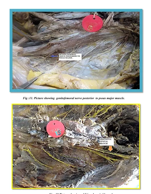
Fig :11. Picture showing genitofemoral nerve posterior to psoas major muscle.
44
FORMATION OF GENITO FEMORAL NERVE.
Genito femoral nerve is formed by the union of communicating branch
from L1 along with the ventral ramus of L2 , within the psoas major muscle
and emerges on its anterior surface along the middle third of psoas major
muscle. It then divides into genital and femoral branches. In the present study,
following observations were made.
Table :9 -RELATION OF GENITO FEMORAL NERVE TO PSOAS MAJOR.
Emerges on No. of specimen Percentage
Anterior surface 48 96%
Posterior surface 2 4%
Genito femoral nerve emerges on the anterior surface of psoas major
muscle in 48 specimen and from the posterior surface in 2 specimens(fig.10).
Table :10 - Mode of origin of genitofemoral nerve.
Mode of origin No.of specimen Percentage
common trunk 45 90%
Separate branches 5 10%
Genito femoral nerve emerges as common trunk in 45 specimens, and as
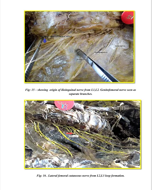
Fig.13- Showing separate genital and femoral branches.Ilioinguinal nerve from L2 nerve root.
45
[image:62.612.127.521.129.347.2]FORMATION OF GENITO FEMORAL NERVE
Table :11-Observation of formation of genitofemoral nerve.
Root valve
Male cases Female cases Right Side %age Left Side %age Right side %age Left side %age
L1,L2 15 30% 15 30% 8 16% 8 16%
L2 1 2% 1 2%
L2,L3 1 2%
Additional genital branch
1 2%
Genito femoral nerve is found to originate from ventral ramus of L1,L2
in 31 specimens (15 on right side and 15 on left side) of male cadaver and 16
specimens in females (8 on right side and 8 on left side). The nerve arises from
L2 ventral ramus in 2 specimens(one on right side and one on left side of male
cadaver) and the nerve originates from ventral rami of L2,L3 in 1 specimen.
An additional genital branch is seen on right side of one male cadaver(fig.12).
Percentage incidence of origin of genito femoral nerve from ventral ramus of
L1,L2 is 62% in male cadaver and 32% in female cadaver. In 4% of cases the
nerve arises from ventral rami of L2. In 2% cases the nerve arises from L2,L3
46
Table: 12-percentage of variant origin.
Origin No .of specimen Percentage
Normal 46 92
Variant 4 8
Genito femoral nerve is found to have normal origin in 92 % of cases
and variant origin in 8 % of cases.
FORMATION OF LATERAL FEMORAL CUTANEOUS NERVE
(LFCN).
Lateral femoral cutaneous nerve ( LFCN) also called as lateral
cutaneous nerve of thigh is formed by the union of branches from dorsal
Fig: 15 – showing origin of ilioinguinal nerve from L1,L2. Genitofemoral nerve seen as separate branches.
Fig : 17- showing origin of lateral femoral cutaneous nerve from femoral nerve.
47
[image:66.612.134.513.141.373.2]OBSERVATIONS OF THE PRESENT STUDY.
Table 13: Formation of LFCN
ROOT VALVE
MALE CASES FEMALE CASES Right side % age Left side % age Right side % age Left side %age
L2,L3 11 22% 14 28% 7 14% 7 14%
L1,L2 1 2% 1 2% 1 2%
L2 1 2%
ARISE FROM FEMORAL NERVE
3 6% 2 4%
DOUBLE LFCN
1 2% 1 2%
Lateral femoral cutaneous nerve originates from ventral rami of L2,L3
in 25 specimens(11on right and 14 on left side) of male cadaver and 14
specimens (7 on right side and 7 on left side) of female cadaver. The nerve
arises from ventral rami of L1,L2 in 3 specimens(one on right side and one on
left side of a male cadaver and one on right side of a female cadaver)(fig 9,13)
.Ventral ramus of L2 gives rise to LFCN in 1 specimen(2%) on right side of
male cadaver. LFCN arises as a branch from femoral nerve in 5 specimens
(10%),(3 on right side and 2 on left side of male cadaver)(fig.17,18). Double
Fig:19- picture showing double lateral femoral cutaneous nerve. Picture also shows absence of iliohypogastric nerve.
48
Table :14 – percentage of variant origin.
Origin of LFCN No.of specimen Percentage
NORMAL 39 78
VARIANT 11 22
Lateral femoral cutameous nerve is found to have normal root valve in
78% cases and in 22 % cases it is found to have variant origin .
FORMATION OF FEMORAL NERVE:
Femoral nerve is the thickest nerve of lumbar plexus. It is formed by
the union of dorsal division of ventral ramus of L2,L3,L4. In the present study
the following observations were made.
Table 15- observation of formation of femoral nerve.
Root valve
Male cases Female cases
R % L % R % L %
L2,L3,L4 17 34 17 34 7 14 8 16
L1,L2,L3 1 2
Femoral nerve was found to be formed from the dorsal divisions of
ventral rami of L2,L3,L4 in 49 specimens. One female cadaver had high
form( prefixed plexus) on left side, with the root valve of L1,L2,L3 ventral
rami . Femoral nerve in 1 specimen was found to be split by fibres of psoas
Fig :21- showing the branches of lumbar plexus and the presence of accessory obturator nerve.
49
FORMATION OF OBTURATOR NERVE.
Obturator nerve is normally formed by the union of anterior division of
ventral ramus of L2,L3,L4. In the present study the following observation was
made.
Table: 16 – observation in formation of obturator nerve.
Root valve
Male cases Female cases
R % L % R % L %
L2,L3,L4 (ventral
division) 17 34 17 34 7 14 8 16
L1,L2,L3 1 2
Obturator nerve is formed from ventral division of L2,L3,L4 in all
specimen expect the prefixed plexus, in which it is formed from ventral
division of L1,L2,L3 on left side of one female cadaver.
ACCESSORY OBTURATOR NERVE.
Accessory obturator nerve is occasionally present, and in such cases,
the accessory obturator nerve is small and is formed by the ventral rami of L3
and L4.
Table : 17- Presence of accessory obturator nerve.
Root valve Male case Female case
R % L % R % L %
50
Accessory obturator nerve was found in 3 specimens in male cadaver
(two on right and one on left side ). In female cadaver accessory obturator
[image:71.612.132.511.201.284.2]nerve was found on left side in 1 specimen.(fig.21).
Table :18- Incidence of accessory obturator nerve.
Presence /absent No .of specimen Percentage
Present 4 8
Absent 46 92
In the present study accessory obturator nerve was present in 4 out of 50
specimens, and was absent in 46 specimens. According to present study
accessory obturator nerve was present in 8% of cases.
51
DISCUSSION
The word plexus, is a temporary union of fibres of adjacent spinal
nerves and this is probably due to multiple origin of nerves, and most of the
branches of the plexus contain fibres from two or more segmental nerves.
According to Furbinger 1909 in Quains Anatomy, on study of
morphology of lumbar plexus the multiple origin is intimately related to the
fusion of myotomes from which the muscles of limbs are derived and in
association with this is the multiple innervations of individual muscles.
Formation of lumbar plexus:
Eisler19 in 1891 has analysed lumbar plexus and has stated that in the
normal condition the first three lumbar nerves enter wholly into the formation
of lumbar plexus while the fifth nerve enters wholly into the formation of
sacral plexus. The fourth nerve is called as nervus furcalis as it is divided
between the two plexus(Quains Anatomy).
In 1893 C.S Sherrington introduced the term prefixed and postfixed for
cases in which the plexus originated more superiorly (rostrally) and more
inferiorly (caudally) for postfixed plexus.
BARDEEN AND ELTING8 1901 used the term proximal and distal in
52
According to Grays Anatomy 41st edition lumbar plexus is present
within the posterior part of psoas major muscle, anterior to lumbar transverse
process and formed by the first three and most of fourth lumbar nerve. The
smaller moiety of fourth nerve joins the fifth lumbar nerve to form lumbosacral
trunk which joins the sacral plexus. The fourth nerve is often called the nervus
furcalis as it is being divided between the lumbar and sacral plexus. In the
present study this pattern is seen in 49 specimens.
The third lumbar nerve is occasionally the nervus furcalis(Gray’s
Anatomy) when the fibre contribution is moved cranially or the fifth nerve is
the nervus furcalis when the fibre contribution is moved caudally.
In the present study third nerve is the nervus furcalis in 1 specimen which
coincides with the illustrations of standard literature.
Situation of lumbar plexus:
In all 50 specimens the lumbar plexus is seen in the posterior part of
psoas major muscle. This coincides with the observation of Farny J.Drolet
P.Girard M21. Who had done 4 cadaveric dissection and demonstrated that the
lumbar plexus is within the posterior part of psoas major muscle rather than
53
[image:75.612.147.505.153.476.2]Normal branching pattern of lumbar plexus.
Fig :2 – Formation of lumbar plexus.
The lumbar plexus which is one of the main nervous pathway supplying
the lower limb is placed in the posterior part of psoas major muscle. The
ventral rami of lumbar spinal nerves pass anterior to transverse process of
corresponding lumbar vertebrae. The upper four lumbar ventral rami along
with a twig from twelfth thoracic ventral ramus (dorsolumbar nerve) form
54
lumbar nerve. First lumbar nerve gives a branch to second lumbar nerve and
then divides into thick iliohypogastric nerve and thin ilioinguinal nerve which
emerges on the lateral border of psoas major muscle.
The second lumbar nerve along with a branch from first lumbar nerve
forms the genito femoral nerve that emerges on the anterior surface of psoas
major muscle. The second, third and fourth lumbar nerve divides into ventral
and dorsal divisions. Dorsal division of second, third and fourth lumbar nerve
join together to form femoral nerve and the ventral divisions join together to
form obturator nerve.
A branch from dorsal division of second and third lumbar nerve form
the lateral cutaneous nerve of thigh. A branch from ventral division of third and
fourth lumbar nerve form the accessory obturator nerve.Smaller part of fourth
lumbar nerve join with fifth lumbar nerve to form the lumbosacral trunk which
55
BRANCHES OF LUMBAR PLEXUS.
ILIOHYPOGASTRIC NERVE L1
ILIOINGUINAL NERVE L1
GENITOFEMORAL NERVE L1 ,L2
LATERAL FEMORAL CUTANEOUS
NERVE
L2,L3
FEMORAL NERVE L2,L3,L4(dorsal division)
OBTURATOR NERVE
L2,L3,L4(ventral
division)
56
ILIOHYPOGASTRIC NERVE.
Observation in the present study:
Table : 19 No.of.specimen-50
Arises from L1 L1 with T12 T12 Absent
No.of plexus 43 5 1 1
Percentage 86% 10% 2% 2%
[image:78.612.152.498.180.267.2]Comparison with observation of previous authors.
Table :20
Authors T12 with L1 L1 T12 Absent
W.Henry Hollingshed 34% 32% -
Kusum R Gandhi 13.3% 86.6% -
Deepti Arora 8.3% 78.3% 13%
Anloague and Huijbregt - - 20.6%
Zacharry klaassen 14% 10% 7%
[image:78.612.123.501.377.540.2]