Does posture affect micturition?
253
0
0
Full text
(2)
(3) 1. ACKNOWLEDGEMENTS. Many people have helped me over a long period of time to produce and streamline the ideas of posture affecting micturition. These thought processes eventually led to the completion of this thesis.. My wife Paula, my children Ben and Tara were supportive during this period and I thank them for their everlasting belief in me.. Audrey Corstiaans RN who has worked with me since I came to Australia, methodically continued our research, and found innovative ways for recruitment, data collection and data saving. Her work remains highly significant in this thesis.. My team who have helped me in many ways including moral support include Sonya Farmer – Urogynaecology nurse from 2004 to 2006, Kim Pattinson – Urogynaecology Nurse form 2008 – 2009, Siva Balakrishnan – Urogynaecology Fellow 2005-2006 and consultant Urogynaecologist in Malaysia for the urodynamics study. Mr Paul Fiadjoe, Urogynaecology Fellow – 2008-2009, Kurinji Kannan- Urogynaecology Subspeciality Trainee 2005 to date.. I wish to acknowledge the invaluable contribution of A/Prof Reinhold Muller who was solely responsible for the statistical analysis of this thesis.. My secretarial staff Barbara Rogers, Jodie Wellington, Narelle Bergin. My initial JCU supervisors who convinced me that ‘this was possible’ – The Late Professor Phil Summers, Professor Alan Nimmo. My current supervisors – Professor Lee Kennedy for putting up with my time lines and my idiosyncrasies. A/Prof Malcolm Frazer my lifelong friend and supervisor for continually believing in me.. I would also like to thank Gurubux Singh and Frank Selwood for making the first Duni prototype in my garage!. Dr Harry Stalewski , Paediatric surgeon for loaning me his uroflowmeter..
(4) 2. A special thanks to Mr Wallace Bowles from NSW , a diehard squatting fanatic who continues to encourage me to promote squatting.. Also a special thanks to Rahul Rane who is taking engineering of squat toilets to a different dimension.. My peers Professor Linda Cardozo, Professor Richard Millard, Professor Shlomo Raz, Professor Hans Peter Deitz and Dr Bernie Haylen for allowing me to use their data, photos and excerpts from their textbooks.. And finally Dr Jay Iyer, a student that a teacher can only hope for, a colleague one could only wish for and a friend one could only dream for, I am truly grateful..
(5) 3. ABSTRACT Posture during toileting and its effect on toileting has been under scrutiny for more than 7000 years. With the advent of the western toilet in the 19 th century and a more closeted approach to toileting, the effects of posture became even more important to study because the visual one to one education of children, regarding toileting became less and less. Wennergren in her study of children demonstrated the value of foot support to relax the pelvic floor during urination in children. [1] Moore in her study showed that a majority of British women will not sit on the toilet outside their house and would ‘hover’ to pass urine. [2] A Taiwanese study showed a similar trend except that young students would precariously perch in a squat over a western toilet than sit on it for hygiene reasons. [3] All these studies stimulated the generation of this thesis. PRINCIPAL OBJECTIVES AND SCOPE OF THE STUDY The principal objectives of these investigations were to study -. The effect of the ‘lean forward’ position on the western toilet (WT) compared to the ‘sit upright position’ during micturition.. -. The effect of the ‘raised knee’ position on the western toilet compared to the ‘lean forward’ position during micturition.. -. The effect of squatting on a custom built Asian toilet compared to ‘lean forward’ position on the western toilet.. What was learnt led to numerous ‘sub-studies’ on squatting which included -. Study of squatting in volunteers leading to a ‘squatability’ index. -. Study of school children and their ability to squat. -. Study of abdominal pressures in squatting and sitting positions at rest and during Valsalva manoeuvre.. -. Study of the levator hiatus during squatting and lying down at rest.. -. Possible design of a retro fit device to aid toileting on the western toilet called Duneze..
(6) 4 METHODOLOGY All toileting parameters were studied using uroflowmetry which was used as standard, as in previous studies. [1-6]. Support for Uroflowmetry, which is a simple non-invasive measurement of urine flow over time and an indication in screening for voiding difficulty, as a screening test for voiding dysfunction has become stronger over time, [7-9] measuring some of the key components of the micturition process. [10] An abnormally slow urine flow suggests a provisional diagnosis of voiding difficulty subject to test repetition, post-void residual bladder volume (PVRBV) measurement and possible voiding cystometry.. Post Void Residual Urine Volume Measurement of post void residual urine volume (PVRBV), the amount of residual urine in the bladder after a voluntary void, is another non-invasive screening test for evaluating voiding dysfunction. Most urologists agree that volumes of 50 mL to 100 mL constitute the lower threshold defining abnormal residual urine volume PVRBV measurement. There are 2 methods of measuring PVRBV: sterile catheterization and bladder ultrasound. Although sterile catheterization provides a urine sample, there are many disadvantages associated with the procedure: it causes patient discomfort, carries a risk of urethral trauma and introducing an UTI, is time-consuming, and may not be necessary. [11]. In contrast, bladder ultrasound can be performed with a portable device. It is noninvasive and time-efficient, minimizes medical waste and supplies, and determines when catheterization is medically appropriate. However, a urine specimen cannot be obtained during this procedure. Portable 3-dimensional ultrasound devices have been shown to provide highly accurate measurement of bladder volume. Coombes and Millard compared the BladderScanTM BVI 2500 series (Diagnostic Ultrasound, Bothell, Wash) with catheterization for the measurement of bladder volume with no significant difference in estimates being demonstrated. The overall accuracy (94%), sensitivity (97%), and specificity (91%) of the BVI 2500+ were encouraging. [11] The current accuracy of modern uroflowmeters in measuring urine voided over time (flow rate) is approximately ± 2–5%, despite the fact that a variety of different physical measurement principles are being used. This accuracy compares favourably with the ± 20–25% for the most accurate ultrasonic techniques for PVR measurement with the potential error using urethral catheterisation being much higher. [12].
(7) 5 After obtaining Institutional ethics approval for all studies, volunteers recruited from nursing staff and medical students participated in these studies coached either by Audrey Corstiaans (AC) or Professor Ajay Rane (AR). SUMMARY OF RESULTS. STUDY ONE:. There was a statistically significant difference in the peak and average urine flow rates in the lean forward position when compared to the sit back posture (p<0.0054 and p<0.0097 respectively). STUDY TWO:. There was a statistically significant difference in uroflowmetric parameters i.e. the peak (p=0.01) and average flow rates (p=0.043), when tested in the lean forward position as compared to the knee raised position respectively. Hence the importance of knee raising or leaning forward with feet stability was deemed equally important when toileting.. STUDY THREE:. This was the most challenging of studies. In summary, only 46% of our volunteers from a cohort of 125 could actually squat (with feet flat for more than 30 seconds). [13] Although not statistically significant, in volunteers who could squat there was a trend to better urine flows especially the “time to maximum flow” (p=0.003) in the squatting position when compared to the lean forward position.. The results of the Study One encouraged us to consider the possibility that an alternative position during toileting would be beneficial in effecting voiding. This lead to the evaluation of Uroflowmetric parameters in the lean forward and raised knee position (Study Two). Encouraged by the results of Study Two we raised the bar even higher and asked patients to squat during the act of voiding. Although no firm conclusions could be drawn from this study the main challenge arose from the fact that less than half of our volunteers could not squat..
(8) 6 PRINCIPAL CONCLUSIONS. The main conclusions derived from these studies are -. Posture on the toilet affects bladder function. -. On the western toilet the lean forward position with foot support is the most optimal. -. Squatting position is difficult to assume in a majority of the population who do not routinely use squat toilets..
(9) 7. TABLE OF CONTENTS Chapter 1. General introduction. 16. Chapter 2. Hypothesis, study parameters and justifications. 35. Chapter 3. Physiology of Pelvic structures during toileting. 44. 3a.. Physiology of Micturition. 45. 3b.. Physiology of Defaecation. 61. Chapter 4. Epidemiology of Incontinence. 78. Chapter 5. The Perfect Pee Study. 102. Chapter 6. The Near Squat Study. 123. Chapter 7. The Squat Study. 133. 7a.. The Squatability study amongst adults and children. 147. 7b.. Abdominal pressures in the sitting and squatting positions. 155. 7c.. Levator hiatus in the supine and squatting position. 161. Chapter 8. Inventions to aid squatting. 172. Chapter 9. General Discussion. 185. References. 189. Appendix A Questionnaires. 203. Appendix B Publicity Material. 205.
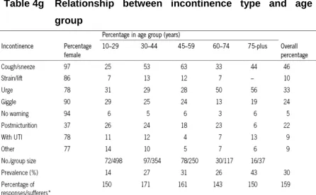
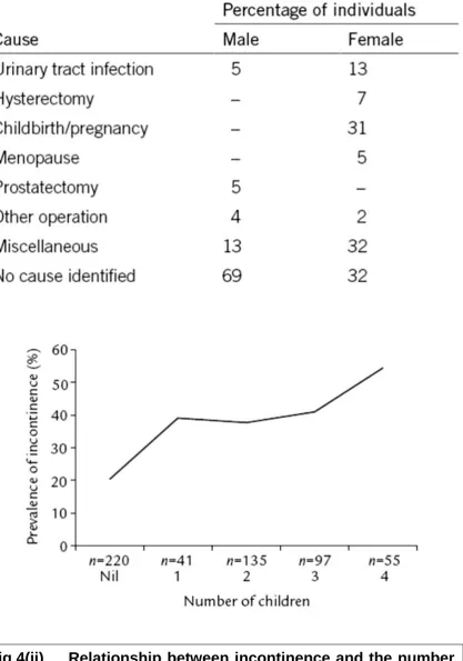
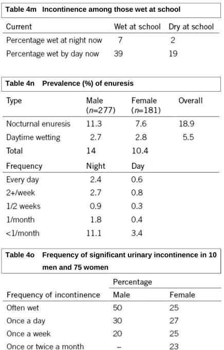
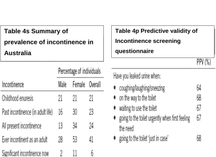
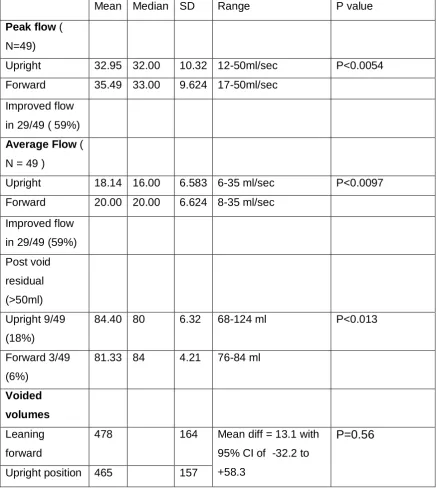
(10) 8. LIST OF TABLES. Table 4a. Frequency of incontinence episodes. 80. Table 4b. Circumstances in which respondents experienced leakage. 80. Table 4c. Quantification of severity of urinary loss in 293 respondents. 81. Table 4d. Percentage frequency and severity of leakage in 293 respondents wet by day. Table 4e. Frequency of nocturnal incontinence, correlated to sex and age group. Table 4f. 81. 84. Prevalence of incontinence and its relationship to age group and gender. 85. Table 4g. Relationship between incontinence type and age group. 85. Table 4h. Cause of leakage identified by 301 individuals who were ‘wet by day or night’. 86. Table 4i. Groups found to be associated with higher rates of incontinence. 88. Table 4j. Adjusted odd ratios for variables associated with leakage of urine in women. Table 4k. Proportion of incontinent women in age cohorts who reported stress, urge or ‘other’ incontinence. Table 4l. 88. 92. Present incontinence among those recalling nocturnal enuresis as a child. 92. Table 4m. Incontinence among those wet at school. 93. Table 4n. Prevalence (%) of enuresis. 93. Table 4o. Frequency of significant urinary incontinence in 10 men and 75 women. Table 4p. Degree of wetness in respondents with significant urinary incontinence. Table 4q. 93. Continence status in patients in 15 of the larger nursing. 95.
(11) 9 homes in Sydney. 95. Table 4r. Degree of incontinence analysed by patient mobility. 97. Table 4s. Summary of prevalence of incontinence in Australia. 97. Table 4t. Predictive validity of Incontinence screening questionnaire. 97. Table 5a. Demographic data. Table 5b. Peak and average flows on micturition on upright and lean forward. 102. positions. 102. Table 5c. Crouching habits of volunteers. 103. Table 6a. Uroflowmetric parameters. 123. Table 7a. Comparative data on Uroflowmetry. 133. Table 7aa. Shows the distribution data against mode of delivery. 143. Table 7ab. Squat by parity. 143. Table 7ac. Shows the distribution data against age. 143. Table 7ad. Squat by age category. 143. Table 7ae. Age, sex and duration of squat in children. 146. Table 7af. Squatting by age. 148. Table 7ag. Squatting by gender. 148. Table 7b. Urodynamic parameters. 153. Table 7c. Levator Hiatus measurements on sitting and squatting. 163.
(12) 10. LIST OF FIGURES. Fig 1(i). “Guardez l’eau” – watch the water!. 19. Fig 1(ii). Thomas “Crapper” advt. 19. Fig 1(iii). Ornate Porcelain toilet (Victorian England). 19. Fig 1(iv). NYC-“CBGB”- Punk Heaven. 21. Fig 1(v). Colca Canyon Andes-home of the “Condor”. 21. Fig 1(vi). Atacama Desert-Humberstone Route 16. 21. Fig 1(vii). London-“Pink eggs are for girls,blue …. 22. Fig 1(viii). Brussels-Square Maurice Raindorf. 22. Fig 1(ix). Alscot Park Manor-England. 24. Fig 1(x). Toilet Apartheid!. 24. Fig 1(xi). All sheep no humans- Kiwiland!. 25. Fig 1(xii). Inside the leading Dunny-Silverton,NSW. 25. Fig 1(xiii). Duneedoo Caravan Park, NSW- A dunny that bitterly divided a village!. 25. Fig 1(xiv). Odoriferous home next to public toilet-India. 27. Fig 1(xv). Hutongs(communal toilets)-Beijing. 27. Fig 1(xvi). Japanese toilet. 27. Fig 1(xvii). Toilets from ancient times. 29. Fig 2(i). Liverpool Normogram for the maximum urine flow rate. 39. Fig 2(ii). Liverpool Normogram for the average urine flow rate. 39. Fig2(iii). Diagram of a urine flow recording with International Continence Society recommended nomenclature. 42. Fig 3a(i). Molecular events leading to smooth muscle contractions. 47. Fig 3a(ii). Sympathetic nervous system events at neuromuscular junction. 47. Fig 3a(iii). Parasympathetic nervous system events at neuromuscular junction. 52.
(13) 11 Fig 3a(iv). Sensory innervation of the bladder and central afferents. 52. Fig 3a(v). Motor supply to the bladder and parasympathetic events. 56. Fig 3a(vi). Overview of Micturition reflexes. 56. Fig 3b(i). The extrinsic innervation of the human colon. 63. Fig 3b(ii). Diagram showing the layers and components of the intestinal wall. 63. Fig3b(iii). Intracolonic pressures leading to spontaneous defaecation by the healthy human colon. Fig 3b(iv). 66. Some of the mechanical processes that facilitate stool expulsion, as illustrated by sequential films of a simulated defaecation of thickened barium during defecation proctography. Fig 3b(v). 69. Responses to distension of the rectum by pressures less than 55 mm Hg. Distension produces passive tension due to stretching of the wall of the rectum, and additional active tension when the smooth muscle in the wall contracts. Fig 3b(vi). 72. Sagittal view of the anorectal area at rest (above) and during straining (below). Note the reduction of the anorectal angle and lowering of the pelvic floor during straining. 72. Fig 4(i). Prevalence of Incontinence and its relationship to age and gender 84. Fig 4(ii). Relationship between incontinence and the number of children. Fig 5(i). The Curtsy Sign. 105. Fig 5(ii). EMG recordings in sitting on a toilet seat. 107. Fig 5(iii). Positions on the toilet seat. 112. Fig 5(iv). Modified sitting-type toilet. 117. Fig 5(v). Voiding positions on public toilet seats. 117. Fig 6(i). CFA poster demonstrating correct sitting position on the toilet. 120. Fig 6(ii). “The Perfect Pee” and “Near Squat”. 120. Fig 6 (iii). ILC Australia-Toiletting Footstool. 126. 86.
(14) 12 Fig 7(i). “The DUNI with my son Ben”. Fig 7(ii). Defecograph showing the anorectal angle as measured in two. 129. different positions of bowel evacuation. 137. Fig 7(iii). American squat toilet with tank (Saline, Michigan). 141. Fig 7(iv). Toilet retrofit installation. 141. Fig 7c (i). The usual acquisition/evaluation screen on Voluson-type systems shows the three orthogonal planes: (a) sagittal, (b) coronal and(c) axial, as well as (d) a rendered volume, which is a semitransparent representation of all gray-scale data in the rendered volume {box delineated in (a), (b) and (c)}.. Fig 7c (ii). The axial plane on (a) magnetic resonance imaging and (b) ultrasound (freehand three-dimensional).. Fig 7c (iii). 160. The levator hiatus (a) at rest and (b) on Valsalva in a young, nulliparous woman with significant pelvic organ descent.. Fig 7c (v). 158. The levator hiatus (a) at rest and (b) on Valsalva in a young, nulliparous woman without significant pelvic organ descent. Fig 7c (iv). 158. 160. The three orthogonal planes: midsagittal (A, top left), coronal (B, top right), and axial (C, bottom left) as well as a semi transparent rendering of all voxels in the region of interest. 161. Fig 8 (i). Duneze. 169. Fig 8 (ii). Nature’s Platform. 171. Fig 8 (iii). LILLY PAD (Lillipad squatting platform). 173. Fig 8 (iv). Sitting position. 175. Fig 8 (v). Squatting position. 175. Fig 8 (vi). The optimum angle between the torso and femurs is 30 degrees for aligning the anal canal with the rectum. 176. Fig 8 (vii). Squat Support features. 176. Fig 8 (viii). IN LEIU. 178.
(15) 13. PUBLICATIONS PRODUCED DURING MY PHD CANDIDATURE Publications. Rane, A., Corstiaans, A. Effect of Postural Changes on Voiding Function. Australian Continence Journal, 1998, 4(4):103. Rane A., Corstiaans A. Does leaning forward improve micturition? Journal of Obstetrics and Gynaecology, November 2000, 26 (3):629-29. Rane A, Corstiaans A.Does micturition improve in the squatting position?J Obstet Gynaecol. 2008, 28(3):317-9.
(16) 14. CONFERENCE PRESENTATIONS 1998 RANZCOG ASM Adelaide PRESENTATION. Does posture influence micturition?. IUGA 2003 POSTER. Posture and micturition – a position. POSTER. trial comparing the near squat. (NSP) to the leaning forward position ( LFP). Rane A, Lim Y, Corstiaans A, 2003 Intraoperative division of TVTa RCT Int J UroGyn October 2003. IUGA 2005 PRESENTATION. Barry C, Dietz HP, Buta S, Lim YN, Greenland H, Rane A et al Imaging the levator hiatus using magnetic resonance imaging and 3 dimensional ultrasound – a comparative study. IUGA 2008 POSTER. Study of intraabdominal pressures in the sitting and squatting positions- A Pilot Study published in Int J Urogyn, Abstract, 2008 (Kannan K, Fiadjoe P, Rane A). POSTER. Rane A, Fiadjoe P, Corstiaans A, Kannan K Int J Urogyn, Abstract,. 2008 Does micturition improve in the squatting. position? IUGA 2009 POSTER. Study of intraabdominal pressures in the sitting and squatting positions- A Study of 89 patients published in Int J Urogyn, Abstract, 2009 (Kannan K, Fiadjoe P, Rane A).
(17) 15. Chapter 1 General introduction. Chapter 2. Hypothesis, study parameters and justifications. Chapter 3. Physiology of Pelvic structures during toileting. 3a.. Physiology of Micturition. 3b.. Physiology of Defaecation. Chapter 4. Epidemiology of Incontinence. Chapter 5. The Perfect Pee Study. Chapter 6. The Near Squat Study. Chapter 7. The Squat Study. 7a.. The Squatability study amongst adults and children. 7b.. Abdominal pressures in the sitting and squatting positions. 7c.. Levator hiatus in the supine and squatting position. Chapter 8. Inventions to aid squatting. Chapter 9. General Discussion.
(18) 16. CHAPTER 1. GENERAL INTRODUCTION.
(19) 17 Posture during micturition and defaecation has been a topic of discussion for centuries. There is a huge body of anecdotal and unscientific literature on this topic. This introduction reflects on this data, and an attempt is made to scientifically study posture, and it’s effect on micturition, in subsequent chapters. Two-thirds of humanity uses the squatting position to answer the call of nature. This fact has generated intense debate both in Orient and the Occident whether the traditional squatting position over the toilet is superior in achieving bowel evacuation to the “more evolved” sitting posture adopted by Western Cultures. In the yogic scriptures of Patanjali, 7000 years ago, there is a description on the use of a Hatha Yoga technique called ‘Mahabandha’ for control of anal and urethral canals through perineal pressure by the heel of the left foot. [14] The lotus position and heel pressure on the perineum to defer micturition are also well enshrined in the teachings of Tai-Chi. Although squatting is the most natural and effective posture for evacuation, the body is able to use other positions in emergencies (like a broken leg). For thousands of years, kings and queens have adopted other positions to distinguish themselves from the “commoners”. [15] Then, in the mid-nineteenth century, at the beginning of the Industrial Revolution, indoor plumbing became universally available. The early industrialists decided (rather arbitrarily) to install throne-like toilets everywhere – to allow ordinary people to feel like kings and queens. Knowing nothing about physiology, they sincerely believed that they were improving people’s lives. Those who felt uncomfortable with this decision were forced to keep silent. (In Victorian England, bodily functions were considered unmentionable). From Great Britain, the most influential country in the world at the time, the fad quickly spread to the rest of Europe, and to North America and Australia. No country wanted to seem “backward” at a time when the world was making such rapid “progress.” [15] Until just a few years ago, the taboo on discussing this subject kept most of the western world in the dark about how the human body was designed to function. The ignorance of the medical profession has been especially regrettable – and has caused much needless suffering. [15] Naylor and Mulley from St Luke’s at Leeds addressed the issue of western commodes and the attitudes of users and carers to them. [16]This study highlighted the existing ignorance amongst carers about the use of commodes and other appliances in aiding toileting..
(20) 18 TOILETTING – A PRIVATE BUT FASCINATING FUNCTION [17]. Toileting has fascinated people the world over. In vernacular English alone there are more than three hundred different names for it like loo, john, dunny, bathroom, WC, washroom, toilet, facilities, little boys’ room etc. Taboo subjects like death, drugs and sex have all provoked deep interest throughout the ages. Toilets are no different. They are indeed a bay window to civilisations and populations. Often they reflect development or lack of, in a given country or region. Toilets are ubiquitous; they transcend all ages, races, religions and social class. Every one of us has to obey the call of nature. Some may say the act is philosophical – emptying the body empties the head! Many people use the toilet to get peace of mind or generate great ideas. Records in the Old Testament tell us of Moses telling his people ‘to go abroad and when they ease themselves , to dig , turn back and cover what has cometh from thee’. Although there was no knowledge of bacteria and sanitation, it was the smell of excrement that drove civilisations to do the ‘job’ away from living space and this by itself may have limited the spread of disease. The oldest known flush toilet dates to the 18 th Century BC in Knossos, Crete, home of King Minos. Sewage disposal was quite rigorous in Jerusalem and Rome. In 800 BC Cloaca Maxima was the first known sewer with running water. The Middle Ages though, saw a fall in sanitation and this contributed to the bubonic plague called Black Death in Europe. Chamber pots were standard in use and the contents usually heaved out of the window with a warning cry “guardez l’eau” in French meaning – watch the water – the word loo remains slang for toilets in Britain.. As the Middle Ages drew to a close, science made the connection between disease and poor sanitation which led to sewers and proper disposal. In 1596, Sir John Harington is known to have designed the first flush toilet for Queen Elizabeth I in Richmond Palace. She is rumoured not to have used it because of the noise! Thomas Crapper dubiously has been given the kudos of inventing the toilet. Far from it Crapper and Co. had their name on a lot of porcelain toilets and American soldiers stationed in the UK during WWI used the word Crapper or Crap when they excused themselves - this word still remains in use! In 1775 Alexander Cummings patented the flush toilet and since then there has been no great changes in the toilet except in bowl design, cistern shapes and the quantity of water used. In a brief tour of the toilets of the.
(21) 19. Fig 1(i) “guardez l’eau” – watch the water!. Fig 1(ii) Thomas “Crapper” advt. Fig 1(iii) Ornate Porcelain toilet (Victorian England).
(22) 20 world, I have picked some geographically salient features continent by continent to describe the effects of population affluence, geography and traditions on toilet designs.. NORTH AMERICAS. The most obvious feature in North American toilets is the amount of water they use; it is the largest amount in the world per flush. The 102 nd U.S. Congress legislated through the Energy Policy Act of 1992 (that came in to effect on 1 st January 1994) that cisterns used no more than 6 litres per flush down from a whopping 19-26 litres per flush. Visitors often complain about the splash back effect due to a high water level. Another important feature is the crescent moon which was cut on top of the doors of outhouses to let light in. This continues to be used even though electricity is ubiquitous. Americans usually avoid the word toilet, and they ask for bathroom, washroom or restroom when they need to go. Amazing toilet designs from North America include toilets in submarines, the international space station, l’urinette (The standing loo for women) in Quebec amongst many others. CENTRAL AND SOUTH AMERICA Impoverishment meant that typically the restrooms or ‘banos’ are small and dirty. There are people who man some for a fee and keep them clean. Of note are the Kuna Indian toilets – which have a narrow plank walk right up to the Pacific Ocean and they let the tides do the cleaning act. Most toilets in this region have thatched palm frond roofs and no sewer systems. EUROPE. Europe seems to be the heart of toilet varieties. Sewerage is universal but the toilet designs and etiquettes are vastly variable. Many are quite exotic and ergonomically designed. [18] Most European countries require you to pay to use facilities in public buildings. Madame Pipi in French or ‘Klofrau’ in German needs to be handed the money before you enter. Perhaps the more scary aspect for the unwary traveller is the Squat toilets in France and southern Europe. Long haired beginners are urged to tie up their hair and require reasonable athletic prowess to squat and avoid splashing trousers, shoes and feet!.
(23) 21. Fig 1(iv) NYC-“CBGB”- Punk Heaven. Fig 1(v) Colca Canyon Andes-home of the “Condor”. Fig 1(vi) Atacama Desert-Humberstone Route 16.
(24) 22. Fig 1(vii) London-“Pink eggs are for girls,blue …. Fig 1(viii) Brussels-Square Maurice Raindorf.
(25) 23 AFRICA. It is obviously assumed that the first act of toileting took place in Africa. Landscape, wild life, dress and customs and economic disparity dictate the types of toilets in this continent. Squat toilets are commoner in North Africa and they get more and more infrequent as one goes south. A door in front of an African toilet is usually without hinges and brightly painted. It is simply propped against the entrance. A clever adaptation in African toilets is the use of the suns’ searing heat to ventilate the toilet. A ventilation pipe runs from the pit below the toilet through the roof. The outer portion of the pipe gets heated by the sun, which causes air and odours from the latrine to be sucked up and expelled from the top of the pipe. Perhaps most relevant is the difference in toilets during apartheid with opulence for the white men and dank, stinky holes for the non – whites. OCEANIA The lingo of the ‘dunny’ and the ‘long drop’ is very specific to Australia and New Zealand. The dual flush toilet is the hallmark for this region. Small flush and big flush for respective jobs! Tales of the outback dunny are plenty with snakes, red back spiders, bats, wombats and even crocs abiding in some dunnies!!. One of the common myths is the direction of the swirl of water in the toilet. The myth is that it swirls in the opposite direction down under. The reality is water does not swirl the other way down under. The direction in which the water swirls depends purely on technicalities like bowl shape and piping. Only in very large bodies of water is the Coriolis effect perceptible. Even a toilet a few kilometres in diameter will not be enough to demonstrate this effect!. Australia is unique in that it has a National Public Toilet Map. Another unique fact for manufacturers for Australian toilets – whilst all over the world these are tested by stuffing condoms with sausages of various sizes and flushed the Australians insist on golf balls stuffed in a condom for testing!! Finally the town of Dunedoo in NSW is split in half – with a pro Big Dunny group and an anti-Big Dunny group!.
(26) 24. Fig 1(ix) Alscot Park Manor-England. Fig 1(x) Toilet Apartheid!.
(27) 25. Fig 1(xi)All sheep no humans- Kiwiland!. Fig 1(xii) Inside the leading DunnySilverton,NSW. Fig 1(xiii) Duneedoo Caravan Park, NSWA dunny that bitterly divided a village!.
(28) 26 ASIA. This continent has perhaps the most varied in cultures and therefore toileting habits. Most Asians use squat toilets. But that’s where the similarity ends. Japanese squat toilets require guest to change footwear before entering the toilet and changing back when exiting. They use plenty of flushable toilet paper.. Chinese use squat toilets but facial tissue paper is used to wipe and flush. The Thai use toilet paper but it must be tossed in the bin next to the toilet. The Indians and Turks prefer washing with their left hand as a substitute for toilet paper.. In India though only a third of the population uses sewered facilities, the rest do it in the open – this has serious health and sanitation consequences and a Non Government Organisation called Sulabh Sanitation is making progress in improving this public health issue.. Finally flushing mechanisms vary considerably. In some there are sensors, buttons, and jets whereas in others there are chains and knobs, and then there is the humble bucket and the floating mug!.
(29) 27. Fig 1(xiv)Odoriferous home next to public toilet-India. Fig 1(xv) Hutongs(communal toilets)-Beijing. Fig 1(xvi) Japanese toilet.
(30) 28 Toilets from Ancient Times Pictures of ancient public toilets tend to confuse westerners, who assume that they were used in the sitting position. This impression is often reinforced by the pose of a comical tourist. But, in reality, these are squat toilets. They are elevated, not for sitting, but because there is an open sewer underneath. The cut-outs in the vertical wall allow people to clean themselves with water, which is done from the front when squatting.. Photo courtesy of Toilets of the World [17] The ancient Romans used the posture shown below on the left. (Togas were more convenient than trousers, and provided some degree of privacy.) The last picture shows a typical tourist. He might be surprised to learn that, except for royalty and the disabled, everyone used the squatting position until the second half of the 19 th century. Highlights in the Evolution of Toilet System – 2500 BC to the 21st Century [19] 2500 BC: In Mohenjo – Daro, there existed highly developed drainage system where waste water from each house flowed into the main drain. 1000 BC: In the Bahrain Island in the Persian Gulf, flush type toilet was discovered. 69 AD: Vespasianus for the first time levied tax on toilets. 1214 AD: Construction for the first time of public toilets manned by scavengers in Europe. 1596 AD: JD Harrington invents W.C. 1668 AD: Edict issued by Police Commissioner Paris, construction of toilets in all houses. 1728 AD: Architect J.F. Brondel argues that attached toilet is ideal. 1739 AD: First separate toilet for men and women appear at a ball in Paris. 1824 AD: First Public Toilet in Paris. 1859 AD: Toilet of Queen Victoria is decorated with gold. 1883 AD: First Ceramic Toilet by Thomas Turiferd for Queen Victoria. 1889 AD: Sewage Treatment for the first time in the world (U.K.). 1959 AD: All surface Toilets abandoned (Paris)..
(31) 29. Fig 1(xvii)-Toilets from Ancient times.
(32) 30 URINARY INCONTINENCE In the larger cities of Asia, many residents have abandoned their traditional customs, believing that the West is more progressive and somehow “superior.” By adopting western toilets, they have unwittingly introduced new diseases into their society. A recent article in the Malaysian newspaper The Star (March 30, 2003) discusses one such ailment: “To squat or not to squat?” That is the question. Actually, your toileting technique may have an effect on urinary incontinence. There is evidence to show that the Asian technique of using the toilet goes a long way to maintaining better pelvic health than the Western technique. [13] A study done in Hong Kong showed that city-dwelling women had more urinary incontinence and bowel problems than country dwelling women who attributed the basic differences in these women, not to their body weight, or how many children they had, but to their toileting habits. [20] In general, women in urban areas use the “sit” method while the rural women use “squat” toilets. ‘Natures’s Platform’ suggests that squatting causes the angle of the pelvis to change and exert higher pressure on the ano-rectum.. When sitting, appropriate relaxation of the pelvic muscles does not occur nor is the angle of the pelvis correct. This further emphasises health gains from adopting the squatting position.[15] The above view is shared by Dr. Stuart Stanton, Chairman of the Continence Foundation and Consultant Urogynaecologist at St. George’s Hospital, London.’ “Squat” toilets are an excellent way for women to exercise their perineum and pelvic floor muscles and control their urinary stream from the age of 2½-3 years onwards. Reports from the developing world suggest that urinary incontinence is much less in women who squat.’ [15] A brief explanation of why sitting toilets increase the risk of incontinence could be as follows: The pelvic floor is a hammock of muscles that supports the abdominal contents, the bladder, rectum and the uterus. Western toilets force the user to strain when evacuating, repeatedly subjecting the pelvic floor to unnatural pressure. The downward pressure constitutes a chronic repetitive stress injury that stretches and weakens the pudendal nerve, responsible for bladder control..
(33) 31 71 women who had delivered at St. Bartholomew’s Hospital, London, were studied by electrophysiological measurements performed on the innervations of the external anal sphincter muscle. Manometry was also performed. [21-25] The investigations were carried out 2-3 days after delivery and repeated in 70% of these women 2 months later. Faecal and urinary incontinence developing after vaginal delivery had always been thought to be due to direct sphincter division or muscle stretching. However the results of this study suggest that in most cases this incontinence results from damage to the innervations of the pelvic floor muscles. [26] To maintain continence, the brain needs to constantly monitor the pressure within the bladder and issue commands to the urethral sphincter muscle. Both functions are impaired when the pudendal nerve is weakened by the descent of the pelvic floor. The following statistics from FocusOnUrology.com [27] show how frequently this occurs: . 17 million Americans are incontinent.. . Women. experience. incontinence. twice. as. often. as. men.. (The gynaecological disorders section explains why.) . 1 in 4 women age 30-59 has experienced an episode of incontinence.. . $16.4 billion is spent every year on incontinence-related care. . $1.1 billion is spent every year on disposable products for adults.. . 50% or more of elderly persons living at home or in long-term care facilities are incontinent.. FocusOnUrology.com attributes incontinence mainly to childbirth, weakened pelvic muscles, hormonal changes associated with menopause, and (in men) prostate surgery. Due to their cultural conditioning, women do not often mention the use of the reclining posture for childbirth. The modern toilet has made women incapable of prolonged squatting, the position designed by nature to protect the pelvic floor during delivery. [28].
(34) 32 Nor does the website FocusOnUrology.com mention the direct effect of using a sitting toilet, which causes the pelvic floor to be pushed downwards each time one strains to evacuate. Based on a conservative estimate that the average person strains four times for each daily evacuation, by the age of 50 the unsupported pelvic floor has been stretched 73,000 times. BA Sikirov discusses straining forces on bowel elimination and its effects on pelvic floor structures. [29] D Sikirov gives a comparison of straining during defaecation in three positions and its implications for human health. [30] An unnatural manoeuvre repeated so many times inevitably causes a “repetitive stress injury.” The pudendal nerve is the main casualty of this unintentional abuse, which renders over 50% of elderly Americans incontinent. An article entitled “The Descent of Women – a Silent Epidemic” by University of Adelaide researchers, in the first comprehensive study of its kind in the world, have found a remarkably high prevalence of pelvic floor disorders in the general population. Most of these complaints were still common among women who had never had a vaginal birth. According to Professor MacLennan, the survey highlights the high prevalence and major social impact of pelvic floor prolapse and incontinence in our society. In his opinion it is a silent epidemic which to some extent is unavoidable, as women continue to give birth and also because those with the problem are often embarrassed to talk or seek help about it. [31] But research by Mr Wallace Bowles on the relevance of the squatting posture has brought a new understanding of how to prevent (and, in many cases, correct) these disorders. Most people with urinary incontinence experience a noticeable improvement within several weeks of commencing to squat for defaecation with complete correction within about 3 months. [32] Anecdotally, a number of women who squat, habitually, for bowel movements and who have experienced pelvic floor trauma and incontinence after the birth of their baby, have regained their continence within about six weeks when they continue to adopt the squat posture for bowel evacuation. Squatting has been linked to prevention of bowel cancer as well. [33] Even children are susceptible to pelvic floor nerve stretch injury. An article entitled “My Child, My Teacher” was published in the Spring 1998 issue of New Vegetarian and Natural Health Magazine. Focusing on the benefits of squatting for children, the article contains numerous reports of bedwetting corrected by this simple change of habit. [34].
(35) 33 A study carried out in Iran attempted to objectively assess the impact of ethnic habits on defecographic measurements. [35] This study emphasises the importance of the wider anorectal angle resulting from defaecating in a squatting type toilet versus the Western toilet. Kate Moore and colleagues studied the phenomenon of “Crouching over the toilet seat” and its prevalence in British gynaecological outpatients and its effect upon micturition. This study highlighted the phenomenon of “hovering over the toilet seat” as opposed to actually being seated upon it and its effect on uroflowmetry measurements and bladder residuals. Given the foregoing background I have attempted to study the effects of posture and its possible impact on micturition..
(36) 34 Chapter 1. General introduction. Chapter 2 Hypothesis, study parameters and justifications Chapter 3. Physiology of Pelvic structures during toileting. 3a.. Physiology of Micturition. 3b.. Physiology of Defaecation. Chapter 4. Epidemiology of Incontinence. Chapter 5. The Perfect Pee Study. Chapter 6. The Near Squat Study. Chapter 7. The Squat Study. 7a.. The Squatability study amongst adults and children. 7b.. Abdominal pressures in the sitting and squatting positions. 7c.. Levator hiatus in the supine and squatting position. Chapter 8. Inventions to aid squatting. Chapter 9. General Discussion.
(37) 35. CHAPTER 2. HYPOTHESES, STUDY PARAMETERS AND JUSTIFICATIONS.
(38) 36 HYPOTHESES. 1. Posture on the western toilet in the lean forward position and the upright position makes no difference to voiding parameters. 2.. Posture on the western toilet with knee elevation and the lean forward position makes no difference to voiding parameters.. 3. Squatting posture on a custom designed toilet and the lean forward position on the western toilet make no difference to voiding parameters. 4. Squatting posture for toileting is possible in all volunteers. 5. All school going children irrespective of gender can adopt the squatting position for toileting in a western setting. 6. There is no difference in abdominal pressures at rest or during Valsalva manoeuvre in the sitting or squatting position. 7.. There is no difference in the levator hiatus on scan in the sitting or squatting position at rest or during Valsalva manoeuvre.. STUDY PARAMETERS. Study parameters included mainly the use of Uroflowmetric data on volunteers, cystometry, vaginal manometry and 3D/4D trans-perineal ultrasound.. Uroflowmetry, the simple, non-invasive measurement of urine flow over time during micturition, has a long history, clear definitions, and a clear purpose in screening for voiding difficulty. Most importantly technical accuracy is reproducible. [36, 37] Support for its use as a screening test for voiding dysfunction has become ever stronger over time.[7, 9] It is recognised as a simple, non-invasive test measuring some of the key final outcomes of the micturition process. [10] Abnormally slow urine flow represents a provisional diagnosis of voiding difficulty subject to test repetition, post-void residual (PVR) measurement and possible voiding cystometry. The current accuracy of modern uroflowmeters in measuring urine voided over time (flow rate) is around ± 2–5%, despite the fact that a variety of different physical principles are being used. This accuracy compares favourably with the ± 20–25% for the most accurate ultrasonic techniques for PVR measurement with the potential error using urethral catheterisation being much higher. [12].
(39) 37 Data interpretation remains the key issue limiting the clinical value of uroflowmetry. This involves an understanding of the normality, relevance and diagnostic potential of the data obtained. The main three clinically relevant parameters are the voided volume and the maximum (MUFR) and average (AUFR) urine flow rates. [38] The voided volume is the area under the urine flow curve. MUFR, the maximum measured value of the urine flow rate, and AUFR, the voided volume divided by the time over which measurable flow occurs (flow time), are different numerical interpretations of the urine flow curve. The clinical use of either flow rate is equally valid. The need for a completely private environment in which women can void for uroflowmetry has long been emphasised as essential. [8] The use of modern uroflowmeters with the levels of accuracy cited above is assumed. Sitting rather than hovering over the toilet to void gives a better urine flow. [2]. Traditionally recommended lower limits of normality in women for the MUFR range between 12 ml/s [39] and 20 ml/s [40] provided 150 ml or sometimes 200 ml has been voided. All these values have been deemed as arbitrary. [41] The strong dependency of urine flow rates on voided volume is recognised most directly and over the widest range of voided volumes by the use of nomograms. The Liverpool (Uroflow) Nomograms [42], published in 1989, using the uroflowmetry data from 249 asymptomatic female volunteers, have become an established reference (Fig. 1 for MUFR, Fig. 2 for AUFR). Under the 10th centile of the nomogram has been determined as having the most useful discriminatory ability (sensitivity 81%, specificity 92%) for the diagnosis of voiding difficulty [43], confirming the findings of a previous study. [44]. All women participating in the study had been given an information leaflet and telephone instructions by a practice nurse or secretary both at the initial booking and at confirmation of the appointment that (i) they should come with a comfortably full bladder; (ii) they should eat and drink normally; (iii) they should not empty their bladder in the hour prior to their appointment. Free uroflowmetry would then be likely to occur 60–90 min following their last void, in an effort to optimise the presenting bladder volume. The clinical and academic utility of reference to nomograms {Fig 2(i), (ii)}. The overall advantages of referring raw uroflowmetry data to established nomograms are: (i) automatic correction of urine flow rates for voided volume; (ii) immediate validation of interpretation for around 89% voided volumes from uroflowmetry studies; (iii) clear delineation of normality over the same range; (iv) the use of centile rankings allows the uroflowmetry data of different populations to be compared; (v) it overcomes.
(40) 38 the dangers of referencing urine flow rates to any one voided volume: e.g. an MUFR of 15 ml/s might be just on the 10th centile Liverpool Nomogram at 200 ml though well below the 5th centile at 400 ml voided volume.. Urine flow traces are complementary to urine flow rates, though their statistical utility is limited by the fact that they cannot be numerically represented except by the flow data. However, abnormally slow but continuous urine flow traces might be due to either a urethral obstruction or to poor detrusor function with pressure flow studies needed to determine the cause. Abnormally interrupted but otherwise normal urine flow might indicate voluntary urethral sphincter contraction, though if it is slow and there is associated abdominal straining to achieve this urine flow, it might again indicate poor detrusor function. Detrusor sphincter dyssynergia is where there is discoordination of the detrusor contraction and urethral relaxation. Generally, urine flow is slow, only occurring with urethral relaxation, with longer interruptions to flow. There is some deterioration in urine flow rates in symptomatic women with age [43, 44] with increasing prolapse with age accounting for much of that deterioration.. In summary, Uroflowmetry is the study of voiding velocity and our Unit measures this electronically. Since Uroflowmetry is usually used to determine obstructive voiding, mean flow rates do not have as much significance as maximal flow rates. Fig 2 (iii) demonstrates a study from a 21-year-old woman with factors that define a normal uroflowmetry. The findings are a bell shaped curve with peak flow rate >25 ml/s and a total flow time <20s. The peak flow rate is reached in <10s and there is no residual urine. An important calculation in uroflowmetry that is not given its due is the residual urine. We consider a residual of < 50 mls as normal but necessarily evaluate this in relation to the volume voided. The test is highly volume dependent and peak flow rates will increase as the volume rises in normal women. JUSTIFICATION For our studies we used uroflowmetry as a minimally invasive and accurate investigation tool which was easy to administer. We followed the example of Moore et al. [2] when they performed studies on women sitting or ‘hovering’ over toilets. Following our first study, numerous studies have utilised uroflowmetry to study the effect of posture during micturition..
(41) 39 39 39 39. Fig 2(i) Liverpool Normogram for the maximum urine flow rate.Reproduced with permission.The equation for (MUFR) Ln(MUFR)=0.511+0.505X Ln(voided volume).Root mean square error=0.340. Fig 2(ii) Liverpool Normogram for the average urine flow rate.Reproduced with permission.The equation for (AUFR) Square root(AUFR)=-0.21+0.869X Ln(voided volume).Root mean square error=0.640.
(42) 40 Bowel studies are inherently difficult to conduct and recruitment for such studies produce small numbers. [29, 35] No new studies have been reported since the midnineties. URODYNAMICS [45-47]. Multichannel. Urodynamic. evaluation. of. the. female. urinary. tract. involves. urethrocystometry, urethral closure pressure profile and voiding pressure studies. Integrating these tests and performing them at various states of bladder fullness as well as in different positions can allow the urodynamicist to obtain a great deal of useful information.. Urodynamics can be used to help counsel patients and to guide treatment plans. For instance, patients who void with a weak or absent detrusor contraction are at an increased risk of postoperative urinary retention after a tension free vaginal sling and patients with a low maximum urethral closure pressure are more likely to have persistent stress incontinence following a transobturator versus a retropubic midurethral sling. As these scenarios indicate, the function of the urethral sphincter and detrusor muscle may affect the decision process when implementing a treatment plan, as well as influencing the counselling of patients on expectations and outcomes. The benefits of the urodynamic evaluation should outweigh the risks, which include time, cost, invasiveness, discomfort for the patient, and the risk of an iatrogenic urinary tract infection.. In our Unit, multichannel urodynamic testing was carried out using a Laborie device (Laborie Medical Technologies) and MediPlus 5400 urethral and abdominal catheters. All procedures were performed in a supine dorsal position with normal saline at room temperature. The filling rate was 50 cm3/min unless this provoked urinary urgency. The abdominal pressure transducer was placed vaginally. Prolapse to or beyond the hymen was reduced manually or rarely a pessary was used for the purpose. Provocative manoeuvres, including water stimulation and cough, were used in an effort to provoke detrusor overactivity or demonstrate stress incontinence. Urethral pressure profilometry was performed manually at a rate of approximately 1 mm/s. Both static and dynamic profiles were performed at cystometric capacity. Recorded urodynamic parameters in addition to the urodynamic tracings were from the uroflowmetry (voided volume, post void residual, maximal and average flow rate, voiding time), cystometrogram (first sensation, first desire to void, strong desire, urgency, capacity, fill rate, detrusor.
(43) 41 overactivity, detrusor overactivity incontinence, urodynamic stress incontinence) and urethral pressure profilometry. Any pertinent documentation regarding sensory urgency or leakage during testing was included. Our unit also performs flexible cystoscopy at the end of urodynamics thus providing an anatomic assessment of the bladder that complements its functional assessment by urodynamics and uroflowmetry.. We also used 3 D/4D ultrasound for examining the levator hiatus on lying and squatting to analyse its effect on the puborectalis. [48, 49].
(44) 434342 42. 43 43. Fig 2(iii) Diagram of a urine flow recording with International Continence Society recommended nomenclature. Basic elements of Maximum flow,mean flow,total flow time,and total voided volume..
(45) 43 Chapter 1. General introduction. Chapter 2. Hypothesis, study parameters and justifications. Chapter 3 Physiology of Pelvic structures during toileting 3a. Physiology of Micturition 3b. Physiology of Defaecation Chapter 4. Epidemiology of Incontinence. Chapter 5. The Perfect Pee Study. Chapter 6. The Near Squat Study. Chapter 7. The Squat Study. 7a.. The Squatability study amongst adults and children. 7b.. Abdominal pressures in the sitting and squatting positions. 7c.. Levator hiatus in the supine and squatting position. Chapter 8. Inventions to aid squatting. Chapter 9. General Discussion.
(46) 44. CHAPTER 3. PHYSIOLOGY OF PELVIC STRUCTURES DURING TOILETING.
(47) 45. CHAPTER 3a. PHYSIOLOGY OF MICTURITION.
(48) 46 INTRODUCTION. The lower urinary tract has two essential functions: the low-pressure storage of urine in a continent reservoir, and the timely expulsion of stored urine in a coordinated, efficient, and complete fashion. These two mutually exclusive functions are ultimately determined by the activity of the smooth and striated musculature of the bladder, urethra, and external urethral sphincter under the control of various neural circuits in the brain and spinal cord. Although a result of complex interplay between both the central and peripheral nervous systems, these functions are also influenced by several anatomic factors such as integrity of the pelvic floor support and dynamic relationship of the bladder and its outlet to various points in the bony pelvis and adjacent organs during voiding. [50] PROPERTIES OF DETRUSOR MUSCLE AND BLADDER WALL. A. Excitation-Contraction Coupling The process of force generation of muscle in response to ligand binding has been termed excitation-contraction coupling. It is a very complex process that results from molecular changes induced by a neurotransmitter crossing the postsynaptic cleft. Smooth muscle cell morphology differs from that of striated muscle in that the major contractile protein in smooth muscle is actin, whereas myosin predominates in striated muscle. Nevertheless, force is ultimately generated by interaction of these two myofilaments. Cardiac muscle and striated muscle have been studied to a much larger extent than smooth muscle, but much of what we know about smooth-muscle physiology comes from the fields of gastroenterology and obstetrics. The molecular and neurological events leading to smooth-muscle contraction are shown in Figure 3a (i),(ii) & (iii)..
(49) 47. Fig 3a(i) Molecular events leading to smooth muscle contractions. Fig 3a(ii) Sympathetic nervous system events at neuromuscular junction.
(50) 48 B. Compliance The ability of the bladder to accommodate increasing volumes of urine at low pressures is termed bladder compliance. In mathematical terms, it is measured as a change in unit volume per change in pressure (C= ∆V/∆P). A bladder that can hold large volumes of urine at low pressures is “highly” compliant. At physiologic rates of filling (<10 ml/min), bladder pressure rarely rises above 10 cm H2O up to a capacity of 400–500 cc. This phenomenon is unique to the bladder as an organ if one considers that bladder smooth muscle must undergo a 100–200% displacement in slack length to create this kind of compliance. Compliance is a product of both the neuromechanical and viscoelastic properties of the bladder wall. The fact that even acutely denervated bladders maintain adequate compliance underscores the importance of the passive viscoelastic properties in maintaining adequate bladder compliance. [50]. The human bladder wall is composed of detrusor smooth muscle interspersed with islands of connective tissue or extracellular matrix (ECM). The ECM is composed of proteins such as collagen, proteoglycans, elastin, and many other molecules that are now being identified. Because bladder muscle does not have a “skeleton” on which to exert force, these ECM proteins are extremely important with regard to energy transmission. They are also crucial to compliance, and any alteration in the composition of the ECM can result in decreased compliance. Such alterations can occur with chronic inflammation, injury, obstruction, or chronic denervation and typically result in increased collagen content and fibrosis. There is no agreement yet on the definition of abnormal compliance values. Ghoniem suggested that a value of, 10 ml/cm H2O is severely impaired compliance and dangerous to the upper urinary tracts, 10–20 ml/cm H2O is moderately impaired and >20 ml/cm H2O is normal. [50] LOWER URINARY TRACT INNERVATION. The pelvic and hypogastric nerves supply the bladder and urethra with efferent parasympathetic and sympathetic neurons, and both convey afferent (sensory) neurons from these organs to the spinal cord. The storage phase of micturition is controlled primarily by sympathetic and voiding phase by parasympathetic, vesicourethral innervation. The somatic innervation is important mainly in regard to the musculature of the pelvic floor and the external or striated urethral sphincter (EUS), and is supplied via efferents in the pudendal nerve. [51-53].
(51) 49 A. Parasympathetic Supply The parasympathetic efferent supply is classically described as originating in the intermediolateral region of the gray matter of the spinal cord segments S 2–4 and emerges as preganglionic fibres in the ventral roots and exits as the pelvic nerve. This nerve courses deep in the pelvis on each side of the rectum as three or four trunks in human. Bilaterally, at a variable distance from the bladder and urethra, the pelvic and hypogastric nerves meet and branch to form the pelvic plexus, sometimes known as the inferior hypogastric plexus, or plexus of Frankenhauser. This is a plexus of freely interconnected nerves in the pelvic fascia that is lateral to the rectum, internal genitalia, and lower urinary organs. Divergent branches of this plexus innervate these pelvic organs. The hypogastric and pelvic nerves also carry afferent autonomic nerve impulses to synapses in the dorsal column of the lumbosacral spinal cord. [51, 52] B. Sympathetic Supply The sympathetic innervation to the lower urinary tract originates in the intermediolateral nuclei of the thoracolumbar spinal cord in segments from T11 through L2 or L3. They traverse the lumbar sympathetic ganglion and join the presacral nerve (superior hypogastric plexus). The hypogastric plexus lies anterolateral to the great vessels at the level of third lumbar to first sacral vertebrae and gives rise to the left and right hypogastric nerves which are really elongated nerve plexuses. These nerve plexuses join the pelvic nerves to form the plexuses of Frankenhauser, from which they spread out to innervate the pelvic organs. [54] As demonstrated by Gilepsie, two nerve bundles extended from the inferior hypogastric plexus (plexus of Frankenhauser), each accompanied by artery derived from the vaginal artery. [55] The first bundle (vesicoureteric plexus) parallels the inferior border of the ureter until it reaches the cardinal ligament, from where, some fibres supply the dorsum of the bladder, while the remaining nerve fibres continue to parallel the ureter to pierce the bladder at the level of the interureteric ridge of the trigone. The destruction of this plexus (vesicoureteric plexus) was found effective in the treatment of women with hypersensitive bladder disorders. The second nerve bundle passes downward to the junction of the urethra with the anterior wall of the vagina. However, more anatomical dissections are needed for this area.. Classically, the autonomic nervous system has been regarded as a two-neuron system composed of two neuron models; preganglionic and postganglionic neurons Figure 3 a(ii). Elbadawi has extensively reviewed the anatomic aspects of the contemporary modifications of classical autonomic nervous system. [56-60] He stated that the.
(52) 50 muscular innervation of the lower urinary tract is derived exclusively from postganglionic neurons of what is called the urogenital short neuron system. Although paraganglia and preganglia exist, actual innervation predominantly emanates from peripheral ganglia that are at a short distance from, adjacent to, or within the organs they innervate, thus the name short. The ganglia are composed of three cell types: cholinergic principal neurons, adrenergic principal neurons, and small intensely fluorescent (SIF) cells. The SIF cells are thought to play an important role in modulation of interganglionic vasomotor function and ganglionic transmission. In addition, there are complex intraganglionic networks of cholinergic and adrenergic fibres. Thus, there is a wide variety of modulating synaptic relays. In addition, postganglionic neurons do not necessarily terminate in the peripheral end organ, but many actually terminate within the ganglia of some systems. C. Somatic Supply The somatic supply arises from motor neurons in the anterior horn of S2, S3, and S4, clustered in an area known as Onuf’s nucleus. There are contradictory views of the neural supply of the striated sphincter. The EUS is composed of an extramural and intramural component that differs physiologically and will be discussed later. However, most authors agree that the striated sphincter, including both components, is innervated only through motor end plates, implying purely somatic innervation, through there may be differences in opinions regarding the actual nerve trunks carrying these fibres. [53, 61] Hollabaugh et al. described an intrapelvic branch of the pudendal nerve that joins the pelvic nerve branch at the level of the proximal urethral sphincter. [62] The morphologic evidence of autonomic innervation of the striated sphincter has not been definitively demonstrated in other species or in human. However, Elbadawi and Atta [60] reported that there is evidence for triple innervation (somatic plus cholinergic and adrenergic autonomic) of the intramural striated sphincter of the male cat. This finding is supported by electrophysiological studies. [63] These conclusions are applicable only to the intramural portion of the striated sphincter..
(53) 51 NEUROTRANSMISSION AND RECEPTORS. A. General In. both. the. parasympathetic. and. sympathetic. systems,. the. preganglionic. neurotransmitter is acetylcholine, which affects nicotinic cholinergic receptors. The primary postganglionic parasympathetic neurotransmitter is also acetylcholine, which affects muscarinic cholinergic receptors, while the postganglionic sympathetic neurotransmitter is a catecholamine, norepinephrine, which affects the adrenergic receptors. Newer scientific data are supporting the existence of many other neurotransmitters and receptors responsible for lower urinary tract function. These include ATP, nitric oxide (NO), dopamine, serotonin, glutamine, gamma amino butyric acid (GABA), various neuropeptides, and prostanoids. SENSORY INNERVATION. Afferent nerve fibres have been demonstrated in the pelvic, pudendal, and hypogastric nerves. [64] In the cat, the afferents subserving the sensation of distension (and active therefore in evoking micturition) are more prominent in muscularis propria layer and are distributed evenly to all regions of the bladder, but the afferents subserving the sensations of pain and conscious touch are more prominent in the submucosa in the regions of trigone and anterior bladder neck. Both pelvic and hypogastric afferent pathways carry nociceptive afferents, whereas afferent pathways from the striated sphincter and from the urethra transmit sensations of temperature, pain, wall distension (urethra), urine passage, and travel in the pudendal nerve. [64] Anatomical and electrophysiological studies have shown that sacral afferent fibres projecting from the bladder to the spinal cord are either myelinated (A-delta with fast conduction up to 30 m/sec) or unmyelinated (C-fibres with slow conduction 0.3 m/sec). [65, 66] Figure 3a (iv) represents a schematic of sensory pathways.. In humans, capsaicin-sensitive nerves have been postulated. A concentrationdependent reduction in first sensation and bladder capacity occurs following acute administration of intravesical capsaicin. It causes desensitization of C-fibre sensory afferents inducing reversible suppression of sensory neuron activity. These pharmacological data support the use of capsaicin or other neurotoxins to treat painful bladder disorders. [67].
(54) 52. Fig 3a(iii) Parasympathetic nervous system events at neuromuscular junction (Ach- Acetylcholine; M1- Muscarinic; NE- Norepinephrine;CCF- Cleveland Clinic Foun)dation). Fig 3a(iv) Sensory innervation of the bladder and central afferents.
(55) 53 Resiniferatoxin (RTX), a substance isolated from the cactus plant Euphorbia resinifera, is 1000 times more potent than capsaicin. In contrast, however, RTX has weaker initial excitatory effects than capsaicin on bladder afferents thus eliciting less discomfort. This agent holds significant promise as an alternative to capsaicin in the treatment of both painful bladder disorders as well as detrusor hyperreflexia. [68, 69] PHYSIOLOGY OF THE EXTERNAL URETHRAL SPHINCTER (EUS). There are two types of muscle fibres in the striated sphincter or EUS. The first one is the strongly reactive fast-twitch muscle fibres, and the second is the weekly reactive slow-twitch muscle fibres. Speed of contraction seems to correlate with histochemical reaction for ATP. Resistance to fatigue is directly related to the intensity of oxidative enzyme staining in the same fibres. Slow-twitch fibres are high in oxidative enzyme activity and relatively fatigue resistant. Fast-twitch fibres may be fatigable or relatively fatigue resistant. [70] The entire intramural (intrinsic) striated sphincter is composed of slow-twitch fibres, whereas the extramural (extrinsic) component consists of both slowtwitch and fast-twitch fibres.. Teleologically, this would be convenient because the intramural striated component would then consist of specialized fibres functionally capable of maintaining tension over prolonged time periods without fatigue. The structure of the extramural component might be related to a role played by this muscle in activity supporting the pelvic viscera and that the slow-twitch fibres are responsible for (background activity) during electromyographic recording. The fast-twitch population of the extramural component is functionally associated with rapid, forceful muscle contraction. It is these fibres then that are recruited to increase the force and speed of contraction of the levator ani during those events that might otherwise cause stress incontinence by raising intraabdominal pressure. [71]. The fast-twitch fibres (fatigable) can convert to slow-twitch fibres by physiotherapy (e.g., electrical stimulation). This is also a theoretical advantage of pelvic floor exercises and behavioural modifications. Some authors explain the success of these therapies for the treatment of urinary incontinence on the basis of changes in the oxidative characteristics of striated muscle. However, other authors have shown that these types of therapies more successfully treat urge incontinence rather than stress incontinence, raising doubt as to the validity of alterations in muscle morphology. [72].
(56) 54 CENTRAL NERVOUS CONNECTIONS OF THE LOWER URINARY TRACT. It has yet to be resolved whether voiding is the result of a segmental reflex arc that is facilitated and inhibited by supraspinal neurologic pathways, or a long routed reflex that is integrated at higher nervous system levels. [73, 74] However, in the cat, it appears that the most fundamental micturition reflex is a spinal reflex occurring largely in the sacral micturition centre (SMC) at S 2–4. [75] The spinal cord itself has complex patterns of facilitation and inhibition that take place among the ascending and descending pathways at the spinal cord level. Above the level of the cord, the (Pontine Micturition Centre (PMC) is located. It is the most important facilitative motor centre for micturition, and it is believed that this centre serves as the final common pathway for all bladder motor neurons. The region is known as Barrigton’s centre and is present in the anterior pons. The cerebellum serves as a major centre for coordinating pelvic floor relaxation and force of detrusor contraction. There are extensive cerebellar interconnections with the brainstem reflex centres. [74, 76]. Above this level, the basal ganglia exert inhibitory function on detrusor contractility. Consequently, detrusor hyperactivity is frequently seen in Parkinson’s disease. The cerebral cortex, particularly the frontal lobes and genu of the corpus callosum, exerts primarily inhibitory influences on the micturition reflex. Thus, facilitative influences that release inhibition occur in the upper cortex and permit the anterior PMC to send efferent impulses through the spinal cord allowing a sacral micturition reflex to occur with resultant bladder emptying. Any lesion in these centres can produce a disturbance in bladder function characterized by a reflex coordinated contraction with complete emptying. [74, 76] A simplified overview of micturition reflexes is shown in Figures 3a (v) & (vi). A. Bradley’s Loop Concept Most of the micturition reflex requires a balanced contribution by all four loops.. Loop I: Cerebral-Brainstem Circuit This loop consists of pathways to and from the frontal lobes to the pontine mesencephalic reticular formation, with contribution from the thalamic nuclei in the basal ganglia and cerebellum. This loop coordinates volitional control of micturition. It matures during infancy, and may account for voluntary control over the micturition reflex in the childhood. This loop integrity can be demonstrated during cystometry by asking the patient to voluntarily suppress detrusor contraction. Interruption of this circuit.
(57) 55 severs the micturition reflex from volitional control, e.g., in brain tumours, trauma, cerebrovascular disease. [77]. Loop II: The Brainstem Sacral Loop This loop consists of pathways from the brainstem (pontine-mesencephalic reticular formation) to the sacral micturition area. Additionally, sensory afferents from the bladder musculature travel directly in the spinothalamic tract to the brainstem without synapsing in the sacral micturition area. These sensory afferent fibres are responsible for the normal sensation of desire to micturate. Loop II is responsible for the occurrence of a coordinated detrusor reflex of adequate duration to produce total evacuation of the intravesical content. Partial interruption of loop II, as in spinal cord injury, results in detrusor reflex of low threshold and the presence of post void residual urine. While abrupt and complete interruption (in spinal shock) produces areflexia and urinary retention; with recovery, uninhibited detrusor reflex contractions appear in the cystogram. [78, 79]. Loop III: Vesical-Sacral Sphincter Loop This loop consists of the detrusor nuclei and pudendal nuclei in the gray matter of the sacral spinal cord with their neurons. Sensory afferents in the detrusor muscle travel the detrusor nucleus and influence the closely located pudendal motor nucleus. Pudendal motor neurons terminate in the striated muscular component of the urethral sphincter. Loop III provides the circuit for coordination of detrusor and urethral muscular activity during voiding. Dysfunction of this loop will be manifested in electromyographic recording as either detrusor sphincter dyssynergia or uninhibited sphincter relaxation. [80].
(58) 56. Fig 3a(v) Motor supply to the bladder and parasympathetic events.
(59) 57. Fig 3a(vi) Overview of Micturition reflexes.
(60) 58 Loop IV: Cerebral-Sacral Loop This loop consists of two components: (a) supraspinal and (b) segmental innervation of the peripheral striated muscle. The supraspinal component consists of sensory pathways originating from muscle spindles and tendon organs in the pelvic floor musculature. These axons course through the posterior column and synapse in the thalamus, to reach the pudendal area of the sensorimotor cortex. From there, the motor fibres originate and travel to terminate by synapsing on motor neurons on the pudendal nucleus in the spinal cord. The segmental portion of the loop consists of sensory axons arising from the muscle spindles and tendon organs, which end by synapsing on pudendal motor neurons. The pudendal neurons give origin to efferent axons to innervate the pelvic floor musculature and to regulate the sensitivity of spindle stretch receptors. Electromyographic evidence of voluntary contraction of the external sphincter demonstrates an intact loop IV. [81, 82] B. Integral Theory of Voiding Reflexes. Mahoney et al. described another concept of micturition as a reflex event that occurs largely in the peripheral autonomic nervous system, permitted to do so by the central nervous system. They proposed 12 reflexes operating among bladder, urethra, brainstem micturition centre, and spinal cord micturition centre. These reflexes could be grouped into four groups according to their function.. 1. Storage-Favouring Reflexes (Four Reflexes) a. Sympathetic-Detrusor Inhibition Reflex (SDIR). The afferent is the pelvic nerve; the efferent is the hypogastric nerve. This reflex is activated by bladder wall stretch during filling and its function is to inhibit detrusor contraction.. b. Sympathetic Sphincter Constrictor Reflex (SSCR). This reflex consists of the same stimulus and pathway as SDIR, but the target organ is the smooth muscle component of the urethral sphincter. It produces an increase in the tone of the sphincter during bladder filling. Together, these two reflexes comprise the “sympathetic stabilizing reflexes” favouring continence of urine.. c. Perineodetrusor Inhibitory Reflex (PDIR). Stimulation of the stretch receptors of the perineum and pelvic floor muscles produces impulses that travel through pudendal nerve afferents to the SMC. Efferent impulses travel via the pelvic nerve, and the function is inhibition of the detrusor contraction..
Figure

+7

Outline
STUDY PARAMETERS
LOWER URINARY TRACT INNERVATION
Cerebral-Sacral Loop
Table 4i Groups found to be associated with higher rates of incontinence
THE BELGIAN STUDY
POSTURE AND MICTURITION – A TRIAL COMPARING THE NEAR SQUAT POSITION (NSP) TO THE LEANING FORWARD POSITION (LFP)
INTRODUCTION:
DISCUSSION :
PELVIC FLOOR 3D/4D ULTRASOUND SCANNING
GENERAL DISCUSSION
Related documents