“A STUDY OF RESPIRATORY DISTRESS IN TERM NEONATES IN EARLY NEONATAL PERIOD”
Dissertation submitted to
THE TAMIL NADU DR. M.G.R. MEDICAL UNIVERSITY In partial fulfillment for the award of the degree of
DOCTOR OF MEDICINE IN
PAEDIATRICS
BRANCH VII
GOVERNMENT THENI MEDICAL COLLEGE AND HOSPITAL THENI
CERTIFICATE
This is to certify that the dissertation entitled “A STUDY OF
RESPIRATORY DISTRESS IN TERM NEONATES IN EARLY
NEONATAL PERIOD” submitted by Dr.P.NIROSHA to the Faculty of Paediatrics, The Tamil Nadu Dr. M.G.R. Medical University, Chennai in partial
fulfillment of the requirement for the award of M.D. Degree Branch VII
(Paediatrics) is a bonafide research work carried out by her under our direct
supervision and guidance.
PROF.DR.M.BALASUBRAMANIAN PROF.DR.NANDINI KUPPUSAMY
M.D., DCH., MD., DCH,
Unit Chief, Professor and Head,
Department of Paediatrics, Department of Paediatrics,
Government Theni Medical College & Government Theni Medical College & Hospital, Theni. Hospital, Theni.
PROF. DR.THIRUNAVUKARASU MD., DA., DEAN
DECLARATION
I Dr.P.NIROSHA solemnly declare that the dissertation titled “A STUDY OF RESPIRATORY DISTRESS IN TERM NEONATES IN EARLY NEONATAL PERIOD” has been prepared by me. This is submitted to The
Tamilnadu Dr.M.G.R. Medical University, Chennai in partial fulfillment of the rules and regulations for the M.D.Degree Examination in Paediatrics.
Place: Theni
ACKNOWLEDGEMENT
It is with immense pleasure and privilege that I express my heartfelt gratitude,
admiration and sincere thanks to my guide Prof. Dr.NANDINI KUPPUSAMY, Professor and Head of the Department of Pediatrics, for her guidance and support
during this study.
I express my sincere thanks and gratitude to Prof. Dr.M.BALASUBRAMANIAN for his support and for his guidance, supervision, constant encouragement and support throughout this study.
I would like to profoundly thank, Associate Prof. Dr.D.SIVAKUMARAN for his timely and able guidance and encouragement while undertaking this study.
I also thank all the members of the Dissertation Committee for their valuable
suggestions.
I gratefully acknowledge the help and guidance received from Asst. Professors Dr.S.SANGEETH, Dr.P.REGUPATHY, Dr.R.ILANGOVAN,
Dr.A.VIDHYA DEVI, Dr.P.PERIYASAMY, Dr.M.KRITHIKA,
Dr.VASANTHAMALAR and Dr.R.JEGADHESH for their constant support and suggestions during this study.
I thank the Dean and the members of Ethical Committee, Government Theni
Medical College and Hospital, Theni for permitting me to perform this study.
I thank all the parents and children who have ungrudgingly lent themselves to
undergo this study without whom this study would not have seen the light of the
GOVERNMENT THENI MEDICAL COLLEGE
THENI,TAMILNADU – 635531
(Affiliated to The Tamilnadu Dr.M.G.R Medical University)
ETHICAL COMMITTEE
CERTIFICATE
Name of the candidate : DR. P.NIROSHA
Course : M.D., PEADIATRICS
Period of study : AUGUST 2015 – JULY 2016
Institution : GOVERNMENT THENI MEDICAL COLLEGE
Dissertation topic : “A STUDY OF RESPIRATORY DISTRESS IN TERM
NEONATES IN EARLY NEONATAL PERIOD”
This is to certify that the Ethical committee, Government Theni Medical
college has accepted you Dissertation Topic and you are permitted to proceed with the
Submission author: Assignment title: Submission title: File name: File size: Page count: Word count: Character count: Submission date: Submission ID:
Digital Receipt
This receipt acknowledges that Turnitin received your paper. Below you will f ind the receipt inf ormation regarding your submission.
The f irst page of your submissions is displayed below.
201417651 Md Pediatrics NIROSHA… 2015-2015 plagiarism
A STUDY OF RESPIRATORY DIST… Theory_aligned.docx
404K 87 10,935 61,845
28-Sep-2016 05:17PM 710211480
28/09/2016 Turnitin Document Viewer
https://www.turnitin.com/dv?o=710211480&u=1055953587&s=&student_user=1&lang=en_us 1/1
The Tamil Nadu Dr.M.G.R.Medical … 20152015 plagiarism DUE 07Nov20… Originality GradeMark PeerMark A STUDY OF
BY 201417651 MD PEDIATRICS NIROSHA.P
15%
SIMILAR OUT OF 0
PAGE: 1 OF 87
ABBREVIATIONS
aEEG- Amplitude Integrated EEG. ANC- Absolute Neutrophil Count BA-Birth Asphyxia
CCHD- Cyanotic Congenital Heart Disease CDH- Congenital Diaphragmatic Hernia CHD- Congenital Heart Disease
COA- Coarctation of Aorta
CPAP-Continuous Positive Airway Pressure CXR-Chest X Ray
DTR- Deep Tendon Reflex
ECMO- Extra Corporeal Membrane Oxygenation ELBW-Extreme Low Birth Weight
EOS- Early Onset Sepsis GA-Gestational Age
HFJV-High Frequency Jet Ventilation
HFOV-High Frequency Oscillatory Ventilation HIE- Hypoxic Ischemic Encephalopathy
HLHS- Hypoplastic Left Heart Syndrome HMD- Hyaline Membrane Disease
ICR- Intercostal Retractions iNO- Inhaled Nitric Oxide
LOS-Late Onset Sepsis
MAS-Meconium Aspiration Syndrome
MODS- Multiple Organ Dysfunction Syndrome MV- Mechanical Ventilation
NICU- Neonatal Intensive Care Unit NPO- Nil per Oral
PDA- Patent Ductus Arteriosus PFO-Patent Foramen Ovale PGE1- Prostaglandin E1
PPHN- Persistent Pulmonary Hypertension of Newborn PROM-Prolonged rupture of Membranes
PS- Pulmonary Stenosis
PVR-Pulmonary Vascular Resistance RDS-Respiratory Distress Syndrome SCR- Sub Costal Retractions
SP B –Surfactant Protein B SP C- Surfactant Protein C
SSRI- Selective Serotonin Reuptake Inhibitor
TAPVC- Total Anomalous Pulmonary Venous Connection TGA- Transposition of Great Arteries
TR- Tricuspid Regurgitation
TTN-Transient Tachypnea of newborn. V/Q- Ventilation Perfusion
CONTENTS
S. NO. CONTENTS PAGE NO.
1 INTRODUCTION 1
2 REVIEW OF LITERATURE 2
3 TITLE OF THE STUDY 35
4 AIM OF THE STUDY 35
5 MATERIALS AND METHODS 36
6 RESULTS AND OBSERVATION 39
7 DISCUSSION 73
8 CONCLUSION 84
9 LIMITATIONS 86
10 FUTURE IMPLICATIONS 86
11 ANNEXURES
Bibliography
Proforma
1
INTRODUCTION
Respiratory distress is one of the most common cause of admission(30- 40%) in
Neonatal Intensive Care Unit and accounts for 20% of neonatal mortality in India(1).
Incidence of respiratory distress varies from 0.7 % to 8.3% of live born babies in
India(2). Babies with respiratory distress are 2-4 times more likely to die than those
without respiratory distress. It results from a variety of disorders of respiratory and
non-respiratory etiology. Among them, transient tachypnoea of newborn, respiratory
distress syndrome and perinatal asphyxia are commonest causes. Although respiratory
distress may represent a benign, self limited process, it may also be the first sign of
sepsis or serious cardiopulmonary disease. The overall incidence of respiratory
distress in term babies is (4.2%). Early diagnosis and management can reduce the
morbidity and mortality in the neonatal period.
Identification of the cause of respiratory distress is important for planning and
provision of facilities for these babies and thereby achieving reduction in neonatal
mortality. In India only very few studies on cause of respiratory distress in term
babies are available. Results of such studies are also variable in different centers.
In this juncture, this study will help us to identify the causes of respiratory distress
2
REVIEW OF LITERATURE
In a study at Birdem titled “Etiology of Respiratory Distress in Newborn – Experience in BIRDEM” conducted by Haquea et al(3), out of 562 admitted cases 192 developed respiratory distress(34%).There was a male predominance with 64% of
cases being males. Mortality rate in this study was 16.7%. Sepsis (40.6%) was the
leading cause of death followed by Birth asphyxia(37.3%).The cause of respiratory
distress was found to be TTN in 83 babies(43.2%), RDS in 58
babies(30.2%),Perinatal asphyxia in 48 babies(25%),septicemia in 31 babies (16.1%),
congenital pneumonia in 23 babies(11.9%), CHD in 20 babies(10.4%), MAS in 3
babies(1.5%) and surgical causes in 4 babies(2.0%).
In a study by Sirageldin MK Abderlrahman et al(4) at Sudan “Neonatal respiratory distress in Omdurman Maternity Hospital” incidence of respiratory distress was 4.83% which constituted 56.5% of the NICU admissions. In this study
also TTN was the commonest cause of respiratory distress(28%), followed by
Sepsis(24%), HMD(15%), CHD(9%), MAS(6%), Other causes (18%). In this study
also males were affected more contributing to 54% of the cases. Overall Mortality rate
was 36% with HMD(13%) as the leading cause of death followed by Sepsis(8%) and
congenital heart disease(5%).
3
and Children Hospital” out of 167 cases, it was found that 75 (44.9%) cases were due to Transient tachypnea of the newborn followed by birth asphyxia in 22(13.2%)
cases; Meconium Aspiration syndrome was found in 16(9.6%) of cases, early sepsis
in 14(8.4%), and Pneumonia in 13(7.8%). Congenital heart diseases was responsible
for 3(7.8%) of cases, while Anemia was found in 7(4.2%) of total cases,
pneumothorax in 5(3%) and Respiratory Distress Syndrome in only 2 (1.2%). Out of
167 cases , 103 (61%) were male babies and 64(39%) were females. Mortality rate in
this study was 9%(15 cases). Sepsis(33%) and Birth asphyxia(33%) were the most
common causes .
In “A Clinical Study of Respiratory Distress In Newborn and its Outcome” study by Santhosh et al(6) at Bangalore out of 553 total admissions 76 were due to
respiratory distress (13.7%). The etiology being TTN in 35 cases(46%), RDS in 24
cases(31.5%), Birth asphyxia in 19 cases(25%), pneumonia and sepsis in 19
cases(25%), MAS in 6 cases(7.8%) , pneumothorax in 2 cases (2.6%) , CHD in 1 case
(1.3%) and laryngomalacia in 1 case(1.3%). Out of 76 study cases, 6 (7.8%) expired.
In Keerti Swarnakar et al(7) study of “Neonatal respiratory distress in early neonatal period and its outcome” out of 855 NICU admissions, 140(16.3%) were due to respiratory distress. The commonest causes of respiratory distress in this study
were transient tachypnea of newborn (TTN) 57 (40.7%), respiratory distress
syndrome (RDS) 24 cases (17.2%), birth asphyxia 16 cases (11.4%) and Meconium
4
predisposing factor associated with the development of TTN and RDS (the most 2
common causes of respiratory distress in this study). The overall mortality rate of
cases of respiratory distress in this study was 22.86%.
In Abhijit Dutta et al(8) study titled “Spectrum of Respiratory Distress in Newborn: A Study from a Tertiary Care Hospital in Kolkata” 152 NICU admissions were due to respiratory distress comprising 6.4% of all inborn admissions
and 14.15% of sick newborns. Transient tachypnea of the newborn (TTN) was the
commonest (32.23%) cause of respiratory distress followed by Pneumonia (24.35%),
MAS (13.15%), Birth asphyxia (12.5%), RDS (7.9%), Cardiovascular (3.3%) and
surgical causes (2.63%).
In a study titled Clinical profile of neonates with respiratory distress by Mamta Bajad et al(9) Hyaline membrane Disease was found to be the commonest cause of
respiratory distress followed by Birth asphyxia, Pneumonia, Meconium aspiration
syndrome, Tracheo-oesophageal fistula, Transient tachypnea of newborn, Congenital
heart disease and others. Among these Mortality was highest in Hyaline membrane
disease 93 (35.49%) followed by Birth asphyxia 54 (22.44%), Pneumonia 44
(18.03%), Congenital heart disease 7 (15.90%), Meconium aspiration syndrome 10
(13.69%), Tracheo-oesophageal fistula 10 (18.50).
In a Descriptive Study of Cases of Respiratory Distress in NICU in Ahmed Maher
Teaching Hospital by Mohd. Zaazou et al(10) the following observations were made.
5
of newborn (TTN) 78 cases (37.9%), Respiratory distress syndrome (RDS) 64 cases
(31%), Meconium aspiration syndrome (MAS) 21 cases (10.2%), and Perinatal
asphyxia 15 cases (7.3%). The study also showed Cesarean section was the most
common predisposing factor associated with the development of TTN and RDS . The
overall mortality rate of cases of respiratory distress in the study was 18.9% and
Cases with Perinatal asphyxia were associated with the highest mortality rate (40%)
followed by RDS (39%), and MAS (19%).
In a study conducted by Manas Ranjan Sahoo et al(11) Clinicoetiological profile and risk assessment of newborn with respiratory distress in a tertiary care centre in South India , Out of 100 newborns admitted with respiratory distress, 90% were of respiratory origin. In that, the most common cause was TTN (32%) but severe distress
was contributed maximum by HMD (44.82% of severe distress).
In a study of Respiratory distress of the term newborn infant by Edwards et al(12) showed the increasing number of term infants delivered by elective cesarean
section has increased the incidence of respiratory distress in neonates. Additionally
the risk decreases with each advancing week of gestation. At 37 weeks, the chances
are three times greater than at 39-40 weeks gestation.
In a study by Kim et al(13) in term neonates titled Early Neonatal Respiratory Morbidities in Term Neonates , A total of 260 term neonates with respiratory distress were enrolled in the study. The average gestational age was 38±1.3 weeks,
6
encountered in term neonates was TTN (n=98, 37.7%), followed by MAS (n=76,
29.2%), Spontaneous Pneumothorax (n=27, 10.4%), PPHN (n=24, 9.2%), Neonatal
Pneumonia (n=19, 7.3%), and RDS (n=16, 6.2%). Incidence of TTN and RDS was higher in neonates aged <39 weeks than in those aged ≥39 weeks. Higher incidence of
spontaneous pneumothorax and RDS was observed in neonates delivered before the
onset of labor. The incidences of TTN, spontaneous pneumothorax, and RDS were
higher in the elective cesarean section group before 39 weeks of gestation.
In a study titled Transient tachypnea of the newborn: predictive factor for prolonged tachypnea by kasap et al(14) found that Male gender, prematurity and delivery by cesarean section were the major risk factors for TTN. Parenteral
furosemide had no effect on the clinical course of the disease.
In a study by Ersch J et al(15) Increasing incidence of respiratory distress in neonates. In the 30 years studied, the proportion of infants hospitalized with Respiratory distress increased from 1.9% to 3.8% of the whole neonatal population
and from 30% to 53% of all infants admitted to a neonatal unit. The use of
Mechanical ventilation decreased from 31% to 16%, nasal CPAP increased from
almost 0% to 26% and surfactant administration increased from 0% to 53% in infants
with hyaline membrane disease. Overall mortality decreased in infants with
Respiratory distress from 15.5% to 3.5%. The incidence of Respiratory distress in
infants admitted to neonatal units doubled over the last 30 years in a geographically
7
birth weight >2500 g and may reflect the corresponding increase in the rate of
caesarean section.
In a study of Neonatal respiratory distress in a reference neonatology unit in Cameroon: a retrospective analysis of prevalence, predictors, etiologies and outcomes by Tochie et al(16), Out of the 703 newborns included in the study 47.5 % of them had Respiratory distress . Acute fetal distress, elective caesarean delivery,
APGAR score < 7 at the 1st minute, prematurity, male gender and macrosomia were
independent predictors of Neonatal Respiratory distress. The major causes were
neonatal infections (31%) and Transient tachypnea of the newborn (25%). Its neonatal
mortality rate was 24.5%, mainly associated with neonatal sepsis and hyaline
membrane disease.
In a study by Chandrasekar et al(17) titled Clinical study of respiratory distress in newborn, Transient tachypnoea of newborn (60%) was the commonest cause of newborn respiratory distress. Development of severe distress was more when onset is
at 6 hours after birth (77%), duration persists more than 24 hours (65.5%) Oxygen
requirement in number of days increases depending on diagnosis TTN 100% for <1
day, MAS 95.4% for 2 days and RDS 100% for 3 days. Ventilation was done in 3
8
RESPIRATORY DISTRESS IN NEONATES
Respiratory distress is a common manifestation in neonates requiring admission.
Its significance ranges from a self limiting process due to a delayed adaptation to the
post natal environment to potentially lethal conditions. The underlying etiology for
distress may be due to respiratory problem or non respiratory pathologies like
neurologic, cardiovascular, metabolic, hematologic, or neuromuscular disorders, as
well as reflection of sepsis, drug withdrawal, and other conditions such as severe
anemia. A good history to identify the possible risk factors, clinical assessment of the
newborn with supporting evidence from investigations helps in tailoring the
management according to the etiology. While general supportive measures, including
provision of supplemental oxygen, thermal support and provision of adequate fluid
and calories are common to all newborns with respiratory distress. Specific
intervention depends on accurate diagnosis. In the majority of infants, the respiratory
illness will be self limited with full recovery, but management and outcome depend
heavily on the underlying cause.
TRANSIENT TACHYPNEA OF NEWBORN
Transient tachypnea of the newborn (TTN) is the commonest cause of respiratory
distress in term newborn. It is usually benign, resolving within 24 to 72 hours. Most
of the affected newborns are born at late preterm or term. It occurs due to delay in the
9
minimal or no retractions, and occasionally cyanosis. The distress and decrease is
oxygen saturation gets alleviated by supplying oxygen with Fio2 <40%.
EPIDEMIOLOGY:
The following are risk factors for TTN:
a) Cesarean section with or without labor
b) Precipitous labour
c) Preterm birth (including late preterm ), at earlier gestations other comorbidities
like Respiratory distress syndrome (RDS) also occur.
d) Male gender(19)
e) Macrosomia
f) Maternal diabetes
g) Family history of asthma
h) Multiple gestations
CLINICAL PRESENTATION:
Onset of distress is within 6 hrs of birth , presenting as increased rate (tachypnea) ,
minimal to no retractions, grunting, nasal flaring and occasionally cyanosis . The
distress is usually mild to moderate getting alleviated with supplemental oxygen at
10
shaped chest. On auscultation bilateral air entry is adequate, crackles may be present.
Distress starts improving and completely resolved within 12 to 24 hours in mild cases
and lasts upto 72 hrs in severe cases.
CHEST X RAY:
Chest radiography in TTN shows retention of lung fluid especially in the perihilar
region (sunburst pattern) due to dilated lymphatics, coarse, fluffy densities indicating
alveolar edema, Hyperinflation with widening of intercostal spaces, fluid collections
at interlobar fissure, occasionally mild cardiomegaly and mild pleural effusions may
also occur. The radiological clearance in TTN starts by 12 to 18 hours and complete
resolution occurs by 48 to 72 hours. This differentiates TTN from meconium
aspiration and pneumonia. CXR also helps in excluding other etiologies like Lung
malformations, respiratory distress syndrome and pneumothorax.
TREATMENT:
Management is essentially supportive by providing supplemental oxygen(20), as
per the requirement. Severe cases may show response to continuous positive airway
pressure (CPAP). If distress persists nasogastric feeds or intravenous fluids may be
needed. Use of Diuretics is not beneficial(21). Although TTN is usually benign,
supplemental oxygen therapy may result in complications. CPAP is associated with
increased risk of air leak. TTN is a benign process with no risk of recurrence and
11
BIRTH ASPHYXIA
Perinatal asphyxia is one of the commonest cause for neonatal admission and
contributes to major morbidity and mortality in newborns(22). It is a global problem
affecting mainly the developing countries. Of the live born babies around 10 %
require resuscitative efforts and extensive resuscitative efforts are needed only in <1%
newborns. Following hypoxia the neuronal injury occurs in two phases, a primary
phase followed by a transient period of recovery with possible therapeutic
interventions and secondary phase(23). Reperfusion following a period of hypoxia
also results in reperfusion injury due to free radical generation.
With improvement in resuscitative measures the proportion of asphyxia leading
onto Hypoxic ischemic encephalopathy is on decline. Still, 5 to 10 / 1000 live born
babies suffer from severe HIE and about one fourth of them are left with permanent
neurological sequelae.
CLINICAL MANIFESTATIONS:
Antenatally IUGR with increased vascular resistance and absent or reversal of end
diastolic flow on Doppler is the earliest indicator for fetal hypoxia.
In the intrapartum period, presence of fetal bradycardia and loss of beat-to-beat
variability occurs in fetal cardiotocograph, Continuous fetal heart monitoring also
shows variable or late decelerations. Especially in near term babies, these are
12
termination of pregnancy to decrease fetal neurological damage and death. Meconium
staining of amniotic fluid is an indicator of fetal distress.
Postnatally, the affected newborn is depressed with a poor tone, absent or weak
cry and absence of repiratory efforts. In the initial hours this hypotonia continues
subsequently changing to hypertonia or normal tone. Pallor, cyanosis, apnea,
bradycardia, and unresponsiveness to stimulation are also signs of HIE. Cerebral
edema occurs in the the next 24 hours leading onto severe central nervous system
depression. This results in seizures, which may be difficult to control with the
conventional anticonvulsants. Although encephalopathy associated with the hypoxia
is a main reason for seizures in these babies yet hypoglycaemia, hypocalcemia and
infection should also be considered.
Other than central nervous system, hypoxia also leads to hypoxic injury of other
vital organs resulting in multi organ dysfunction. Kidney is the most common organ
to be involved resulting in acute kidney injury, followed by cardiac failure and
cardiogenic shock, persistent pulmonary hypertension and gastrointestinal perforation.
DIAGNOSIS :
Imaging and electroencephalography are the principal investigations. MRI is
preferred to CT to delineate the extent of injury(24). Amplitude-integrated
13
This procedure provides useful data in the therapeutic window period hence helpful in
guiding therapy and prognosis.
TREATMENT:
Whole body (systemic) or selective cerebral therapeutic hypothermia reduces
mortality or major neurodevelopmental impairment in term and near-term infants with
HIE(26). Systemic hypothermia may result in more uniform cooling of the brain and
deeper CNS structures, hence reduces neurological damage. But it is associated with
various complications. Phenobarbitone is the first line antiepileptic for seizures.
Phenytoin or lorazepam is used in refractory seizures. Status epilepticus, multifocal
seizures and multiple anticonvulsant medications during therapeutic hypothermia, is
associated with a poor prognosis.
Managing the multiorgan dysfunction asscociated with the condition is important.
Hyperthermia leads to adverse neurological outcome hence should be avoided. Other
supportive measures like adequate ventilation, BP, pH should also be ensured. Use of
antibiotics to control infections is also needed.
PROGNOSIS:
The outcome of HIE, varies from complete recovery in milder cases to severe
neurological damage and death in severe cases. The result of investigations like MRI
and EEG are also useful in predicting prognosis. All survivors of moderate to severe
14
MECONIUM ASPIRATION SYNDROME
The passage of meconium in utero occurs in the setting of acute or chronic
hypoxia and/or infection.This can result in aspiration of meconium in the intrapartum
or postpartum period, by the gasping fetus or newborn respectively(27).
Meconium-stained amniotic fluid is found in 10-15% of births and usually occurs in term or
postterm infants. Meconium aspiration syndrome (MAS) develops in 5% of such
infants(28); 30% require mechanical ventilation and 3-5% die. Meconium causes
lung injury by stimulation of cytokines and release of inflammatory mediators(29).
Aspirated meconium also causes inhibition of surfactant function(30). The infants are
meconium stained and may be depressed and require resuscitation at birth.
CLINICAL MANIFESTATIONS:
Respiratory distress within the first few hours of birth, which manifests as
increased respiratory rate, retractions, grunting, and cyanosis in severely affected
newborns. Partial obstruction of airways causes air trapping by ball valve mechanism
which may result in air leak syndromes. Hyperinflation of certain areas of the lung
may lead to overdistended barrel shaped chest. Mild to moderate cases improves
within 72 hrs, but when the disease is severe requiring mechanical ventilation, it
carries a high risk for mortality. Tachypnea may persist for several days or even
15 CHEST RADIOGRAPH:
The classic roentgenographic findings are Diffuse, asymmetric patchy
infiltrates,areas of consolidation, often worse on the right, Hyperinflation and
Flattening of the diaphragm. A normal chest radiograph in a newborn with severe
hypoxia with no cardiac anomaly should arouse the suspicion of pulmonary
hypertension.
PREVENTION OF MAS:
Risk factors for mecoinum aspiration include:
1. Pre-eclampsia
2. Chronic diseases affecting cardiovascular or respiratory system.
3. Small for gestational age
4. Post term pregnancy.
Monitoring of all high risk mothers for fetal well being is essential for early
identification of meconium passage.The risk of meconium aspiration may be
decreased by rapid identification of fetal distress and initiation of prompt delivery.
TREATMENT OF MAS:
Treatment of the MAS includes supportive care and oxygen therapy for respiratory
16
Oxygen Supplementation: Free flow oxygen given in mild cases
CPAP: When the requirement of Fi02 exceeds beyond 40%, CPAP should be considered. CPAP is useful in reducing the distress, but the pressures must be tailored
for each baby as CPAP may aggravate air trapping increasing the risk of air leak
syndromes.
Mechanical ventilation: Babies with severe disease often require mechanical ventilation. While ventilating a baby with MAS high Peak inspiratory pressures are
needed. PEEP should be minimum and expiratory time must be adequate to decrease
the air trapping.
Extracorporeal membrane oxygenation (ECMO) may be needed for babies with refractory respiratory failure.
Surfactant: Meconium inhibits surfactant activity. Surfactant instillation may improve oxygenation and reduce pulmonary complications and the need for
ECMO(31). But routine use of surfactant to treat MAS is not advised. However, it
may be helpful in babies detoriating inspite of standard therapy.
COMPLICATIONS:
Air leak syndromes (Pneumothorax or pneumomediastinum) complicate around 15% to 30% of babies with Meconium aspiration syndrome. Chances of air leak are
17
newborn complicates around 30% cases and increases the mortality associated with
MAS.
PERSISTANT PULMONARY HYPERTENTION OF NEWBORN
Persistent pulmonary hypertension of the newborn (PPHN) is a condition that
occurs due to defect in the transition from fetal circulation to neonatal circulation
resulting in increased pulmonary resistance with right to left shunt across foramen
ovale or ductus arteriosus(32). This persistence of fetal circulation results in decrease
in blood flow to lungs and thereby decreases oxygenation at the pulmonary vascular
bed resulting in systemic hypoxia.
EPIDEMIOLOGY:
Term and post term babies are at risk of developing Persistent pulmonary
hypertension of the newborn (PPHN). The incidence ranges from 1/500 to 1,500 live
birth. Risk factors are Perinatal asphyxia, Meconium aspiration, EOS, Respiratory
distress syndrome, hypoglycemia, polycythemia, use of drugs like NSAIDS(33),
SSRIs in third trimester leading to constriction of the ductus arteriosus , and
pulmonary hypoplasia associated with anomalies like CDH, Potters sequence etc.
PPHN is mostly idiopathic. But few may have decrease in levels of arginine and nitric
18 CLINICAL MANIFESTATIONS:
In PPHN, onset of distress is usually immediately following birth or within the
first 12 hours of life. Depending on the likely etiology the newborn may have severe
cyanosis and increased respiratory rate with minimal retractions as in the case of
polycythemia, hypothermia, hypoglycemia or marked distress in the form of grunting,
nasal flaring, retractions and shock in cases of MAS, CDH, Hypoplasia of lung. The
resultant myocardial dysfunction may culminate in cardiogenic shock. Hypoxia is
usually more severe than expected.
DIAGNOSIS:
PPHN must be a differential diagnosis whenever a cyanotic baby is investigated.
A. The other possible differentials of a cyanotic newborn include Congenital heart
disease, Parenchymal lung disease and Sepsis.
B. The affected newborn usually presents with respiratory distress and cyanosis.
Some may show difference in extent of cyanosis in the preductal and postductal
regions. Precordial pulsations, systolic murmur of TR, accentuated S2 are other
findings in PPHN.
C. A PaO2 or SpO2 gradient of 10% or more between preductal (right upper limb)
and postductal (lower extremity) indicates right to left shunt. Ruling out structural
19
D. CXR is either normal or reveals the associated anomalies like pulmonary
hypoplasia.
E. An echo is mandatory to demonstrate right to left shunting and rule out structural
anomaly.
TREATMENT
PPHN is an emergency needing urgent measures to correct hypoxia and improve
perfusion to lungs. Failure to achieve this results in multiorgan dysfunction. Providing
supportive measures helps in transition from the fetal circulation to neonatal
circulation.
Supplemental oxygen: Oxygen supplementation forms the mainstay of therapy to decrease the pulmonary vascular resistance. Maintaining saturation between 90 to
98% provides adequate oxygenation avoiding the ill effects of hyperoxia.
Intubation and mechanical ventilation: Indications for mechanical ventilation in PPHN are: Persistent hypoxia inspite of maximum oxygen supplementation, Respiratory failure as evidenced by increased Paco2 and decreased pH. Since the
disease course is labile and outcome is variable, careful tapering of oxygen support is
essential.The ventilator settings are adjusted, based on the underlying pathology
leading to PPHN.In diseases involving the lung parenchyma, High-frequency
20
especially in MAS. Also HFO plays a useful role in giving nitric oxide therapy in
such cases(35).
Inhaled nitric oxide: Nitric oxide is synthesized by the vascular endothelium. Inhaled nitic oxide exerts its effect at the pulmonary vasculature causing its dilation
with minimal or no systemic effect, thereby decreasing pulmonary vascular resistance
selectively. It is also useful in reducing the need for extracorporeal membrane
oxygenation. Methemoglobinemia is a serious toxicity at higher doses.Another
serious complication observed is rebound hypoxia which occurs in case of sudden
discontinuation of nitric oxide rather than tapering and stopping the drug after
ensuring adequate oxygenation with minimal dose of iNO and Fio2 < 50% (36).
ECMO: Its is a life saving treatment in around 75 to 85% cases carrying good prognosis even in cases failed to improve with inhaled nitic oxide therapy.
PROGNOSIS:
Survival depends on the pathology underlying the development of persistent
pulmonary hypertension on newborn, associated conditions and the need for ECMO.
CONGENITAL HEART DISEASE
Congenital heart disease (CHD) is one of the commonest reason for admission to
NICU with incidence upto 8 /1,000 live births(37) and leads to morbidity and
mortality in the newborn. About 15% of these lesions are lethal in the newborn period
21 Congenital heart diseases are classified into:
1. Acyanotic Congenital heart diseases and
2. Cyanotic congenital heart diseases (CCHD)
CCHD are further subdivided into diseases with:
a. Normal to increased blood flow to the lung flow with intracardiac mixing
(complete or incomplete) and
b. Decreased pulmonary blood flow with intra cardiac right to- left shunt
CLINICAL EVALUATION:
Onset of symptoms varies in different cardiac lesions, being influenced by the
changes occurring in the circulation after birth. Some lesions may not be evident
before discharge. Hence identifying these conditions early is a difficult task. Many
findings of cardiac disease like hypotension, hypoxia etc may also occur as a
complication of various newborn conditions, making the diagnosis even more difficult
especially in preterm babies.
Few symptoms pointing to cardiac illness include: Respiratory distress in the form
of increased rate, retractions, difficulty in feeding in the form of suck rest suck cycle,
forehead sweating, and lethargy. Cyanosis is evident in cyanotic heart diseases, pallor
22
symptoms are non specific and are not pathognomonic of cardiac lesions alone, and in three fourth of the newborns, these symptoms doesn’t occur.
Cyanosis is an important finding of cyanotic heart disease but is difficult to
appreciate in first 2 days of life as well as in darkly pigmented newborns. Pulse
oximetry is useful in such cases, with a post ductal oxygen saturation <95% indicating
the possibility of CHD.
Respiratory disorders also result in central cyanosis making the distinction of these
two conditions difficult. The hyperoxia test(38) is valuable in ruling out pulmonary
disorders and establishing an underlying cardiac disorder. Decreased respiratory
drive from CNS due to diseases like Hypoxic ischemic encephalopathy may also
result in cyanosis due to hypoxemia.
MANAGEMENT:
Establishing the diagnosis early is vital for provision of immediate measures like
PGE1 which increases the likely hood of survival(39). Diagnosis is based on clinical
suspicion, Chest Radiograph, Electrocardiography and Echocardiography. Treatment
is essentially guided by the underlying cardiac pathology. Supportive measures,
supplemental oxygen, antifailure measures if needed and planning for surgery are the
23
NEONATAL SEPSIS
Sepsis complicates almost all disease process affecting the newborn and is a major
cause for neonatal morbidity and mortality.
TYPES:
Based on the onset it is categorised into
I. Early onset Sepsis (EOS) - onset within 72 hours ( some studies upto 7 days)
of life.
II. Late Onset Sepsis.(LOS) - after 4th or 8th day of life.
This is important to suspect the possible risk factor and thereby the organisims
leading to sepsis and helps in choosing the appropriate antibiotic regimen.
EOS is mostly due to maternal vaginal flora and the perinatal risk factors, whereas
LOS is mostly due to environmental factors leading to infection by hospital or
community acquired organism(40). Hence the pathogens leading to sepsis varies with
time. Till few years ago, Strep. pneumoniae and Group A streptococcus were the most
common organisms causing sepsis. This has changed and Group B streptococcus and
E.coli are the common pathogens at present(41). These are the likely organisms in
most of the developed countries, data regarding developing countries are lacking but
Group B streptococcus is less common whereas Klebsiella species and
24 CLINICAL MANIFESTATIONS:
There is no typical feature for sepsis. The manifestations are nonspecific:
i. Fever or Hypothermia
ii. Poor feeding / Lethargy
iii. Irritable / Inconsable cry
iv. Respiratory distress and apnea
v. Vomiting and Jaundice
These features may also be present in encephalopathy and inborn errors of
metabolism also making the exact diagnosis of sepsis difficult in regions with poor
lab facilities. Definition of sepsis is by presence of bacteremia in blood culture. Other
infectious foci like pneumonia, meningitis and UTI may also be present.
Presence of risk factors and symptoms suggesting of sepsis point towards the
diagnosis. Maternal fever, chorioamnionitis, PROM, maternal UTI suggest EOS.
Premature and low birth weight babies succumb to sepsis more often.
DIAGNOSIS:
Blood culture showing bacteremia is the gold standard. But it lacks sensitivity.
Additional lab investigations for sepsis are: C reactive protein, ESR, leucocyte count,
25 TREATMENT:
On identifying the risk factors, Empiric treatment is initiated, sepsis screen
investigations are sent and antibiotics are continued till the results turn out to be
negative. The most common antibiotic regimen for empirical treatment includes
Ampicillin and an Aminoglycoside which provides effective coverage against both
Gram-positive and Gram-negative organisms, hence showing good response for the
common etiological agents. Empirical antibiotic regimen should be decided according
to the common agents prevalent in the particular region. Antibiotics are changed
according to the sensistivty pattern once the culture reports are available. Strategies
should be applied at the antepartum, intra partum and postnatal levels for effective
control of infection(42).
NEONATAL PNEUMONIA
Pneumonia is a killer disease and contributes to majority of the neonatal mortality
globally(43). It may also result in stillbirth. The causative agent primarily differs
based on the time of acquisition of infection. In the early neonatal period Gram
positive organisms are the most common cause for pneumonia mainly Group B
streptococci, whereas in the late neonatal period by Gram negative organisms like
26
The severity of pneumonia is related to the time of acquisition and birth weight.
Pneumonia with onset in early neonatal period and those occurring in low birth
weight babies are more severe with high mortality rates(45).
EPIDEMIOLOGY OF NEONATAL PNEUMONIA:
Based on the time of onset pneumonia is categorised into
I. Early onset pneumonia (<7 days) and
II. Late onset pneumonia (>7 days)
Maternal infection spreads across the placenta to the fetus or the organisms present
in the amniotic fluid enters the lungs as the fetus aspirates the fluid during delivery.
TORCH infections, TB, Retrovirus and Listeria can also spread from the mother to
the fetus. Thus the organisms which gained entry into the lungs, colonize, replicate
and establish infection leading onto Pneumonia.
ETIOLOGY:
Early onset neonatal pneumonia: Pneumonia occurring in the first week of life is
usually a result of maternal infection like chorioamnionitis or aspiration of amniotic
fluid containing the infectious agent leading to invasion of the lungs by the infectious
organisms resulting in pneumonia. Hence organisms in maternal genitourinary tract
like Group B Streptococcus, Staph aureus and S. pneumoniae are the most common
27 DIAGNOSIS:
Respiratory distress with onset in early neonatal period manifesting as increased
repiratory rate, SCR, ICR, expiratory grunt , chest indrawing and dyspnea with
features suggesting sepsis like refusal of feeds, lethargy, diminished DTR , sluggish
neonatal reflexes, temperature instability should arouse the likelyhood of infectious
etiology for the respiratory distress possibly pneumonia. CXR findings suggesting
pneumonia include consolidation of a lobe or segment of the lung, patchy infiltrates,
air bronchogram and haziness of lung. Some of the findings may also occur in TTN,
RDS etc, but the persistence of these findings and no radiological clearance within 48
hrs, suggest pneumonia.
TREATMENT:
Empirical antibiotics must be initiated till the culture sensitivity reports are
available.
Antibiotics: A combination of penicillin (Ampicillin- 50mg/kg bd) and aminoglycoside (gentamicin- 5mg/kg od).
Oxygen : Oxygen supplementation is essential when there is hypoxemia(46). Fio2 should be titrated based on saturation to avoid hypoxic as well as hyperoxic injury.
28
Mechanical ventilation and CPAP: Severe cases may not respond to supplemental oxygen and require Continuous positive airway pressure or Mechanical ventilation.
RESPIRATORY DISTRESS SYNDROME
Respiratory distress syndrome is a condition mostly affecting preterm babies.
Gestational age is the major risk factor, lesser the gestational age more the risk and
vice versa. Next important determinant is birth weight, which also bears an inverse
relation with ELBW and VLBW babies being more susceptible.
The risk percentage for gestational age is as follows:
I. <28 wk - 60 to 80%
II. 32-36wk - 15 to 30%
III. >37 wk - rare.
Other risk factors are: Gestational diabetes, multiple pregnancy, LSCS, perinatal
asphyxia, precipitous delivery, cold stress, and history of sibling previously affected.
Respiratory distress syndrome occurs due to deficiency of surfactant in the lung.
Surfactant is the substance which lowers the surface tension at alveoli and prevents
collapse of the lung. Out of the 4 Surfactant- associated proteins (A to D), B (SP-B)
and to a minimal extent (SP-C) are vital for adequate surfactant function. Rarely
mutations affecting these surfactant proteins( B and C)(47) and ATP-binding cassette
transporter A3 (ABCA3) genes result in lethal form of Respiratory Distress Syndrome
29 CLINICAL MANIFESTATIONS:
Onset is within 6 hrs of life or sometimes later and presents as Increased
respiratory rate, SCR and / or ICR, Nasal flaring, Grunting, Cyanosis in preterm newborns. The distress worsens in the initial 24 hours followed by stabilization and
recovery within 72 to 96 hours, which is heralded by spontaneous diuresis(48).
Therapeutic intervention modifies this natural course.
DIAGNOSIS:
Diagnosis is established by clinical manifestations, typical chest radiograph
picture and ABG. Typical chest radiograph findings include;
1. Reticulo nodular pattern.
2. Air bronchogram
3. Ground glass appearance / white out lung.
Initial hypoxia is followed by hypercarbia and variable metabolic acidosis.
PREVENTION:
i. Avoiding early term and near term elective section.
ii. Suspecting lung immaturity and administration of therapies to improve the lung
30
iii. Antenatal corticosteroids should be administered to pregnant mothers with < 34
week gestational age to decrease the incidence of RDS and death associated with the
condition(49).
TREATMENT:
Treatment is based on the severity of the disease.
Mild cases : Supportive therapy and oxygen supplementation.
Moderate to severe cases : CPAP and Surfactant therapy.
Continuous positive airway pressure (CPAP) delivers constant pressure and
thereby maintains the functional residual capacity of the lung. This decreases the
chance of occurence of atelectasis and also reverses microatelectasis.
Surfactant therapy is indicated for babies with severe distress who require
mechanical ventilation and for extremely preterm babies (GA< 28 wks), Surfactant is
given by INSURE technique. Term babies may also require surfactant as in case of
infant of diabetic mother and meconium aspiration syndrome in which surfactant
function is deficient. Recent studies support the administration of CPAP alone without
surfactant instillation for the treatment of RDS. The results are comparable to those
receiving mechanical ventilation and surfactant. Hence role of CPAP is promising in
31 PROGNOSIS:
Prognosis is determined by various factors.
I. Gestational age: More the gestational age better the outcome.
II. Occurrence of other comorbid conditions leading to RDS like: Gestational
diabetes, perinatal asphyxia, MAS etc.
III. Other complications of preterm infants like hypoglycaemia, hypocalcemia,
sepsis etc.
IV. Mortality as a result of Respiratory Distress Syndrome is relatively low, most
of the times, death occurs due to other complications affecting the preterm
babies like sepsis, IVH or pulmonary hemorrhage.
CONGENITAL DIAPHRAGMATIC HERNIA
CDH is a condition characterised by herniation of abdominal contents through a
defect in the diaphragm into the chest and associated hypoplasia of the lung.
This is due to defect in embryogenesis when the structures forming diaphragm are
not formed adequately or fail to fuse resulting in defect in diaphragm. Since the
abdominal viscera herniated into the chest, development of lung is disturbed due to
the mechanical effect of the intestines, resulting in hypoplasia of the lung which is the
32 EPIDEMIOLOGY:
Incidence is around 1 per 2500 to 3000 live births. It also carries chances of
recurrence into the subsequent pregnancies. Left side (80 to 90%) is affected more
than the right side. Male and female are equally affected but right side defect found to
be more common in males(50).
Types of diaphragmatic hernias:
i. The posterior Bochdalek hernia (95%)
ii. The anterior Morgagni hernia (5%)
CLINICAL MANIFESTATIONS:
Onset of distress is usually immediately following birth. On auscultation there is
decreased air entry on the affected side with shifting of mediastinal contents to
opposite side. At times bowel sound may be heard on the affected side which is
specific for the condition. As the abdominal contents herniated into the thorax,
abdomen appears scaphoid.
DIAGNOSIS:
Diagnosis is established by the clinical findings and xray findings. Chest Xray shows bowel in the chest but if air hasn’t entered the bowel, a water density mass with
33 TREATMENT:
Providing oxygen by endotracheal intubation and mechanical ventilation is
essential. Bag and mask ventilation and CPAP must be avoided as air entering the
intestines during the procedure worsens the condition. A naso- or oro-gastric tube is
rapidly placed for gastric decompression.
CDH is no longer considered a surgical emergency, as the basic reason for
morbidity and mortality is hypoplasia of the lung which is not reversed by surgery.
Attempting surgery immediately after birth when the newborn is not even
hemodynamically stable, increases the chances of death. Hence recently, Surgery is
adviced after 3 to 7 days after stabilising the newborns general condition for a better
outcome(51,52).
EXTRAPULMONARY AIR LEAKS
Airleak syndromes include pneumothorax, pneumopericardium,
pneumoperitoneum and pneumomediastinum. Of these symptomatic pneumothorax,
usually unilateral is more common with an incidence of 1 to 2 % which further
increases with MAS, RDS and mechanical ventilation (53).
CLINICAL MANIFESTATIONS
Clinical manifestations are sudden onset respiratory distress, with diminished
34
the unaffected side, leading to shifting of apical impulse. Transillumination is a useful
bedside test for diagnosing the condition.
DIAGNOSIS
In suspected newborns diagnosis is confirmed by chest Xray, with the edge of the
collapsed lung standing out in relief against the pneumothorax; pneumomediastinum
is signified by hyperlucency around the heart border and between the sternum and the
heart border.
TREATMENT
Mild, asymptomatic cases can be managed conservatively. In severe cases
causing respiratory or cardiovascular compromise, emergency aspiration using a soft
small catheter introduced with a needle is indicated (53). Either immediately or after
catheter aspiration, intercostal tube insertion should be done or air drained into the
underwater seal.
A pneumopericardium requires urgent evacuation of entrapped air. Severe
localized interstitial emphysema may respond to selective bronchial intubation.
Judicious use of sedation in an infant fighting a ventilator may reduce the risk of
35
STUDY
TITLE:
A STUDY OF RESPIRATORY DISTRESS IN TERM NEONATES IN EARLY NEONATAL PERIOD
AIMS AND OBJECTIVES:
OBJECTIVE: To identify the etiological factors of respiratory distress in early
neonatal period and its immediate outcome.
36
MATERIALS AND METHODS
:STUDY AREA:
Sick NeoNatal Ward, Department of Neonatology, Govt Theni Medical College
Hospital
STUDY POPULATION:
Inclusion Criteria:
1. Full term neonates with respiratory distress GA(>37 wks)
2. Both Inborn and Outborn
Exclusion Criteria:
Age at admission >7 days
STUDY DURATION:
August 2015 to July 2016
METHODOLOGY:
Full Term (Gestational Age 37 weeks and above) Neonates (both inborn and
outborn) admitted in SNN Ward of GOVERNMENT THENI MEDICAL COLLEGE
HOSPITAL, within the above specified period, with respiratory distress, will be
consecutively recruited into the study after getting informed consent from the parents.
37 1.Respiratory Rate >60/min.
2.Subcostal /intercostal retractions.
3. Expiratory grunt.
Severity is assessed using Downe’s scoring:
SCORE 0 1 2
Respiratory rate <60 60-80 >80/apneic episode
Cyanosis None In room air In 40% oxygen
Retractions None Mild Moderate to severe
Grunting None
Audible with
Stethoscope
Audible without
Stethoscope
Air entry Clear Decreased Barely audible
At birth, weight is recorded and a detailed physical examination is performed to
detect congenital anomalies.
A special questionnaire is designed for the purpose of the study. The following information will be taken: name, age at admission, sex, date of admission and date of
38
Neonatal data includes: body weight, gestational age according to the date of last
menstrual period of the mother antenatal ultrasound or New Ballards score.
Factors related to labor and delivery assessed includes: Mode of delivery (vaginal or
LSCS or assisted), place of delivery (Inborn or Outborn), complications (prolonged
rupture of membranes >18 hr, prolonged labor>18hrs, meconium staining of liquor,
antepartum hemorrhage and others)
Maternal information recorded includes: age (high risk group≤18 yr or ≥35 yr and low
risk group 19-34 yr), parity (which is divided into risk group=P0 or ˃P4 and normal
group = P1-4), any medical disease complicating pregnancy. This information will be
reviewed retrospectively from the clinical records.
The final diagnosis of clinical conditions producing respiratory distress will be based
mainly on careful scrutiny of the history, clinical and radiological findings.
INVESTIGATIONS:
Chest X ray is done in all cases. Complete blood counts, CRP, Blood-Culture and
39
[image:49.612.92.533.389.671.2]RESULTS AND OBSERVATION
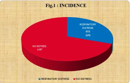
TABLE 1 : INCIDENCE
VARIABLE NUMBER
TOTAL NO. OF ADMISSION 2152
RESPIRATORY DISTRESS IN TERM 655
INCIDENCE 30.4%
Out of 2152 cases admitted during the study period, 655 term newborns were
admitted with respiratory distress. The incidence is 30.4%.
RESPIRATORY DISTRESS
655 30%
NO DISTRESS 1497
Fig.1 : INCIDENCE
40
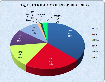
TABLE 2: ETIOLOGY OF RESPIRATORY DISTRESS (n=655)
ETIOLOGY NUMBER PERCENTAGE
TTN 242 36.95
BIRTH ASPHYXIA 144 21.98
MAS 93 14.20
CHD 78 11.91
SEPSIS&PN 73 11.14
RDS 1 0.16
SURGICAL 7 1.07
OTHERS 24 2.59
TOTAL 655 100
In the total 655 cases, TTN was the commonest cause of respiratory distress found
in 242 cases(36.95%), Birth asphyxia was the second common cause found in 144
cases(21.98%), followed by MAS in 93 cases(14.2%), CHD in 78 cases(11.91%),
Sepsis and Pneumonia in 73 cases (11.14%), RDS in 1 case(0.16%), 7(1.07%) were
41
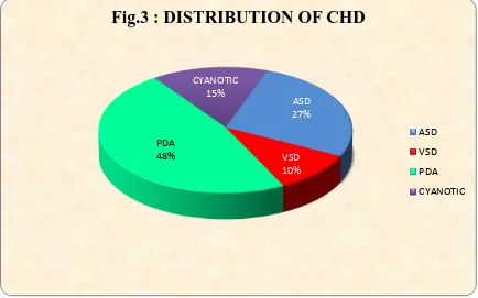
TABLE 3: DISTRIBUTION OF CONGENITAL HEART DISEASES
CAUSE NUMBER PERCENTAGE
ASD 21 26.92
VSD 8 10.26
PDA 37 47.44
CYANOTIC 12 15.38
TOTAL 78 100
TTN 242 37% BA 144 22% MAS 93 14% CHD 78 12% SEPSIS 64 10% PN 9 1% RDS 1 0% SURGICAL 7 1% OTHERS 17 3%
Fig.2 : ETIOLOGY OF RESP. DISTRESS
42
Out of the 78 babies with Congenital heart disease, PDA was the commonest cause
found in 37 babies (47.44%), ASD was found in 21 babies (26.92%), VSD was found
[image:52.612.91.525.173.444.2]in 8 babies (10.26%) and Cyanotic heart diseases were found in 12 cases (15.38%).
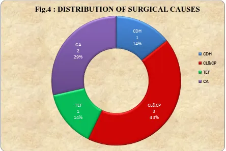
TABLE 4: DISTRIBUTION OF SURGICAL CAUSES
CAUSE NUMBER PERCENTAGE
CDH 1 14.29
CL&CP 3 42.85
TEF 1 14.29
CHOANAL
ATRESIA 2 28.57
ASD 27%
VSD 10%
PDA 48%
CYANOTIC 15%
Fig.3 : DISTRIBUTION OF CHD
ASD
VSD
PDA
43
Out of the 7 surgical causes, 1 was CDH, 3 were Cleft lip and cleft palate, 1 was
Tracheo-esophageal fistula and 2 were Choanal Atresia.
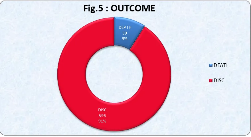
TABLE 5: DISTRIBUTION OF OUTCOME (n=655)
OUTCOME NUMBER PERCENTAGE
DISCHARGE 596 90.99 %
DEATH 59 9.01
TOTAL 655 100%
CDH 1 14%
CL&CP 3 43% TEF
1 14%
[image:53.612.90.534.141.438.2]CA 2 29%
Fig.4 : DISTRIBUTION OF SURGICAL CAUSES
CDH
CL&CP
TEF
44
Out of 655 term newborns with respiratory distress 596 were discharged, 59 died.
Mortality being 9.09%.
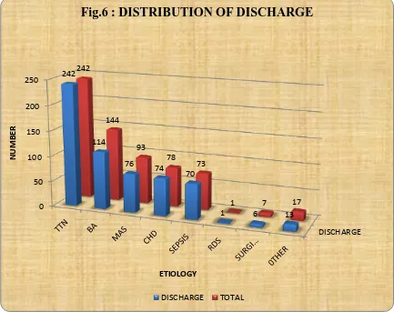
TABLE 6: ETIOLOGYWISE DISTRIBUTION OF DISCHARGE: (n=596)
ETIOLOGY TOTAL DISCHARGED PERCENTAGE
E
TTN 242 242 100
BIRTH
ASPHYXIA
144 114 79.17
MAS 93 76 81.72
CHD 78 74 94.87
SEPSIS&PN 73 70 95.89
RDS 1 1 100
SURGICAL 7 6 85.71
OTHERS 17 13 76.47
TOTAL 655 596 90.99
DEATH 59 9%
[image:54.612.92.532.141.381.2]DISC 596 91%
Fig.5 : OUTCOME
DEATH
45
Out of the total 655 cases admitted, 596 were discharged. All the 242 cases of
TTN were discharged, 114 out of 144 Birth asphyxia cases, 76 out of 93 MAS cases,
74 out 78 CHD cases, 70 out of 73 Sepsis and pneumonia cases, 1 RDS case, 6 out of
7 cases due to surgical causes and 13 out of 17 cases due to Other cases were
discharged.
DISCHARGE 0
50 100 150 200
250 242
114
76 74
70
1 6 13
242
144
93 78
73
1 7 17
N
UM
B
ER
[image:55.612.94.527.314.657.2]ETIOLOGY
Fig.6 : DISTRIBUTION OF DISCHARGE
46
TABLE 7: ETIOLOGY WISE DISTRIBUTION OF DEATH (n=59)
ETIOLOGY DEATH PERCENTAGE
TTN 0 0
BIRTH ASPHYXIA 30 50.85
MAS 17 28.81
CHD 4 6.78
SEPSIS & PN 3 5.08
RDS 0 0
SURGICAL 1 1.69
OTHERS 4 6.79
TOTAL 59 100
Out of total 655 cases included in the study, 59 died with a mortality rate of
9.09 %. Birth asphyxia was the main cause for mortality contributing to 50 % deaths.
MAS was the second common cause for mortality found in 28% cases. CHD was the
cause of death in 6.7%cases, Sepsis and Pneumonia were found as the cause of death
in around 5% cases, surgical causes in 1.6% and other causes were found in 6.79%
cases.
47
[image:57.612.91.536.73.367.2]
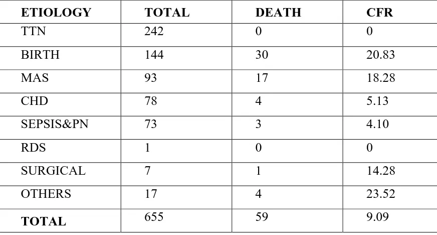
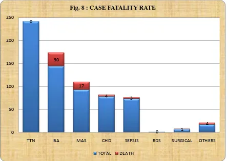
TABLE 8: CASE FATALITY RATE
ETIOLOGY TOTAL DEATH CFR
TTN 242 0 0
BIRTH
ASPHYXIA
144 30 20.83
MAS 93 17 18.28
CHD 78 4 5.13
SEPSIS&PN 73 3 4.10
RDS 1 0 0
SURGICAL 7 1 14.28
OTHERS 17 4 23.52
TOTAL 655 59 9.09
[image:57.612.97.533.441.675.2]TTN 0% BA 30 50% MAS 17 28% CHD 4 7% SEPSIS&PN 3 5% RDS 0% SURGICAL 1 2% OTHERS 5 8%
Fig.7 : DISTRIBUTION OF DEATH
48
Out of 242 TTN cases admitted, all the cases were discharged and no death was
found, with CFR of 0%. Highest Case fatality rates were associated with Birth
asphyxia (20.83%), Pneumonia(22.22%) , Surgical causes(14.28%) and other
[image:58.612.91.534.201.516.2]causes(23.52%). Case fatality rate in MAS was 18.28% and CHD was 5.13%.
TABLE 9: GENDER DISTRIBUTION OF NEONATES(n=655)
GENDER NUMBER PERCENTAGE
Male 416 63.53
Female 239 36.47
Total 655 100
0
30
17
4 3
0 1
4
0 50 100 150 200 250
TTN BA MAS CHD SEPSIS RDS SURGICAL OTHERS
Fig. 8 : CASE FATALITY RATE
49
Out of 655 term newborns with respiratory distress 416 were male, 239 were female. There is a male preponderance.
Table 10 :ETIOLOGY WISE GENDER DISTRIBUTION(n=655)
ETIOLOGY MALE FEMALE
TTN 153 89
BA 86 58
MAS 64 29
CHD 50 28
SEPSIS&PN 48 25
RDS 1 0
SURGICAL 5 2
OTHERS 9 8
TOTAL 416 239
MALE 416 64% FEMALE
239 36%
Fig.9 : GENDER DISTRIBUTION OF NEONATES
MALE
50
Out of the 242 TTN cases 153 were males 89 were females with a male
preponderance. In Birth asphyxia 86 were males, 58 were females. In MAS 64 were
males, 29 were females. In CHD 50 were males 28 were females. In Sepsis and
Pneumonia 48 were males and 25 were females, 5 were males and 2 were females in
surgical causes. In RDS 1 was male. Other causes for distress were found in 9 males
and 8 females.
153 86
64 50 48 1
5 14
89 58
29 28 25
0 2
10
0 50 100 150 200 250
TTN BA MAS CHD SEPSIS RDS SURGICA
L OTHERS
Fig.10 ETIOLOGY WISE GENDER DISTRIBUTION
51
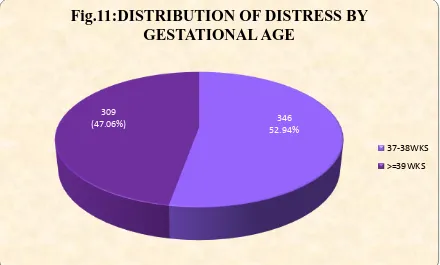
TABLE 11: DISTRIBUTION OF RESPIRATORY DISTRESS BY
GESTATIONAL AGE (n=655)
GESTATIONAL AGE No. PERCENTAGE
37-38 wks(ET) 346 52.94
≥39 wks(LT) 309 47.06
Total 655 100
Out of the 655 term cases, 346 were early term and 309 were late term with more cases of respiratory distress in early term.
346 52.94% 309
[image:61.612.92.532.384.649.2](47.06%)
Fig.11:DISTRIBUTION OF DISTRESS BY
GESTATIONAL AGE
37-38WKS
52
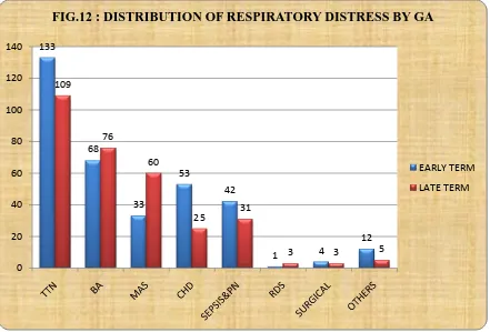
TABLE 12: ETIOLOGY WISE DISTRIBUTION OF RESPIRATORY DISTRESS BY GESTATIONAL AGE (n=655)
ETIOLOGY EARLY TERM LATE TERM
TTN(242) 133 109
BIRTH
ASPHYXIA(144)
68 76
MAS(93) 33 60
CHD(78) 53 25
SEPSIS&PN(73) 42 31
RDS(1) 1 0
SURGICAL(7) 4 3
OTHERS(17) 12 5
TOTAL(655) 346 309
Out of the 242 TTN cases, 133 were early term and 109 were late term, out of 144
birth asphyxia cases, 68 were early term and 76 were late term, out of 78 CHD cases,
53 were early term and 25 were late term, out of 73 sepsis and pneumonia cases 42
were early term and 31 were late term, out of 7 cases due to surgical causes 4 were
early term and 3 were late term , In RDS 1 case was early term, out of 14 cases due to
other causes 12 were early term and 5 were late term. Thus TTN, CHD, SEPSIS and
53
TABLE 13: WEIGHT DISTRIBUTION OF NEONATES (n=655)
BIRTH WEIGHT No. PERCENTAGE
<2.5 or >4 KG 135 20.67
2.5 – 4 KG 520 79.33
TOTAL 655 100
Out of the total 655 cases, 135 babies (20.67%) had a birth weight < 2.5 kg or
> 4 kg. 520 babies (79.3%) had birth weight between 2.5 to 4 kg. 133
68
33
53
42
1 4
12 109
76
60
25 31
3 3 5
0 20 40 60 80 100 120 140
FIG.12 : DISTRIBUTION OF RESPIRATORY DISTRESS BY GA
EARLY TERM
54 TABLE 14: MODE OF DELIVERY
MODE NO. PERCENTAGE
LSCS 341 52.06
VAGINAL
(LN+FORCEPS) 314 47.94
TOTAL 655 100
Out of the total 655 cases, 341 were delivered by LSCS (52.06%) and 314 cases
were delivered by vaginal delivery ( 47.94%), with more LSCS cases.
123, 19% 520, 79%
12, 2%
Fig.13 : BIRTH WEIGHT
<2.5
2.5-4
55
TABLE 15: ETIOLOGYWISE DISTRIBUTION OF MOD (n=655)
ETIOLOGY LSCS VAGINAL
TTN(242) 145(59.91%) 97(40.09%)
BA(144) 48(33.33%) 96(66.67%)
MAS(93) 53(56.99%) 40(43.01%)
CHD(78) 48(61.54%) 30(38.46%)
SEP & PN(73) 33(45.2%) 40(54.8%)
RDS(1) 0(0%) 1(100%)
SURGICAL(7) 5(71.4%) 2(28.6%)
OTHERS(17) 9(52.94%) 8(47.06%)
TOTAL(655) 341 314
LSCS 341 52% VAGINAL
314 48%
Fig.14 : MODE OF DELIVERY
LSCS