V I E W P O I N T
Open Access
Complications of regional citrate
anticoagulation: accumulation or overload?
Antoine G. Schneider
1,2*, Didier Journois
3and Thomas Rimmelé
4,5Abstract
Regional citrate anticoagulation (RCA) is now recommended over systemic heparin for continuous renal replacement therapy in patients without contraindications. Its use is likely to increase throughout the world. However, in the absence of citrate blood level monitoring, the diagnosis of citrate accumulation, the most feared complication of RCA, remains relatively complex. It is therefore commonly mistaken with other conditions. This review aims at providing clarifications on RCA-associated acid-base disturbances and their management at the bedside. In particular, the authors wish to propose a clear distinction between citrate accumulation and net citrate overload.
Keywords:Regional citrate anticoagulation, Continuous renal replacement therapy, Acute kidney injury, Citrate accumulation, Complications of therapy, Metabolic alkalosis
Introduction
Anticoagulation is required during continuous renal replacement therapy (CRRT) to maintain circuit patency. Heparin has historically been the standard choice for anticoagulation [1]. Unfortunately, in fear of bleeding complications, heparin is often administered at sub-therapeutic doses and frequently interrupted for procedures. The resulting anticoagulation is commonly insufficient, lead-ing to poor filter life [2].
Regional citrate anticoagulation (RCA) is an appealing alternative since it provides excellent anticoagulation within the circuit without increasing the risk of bleeding [2, 3]. In randomized controlled trials [4–6] and meta-analyses [7, 8], RCA has been shown to increase filter life and decrease the rate of complications, therapy interrup-tions, and costs compared with heparin. RCA has been used in large tertiary centers with very low complication rates [9]. RCA is now recommended as the first line antic-oagulation strategy for CRRT in patients without contrain-dications [10].
Given these recommendations, RCA is likely to be gradually adopted in an increasing number of centers, including smaller and non-academic hospitals with less experience with CRRT. The implementation of RCA
requires particularly strict protocols and specific training of both medical and nursing staff. Indeed, unguided RCA might lead to potentially disastrous complications off-setting its potential benefits. Currently published literature might lead to some confusion regarding the interpretation of RCA complications, in particular regarding acid-base derangements.
This viewpoint aims at providing clarifications on citrate-associated acid-base disturbances and their man-agement at the bedside. In particular, the authors wish to propose a clear distinction between citrate accumula-tion and citrate overload, two intertwined noaccumula-tions, which are commonly confused.
General principles
Principles of citrate anticoagulation
Citrate (C6H5O7) is an organic acid. It is commonly used as an anticoagulant as trisodium citrate and, for stored blood products, as acid citrate dextrose (ACD). Citrate anticoagulant properties are related to its high affinity for the divalent calcium ion (Ca++). The addition of cit-rate to blood results in the formation of citcit-rate–calcium complexes (CCC), effectively decreasing the level of ion-ized free calcium. Ionion-ized magnesium is also chelated by citrate but to a lesser extent. Since calcium is a mandatory co-factor of most enzymes of the coagulation cascade, citrate-mediated decrease in plasma calcium
* Correspondence:antoine.schneider@chuv.ch
1Adult Intensive Care Unit, Centre Hospitalier Universitaire Vaudois (CHUV), 46 avenue du Bugnon, 1011 Lausanne, Switzerland
2Université de Lausanne, UNIL, Lausanne, Switzerland
Full list of author information is available at the end of the article
levels below 0.35 mmol/l results in very effective antic-oagulation (Fig. 1) [11, 12].
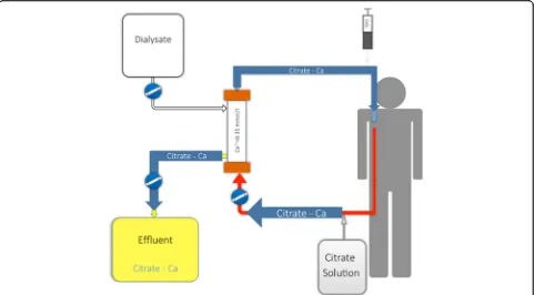
Numerous protocols for RCA have been proposed and tested [13, 14]. They differ by solution type (ACD, triso-dium citrate, diluted citrate solutions) and CRRT modal-ity (continuous veno-venous hemofiltration (CVVH), continuous veno-venous hemodialysis (CVVHD), con-tinuous veno-venous hemodiafiltration (CVVHDF)). All these protocols require pre-filter administration of a cit-rate solution at the required dose to reach approximately 3 to 4 mmol of citrate per liter of blood in the circuit. Such a dose is usually sufficient to decrease ionized cal-cium to the target range (0.2 to 0.35 mmol/l according to the protocol used). Post-filter calcium is monitored to ensure adequate anticoagulation and permit citrate dose adjustment according to pre-defined models. In current CRRT machines, citrate administration rate is coupled with blood flow, minimizing the risk of variation in cit-rate concentration. A calcium chloride solution needs to be administered either at the end of the circuit or dir-ectly through a separated central line to compensate for calcium loss in the effluent in the form of CCC (Fig. 2). Calcium reinfusion rate is adjusted according to sequen-tially measured systemic ionized calcium level (targeting physiological range). After the initiation phase, regular monitoring (every 6 h) of post-filter, systemic, and total calcium levels (with total/ionized ratio calculation) should be performed. Since, according to the compos-ition of dialysate/replacement fluids used, magnesium might also need to be supplemented, daily monitoring of serum magnesium levels is also advisable.
Citrate clearance and metabolism
As depicted in Fig. 2, a large portion of CCC is removed through the hemofilter [15]. CCC clearance is very high
(up to 60%) due to their low molecular weight (298 Daltons) associated with their high hydrosolubility con-ferred by the negative charge of a free carboxylate rad-ical. Their sieving coefficient is 1.0. The clearance must be maintained as high as possible to minimize the administration of citrate to the patient. This clearance increases with the dialysate flow (the higher the dialysate flow, the higher the clearance). In convective modes, cit-rate’s clearance is dependent on filtration flow (the higher the filtration flow, the higher the clearance). CCC which are not removed through the hemofilter return to the patient. They are metabolized in the liver, muscle, and kidney fitting into the Krebs (citric acid) cycle. Under normal conditions, citrate’s half-life is approxi-mately 5 minutes. The process generates energy (2.48 KJ or 593 calories per mmol of citrate), releases sodium as well as calcium ions [16].
Citrate and acid-base balance
Acid-base consequences of RCA are often reduced to bicarbonate generation by citrate metabolism. Unfortu-nately, this simplification is inaccurate and correct un-derstanding of citrate’s effect on acid-base balance requires the use of Stewart’s global approach [17–19]. Briefly, according to this approach, blood pH is mainly determined by three variables: PaCO2, strong ion differ-ence (SID), and weak acids concentration. Citrate belongs to the weak acid category and its effect should be to markedly acidify a solution. Its three carboxylate radicals have respective pKa values of 5.21, 4.28, and 2.92 at 25 °C) [20]. However, in plasma, unless the cal-cium level is extremely low (to levels incompatible with life), citrate is only present in the form of CCC. In that form, its acidifying capacity is limited by the binding of ionized calcium to two adjacent carboxylates, leaving only one residual anionic charge (Fig. 3). Circulating CCC therefore lead to mild plasma acidification. Under normal conditions, this effect is negligible since CCC are rapidly cleared from the blood.
However, the acid-base impact of RCA is not limited to the effect of citrate itself. Indeed, the composition and amount of dialysis/substitution fluid used are of major importance. Many citrate solutions have a high sodium content (three Na+ for one citrate molecule). This net sodium administration tends to increase plasma SID leading to plasma alkalinization.
Overall, when citrate catabolism is normal, RCA leads to plasma alkalinization. This alkalinizing effect is max-imal with trisodium citrate solutions and less marked with ACD solutions (which have a low sodium content). To some extent, this alkalinization is desirable as it buffers acute kidney injury associated-acidosis and normalizes pH. As discussed in further sections, in some clinical
Fig. 1“ON-OFF”anticoagulation effect of ionized hypocalcemia. The
[image:2.595.58.290.524.684.2]situations where citrate catabolism is markedly impaired, CCC tend to accumulate, generating a mild acidosis.
Citrate accumulation and alternative diagnoses
Citrate accumulation is a feared and potentially lethal complication of RCA. Fortunately, when a strict protocol is followed, it is rarely encountered [9]. In order to avoid unnecessary therapy interruptions, it is essential for the
clinician to distinguish citrate accumulation from other situations resulting in acid-base disturbance during RCA: citrate net overload and insufficient trisodium-citrate delivery. The main differences between these entities are summarized in Table 1.
Citrate accumulation
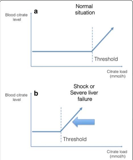
The body’s capacity to metabolize citrate is saturable (Fig. 4). If citrate administration exceeds this capacity, residual citrate, in the form of CCC remain in blood. In the absence of a routinely available assay for citrate blood level, citrate accumulation can only be suspected through indirect signs. The most reliable sign for citrate accumulation is probably an increased total/ionized cal-cium (Ca/Ca++) ratio. Indeed, an increase in this ratio demonstrates an increase in the serum level of anion-bound calcium, which in the context of RCA is almost synonymous with circulating CCC. A cut-off value of 2.5 is usually recognized as indicative of significant accumu-lation but a trend towards this value is highly indicative of ongoing accumulation.
Other signs are commonly observed during citrate accumulation. These signs should not be considered as diagnostic criteria but represent warning signs of poten-tial citrate accumulation. Of those, an increase in cal-cium substitution needs might suggest the absence of
Fig. 2Schematic view of a CRRT circuit with regional citrate administration in CVVHD mode. Alternative modes can be used (postdilution CVVH, combined pre- and postdilution CVVH, CVVHDF, etc.) according to the protocol used. Citrate solution is administered at the beginning of the CRRT circuit. It forms citrate–calcium complexes, which are largely removed from the blood at the level of the filter. Only complexes which are not removed through the hemofilter return to the patient’s blood and need to be metabolized
[image:3.595.58.539.88.354.2] [image:3.595.56.291.538.665.2]CCC-bound calcium release and should prompt particu-lar attention from clinicians. In overt citrate accumula-tion, hypocalcemia is usually observed, potentially leading to severe complications. Similarly, recurrence of high anion gap metabolic acidosis and increased serum lactate levels are also frequently observed concomitant to citrate accumulation. These anomalies are not
thought to be secondary to citrate accumulation itself but rather to a common primary process impairing the tricarboxylic acid cycle, reducing citrate metabolism, and limiting pyruvate metabolism leading to lactate gener-ation. Accumulated CCC participate in the elevated anion gap as well as to a strong ion gap. Pathophysiologic conse-quences of CCC accumulation are presented in Fig. 5.
Net citrate overload
[image:4.595.55.537.98.279.2]Net citrate overload is a common, benign, and easy to manage complication of RCA. Citrate overload is a situ-ation in which the organism’s capacity to metabolize cit-rate is not reached and all citrate–calcium complexes are metabolized (Fig. 4). The concomitant net load of sodium ions leads to plasma alkalinization through an increased SID. No increase in total/ionized calcium is ob-served and ionized calcium levels remain normal. Net Table 1Citrate accumulation and alternative diagnoses: summary table
Citrate accumulation Citrate net overload Insufficient trisodium citrate delivery
Mechanism Incomplete citrate metabolism: persistence of circulating citrate–calcium complexes in the blood
Excess citrate administration relative to buffer requirements
Insufficient alkalotic load administered to the patient to adequately buffer acute kidney injury-associated acidosis
Diagnosis
Acid-base Metabolic acidosis Metabolic alkalosis Metabolic acidosis
Catot/Cairatio Increased (>2.5) Normal (< 2.5) Normal (< 2.5)
Other Increased need for calcium substitution Trend for a decreased ionized calcium level
None None
Appreciation Potentially lethal (via severe hypocalcemia) Benign and easy to fix Benign and easy to fix
Incidence Rare Common Rare
Management Decrease blood flow or increase dialysate flow rate (if mild) Consider alternative anticoagulation strategy
Decrease blood flow or increase dialysate flow rate
Increase blood flow or decrease dialysate flow rate
Fig. 4Theoretical relationship between blood citrate level and citrate load.aAn increase in citrate load is not associated with an increase in blood citrate level until a threshold is reached. This threshold corresponds to the body’s capacity to metabolize citrate.bCertain circumstances, such as severe liver failure or circulatory shock, might result in a lower threshold
[image:4.595.57.291.385.665.2] [image:4.595.303.539.525.713.2]citrate overload is a sign of excessive citrate administra-tion or, more frequently, of low clearance in the hemofilter.
Insufficient trisodium-citrate delivery
Insufficient trisodium-citrate delivery is a situation where the alkalotic load administered to the patient is insufficient to adequately buffer acute kidney injury-associated acid-osis, resulting in residual metabolic acidosis. This may happen if blood flow is set too low proportional to dialys-ate flow.
In this situation, the observed metabolic acidosis should not be interpreted as resulting from citrate accu-mulation. On the contrary, the adequate response should be toincreasethe blood flow or todecreasethe dialysate flow. Key elements here are the normal total/ionized cal-cium ratio and calcal-cium substitution rate.
Situations at risk of citrate accumulation or net overload Some situations lead to increased citrate delivery or de-creased metabolizing capacity. According to the extent of this process, and the patient’s ability to metabolize CCC, it might lead to citrate accumulation or net overload (Fig. 4).
Excess citrate delivery to the patient
Accidental excess citrate infusion might occur in cases of incorrect circuit setup (e.g.,post-filter citrate adminis-tration) or administration of citrate when the blood pump is stopped. These issues are now unlikely with new generation CRRT devices with built-in citrate mod-ules designed to prevent handling errors and increase safety. In particular, citrate administration is coupled with the blood pump. Such devices make use of specific tubes and connections as well as color codes, minimiz-ing the risk of errors durminimiz-ing circuit setup and use.
Citrate removal at the hemofilter level can be impaired, resulting in excess citrate delivery to the patient. Such an issue might occur in CVVH mode when the ultrafiltration rate is set too low or in CVVHD mode when an insuffi-cient dialysate rate is set. These complications should be prevented by adherence to a strict protocol. Rapid loss of clearance at the filter level is occasionally observed in some patients with early clogging of the membranes. In such situations, the patient’s citrate delivery is higher than expected by the mathematical model driving the pumps and overload might occur. In this case, rapid replacement of the circuit is necessary. Of note, such a situation is un-likely to occur in CVVH mode, since early clogging would be identified by an increased transmembrane pressure.
Most of these situations may be prevented by medical and nursing education and their frequency should decrease with increasing experience.
Decreased citrate metabolization
In some situations citrate metabolization is decreased (Fig. 4b). A patient’s capacity to metabolize citrate is a dynamic process depending on baseline characteristics and hemodynamic status as well as mitochondrial func-tion. Therefore, such situations are difficult to predict a priori, but some groups of patients should be considered at risk.
Patients with acute liver failure or acute-on-chronic liver failure have classically been described as having decreased citrate metabolizing capacity. However, recent literature has suggested that most patients in these situa-tions could process citrate anyway and that classic markers of liver function were poor predictors of the risk of citrate accumulation [21–23]. As depicted in Fig. 4b, the ability of these patients to metabolize citrate is not null but simply decreased. Therefore, a protocol associated with low citrate delivery (normal or mildly reduced doses associated to increased clearance) to the patient is likely to be tolerated in most situations.
Patients with circulatory shock are likely to have a de-creased oxygen delivery to the cells with dede-creased Krebs cycle activity due to reduced activity of the mitochondrial oxidation chain. Similarly, some commonly encountered intoxications (biguanides (e.g., metformin), cyclosporine, paracetamol, trichloroethylene, or propofol) can lead to mitochondrial blockage and decrease citrate metaboliza-tion capacity [24]. In these situametaboliza-tions, a transient decrease in citrate metabolizing capacity is likely.
Since all these situations are typically associated with elevated serum lactate levels, such measurement is an important indicator of the body’s capacity to metabolize citrate. However, the lactate threshold above which RCA should not be used remains to be determined.
Management
When either citrate accumulation or overload is sus-pected, the net citrate load finally administered to the patient must rapidly be decreased. According to the protocol used, this can be obtained either by 1) decreas-ing the blood flow rate (decreases intake through blood flow–citrate coupling) or 2) increasing the dialysate rate (CVVHD) or the filtration rate (CVVH) (increases removal), or 3) decreasing the targeted citrate concen-tration within the filter.
reduction. Normalization of the pH, however, is a slow process and its correction requires time.
Minimizing the risk of citrate accumulation
A strict protocol for RCA should be applied in all cen-ters for all patients. Particular care should be taken in patients with suspected decreased citrate metabolization capacity (acute liver failure, circulatory shock, and intox-ications). In centers with limited experience with the technique, RCA should probably be considered as con-traindicated in such patients.
In addition to close monitoring of ionized calcium (post-filter for efficacy and systemic for safety), regular assessments of total/ionized Ca2+ and pH must be performed.
In general, well-designed protocols should aim to minimize citrate delivery to patients. This goal can be achieved by combining several measures:
1. Limited blood flow should be used. Indeed, since citrate administration is coupled to blood flow, lower blood flow means less need for citrate. This can easily be achieved in diffusion-based modes. Of note, in diffusive modes, low blood flows do not translate into low blood purification for two reasons: 1) di-alysate rate remains the limiting factor and 2) high flux membranes are preferred for RCA, allowing im-portant clearance even with reduced blood flows. Most protocols using diffusive modes would recom-mend blood flow between 80 and 150 ml/min. Purely convective techniques can be used but with a higher risk of metabolic complications. Indeed, the combination of low blood flows (to limit citrate ad-ministration) and high filtration rates (to optimize CCC clearance) would lead to high filtration frac-tion, increasing the risk of membrane clogging and decreased CCC clearance. This issue can be mini-mized if diluted citrate solutions are used as predilution.
2. High dialysate/filtration rates should be favored to increase citrate removal.
Conclusions
Based on recent recommendations, the use of RCA is likely to increase dramatically throughout the world. RCA protocols should aim to minimize the amount of net citrate load delivered to the patient. In case of acid-base derangement during RCA, clinicians should be able to differentiate a benign citrate net overload (alkalosis) from a life-threatening accumulation (elevated Ca/Ca++, increase need for calcium substitution, and trend toward acidosis), which should prompt therapy tuning and early termination. The distinction between these two entities
is crucial to enable adequate patient surveillance while receiving RCA.
Abbreviations
ACD:Acid citrate dextrose; CCC: Citrate–calcium complexes; CRRT: Continuous renal replacement therapy; CVVH: Continuous veno-venous hemofiltration; CVVHD: Continuous veno-venous hemodialysis; CVVHDF: Continuous veno-venous hemodiafiltration; RCA: Regional citrate anticoagulation; SID: Strong ion difference
Acknowledgements
None.
Funding
None
Availability of data and materials
Not applicable.
Authors’contributions
AS reviewed the literature and drafted the review and DJ and TR critically reviewed the manuscript. All authors read and approved the final manuscript.
Ethics approval and consent to participate
Not applicable.
Consent for publication
Not applicable.
Competing interests
AS received speaker honoraria from Fresenius Medical Care and Baxter Healthcare Corp. and consulting honoraria from B. Braun Melsungen AG. DJ has no competing interests to disclose. TR received speaker honoraria from Fresenius Medical Care, Baxter Healthcare Corp., and Bellco-Medtronic.
Publisher’s Note
Springer Nature remains neutral with regard to jurisdictional claims in published maps and institutional affiliations.
Author details 1
Adult Intensive Care Unit, Centre Hospitalier Universitaire Vaudois (CHUV), 46 avenue du Bugnon, 1011 Lausanne, Switzerland.2Université de Lausanne, UNIL, Lausanne, Switzerland.3Anesthesiology and Intensive Care Medicine, Cochin Hospital, Assistance Publique Hôpitaux de Paris, René Descartes University, Paris, France.4Anesthesiology and Intensive Care Medicine, Edouard Herriot Hospital, Hospices Civils de Lyon, Lyon, France.5EA 7426 (Université Claude Bernard Lyon 1–Hospices Civils de Lyon–bioMérieux) “Pathophysiology of Injury-induced Immunosupression–PI3”, Joint Research Unit, Edouard Herriot Hospital, Lyon, France.
Received: 31 July 2017 Accepted: 31 October 2017
References
1. Uchino S, Bellomo R, Morimatsu H, Morgera S, Schetz M, Tan I, Bouman C, Macedo E, Gibney N, Tolwani A, et al. Continuous renal replacement therapy: a worldwide practice survey. The beginning and ending supportive therapy for the kidney (B.E.S.T. kidney) investigators. Intensive Care Med. 2007;33(9):1563–70.
2. Joannidis M, Oudemans-van Straaten HM. Patency of the circuit in continuous renal replacement therapy. Crit Care. 2007;11(4):218. 3. Mehta RL, McDonald BR, Aguilar MM, Ward DM. Regional citrate
anticoagulation for continuous arteriovenous hemodialysis in critically ill patients. Kidney Int. 1990;38(5):976–81.
4. Stucker F, Ponte B, Tataw J, Martin PY, Wozniak H, Pugin J, Saudan P. Efficacy and safety of citrate-based anticoagulation compared to heparin in patients with acute kidney injury requiring continuous renal replacement therapy: a randomized controlled trial. Crit Care. 2015;19:91.
for continuous renal replacement therapy in critically ill adults. Crit Care Med. 2015;43(8):1622–9.
6. Schilder L, Nurmohamed SA, Bosch FH, Purmer IM, den Boer SS, Kleppe CG, Vervloet MG, Beishuizen A, Girbes AR, Ter Wee PM, et al. Citrate
anticoagulation versus systemic heparinisation in continuous venovenous hemofiltration in critically ill patients with acute kidney injury: a multi-center randomized clinical trial. Crit Care. 2014;18(4):472.
7. Zhang Z, Hongying N. Efficacy and safety of regional citrate anticoagulation in critically ill patients undergoing continuous renal replacement therapy. Intensive Care Med. 2012;38(1):20–8.
8. Bai M, Zhou M, He L, Ma F, Li Y, Yu Y, Wang P, Li L, Jing R, Zhao L, et al. Citrate versus heparin anticoagulation for continuous renal replacement therapy: an updated meta-analysis of RCTs. Intensive Care Med. 2015;41(12):2098–110.
9. Khadzhynov D, Schelter C, Lieker I, Mika A, Staeck O, Neumayer HH, Peters H, Slowinski T. Incidence and outcome of metabolic disarrangements consistent with citrate accumulation in critically ill patients undergoing continuous venovenous hemodialysis with regional citrate anticoagulation. J Crit Care. 2014;29(2):265–71.
10. KDIGO. Kidney disease: improving global outcomes (KDIGO) acute kidney injury work group (2012) KDIGO clinical practice guidelines AKI: AKI definition. Kidney Int. 2012;2(1):19–36.
11. James MF, Roche AM. Dose-response relationship between plasma ionized calcium concentration and thrombelastography. J Cardiothorac Vasc Anesth. 2004;18(5):581–6.
12. Calatzis A, Toepfer M, Schramm W, Spannagl M, Schiffl H. Citrate anticoagulation for extracorporeal circuits: effects on whole blood coagulation activation and clot formation. Nephron. 2001;89(2):233–6. 13. Morgera S, Schneider M, Slowinski T, Vargas-Hein O, Zuckermann-Becker H,
Peters H, Kindgen-Milles D, Neumayer HH. A safe citrate anticoagulation protocol with variable treatment efficacy and excellent control of the acid-base status. Crit Care Med. 2009;37(6):2018–24.
14. Tolwani AJ, Prendergast MB, Speer RR, Stofan BS, Wille KM. A practical citrate anticoagulation continuous venovenous hemodiafiltration protocol for metabolic control and high solute clearance. Clin J Am Soc Nephrol. 2006;1(1):79–87.
15. Balik M, Zakharchenko M, Otahal M, Hruby J, Polak F, Rusinova K, Stach Z, Vavrova J, Jabor A. Quantification of systemic delivery of substrates for intermediate metabolism during citrate anticoagulation of continuous renal replacement therapy. Blood Purif. 2012;33(1-3):80–7.
16. Balik M, Zakharchenko M, Leden P, Otahal M, Hruby J, Polak F, Rusinova K, Stach Z, Tokarik M, Vavrova J, et al. Bioenergetic gain of citrate anticoagulated continuous hemodiafiltration–a comparison between 2 citrate modalities and unfractionated heparin. J Crit Care. 2013;28(1):87–95. 17. Stewart PA. Modern quantitative acid-base chemistry. Can J Physiol
Pharmacol. 1983;61(12):1444–61.
18. Adrogue HJ, Madias NE. Assessing acid-base status: physiologic versus physicochemical approach. Am J Kidney Dis. 2016;68(5):793–802. 19. Nagaoka D, Nassar Junior AP, Maciel AT, Taniguchi LU, Noritomi DT,
Azevedo LC, Neto LM, Park M. The use of sodium-chloride difference and chloride-sodium ratio as strong ion difference surrogates in the evaluation of metabolic acidosis in critically ill patients. J Crit Care. 2010;25(3):525–31. 20. Goldberg RNKN, Lennen RM. Thermodynamic quantities for the ionization
reactions of buffers. J Phys Chem Ref Data. 2002;31(2):231–370. 21. Schultheiss C, Saugel B, Phillip V, Thies P, Noe S, Mayr U, Haller B,
Einwachter H, Schmid RM, Huber W. Continuous venovenous hemodialysis with regional citrate anticoagulation in patients with liver failure: a prospective observational study. Crit Care. 2012;16(4):R162. 22. Slowinski T, Morgera S, Joannidis M, Henneberg T, Stocker R, Helset E,
Andersson K, Wehner M, Kozik-Jaromin J, Brett S, et al. Safety and efficacy of regional citrate anticoagulation in continuous venovenous hemodialysis in the presence of liver failure: the Liver Citrate Anticoagulation Threshold (L-CAT) observational study. Crit Care. 2015;19:349.
23. De Vico P, Messino V, Tartaglione A, Beccaris C, Buonomo C, Talarico D, Prati P, Sabato AF, Colella DF. Safety and efficacy of citrate anti-coagulation continuous renal replacement therapies in post-cardiac surgery patients with liver dysfunction. Ther Apher Dial. 2015;19(3):272–8.
![Fig. 1 “ON-OFF” anticoagulation effect of ionized hypocalcemia. Thegrey zone corresponds to the area of adequate anticoagulation.Target values indicated are only indicative and depend on theprotocol used [12]](https://thumb-us.123doks.com/thumbv2/123dok_us/8348849.308588/2.595.58.290.524.684/anticoagulation-hypocalcemia-corresponds-adequate-anticoagulation-indicated-indicative-theprotocol.webp)