R E S E A R C H
Open Access
Determinants of long-term outcome in ICU
survivors: results from the FROG-ICU study
Etienne Gayat
1,28*, Alain Cariou
2, Nicolas Deye
3, Antoine Vieillard-Baron
4, Samir Jaber
5, Charles Damoisel
1,
Qin Lu
6, Xavier Monnet
7, Isabelle Rennuit
8, Elie Azoulay
9, Marc Léone
10, Heikel Oueslati
1, Bertrand Guidet
11,
Diane Friedman
12, Antoine Tesnière
13, Romain Sonneville
14, Philippe Montravers
15, Sébastien Pili-Floury
16,
Jean-Yves Lefrant
17,18, Jacques Duranteau
19, Pierre-François Laterre
20, Nicolas Brechot
21, Karine Chevreul
22,
Morgane Michel
23, Bernard Cholley
24, Matthieu Legrand
1, Jean-Marie Launay
24, Eric Vicaut
25, Mervyn Singer
26,
Matthieu Resche-Rigon
27†and Alexandre Mebazaa
1†Abstract
Background:Intensive care unit (ICU) survivors have reduced long-term survival compared to the general population. Identifying parameters at ICU discharge that are associated with poor long-term outcomes may prove useful in targeting an at-risk population. The main objective of the study was to identify clinical and biological determinants of death in the year following ICU discharge.
Methods:FROG-ICU was a prospective, observational, multicenter cohort study of ICU survivors followed 1 year after discharge, including 21 medical, surgical or mixed ICUs in France and Belgium. All consecutive patients admitted to intensive care with a requirement for invasive mechanical ventilation and/or vasoactive drug support for more than 24 h following ICU admission and discharged from ICU were included. The main outcome measure was all-cause mortality at 1 year after ICU discharge. Clinical and biological parameters on ICU discharge were measured, including the circulating cardiovascular biomarkers N-terminal pro-B type natriuretic peptide, high-sensitive troponin I, bioactive-adrenomedullin and soluble-ST2. Socioeconomic status was assessed using a validated deprivation index (FDep).
Results:Of 1570 patients discharged alive from the ICU, 333 (21%) died over the following year. Multivariable analysis identified age, comorbidity, red blood cell transfusion, ICU length of stay and abnormalities in common clinical factors at the time of ICU discharge (low systolic blood pressure, temperature, total protein, platelet and white cell count) as independent factors associated with 1-year mortality. Elevated biomarkers of cardiac and vascular failure independently associated with 1-year death when they are added to multivariable model, with an almost 3-fold increase in the risk of death when combined (adjusted odds ratio 2.84 (95% confidence interval 1.73–4.65),p< 0.001).
Conclusions:The FROG-ICU study identified, at the time of ICU discharge, potentially actionable clinical and biological factors associated with poor long-term outcome after ICU discharge. Those factors may guide discharge planning and directed interventions.
Trial registration:ClinicalTrials.gov NCT01367093. Registered on 6 June 2011.
Keywords:Post-intensive care syndrome, Long-term survival, Biomarkers, Score, Discharge
* Correspondence:etienne.gayat@aphp.fr †Equal contributors
1Department of Anesthesiology, Critical Care and Burn Unit, Hôpitaux
Universitaires Saint Louis—Lariboisière, Assistance Publique—Hôpitaux de Paris, Université Paris Diderot—Paris 7, Sorbonne Paris Cité, UMR-S 942, INSERM, Paris, France
28Department of Anesthesiology and Intensive Care, University Paris Diderot,
INSERM UMR-S 942, Saint Louis—Lariboisière University Hospitals, 2 rue Ambroise Paré, 75010 Paris, France
Full list of author information is available at the end of the article
Background
Survivors of critical illness will face a period of increased risk of reduced long-term survival and impaired quality of life compared to the general population [1]. This period, lasting several years, is associated with an increased risk of posttraumatic stress, depression, cognitive impairment and physical weakness, all grouped under the entity“ post-intensive care syndrome”(PICS) [2].
To reduce the mortality rate of intensive care unit (ICU) survivors, it is important to identify the group of patients who have a higher probability of death in the year follow-ing ICU discharge and to recognize the adjustable factors associated with mortality. Although data have been pub-lished regarding the long-term outcome of ICU patients, there are no recommendations for the long-term manage-ment of these patients. Only experts’opinions have been published [2, 3]. Some studies have demonstrated that mortality rates among ICU survivors are higher compared to the general population [4–8] and that an ICU stay im-pacts on patients’quality of life [9] and disability [10, 11]. Moreover, other studies [5, 6] found that this over-risk of mortality is sustained after 5–15 years of follow-up. Three studies [4, 7, 8] reported a worse survival rate for ICU pa-tients compared to an age-matched control population in the years following ICU discharge. Although we under-stand that age, comorbidity burden and severity of acute illness are important predictors of late mortality as described previously [12], we know less about clinical and laboratory values at the time of ICU discharge.
The transition of care from ICU to ward and, eventually, to home is a complex process with many challenges. We hy-pothesized that clinical and biological abnormalities present on the day of ICU discharge are associated with worse long-term outcome. In particular, we hypothesized that ICU sur-vivors are at long risk of increased cardiovascular events, as suggested previously [13]. Among biological abnormalities, we focused on circulating cardiovascular biomarkers, namely N-terminal pro-B type natriuretic peptide (NT-proBNP), high-sensitive troponin I (hs-TnI), bioactive-adrenomedullin (bio-ADM) and soluble-ST2 (sST2). The choice of those four biomarkers was guided by their relative function, with NT-proBNP a marker of cardiac congestion, hs-TnI a marker of cardiac injury, sST2 a marker of cardiac remodeling and bio-ADM a marker of vascular dysfunction.
Accordingly, the FROG-ICU (French and European Outcome reGistry in Intensive Care Units) study aimed to identify clinical and biological (including cardiovascu-lar biomarkers) parameters associated with long-term outcome in ICU survivors.
Methods Study design
FROG-ICU was a prospective, observational, multicenter cohort study in which survivors of critical illness were
followed up for up to 1 year post ICU discharge. The study was conducted in France and Belgium in accord-ance with Good Clinical Practice (Declaration of Helsinki 2002) and Ethical Committee approvals (Comité de Protection des Personnes—Ile de France IV, IRB n°00003835 and Commission d’éthique biomédicale hospitalo-facultaire de l’hôpital de Louvain, IRB n° B403201213352). It is registered on ClinicalTrials.gov (NCT01367093). Patients were included from August 2011 to June 2013. Details of design and methods have been published previously [14]. All patients admitted to any of the participating centers during the recruitment period who met the eligibility criteria and survived their ICU stay had a clinical examination and biological tests performed at discharge from the ICU, and were followed up for 1 year through telephone calls and postal ques-tionnaires at 3, 6 and 12 months.
Participants
The study involved 21 medical, surgical or mixed ICUs in 14 university hospitals. Inclusion criteria were: inva-sive mechanical ventilation support for at least 24 h and/or treatment with a vasoactive agent (except dopa-mine) for more than 24 h. Noninclusion criteria were: age younger than 18 years old; severe head injury (initial Glasgow Coma Scale≤8), brain death or a persistent vegetative state; pregnancy or breastfeeding; transplant-ation in the past 12 months; moribund patient; and/or no social security coverage. The Ethical Committees waived the need for written consent; all patients and/or next of kin were informed and oral consent was docu-mented in the patients’ medical records by the investigator.
Study objectives
The primary purpose of the FROG-ICU study was to as-sess the incidence of all-cause mortality in the year fol-lowing ICU discharge, and to identify independent factors associated with mortality. The main secondary objective of FROG-ICU was to evaluate the association between circulation cardiovascular biomarkers levels at discharge and 1-year mortality.
Data collection
dysfunction [15] (bio-ADM; Adrenomed GmbH, Hen-nigsdorf, Germany); and cardiac stress (sST2; Eurobio, Critical Diagnostics, San Diego, CA, USA) which prog-nosticates for cardiovascular death [16, 17]. The deprivation index (FDep) was used as a measure of so-cioeconomic inequalities in health status. The FDep is based on the patients’ residential zip codes and was specifically developed for the French context using the following four variables to compute a single composite index: median household income, percentage of high school graduates in the population aged≥15 years, per-centage of blue-collar workers in the active population and unemployment rate [18].
Statistical analysis
Results are expressed as median (interquartile range (IQR)) or count (percentage) as appropriate. The primary analysis examining factors associated with 1-year mortality was based on analysis of the clinical and biological vari-ables measured in patients discharged alive from the ICU. Marginal associations between single variables and 1-year mortality were assessed by a Wilcoxon rank-sum test for quantitative variables and the chi-square test for qualita-tive variables. Multivariable logistic regression was used to determine a set of variables independently associated with 1-year mortality. Variables associated with outcome at a 0.05 level and with less than 20% of missing data were considered within the multivariable model. The log-linearity of the quantitative variables was evaluated systematically, and, if appropriate, variable transformation was performed. Log-linearity of the association between continuous variables and the outcome was checked using a cubic spline and the Wald test. Cutoff values were de-rived from the plots of the effect according to the value of the variable of interest. Missing values were handled by multiple imputation by chained equations (MICE) [19]. All variables selected for the multivariable model were considered in the imputation model. A total of 51 imputed samples was generated using 15 iterations of the chained equation process. A selection model process was per-formed using a backward stepwise approach with stopping rules based on a cutoff at 0.05 forpvalues. At each step of the selection, inference was combined from the sets of im-puted samples using Rubin’s rules [20]. The existence of any colinearities was observed, and a test of goodness of fit was performed using the Hosmer–Lemeshow test on the complete case model [21]. Measures of association consisted of odds ratios (ORs) and their confidence inter-vals (CIs) at 95% estimated using Rubin’s rules. The pre-dictive power of the four biomarkers of interest was assessed using receiver operating curve (ROC) analyses. The area under the ROC (AUC) was estimated for each biomarker. For both the clinical model and the clinical model including biomarker information, the AUCs were
estimated from the sets of imputed samples using Rubin’s rules. The latter two were compared using the Delong test. As it is now recognized that highlighting a statistically sig-nificant association between new biomarkers and patient outcomes is not sufficient to demonstrate the interest of these biomarkers in terms of risk prediction [22–24], we used the proposed methodology of Pencina et al. [23], which has been used in multiple articles of application. The net reclassification improvement (NRI) and integrated discrimination improvement (IDI) of each biomarker added to the full clinical model will be calcu-lated, and comparisons between different biomarkers will be performed [23].
Calculating the number of subjects required was based on the primary endpoint; that is, the risk factors associ-ated with 1-year all-cause mortality. Study of the litera-ture and preliminary studies conducted in December 2009 in 14 participating centers led us to estimate a 1-year mortality after ICU discharge of 18%. To ensure de-tection with a power of 80% for the dede-tection of binary prognostic factors with a prevalence of 33% and an ex-pected OR of 1.5 in a population with a probability of death in the year following ICU discharge of approxi-mately 18%, 1636 patients were required [25]. Assuming a 10% rate of refusal and/or loss to follow-up, the num-ber of patients to be enrolled was raised to 1800. Finally, since the expected in-ICU mortality rate was 25%, the total number of patients included in the study was 2250.
p< 0.05 was considered statistically significant. All statis-tical analyses were performed using R statisstatis-tical software version 3.1.1 or above (The“R”Foundation for Statistical Computing, Vienna, Austria).
[image:3.595.306.539.500.703.2]Results
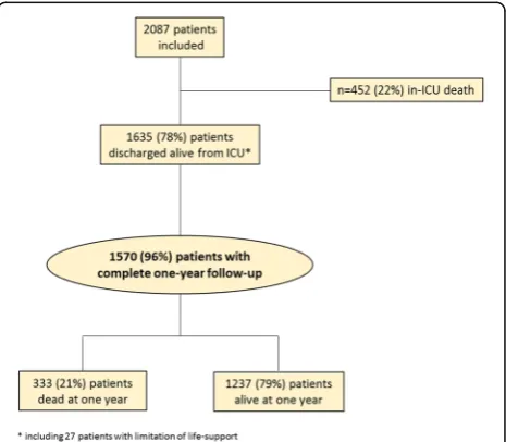
Of the 2087 ICU patients who consented to participate in the FROG-ICU study, 1570 were discharged from the
[image:4.595.60.539.100.651.2]ICU and followed up for 1 year (Fig. 1). Patient charac-teristics are presented in Table 1. ICU mortality was 22%. Median ICU and hospital lengths of stay for ICU Table 1Patient characteristics
Variable % of
missing value
Studied patients 1 year post ICU pvalue
(n= 1570) Survivors (n= 1237) Nonsurvivors (n= 333)
Age (years) 0.0 61 (49; 73) 58 (47; 70) 71 (61; 79) < 0.01
Male gender 0.0 1000 (63.7) 782 (63%) 218 (66%) 0.48
BMI (kg/m2) 37.5 26 (23; 31) 26 (23; 31) 26 (23; 31) 0.96
Charlson score 0.1 3 (1; 4) 2 (1; 4) 4 (3; 6) < 0.01
Deprivation index (FDep) 10.8 −0.6 (−1.6; 0.3) −0.6 (−1.6; 0.3) −0.6 (−1.5; 0.4) 0.44
SOFA score at admission 37.2 6 (4; 9) 7 (4; 10) 8 (5; 11) 0.11
SAPS II score at admission 0.1 46 (34; 59) 45 (33; 58) 51 (40; 65) < 0.01
Main cause of admission < 0.01
Septic shock 0.1 339 (22%) 244 (20%) 95 (29%)
Acute respiratory failure 303 (19%) 230 (19%) 73 (22%)
Acute neurological disorder 241 (15%) 210 (17%) 31 (9%)
Out-of-hospital cardiac arrest 117 (8%) 102 (8%) 15 (5%)
In-ICU management
In-ICU LOS (days) 0.0 12 (7; 21) 12 (7; 20) 13 (7; 24) 0.03
In-hospital LOS (days) 0.1 26 (15; 43) 25 (15; 43) 28 (16; 47) 0.05
Tracheotomy 0.0 241 (15%) 181 (15%) 60 (18%) 0.13
RRT 0.0 286 (18%) 202 (16%) 84 (25%) < 0.01
Inotrope/vasopressor 0.0 1151 (73%) 888 (72%) 263 (79%) < 0.01
RBC 0.0 676 (43%) 490 (40%) 186 (56%) < 0.01
FFP 0.0 236 (15%) 171 (14%) 65 (20%) < 0.01
Status at discharge
SBP (mmHg) 12.9 125 (111; 139) 125 (112; 139) 122 (108; 139) 0.03
DBP (mmHg) 16.7 68 (59; 76) 69 (60; 77) 64 (55; 73) < 0.01
HR (bpm) 14.1 90 (79; 101) 90 (79; 100) 89 (79; 101) 0.41
Atrial fibrillation 10.0 297 (21%) 265 (21%) 32 (21.2) 0.96
Temperature (°C) 10.3 37.1 (36.8; 37.5) 37.1 (36.8; 37.5) 37 (36.6; 37.4) < 0.01
Sodium (mmol/l) 3.1 139 (136; 142) 139 (136; 142) 139 (136; 142) 0.6
Potassium (mmol/l) 8.3 3.9 (3.6; 4.2) 3.9 (3.6; 4.2) 4.0 (3.6; 4.2) 0.36
Creatinine (μmol/l) 3.7 66 (51; 95) 64 (50; 87) 80 (57; 131) < 0.01
eGFR (ml/min/1.73 m2) 3.7 91 (51.2; 110) 110 (75; 146) 79 (46; 119) < 0.01
Lactate (mmol/l) 58.3 1.0 (0.7; 1.3) 1.0 (0.7; 1.3) 1.1 (0.8; 1.4) < 0.01
WBC count (/mm3) 13.6 9600 (7015; 13,100) 9500 (7000; 12,952) 10050 (7342; 13,962) 0.04
Hemoglobin (g/dl) 13.6 10.0 (9.0; 11.2) 10.2 (9.1; 11.3) 9.6 (8.7; 10.6) < 0.01
Platelets count (/mm3) 12.9 291,500 (181,750; 432,500) 308,500 (191,000; 457,000) 240500 (137,500; 347,750) < 0.01
Bilirubin (mmol/l) 63.2 11 (7; 20) 10 (7; 19) 14 (9; 36) < 0.01
Glycemia (mmol/l) 16.8 6.8 (5.7; 8.3) 6.7 (5.7; 8.2) 7.1 (5.9; 8.7) 0.01
Total protein (g/L) 18.6 62 (56; 69) 63 (57; 69) 60 (52; 66) < 0.01
Results expressed as count (percentage) or median (interquartile range)
BMI body mass index,SOFASequential Organ Failure Assessment,SAPSSimplified Acute Physiology Score,ICUintensive care unit,LOSlength of stay,RRTrenal replacement therapy,RBCred blood cell transfusion,FFPfresh frozen plasma transfusion,SBPsystolic blood pressure,DBPdiastolic blood pressure,HRheart rate,
survivors were 12 (IQR 7; 21) and 26 (IQR 15; 43) days, respectively. Details of patients’ comorbidities are pre-sented in Additional file 1: Table S1. The main reasons for ICU admission were septic shock (22%), acute re-spiratory failure (19%), acute neurological disorder (15%) and out-of-hospital cardiac arrest (8%). On admission, the Sequential Organ Failure Assessment (SOFA) score was 6 (IQR 4; 9) and the Simplified Acute Physiologic Score (SAPS) II was 46 (IQR 34; 59).
Clinical and biological characteristics at the time of ICU discharge were generally in the normal range (Table 1), except for hemoglobin (median value 10 g/dl). Patients were mostly discharged to a ward (n= 976, 50%) or step-down unit (n= 269, 14%).
Determinants of 1-year survival after ICU discharge Of the 1570 ICU survivors, 333 (21%) died during the year following ICU discharge, including 123 (8%) during the index hospitalization (Additional file 1: Figure S1). Univariate analysis revealed that the 333 nonsurvivors at 1 year post ICU discharge had a greater degree of illness severity at ICU admission and more comorbidities (Table 1, Additional file 1: Table S1). One-year nonsurvi-vors were more likely to have septic shock as the cause of admission. While in the ICU, 1-year nonsurvivors re-quired more renal replacement therapy, inotropes/vaso-pressors and transfusion than survivors. On ICU discharge, nonsurvivors had lower blood pressure and residual organ dysfunction than survivors. Yet renal function was more profoundly altered in nonsurvivors with a higher serum creatinine and lower eGFR at ICU discharge (Table 1).
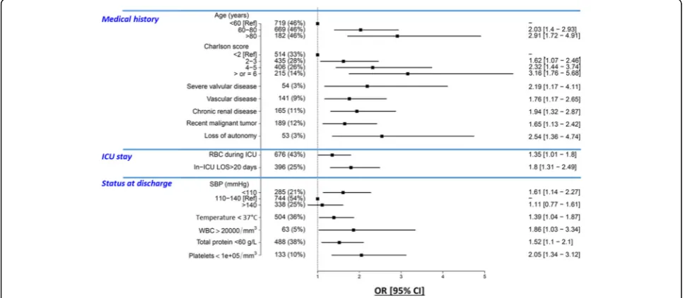
Multivariable analysis identified 14 independent pre-dictors of post-ICU survival (Fig. 2). Odds ratios of sig-nificantly associated variables are presented in Additional file 1: Table S2. Linearity of the association between continuous variables in the multivariable model and the outcome is depicted in Additional file 1: Figure S2. The area of the ROC curve for the multivariable model was 0.787 (95% CI 0.759–0.815). Age and comor-bidities (Charlson comorbidity score, vascular disease, severe valvular disease, chronic kidney diseases, cancer and loss of autonomy) were associated with a greater 1-year risk of death. At ICU discharge, five clinical vari-ables (low values of systolic blood pressure, body temperature, total protein and platelet counts, and a high white blood cell count) were associated with an in-creased post-ICU risk of death. With respect to their ICU stay, red blood cell transfusion and prolonged ICU length of stay were associated with higher risk of 1-year post-ICU mortality. Of note, AUCs of SOFA at admis-sion and SAPS II were 0.574 (95% CI 0.531–0.619) and 0.605 (95% CI 0.572–0.64) respectively; both were sig-nificantly lower than the AUC of the clinical score.
Association between cardiovascular biomarkers at discharge and 1-year survival after ICU discharge
At the time of ICU discharge, 1-year nonsurvivors had elevated levels of all measured cardiovascular biomarkers (Table 2). As depicted in Additional file 1: Figure S3, the association between the level of biomarkers at discharge and the outcome was not linear in all cases. After di-chotomization according to the median value, elevated biomarkers of cardiac (NT-proBNP, sST2) and vascular
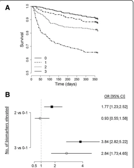
[image:5.595.56.541.483.694.2](bio-ADM) failure were independently associated with 1-year death when they are added to the multivariable model, with an almost 3-fold increase in the risk of death when combined (adjusted OR 2.84 (95% CI 1.73– 4.65), p< 0.001) (Fig. 3). Of note, the association between elevated hs-TnI and 1-year mortality did not remain significant after adjustment. Although only NT-proBNP, bio-ADM and sST2 significantly improve the
c-statistic of the clinical model, reclassification analyses showed that all cardiovascular biomarkers, including hs-TnI, improve predictive power of the multivariable model (Table 2).
Discussion
The FROG-ICU study confirmed the substantial number of vulnerable patients among ICU survivors. More im-portantly, FROG-ICU identified clinical and biological
factors at the time of ICU discharge that were associated with an increased risk of long-term death.
We found that the 1-year mortality rate in ICU survi-vors was roughly 20%, a figure comparable to that already described [8, 26–29]. The FROG-ICU study confirmed that increasing age and number of comorbidities are inde-pendently associated with an increased long-term risk of death [30]. In contrast to previous findings [31], with the exception of blood transfusion and prolonged length of ICU stay, we found no“in-ICU”factor was associated with an increased risk of post-ICU death. Indeed, the reason for ICU admission, illness severity scores at admission and/or use of invasive therapy, factors known to be associ-ated with ICU mortality, were not associassoci-ated with worse long-term outcomes in our 1570 consecutive ICU survi-vors, as described recently [32].
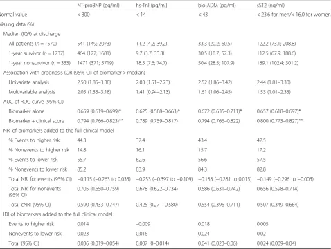
[image:6.595.60.539.98.458.2]A major strength of the FROG-ICU study is the provision of a comprehensive clinical and biological evaluation of Table 2Performance of cardiovascular biomarkers measured at ICU discharge for the prediction of 1-year post-ICU survival
NT-proBNP (pg/ml) hs-TnI (pg/ml) bio-ADM (pg/ml) sST2 (ng/ml)
Normal value < 300 < 14 < 43 < 23.6 for men/< 16.0 for women
Missing data (%)
Median (IQR) at discharge
All patients (n= 1570) 541 (149; 2073) 11.2 (4.2; 39.2) 33.3 (20.2; 60.5) 122.2 (73.1; 208.8)
1-year survivor (n= 1237) 464 (127; 1681) 9.7 (3.7; 33.8) 30.5 (18.7; 52.3) 112.5 (67.9; 188.6)
1-year nonsurvivor (n= 333) 1471 (371; 5719) 18.5 (7.6; 74.7) 50.4 (28.5; 107.9) 189.1 (102.4; 301.2)
Association with prognosis (OR (95% CI) of biomarker > median)
Univariate analysis 2.50 (1.85–3.38) 2.03 (1.51–2.73) 2.52 (1.86–3.42) 2.44 (1.81–3.30)
Multivariable analysis 2.05 (1.33–3.18) 1.41 (0.94–2.13) 1.61 (1.06–2.45) 1.53 (1.01–2.33)
AUC of ROC curve (95% CI)
Biomarker alone 0.659 (0.619–0.699)* 0.625 (0.588–0.663)* 0.672 (0.635–0.711)* 0.657 (0.618–0.697)*
Biomarker + clinical score 0.794 (0.766–0.823)** 0.789 (0.759–0.817) 0.794 (0.766–0.822) 0.800 (0.773–0.827)**
NRI of biomarkers added to the full clinical model
% Events to higher risk 44.3 37.4 43.4 42.5
% Nonevents to higher risk 14.8 16.1 15.7 17.2
% Events to lower risk 55.7 62.6 56.6 57.5
% Nonevents to lower risk 85.2 83.9 84.3 82.8
Total NRI for events (95% CI) −0.115 (−0.263 to 0.033) −0.253 (−0.397 to−0.109) −0.133 (−0.281 to 0.015) −0.149 (−0.296 to−0.003)
Total NRI for nonevents (95% CI)
0.705 (0.650–0.759) 0.678 (0.622–0.734) 0.686 (0.631–0.742) 0.656 (0.598–0.714)
Total cNRI (95% CI) 0.590 (0.433–0.747) 0.425 (0.271–0.580) 0.554 (0.396–0.711) 0.507 (0.349–0.664)
IDI of biomarkers added to the full clinical model
Events to higher risk 0.014 –0.009 0.018 0.005
Nonevents to lower risk 0.023 0.016 0.024 0.02
Total (95% CI) 0.036 (0.019–0.054) 0.007 (0–0.014) 0.041 (0.023–0.06) 0.024 (0.009–0.04)
NT-proBNPN-terminal pro-B type natriuretic peptide,hs-TnIhyper-sensitive troponin I,bio-ADMbio-adrenomedullin,sST2soluble ST2,IQRinterquartile range,AUC area under the curve,ORodds ratio,ROCreceiver operating curve,CIconfidence interval,NRInet reclassification improvement,IDIintegrative
discrimination improvement
*p< 0.05 corresponding to Wilcoxon test comparing survivors to nonsurvivors
patients at the time of ICU discharge to assess risk predic-tion for subsequent poor outcomes. FROG-ICU demon-strated that hypotension and symptoms of persisting inflammation (abnormal temperature, protein, platelet and WBC count) were risk factors for a poor post-ICU out-come. FROG-ICU further showed that elevated biomarkers of impaired cardiac (NT-proBNP and sST2) and vascular (bio-ADM) function strikingly improved the prediction of post-ICU risk of death. Altogether, these data demonstrate that evidence of cardiovascular and/or inflammation abnor-malities on ICU discharge is associated with, and likely leads to, a poor post-ICU outcome. Specific causes of death need to be ascertained but may be related to accelerated atheroma and plaque formation in the heart, brain or other organs, or repeated bouts of infection related to immunosuppression resulting from persisting inflamma-tion. Those results are consistent with other work suggesting that the level of residual inflammation at discharge for patients with sepsis is associated with subsequent mortality [33].
Limitations of the study
Sixty-five (4%) patients discharged alive from the ICU were not assessed at 1 year. Although the number is small, this could have affected the accuracy of our re-sults. We cannot assess the risk of readmission after ICU discharge as this information was not recorded prospectively. More broadly, we had no information on patient management (e.g., drug therapy, rehabilitation, psychologist support) after ICU discharge. This may also have contributed to patient vulnerability and needs to be further explored. In addition, while we described clinical and biological variables independently associ-ated with 1-year mortality in ICU survivors, other im-portant parameters need to be considered when discharging a patient from the ICU, such as the amount of nursing care. Some potential predictors of post-ICU outcome were not considered in the present study; in particular, only comorbidities were considered but no frailty score. Moreover, because of the French law, we were not allowed to include patients with no social se-curity coverage, which may limit the external validity of our results. Biological collection was performed when the patient physically left the ICU and not at the time the patient was considered dischargeable from the ICU, which is more tightly linked to the physiologic status of the patient. However, our approach reflects the real-life management of ICU discharge. Although the study was multicentric and conducted in two European countries, only one center outside France included patients; this may limit the external validity of our results. Finally, despite the fact that a sample size calculation was per-formed, factors that were weakly associated with the 1-year risk of death could not be identified due to insuffi-cient study power. Of note, the main aim of the study was to identify an explanatory model. Thus, the object-ive of our variable selection procedure was to identify the factors most strongly associated with mortality at 1 year and not to establish a prognostic score that would have to be validated.
Conclusions
Our findings suggest recommending a comprehensive clinical examination and targeted biological testing, including biomarker measures in ICU survivors, to guide personalized discharge long-term planning. Fu-ture trials should assess whether actions targeting the pathophysiology underlying the abnormal cardiac or vascular biomarkers may translate into improved post-ICU outcomes. In summary, the FROG-ICU study confirmed the striking prevalence of death at 1 year after ICU discharge. The FROG-ICU study fur-ther identified clinical and biological factors that may guide personalized discharge planning.
[image:7.595.58.292.88.381.2]Additional file
[image:8.595.57.289.376.667.2]Additional file 1: Figure S1. showing Kaplan–Meier curves for 1-year mortality after discharge from the ICU,Figure S2.showing plots of restricted cubic spline of continuous variables included in the multivariable model,
Figure S3.showing plots of restricted cubic spline of continuous variables included in the multivariable model,Table S1.presenting details on comorbidities and chronic treatment, andTable S2.presenting ORs (with 95% CI) for variables significantly associated with 1-year mortality in univariate analysis and in multivariable analysis (DOCX 110 kb)
Abbreviations
AUC:Area under the curve; bio-ADM: Bio-adrenomedullin; CI: Confidence interval; eGFR: Estimated glomerular filtration rate; FROG-ICU: French and European Outcome reGistry in Intensive Care Units; hs-TnI: Hyper-sensitive troponin I; ICU: Intensive care unit; IDI: Integrative discrimination index; IQR: Interquartile range; NRI: Net reclassification index; NT-proBNP: N-terminal pro-B type natriuretic peptide; OR: Odds ratio; PICS: Post-intensive care syndrome; ROC: Receiver operating curve; SAPS: Simplified Acute Physiologic Score; SOFA: Sequential Organ Failure Assessment; sST2: Soluble ST2; WBC: white blood cell
Acknowledgements
The authors are particularly grateful to Marie-Céline Fournier who coordinated organizational aspects of the study. They also thank the Centre de Recherche Clinique (CRC) of Lariboisière University Hospital for support.
Investigators for the FROG-ICU study
Funding
The FROG-ICU study was funded by the Programme Hospitalier de la Recherche Clinique (AON 10-216) and by a research grant from the Société Française d’Anesthésie—Réanimation.
Abbot, Sphingotec, Roche Diagnostics and Critical Diagnostics provided unrestricted free kits to Assistance Publique—Hôpitaux de Paris to conduct biomarker analyses.
Availability of data and materials
AM had full access to all data in the study and takes responsibility for the integrity of the data and the accuracy of the data analysis.
Authors’contributions
Study concept and design: EG, AM, MR-R, EV. Acquisition of data: AC, ND, AV-B, SJ, CD, QL, XM, IR, EA, MLé, HO, BG, DF, AT, RS, PM, SPF, J-YL, JD, P-FL, NB, XR. Analysis and interpretation of data: EG, AM, KC, MM, J-ML, MR-R. Drafting of the manuscript: EG, AM. Critical revision of the manuscript for important intellectual content: BC, J-ML, BG, AV-B, MLe, MS. Statistical analysis: EG, MR-R. Obtained funding: EG, AM, EV. Administrative, technical or material support: EG, AM, EV. Study supervision: EG, AM. All authors read and approved the final manuscript.
Ethics approval and consent to participate
The study was conducted in France and Belgium in accordance with Good Clinical Practice (Declaration of Helsinki 2002) and Ethical Committee approvals (Comité de Protection des Personnes—Ile de France IV, IRB n° 00003835 and Commission d’éthique biomédicale hospitalo-facultaire de l’hôpital de Louvain, IRB n°B403201213352).
Consent for publication
Not applicable
Competing interests
EG received research grant from Sphingotec, and consultancy fees from Magnisense and Roche Diagnostics. AM received speaker’s honoraria from Abbott, Novartis, Orion, Roche and Servier, and fees as a member of the advisory board and/or Steering Committee from Cardiorentis, Adrenomed, MyCartis, Neurotronik and Sphyngotec. The remaining authors declare that they have no competing interests.
Publisher’s Note
Springer Nature remains neutral with regard to jurisdictional claims in published maps and institutional affiliations.
Author details
1Department of Anesthesiology, Critical Care and Burn Unit, Hôpitaux
Universitaires Saint Louis—Lariboisière, Assistance Publique—Hôpitaux de Paris, Université Paris Diderot—Paris 7, Sorbonne Paris Cité, UMR-S 942, INSERM, Paris, France.2Medical Intensive Care Unit, Cochin University Hospital, Assistance Publique—Hôpitaux de Paris, Paris Descartes University, Paris Cardiovascular Research Center-INSERM U970 (PARCC), Paris Sudden Death Expertise Center, Paris, France.3Medical Intensive Care Unit, Hôpitaux Universitaires Saint Louis—Lariboisière, Assistance Publique—Hôpitaux de Paris, Université Paris Diderot—Paris 7, Sorbonne Paris Cité, UMR-S 942, INSERM, Paris, France.4Intensive Care Unit, University Hospital Ambroise Paré, Assistance Publique—Hopitaux de Paris, 26930 Boulogne-Billancourt, France. 5Intensive Care Unit, Anaesthesia and Critical Care Department, Saint Eloi
Teaching Hospital, Centre Hospitalier Universitaire Montpellier, Montpellier University, Montpellier, France.6Multidisciplinary Intensive Care Unit, Department of Anesthesiology and Critical Care Medicine, La Pitié-Salpêtrière Hospital, Assistance Publique Hôpitaux de Paris, UPMC Paris 6, Paris, France. 7Medical Intensive Care Unit, Bicêtre Hospital, Paris-Sud University Hospitals,
Inserm UMR_S999, Paris-Sud University, Le Kremlin-Bicêtre, France. 8
Department of Anesthesiology and Critical Care, Beaujon Hospital, Assistance Publique Hôpitaux de Paris University, Clichy, France.9Medical Intensive Care Unit, Hôpital Saint-Louis, ECSTRA Team, Biostatistics and Clinical Epidemiology, UMR 1153 (Center of Epidemiology and Biostatistics Sorbonne Paris Cité, CRESS), INSERM, Université Paris Diderot Sorbonne, Paris, France.10Service d’anesthésie et de réanimation, Hôpital Nord, Assistance Publique—Hôpitaux de Marseille, Aix Marseille Université, Marseille, France. 11Service de Réanimation Médicale, Hôpital Saint-Antoine, Assistance
Publique—Hôpitaux de Paris, Université Pierre et Marie Curie, Paris, France. 12General Intensive Care, Raymond Poincaré University Hosptal, Assistance
Publique—Hopitaux de Paris, Garches, France.13Department of Anesthesiology and Intensive Care, Cochin University Hospital, Assistance
Center Investigators
Hopital Lariboisiere (Paris) N Deye, C Fauvaux, A Mebazaa, C Damoisel, D Payen
Hopital Saint Louis (Paris) E Azoulay, AS Moreau, L Jacob, O Marie
Hopital Bichat (Paris) M Wolf, R Sonneville, R Bronchard
Hopital Beaujon (Clichy) I Rennuit, C Paugam
Hopital Cochin (Paris) JP Mira, A Cariou, A Tesnieres
Hopital Bicetre (Le Kremlin-Bicetre)
N Dufour, N Anguel, L Guerin, J Duranteau, C Ract
Chu De Marseille (Marseille) M Leone, B Pastene
Hopital Raymond Poincare (Garches)
T Sharshar, A Fayssoyl
Hopital Saint-Antoine J-L Baudel, B Guidet
Hopital De La Pitie—Salpetriere (Paris)
Q Lu, W Jie Gu, N Brechot, A Combes
Chu St Eloi (Montpellier) S Jaber, A Pradel, Y Coisel, M Conseil
Hopital Ambroise Pare (Boulogne)
A Veillard Baron, L Bodson
Chu Caremeau (Nimes) Jy Lefrant, L Elotmani, A Ayral, S Lloret
Hopital Jean Minjoz (Besançon)
S Pily-Flouri, Jb Pretalli
Publique—Hôpitaux de Paris, Paris Descartes University, Paris Cardiovascular Research Center-INSERM U970 (PARCC), Paris Sudden Death Expertise Center, Paris, France.14Department of Intensive Care Medicine and Infectious Diseases, Univ Paris Diderot, Sorbonne Paris Cité, Assistance Publique—Hôpitaux de Paris, Hôpital Bichat-Claude, Paris, France. 15Department of Anesthesiology and Intensive Care, Bichat University
Hospital, Assistance Publique—Hôpitaux de Paris, Université Paris Diderot—Paris 7, Sorbonne Paris Cité, Paris, France.16Department of Anesthesiology and Intensive Care Medicine, University Hospital of Besancon, 25000 Besancon, France.17Department of Anesthesiology, Emergency and Critical Care Medicine, Nimes University Hospital, 30029 Nîmes, France. 18
Physiology Department, EA 2992, Faculté de Médecine de Nîmes, Université Montpellier 1, 30029 Nîmes, France.19Département
d’Anesthésie-Réanimation, Hôpital de Bicêtre, Université Paris-Sud, Hôpitaux Universitaires Paris-Sud, Assistance Publique—Hôpitaux de Paris, Le Kremlin Bicêtre, Paris, France.20Medical–Surgical Intensive Care Unit, Cliniques Saint-Luc, Brussels, Belgium.21Medical Intensive Care Unit, Hôpital Pitié-Salpêtrière, Assistance Publique—Hôpitaux de Paris, Sorbonne Pierre-Marie Curie University Paris, INSERM, UMRS_1166-ICAN, Institute of Cardiometabolism and Nutrition and CIC 1421—Paris Est, Paris, France. 22URC-Eco, Assistance Publique—Hôpitaux de Paris, Sorbonne Paris Cité,
Université Paris Diderot, ECEVE, INSERM, Paris, France.23Department of Anesthesiology and Critical Care Medicine, Hôpital Européen Georges Pompidou, APHP, Université Paris Descartes, Sorbonne Paris Cite, Paris, France.24Service de Biochimie, Hôpitaux Universitaires Saint
Louis—Lariboisière, Assistance Publique—Hôpitaux de Paris, Université Paris Diderot—Paris 7, Sorbonne Paris Cité, UMR-S 942, INSERM, Paris, France. 25
Unité de Recherche Clinique, Hôpitaux Universitaires Saint
Louis—Lariboisière, Assistance Publique—Hôpitaux de Paris, Université Paris Diderot—Paris 7, Sorbonne Paris Cité, UMR-S 942, INSERM, Paris, France. 26Bloomsbury Institute of Intensive Care Medicine, University College
London, Cruciform Building, Gower St, London WC1E 6BT, UK.27Service de Biostatistique et Information Médicale, Hôpitaux Universitaires Saint Louis—Lariboisière, Assistance Publique—Hôpitaux de Paris, Université Paris Diderot—Paris 7, Sorbonne Paris Cité, ECSTRA Team, INSERM, Paris, France. 28
Department of Anesthesiology and Intensive Care, University Paris Diderot, INSERM UMR-S 942, Saint Louis—Lariboisière University Hospitals, 2 rue Ambroise Paré, 75010 Paris, France.
Received: 7 August 2017 Accepted: 8 December 2017
References
1. Winters BD, Eberlein M, Leung J, Needham DM, Pronovost PJ, Sevransky JE. Long-term mortality and quality of life in sepsis: a systematic review. Crit Care Med. 2010;38(5):1276–83.
2. Needham DM, Davidson J, Cohen H, Hopkins RO, Weinert C, Wunsch H, Zawistowski C, Bemis-Dougherty A, Berney SC, Bienvenu OJ, et al. Improving long-term outcomes after discharge from intensive care unit: report from a stakeholders’conference. Crit Care Med. 2012;40(2):502–9. 3. Desai SV, Law TJ, Needham DM. Long-term complications of critical care.
Crit Care Med. 2011;39(2):371–9.
4. Wright JC, Plenderleith L, Ridley SA. Long-term survival following intensive care: subgroup analysis and comparison with the general population. Anaesthesia. 2003;58(7):637–42.
5. Cuthbertson BH, Rattray J, Campbell MK, Gager M, Roughton S, Smith A, Hull A, Breeman S, Norrie J, Jenkinson D, et al. The PRaCTICaL study of nurse led, intensive care follow-up programmes for improving long term outcomes from critical illness: a pragmatic randomised controlled trial. BMJ. 2009;339:b3723.
6. Williams TA, Dobb GJ, Finn JC, Knuiman MW, Geelhoed E, Lee KY, Webb SA. Determinants of long-term survival after intensive care. Crit Care Med. 2008; 36(5):1523–30.
7. Flaatten H, Kvale R. Survival and quality of life 12 years after ICU. A comparison with the general Norwegian population. Intensive Care Med. 2001;27(6):1005–11.
8. Niskanen M, Kari A, Halonen P. Five-year survival after intensive care—comparison of 12,180 patients with the general population. Finnish ICU Study Group. Crit Care Med. 1996;24(12):1962–7.
9. Hofhuis JG, Spronk PE, van Stel HF, Schrijvers GJ, Rommes JH, Bakker J. The impact of critical illness on perceived health-related quality of life during
ICU treatment, hospital stay, and after hospital discharge: a long-term follow-up study. Chest. 2008;133(2):377–85.
10. Hodgson CL, Udy AA, Bailey M, Barrett J, Bellomo R, Bucknall T, Gabbe BJ, Higgins AM, Iwashyna TJ, Hunt-Smith J, et al. The impact of disability in survivors of critical illness. Intensive Care Med. 2017;43(7): 992–1001.
11. Dinglas VD, Aronson Friedman L, Colantuoni E, Mendez-Tellez PA, Shanholtz CB, Ciesla ND, Pronovost PJ, Needham DM. Muscle weakness and 5-year survival in acute respiratory distress syndrome survivors. Crit Care Med. 2017;45(3):446–53.
12. Garland A, Olafson K, Ramsey CD, Yogendran M, Fransoo R. Distinct determinants of long-term and short-term survival in critical illness. Intensive Care Med. 2014;40(8):1097–105.
13. Yende S, Linde-Zwirble W, Mayr F, Weissfeld LA, Reis S, Angus DC. Risk of cardiovascular events in survivors of severe sepsis. Am J Respir Crit Care Med. 2014;189(9):1065–74.
14. Mebazaa A, Casadio MC, Azoulay E, Guidet B, Jaber S, Levy B, Payen D, Vicaut E, Resche-Rigon M, Gayat E. Post-ICU discharge and outcome: rationale and methods of the The French and euRopean Outcome reGistry in Intensive Care Units (FROG-ICU) observational study. BMC Anesthesiol. 2015;15:143.
15. Marino R, Struck J, Maisel AS, Magrini L, Bergmann A, Di Somma S. Plasma adrenomedullin is associated with short-term mortality and vasopressor requirement in patients admitted with sepsis. Crit Care. 2014;18(1):R34.
16. Yancy CW, Jessup M, Bozkurt B, Butler J, Casey Jr DE, Drazner MH, Fonarow GC, Geraci SA, Horwich T, Januzzi JL, et al. 2013 ACCF/AHA guideline for the management of heart failure: a report of the American College of Cardiology Foundation/American Heart Association Task Force on Practice Guidelines. J Am Coll Cardiol. 2013;62(16):e147–239.
17. Lassus J, Gayat E, Mueller C, Peacock WF, Spinar J, Harjola VP, van Kimmenade R, Pathak A, Mueller T, Disomma S, et al. Incremental value of biomarkers to clinical variables for mortality prediction in acutely decompensated heart failure: the Multinational Observational Cohort on Acute Heart Failure (MOCA) study. Int J Cardiol. 2013;168(3):2186–94. 18. Rey G, Jougla E, Fouillet A, Hemon D. Ecological association between a
deprivation index and mortality in France over the period 1997–2001: variations with spatial scale, degree of urbanicity, age, gender and cause of death. BMC Public Health. 2009;9:33.
19. Harrell Jr FE, Lee KL, Mark DB. Multivariable prognostic models: issues in developing models, evaluating assumptions and adequacy, and measuring and reducing errors. Stat Med. 1996;15(4):361–87.
20. Rubin D. Multiple Imputation for Nonresponse in Surveys. New York: Wiley; 1987. 21. Hosmer D, Lemeshow S. Applied Logistic Regression. New York: Wiley; 2000. 22. Hlatky MA, Greenland P, Arnett DK, Ballantyne CM, Criqui MH, Elkind MS, Go AS, Harrell Jr FE, Hong Y, Howard BV, et al. Criteria for evaluation of novel markers of cardiovascular risk: a scientific statement from the American Heart Association. Circulation. 2009;119(17):2408–16.
23. Pencina MJ, D’Agostino Sr RB, D’Agostino Jr RB, Vasan RS. Evaluating the added predictive ability of a new marker: from area under the ROC curve to reclassification and beyond. Stat Med. 2008;27(2):157–72.
24. Cook NR. Use and misuse of the receiver operating characteristic curve in risk prediction. Circulation. 2007;115(7):928–35.
25. le Cessie S, van Houwelingen HC. Testing the fit of a regression model via score tests in random effects models. Biometrics. 1995;51(2):600–14. 26. Rockwood K, Noseworthy TW, Gibney RT, Konopad E, Shustack A, Stollery D,
Johnston R, Grace M. One-year outcome of elderly and young patients admitted to intensive care units. Crit Care Med. 1993;21(5):687–91. 27. Bagshaw SM, Mortis G, Doig CJ, Godinez-Luna T, Fick GH, Laupland KB.
One-year mortality in critically ill patients by severity of kidney dysfunction: a population-based assessment. Am J Kidney Dis. 2006;48(3):402–9. 28. Orwelius L, Nordlund A, Nordlund P, Simonsson E, Backman C, Samuelsson
A, Sjoberg F. Pre-existing disease: the most important factor for health related quality of life long-term after critical illness: a prospective, longitudinal, multicentre trial. Crit Care. 2010;14(2):R67.
29. Braun A, Chang D, Mahadevappa K, Gibbons FK, Liu Y, Giovannucci E, Christopher KB. Association of low serum 25-hydroxyvitamin D levels and mortality in the critically ill. Crit Care Med. 2011;39(4):671–7.
31. Wunsch H, Guerra C, Barnato AE, Angus DC, Li G, Linde-Zwirble WT. Three-year outcomes for Medicare beneficiaries who survive intensive care. JAMA. 2010;303(9):849–56.
32. Herridge MS, Chu LM, Matte A, Tomlinson G, Chan L, Thomas C, Friedrich JO, Mehta S, Lamontagne F, Levasseur M, et al. The RECOVER Program: disability risk groups and 1-year outcome after 7 or more days of mechanical ventilation. Am J Respir Crit Care Med. 2016;194(7):831–44. 33. Yende S, D’Angelo G, Kellum JA, Weissfeld L, Fine J, Welch RD, Kong L,
Carter M, Angus DC, Gen IMSI. Inflammatory markers at hospital discharge predict subsequent mortality after pneumonia and sepsis. Am J Respir Crit Care Med. 2008;177(11):1242–7.
• We accept pre-submission inquiries
• Our selector tool helps you to find the most relevant journal • We provide round the clock customer support
• Convenient online submission • Thorough peer review
• Inclusion in PubMed and all major indexing services • Maximum visibility for your research
Submit your manuscript at www.biomedcentral.com/submit