Rapid Antigenic-Type Replacement and DNA Sequence
Evolution
of
Canine Parvovirus
COLIN R.
PARRISH,'*
CHARLES F. AQUADRO,2M. LISASTRASSHEIM,1 JAMES F. EVERMANN,3 JEAN-YVES SGRO,4 ANDHUSSNI 0. MOHAMMED5JamesA. Baker Institute,' Section ofEpidemiology,5 New YorkState College ofVeterinaryMedicine, andSectionof
Geneticsand Development,2 Biotechnology Building, Cornell University, Ithaca, New York 14853; Washington AnimalDisease DiagnosticLaboratoryandDepartment ofVeterinary Clinical Medicine andSurgery, College
of Veterinary Medicine, Washington State University, Pullman, Washington 991643; andInstitute
for Molecular Virology, University of Wisconsin-Madison, Madison, Wisconsin 537064
Received 28 June 1991/Accepted 19 August 1991
Analysis of canine parvovirus (CPV) isolateswithapanelof monoclonal antibodies showed that after1986,
most viruses isolated from dogs in many parts ofthe United States differed antigenically from the viruses isolated priortothat date. Thenewantigenictype(designatedCPV type2b)haslargely replacedtheprevious antigenic type (CPV type 2a) among virus isolates from the United States. This represents the second occurrenceofanewantigenictypeof this DNAvirus sinceitsemergencein1978,astheoriginalCPV type(CPV
type 2) had previously been replaced between 1979 and 1981 by the CPV type 2a strain. DNA sequence
comparisonsshowed that CPVtypes2b and 2a differedbyasfewastwononsynonymous(aminoacid-changing)
nucleotide substitutions in the VP-1 and VP-2 capsid proteingenes. One mutation, resulting inanAsn-Asp
differenceatresidue 426intheVP-2sequence, wasshown by comparisonwithaneutralization-escapemutant selected with a non-CPV type 2b-reactive monoclonal antibody to determine the antigenic change. The
mutation selected bythat monoclonalantibody,aHis-TyrdifferenceinVP-2amino acid 222,wasimmediately
adjacenttoresidue 426inthe three-dimensional structureof the CPVcapsid. The CPV type 2b isolatesare
phylogenetically closely relatedtothe CPVtype2a isolatesandareprobably derived froma commonancestor.
Phylogenetic analysis showedaprogressiveevolutionawayfrom the original CPVtype.This pattern of viral evolutionappears most similar to thatseen insomeinfluenza A viruses.
Canineparvovirus (CPV)is arecently emerged pathogen
ofdogswhich wasfirstobserved in 1978. Theoriginal 1978 strain is here designated CPVtype2(CPV-2) todistinguish
it from subsequent variant strains. No evidence for the
existence ofaCPV-likeparvovirusindogs priorto1974 has
beenreported. During 1978, CPV-2 becameglobally
distrib-uted (for areview, see reference 36), andCPV-like viruses are now endemic in mostpopulations of domestic and wild
canids. We have previously shown that around 1979, a
variant CPV strain (designated CPV type 2a [CPV-2a])
becamewidespread (36, 37).That strain differedfrom CPV-2
in that it lostatleastoneepitoperecognized by monoclonal
antibodies (MAbs) and gained a new specific epitope. By 1981, the CPV-2a strain was the virus most frequently
isolated from domestic dogs with clinical disease in the United States, Japan, Denmark, andAustralia, and CPV-2
wasrarely seenafter that time (36, 43).
Variation is seen among many different viruses, and in some casesvirusesmayevolveovertimetogiverisetonew
genetic or antigenic types, while among other viruses the
types or subtypes are relatively stable (for examples, see
references 2, 15, 20, 22, 23, 26, 35, 44, 45, and 50). The sequential evolution ofnewantigenic types(antigenic drift)
appears uncommon but has been studied extensively as a feature of theepidemiology and evolution ofinfluenza virus isolates (1, 11-13, 16-18, 40). Genetic variation iscommon
among viruses, probably because of their large effective population sizes, their rapid replication, and their selection bythehost. Therapid evolution of RNA viruses is reported
* Correspondingauthor.
tobedue,atleast inpart, totheerror-prone natureof their
RNA-dependent RNApolymerases (11, 46).
Parvoviruses contain about5,000bases ofsingle-stranded
DNA and encodetwostructural(VP-1andVP-2)andoneor
twononstructural (NS-1 andNS-2) proteingenes (7). VP-1
and VP-2 aretranslated fromdifferently spliced productsof the RNA transcribed from the p40 promoter, and the two
proteinshave mostof theirsequences in common, thegene
being referred to here as the VP-1/VP-2 gene. The three-dimensional molecular structure of CPV has been deter-minedby X-ray crystallography,and it shows that thecapsid comprisesatotalof 60copiesof theVP-1 and VP-2proteins (48). The surface features of thecapsidinclude aprominent
region surrounding the threefold axis of symmetry (the
"threefold spike"), a circular canyon around the fivefold axisofsymmetry,andadepressionordimpleonthe twofold axis ofsymmetry.Definedantigenic epitopesareaffectedby
residueson the threefoldspike (30, 33, 48).
In this study, we have examined manyrecentisolates of CPV and have shown that afurther antigenic variant
(des-ignated CPV type 2b [CPV-2b]) emerged around 1984 and that the CPV-2b antigenic type subsequently replaced
CPV-2aasthecauseof CPV disease inmanyregions ofthe United States. The molecular basis of that antigenic varia-tionwasdetermined, and the molecularevolutionarytrends of theCPVs wereexamined.
MATERIALS AND METHODS
Cells and viruses. Cells of the feline NLFKline,a deriv-ativeof the Crandell felinekidneycell line(8),weregrownin
a50% mixtureofMcCoy'sSAandLeibovitzL15mediawith
6544 CopyrightX 1991,American Society for Microbiology
on November 10, 2019 by guest
http://jvi.asm.org/
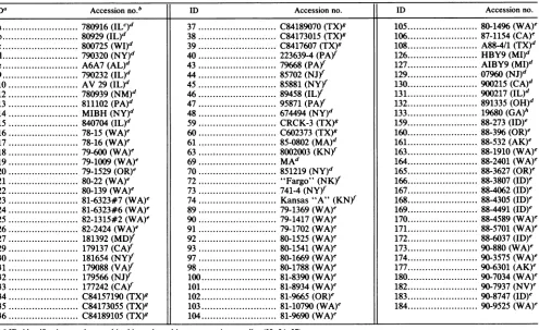
TABLE 1. Identification numbers,sources, andorigins of virus isolates
IDa Accessionno."b ID Accessionno. ID Accessionno.
a.780916(ILC)d 37... C84189070(TX)g 105.80-1496(WA)e
b.80929(IL)d 38... C84173015(TX)g 106.87-1154(CA)e
c.800725(WI)d 39... C8417607 (TX)g 108.A88-4/1
(TX)d
d.790320
(NY)d
40... 223639-4(PA)Y 126.HBY9(MI)d
e.A6A7(AL)d 43... 79668
(PA)f
127.AIBY9(MI)d
9...790232(IL)d 44...85702
(NJ)f
129...07960(NJ)d10 .AV29(IL)d 45 .85881(NY)f 130.900215(CA)d
12 .780939
(NM)d
46... 89458(IL)f
131.900217(IL)"13 .811102
(PA)d
47... 95871(PA)f
132.891335 (OH)"14.MIBH
(NY)d
48... 674494(NY)d
133.19680(GA)h15.840704
(IL)"d
59... CRCK-3(TX)g 159.88-273 (ID)e16 .78-15(WA)e 60... C602373 (TX)g 160.88-396(OR)e
17.78-16 (WA)e 61... 85-0802(MA)d 161... 88-532(AK)e
18 .79-600 (WA)e 63....; ... 8002003(KN}" 163... 88-1910(WA)'
19.79-1009(WA)e 69.MAd... 164... 88-2401(WA)'
20 .79-1529(OR)e 70 .851219
(NY)d
165... 88-3627(OR)e21.80-22(WA)e 72."Fargo"
(NK)f
166... 88-3807(ID)e22 .80-139(WA)' 73.- 741-4---(NY)" 167.88-4062(ID)'
23.81-6323#7(WA)e 74 .Kansas"A" (KN) 168... 88-4305(ID)'
24.81-6323#6(WA)e 89 .79-1369(WA)e 169... 88-4491(ID)e
25.82-1315#2(WA)e 90 .79-1417(WA)e 170... 88-4589(WA)'
26 .82-2424(WA)e 91.79-1702(WA)e 171... 88-5701(WA)'
27.181392
(MD)f
92 .80-1525(WA)e 172... 88-6037(ID)e29 .179137
(CA)Y
93.80-1541(WA)e 173... 90-880(WA)e30.181654(NY)" 97.80-1669(WA)e 174... 90-3575(WA)'
31.179088
(VAY)
98 .80-1788(WA)e 177... 90-6301(AK)32 .179566(NJ)" 100.81-8390(WA)e 180... 90-7034(WA)'
33 .177242
(CAY)
101 .81-8934(WA)e 182... 90-7937(NV)'34.C84157190(TX)g 102 .81-9665(OR)e 183... 90-8747(ID)e
35 .C84173055 (TX)g 103 .81-10790(WA)e 184... 90-9525(WA)'
36 .C8418910 C84189105(TX)g 104 .81-9690(WA)'
a ID, identification number used in this study and in some previous studies (32, 34, 37). b Original accession number from diagnostic laboratory or code number.
cState abbreviations: AK,Alaska; AL, Alabama; CA,
California;
GA, Georgia; ID, Idaho; IL,Illinois; KN, Kansas; MA, Massachusetts;MD,Maryland;MI, Michigan; NJ, New Jersey; NM, New Mexico; NV, Nevada; NY, New York; OH, Ohio; OR, Oregon; PA, Pennsylvania; TX, Texas; VA,Virginia; WA, Washington; NK, not known.dSubmittedtoJamesA.Institute, New York State College of Veterinary Medicine.
'SubmittedtoWashingtonAnimal DiseaseDiagnostic Laboratory.
fSubmittedtoNewYork StateVeterinary Diagnostic Laboratory (obtained from E. J. Dubovi). g SubmittedtoTexasVeterinaryDiagnosticLaboratory (obtained from R.A.Crandell).
h SubmittedtoUniversity ofGeorgia,VeterinaryDiagnostic and Investigational Laboratory(obtainedfromA.R.Purcell).
5% fetal bovine serum. For virus propagation, cells were
seededat adensity of2 x
104/cm2
24hpriortoinoculation. Felineprimary fetal heart lungcells were grown in Eagle's minimal essential medium with10%
fetalbovine serum.The sources and the identification numbers of the virus isolates and the isolates' origins within the United States
are listed in Table 1. Field strains of CPV were obtained from veterinary diagnostic laboratories or from canine
feces ortissue samples submitted directly to our
laborato-ries. Viruses wereantigenically typeddirectly from clinical specimens if sufficient hemagglutination (HA) titer was
present;
otherwise,
viruses were propagated during fromonetofivepassagesinNLFKorfeline fetalheart
lung
cellspnor to
testing.
Prototype strains of CPV-2(CPV-d)
andfelinepanleukopeniavirus(FPV)(FPV-b)wereisolated from
infectious plasmid clones ofthose virusesafter transfection intoNLFKcells,and theviruseswerethenpassagedtwo to threetimestoprepare stocks, asdescribedpreviously (29). Antigenictypesofall viruses listed in Table 1 are shown in
Fig. 1.
MAb typing and analysis. A
panel
of 23 MAbsprepared
against either CPV-b, FPV-c, or CPV-39 (32, 34, 37) was
used for
antigenic typing
(see Table2).
Antibodies wereprepared as tissue culture supernatant fluids of hybridoma
cell lines grown in Dulbecco's minimal essential medium with nonessential amino acids and 20% fetal bovine serum.
Antigenic typing
wasperformed by using
theHA inhibition(HI)
assay, with 8 HA units of virusantigen
in barbital-acetate-buffered saline(pH
6.2)
and0.5% rhesus macaqueerythrocytes (29, 32).
Twofold dilutions of 25RI
of thehybridoma
culturesupernatant
wereprepared
and incubatedwith 25
R1
ofantigen
for1 hat roomtemperature, andthen
50 ,u of0.5%
erythrocytes
was added and theplates
wereincubated at
4°C
for atleast 8 h. HI titers were readasthe lastantibody
dilutioninhibiting
HAby
>50%.Escape
mutantselection andanalysis.
ACPV-d stock fromplasmid-derived
virus wasprepared
in NLFK cell culturesand then was concentrated 40x
by
ultracentrifugation
at100,000 x gfor3 h. Viruswasincubated with MAb I for1h
at
37°C,
inoculated ontoNLFK cells in a75-cm2
flask,
andcultured fortwoblindpassages in the presenceofMAbI. As thederivation ofCPVfrom
single plaques
isdifficult,
DNAwas cloned
by
preparing
arecombinant infectiousplasmid
from the mutant virus DNA to ensure that a
single
viralsequence was
analyzed.
Thereplicative-form
DNA ofthe MAb I-selected virus recoveredby
a modified Hirtextrac-tion
procedure (33)
waspurified by
electroelution.ThePflMI
(nucleotide [nt]
2814,
54.5 map units[m.u.])-to-EcoRV (nt
on November 10, 2019 by guest
http://jvi.asm.org/
[image:2.612.68.563.91.394.2]Year of Virus Isolation
1978 1979 1980 1981 1982 1983 1984 1985 1986 1987 1988
CPV- a,b, 12 d, 9, 10 C,21, 22 103,104 48 165,167
type2
|16,17
18,19,20 59,60,63type2 ~~89,90,91
CPV- e 92,93,97 13,23,24 25,26 14,27,29 15,34,35 61,69,70 72,73,74 164,171 177,130
type2a 98,105 43,44,45 30,31,32 37,38,40
46,47,100 33
101,102
CPV-type 2b
36,39 106 108, 159, 160
161, 163, 166 168,169,170 172
[image:3.612.59.553.81.237.2]129,132 126,127,131 133,173,174 180,182,183 184
FIG. 1. Antigenictypesof CPV isolates collected from 1978 through 1990 from clinically ill dogs in the United States. The numbersor lettersare ourcatalogue designations and refertothe virusisolates described in Table 1. Virusesweretyped by the panel of MAbs shown inTable 2, andeach virus showed thesamepattemnwith thepanel of MAbsastheprototypevirus of thattype(CPV-d, CPV-15, and CPV-39).
4011, 77.6 m.u.) fragmentwasisolated and used to replace the same sequence from the plasmid clone of CPV-d (29).
The recombinant plasmid was purified by CsCl gradient
centrifugation and transfected into NLFK cells, and the antigenic type of the virus reisolated was determined by
MAb typing.
DNAsequencing and analysis. Replicative-form DNAwas
isolated from culturesof CPV-39 and CPV-133. The 21.3-to-77.6-m.u. and74.4-to-95.4-m.u. sequenceswere cloned into
M13vectorsmpl8 and mpl9 (51) after digestion with EcoRI
and EcoRV or HaeIII. Sequences from both strands were
determined by using a series of synthetic oligonucleotide
primers complementary to various positions in the CPV
sequences(29, 30, 41, 47) (Fig. 2B).
The PflMI-EcoRV region of the recombinant plasmid containing that region derived from the MAb I-selected
mutant was sequenced in both directions by using someof
thesameprimersafterdenaturing the plasmid with alkali(14,
47).
Phylogeneticanalysis of viralsequences.Phylogenetic
anal-ysis of the completeVP-1NVP-2sequences wasperformed by
themethod of Fitch(10). Themostparsimonioustreeof the sequences was obtained by comparison of the variable
positions among the reported sequences of CPV-2 isolates CPV-d (29, 30), a1978 Belgium isolate of CPV L78-778 (5, 31), and CPV-Norden (38) (isolated in 1978) (21); CPV-2a strains CPV-15 and CPV-31 (30, 37); and the CPV-39 and CPV-133 sequences (Fig. 2A). A published sequenceof the VP-1/VP-2 gene ofa 1978 CPV-2 isolate (39) contained a
numberofsequencingerrorswhich made its analysis
uncer-tain(30), and itwasnotincluded in this study. Thechanges uniquetothatsequencewould place that virus furtherouton
theCPV-2branchof the phylogeny and wouldnotalter the phylogenies of the other viruses (30). The rateofsequence
variation of the VP-1/VP-2 gene was estimated after the
plotting of the number of variant positions, determined from thetotal branchlengths measured from therootnode of the
CPVs.
Nucleotide sequence accession number. Sequences have
been deposited in Genbank, and accession numbers are
M74849 and M74852.
RESULTS
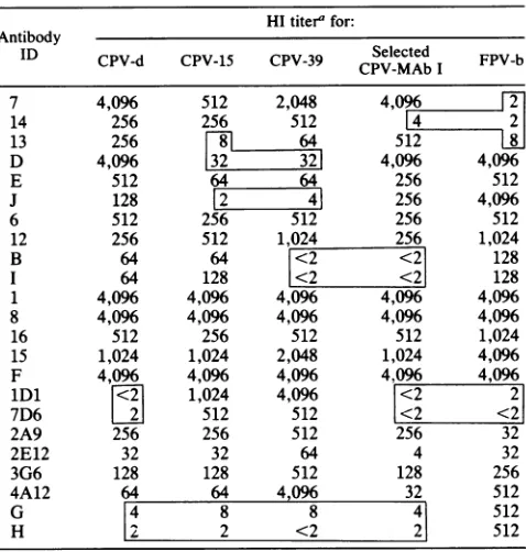
The HI titers of various antigenic types ofvirus withthe panel of 23 MAbs are shown in Table 2, along with the reactivities of the MAb I-selected virus andanFPVisolate. This shows that CPV-2b retains boththe CPV-specific and the CPV-2a-specific epitopes (recognized by MAb 7 or 14 andby MAb lDlor7D6,respectively) but loses its
reactiv-(A)
2000 3000 4000
VP-1 VP-2
CPV- r-t 333~35 3685 I71
2343 3045 3088 3699 3909 4062 4449 4538
Type-2 CPV-d, L78-778 G A C T G C G G A A A G T
Met
lie
AlaAspAspAsn Asn Val CPV-Norden G A C T G C G T G G A G TMet tle AlaAsp yr A spAsn Val
Type-2a CPV-15 G T C C G G T G GAA A T Leu Thr GyTyr As As Asn lle LCPV-31 G T C C A G T G G A A A T
Leu Thr GyGlyTyrAs Asp Asn tIe
Type-2b CPV-39 G T C C G G T G G A G G T Leu Thr GlyTyrAspAs Asp Val CPV-133 A T T C G G T G GA G G C
Aminoacid ILeul". Thr GlyTyrAsp P
Vetl
in VP-2 87 101 300 305 367 375 426 555(B)
Eco RI
(1100) 2000
Haelli EcoRV Hae III
30W \>400 (4888)
--- 40
4 4 -4 -4 4
4 -4- 4 4- 4
FIG. 2. (A) Variantsequencesin the VP-1/VP-2genesofCPV-2, CPV-2a,and CPV-2b isolates.SequencesofCPV-d, CPV-Norden, Belgium isolate L78-778, CPV-15,and CPV-31 have beenpreviously reported (29-31, 38),and CPV-39(1984 isolate)and CPV-133(1990 isolate)weresequencedasshown here.Only positionswhich varied
areshown. Variantpositions areboxed; coding changesareboxed
byasolid line. Nucleotidesarefromthecomplete CPV sequence
(29); amino acidsareresidues in the VP-2sequence. (B) Strategy used in the sequencing of CPV-39 and CPV-133. Clones were prepared from each virus by using the EcoRI, EcoRV,orHaeIII site toinsert DNA sequencesinto M13 vectors. A series ofsynthetic oligonucleotide primerscomplementarytoeach of the DNA strands
wasused forsequencing.
1989 1990
on November 10, 2019 by guest
http://jvi.asm.org/
[image:3.612.314.548.410.605.2]TABLE 2. MAb reactivitiesof various virus isolatesas
determinedby HI assay
HItiter' for: Antibody
ID CPV-d CPV-15 CPV-39 Selected FPV-b CPV-MAb I
7 4,096 512 2,048 4,096 2
14 256 256 512 4 2
13 256
t
5128L
D 4,096 32 321 4,096 4,096
E 512 64 64 256 512
J 128 E2 41 256 4,096
6 512 256 512 256 512
12 256 512 1,024 256 1,024
B 64 64 <2 <2 128
I 64 128 <2 <2 128
1 4,096 4,096 4,096 4,096 4,096
8 4,096 4,096 4,096 4,096 4,096
16 512 256 512 512 1,024
15 1,024 1,024 2,048 1,024 4,096
F 4,096 4,096 4,096 4,096 4,096
lDl
[2]
1,024 4,096 <2 27D6 L2 512 512 <2 <2
2A9 256 256 512 256 32
2E12 32 32 64 4 32
3G6 128 128 512 128 256
4A12 64 64 4,096 32 512
G 4 8 8 4 512
H | 2 <2 2 512
'HItiters aregiven asreciprocals of last antibody dilutions inhibitingHA. Numbers in boxes are titerswhich differ by >10-fold from those of at least one otherprototype virus.
ity with MAbs B and I. All isolates showed the same
antigenictypes as oneofthe prototype viruses describedin this paper, and no intermediateoralternate antigenic types were observed.
Testing 92 isolates collected duringeach year from 1978 through 1990showed that the prevalence of viruses with the various antigenic types changed over time (Fig. 1). Along
with the change from CPV-2 to CPV-2a between 1979 and
1981,a change to the CPV-2b antigenic type was observed among the viruses collected after 1984. The CPV-2b
anti-genictypewasfirst seen amongvirusisolates from Texas in
1984; by 1988, CPV-2b was the common antigenic type
isolated from dogs in many parts of the United States (Fig. 1). The trend for isolating particular strains of CPV in
specifictime intervalswas evaluatedusingthe x2statistical test,with theprobability of isolatingaparticularCPV strain in a specific period being estimated by polychotomous
logistical regression analysis. The model was specified as
follows:
P(CPV2i)
= 1/[1 +exp-f(13L
Xi)],
whereP(CPV2i)
is the probability of a CPV isolate being either CPV-2a orCPV-2b inaspecific period (Xi) (1978to1980, 1981 to1986, 1987 to 1990) and
P
is the change in the probability ofisolating a specific CPV strain in a particular period (X). Results inTable 3 confirm the significance of the trend for strain replacement, as it was much less likely that CPV-2 would be isolated during recent years (1981 to 1990) than
previously(1978 to 1980), and itwas morelikelythatCPV-2a would be isolatedduring the period from 1981 to 1986than
duringanyotherperiod.
Sequence differences in the VP-1/VP-2 genes of CPV-39
(isolatedin 1984 in Texas) and CPV-133(isolated in 1990 in
Georgia)areshown andcomparedwith sequences ofCPV-2 and CPV-2a isolates in Fig. 2A. CPV-39 differs from the
TABLE 3. Resultsofpolychotomous logistical regression
analysis ofvirus straindistribution,comparinglikelihood ofisolating CPV-2aand CPV-2btothatofisolating
CPV-2 in three periodsexamined
Outcome
Outcomeriod
or Regression Probabilityofisolation of:coeffcent SE
period coefficient CPV-2 CPV-2a CPV-2b
Outcome
CPV-2 0
CPV-2a 3.52 0.62
CPV-2b 1.37 0.50
Period
1978-1980 0 0.80 0.17 0.03
1981-1986 -5.36 0.78 0.02 0.86 0.12
1987-1990 -2.56 0.62 0.07 0.13 0.79
CPV-2a isolates in a minimum of two nonsynonymous
nucleotide substitutions in theVP-1/VP-2 gene, while CPV-133 differs from those isolates in threefurther synonymous
(non-amino
acid-changing) nucleotide substitutions.ThetwoCPV-2b-specific coding changes
resulted in differences in amino acids 426 and 555 in the VP-2 sequence. Thesubsti-tution of amino acid 555
represented
a reversion to orretentionofthe sequence ofCPV-2, and
only
thedifferenceat amino acid 426 represented a replacement
unique
toCPV-2b.
An
antigenic
variantof CPV-d was selected with MAbI,
and thePflMI-EcoRV region
recoveredfromthereplicative-formDNAof that viruswasusedto
replace
thehomologous
sequence in the CPV-dplasmid
clone. The virus reisolated did not react with MAbs B and I(which
did not reactwith CPV-2bstrains),
and it had also lostreactivity
toCPV-specific
MAb 14(Table
2). Theonly
sequence difference in the MAb-selected viruswasaC-T substitutionatnt 3450 in thecomplete
CPV sequence(29),giving
aHis-Tyr
substitu-tion at VP-2 amino acid 222.Examinationof the molecularstructure ofCPV-2 showed thatmost
(six
ofeight) of theamino acid differences amongCPV-2,
CPV-2a,
and CPV-2b sequencesareexposed
onthe surface of thecapsid (48)
(Fig. 3).
The surface structure of thevirus at thethreefold axis of symmetry is shown inFig.
3AandBand indicates that the variablepositions
arelocated in threeregions
onthesurface of the viruscapsid.
Figure
3C shows thatresidue 87 ofoneVP-2 subunit is close(0.8to1.0nm) to residues 300 and 305 of the
neighboring
VP-2 mole-cule and that those differences may therefore all affectoneepitope.
Figures
3B and D show that residue 426 is locatedatthe top of the threefold
spike,
the threesymmetrically
arranged
426residuesbeing only
2.5 nm apart in thestruc-ture.Residue 375 is locatedonthesideof the threefold
spike
opposite
residues87, 300,
and 305(Fig.
3C).The
phylogenetic relationships
among the threeCPV-2,
two
CPV-2a,
and two CPV-2b VP-1/VP-2 sequences fromFig. 2Aare shownas a "one-trunk"tree in
Fig.
4.The rootof the tree was derived from the common node of the
FPV-mink enteritis virus
(MEV)-raccoon
parvovirus (RPV)
sequences, aspreviously
determined(30).
Thephylogeny
predicts
three ancestral viruses(designated A, B,
and C inFig.
4),eachof which differsby
only
one ortwonucleotides from the viral sequences determined.The CPV
phylogeny
has a total branchlength
of 13nucleotide
changes
among the sevenviruses,
the mostdistantly
related viruses(CPV-Norden
andCPV-133)
on November 10, 2019 by guest
http://jvi.asm.org/
[image:4.612.68.307.99.350.2] [image:4.612.68.308.99.349.2] [image:4.612.322.568.118.235.2]A
.. _
q
e # g9is.
B
D
C
on November 10, 2019 by guest
http://jvi.asm.org/
FIG. 3. (A) Stereo view of the CPV-2 capsid shown bygrid-mesh surfacing, indicating differences between CPV-2 and CPV-2a which are exposed on thesurface of the particles. The view is along the threefold axis. The triangular outline depicts the theoretical crystallographic asymmetric unit. Variant residues are represented by dotted spheres (van der Waals radius of each atom). Residues 87 (Met-Leu), 300 (Ala-Gly), and 305 (Gly-Thr) are located on the flanks of the threefold spike, while residue 555 (Val-Ile) is within the dimple, adjacent to the other three changes (see the closer view[C]). (B) Same structure as in panel A but showing the location of residues which differed between CPV-2a and CPV-2b strains. Residue 426 (Asn-Asp) isonthe peakof the threefold spike. Residue555(Ile-Val)reverts tothe CPV-2sequence (seecloser view [D]).(C) View of the differences between CPV-2 and CPV-2a, viewed down the threefold axis. The C-alpha tracings of the threefold related proteins are shown as solid, dotted, and dashed lines, with the altered residues represented by dotted spheres (van der Waals radius ofeach atom). Residue 375 (Asn-Asp) is not exposed directly on the surface, but its mutation affects the pH dependence of HA. The residue is locatedunderneathresidues 320 and 321 of athreefold related protein. (D) Similar to panel C but showing thepositionof residue 426(Asn-Asp) (van der Waals radius of the atoms), which differs between CPV-2a andCPV-2b. Adjacent residues222(His, mutatedtoTyr inthe MAb I escapemutant virus) and 439 (Lys) are shown as line tracings.
fering by 10 nucleotides in the VP-1/VP-2 gene (0.44%). All six nucleotide changes which separate the distinct antigenic types of virus within the phylogeny were nonsynonymous changes, while only two of seven of the differences among viruses of the same antigenic type were nonsynonymous differences. Plotting the number of changes against time leads to an estimate of the rate of retained nucleotide sequence substitution of 0.382 (standard error = 0.064) nt per year in theVP-1/VP-2 gene over the 12 years sampled, equivalent to 1.69 x 10⁻⁴/nt/year(Fig. 5).
DISCUSSION
We demonstrated here the continuing evolution of the
antigenic type of CPV. After 1983, anantigenically variant
virus which differedfrom both CPV-2 and CPV-2a emerged,
20-
18-
16- 14-Number
Nucleotide Changes
12- 10-133
3S 31
is
39 15 IR
CR d,17tNorden
3Ra is
1
6R 6S
2.
[image:6.612.122.258.410.618.2]FPV,MEV, RPV
FIG. 4. One-trunkphylogenetictreeoftheCPVs,derived from thesequencesof theVP-1/VP-2genes shown inFig.2A. Themost
parsimonious tree was calculated by the method of Fitch (10). Virusesareindicatedbytheirisolatenumbers(see Table 1)except
for theNorden isolate (38) andthe L78-778 isolate from Belgium (L78). The basal node of the CPV isolates wasderived from the FPV, MEV, and RPV sequences, as previously reported (30). Branchlengthsareproportionaltothenumber ofchanges,and the number andtypeofsequencechangesalong each branch of thetree
areindicated(R, replacement change;S,silentchange). A, B,and C indicatehypotheticalancestral viruses that havesequencesin com-monwithtwoormoreofthe viruses.
and we designatethat newantigenic type CPV-2b to
distin-guishit from theprevious types. By 1988, CPV-2b was the
predominantvirus type (Fig. 1). CPV-2b was characterized
by the loss ofaneutralizingepitope recognizedby two MAbs
(Table 2), and it also differed from CPV-2a in two coding sequencechanges inthe VP-1/VP-2 genes. Although many
oftheisolateswerefrom Washingtonstate, the same
varia-tion occurred in other distant areas of the United States
(Table 1), and we have also observed the CPV-2b antigenic type for a CPV isolate collected in France in 1984 (28). Analysisofthe data inFig. 1indicated that there was ahigh
probability that this represents antigenic drift with strain
replacement and was not an artifact of the sampling of variants (Table3).
Thesuccessivereplacement of CPV-2 by CPV-2aand then CPV-2b waslikely due to selection to escape canine
immu-nity, asthechanges in neutralizing epitopeswerethe mark-ers used todefinethestrains. However, we cannot ruleout
the possibilitythat some of theadvantage of the newvirus
strains is due to factors other than immune selection. We have shown thatsurface residues in theVP-1/VP-2 proteins
areimportantindeterminingthehostranges of these viruses
(29, 30, 33), and it is possible that some of the surface changes also represent a further adaptation of CPV for
optimal replication inand spreadbetween dogs. For
exam-ple, VP-2 residues 87, 300, and 305, which differ between CPV-2 and the later virus types, are all very close in the
Changes
31 -39 15
1972 1976 1980 1984 1988 1992
Yearof VirusIsolation
FIG. 5. Plot of the number of nucleotide substitutions in the
VP-1NP-2gene versus time of isolation of various CPV isolates. The number of changes was calculated from the hypothetical
common ancestor of the CPVs (virus A in Fig. 4). The linear regressionwascalculatedas the best fittothedata.
on November 10, 2019 by guest
http://jvi.asm.org/
[image:6.612.327.559.507.678.2]structureandmay affect a single epitope. The new CPV-2a-and CPV-2b-specific epitope is also most likely determined
by one or more of those changes, as VP-2residue 101 was
not exposed on thesurface ofthe capsid and theother two
changes either were present in FPV (residue 375) (30) or were not present inCPV-2b(residue 555). The epitope which is affected by those changes is also affected in a mutant virus (CPV 102/10) (33). The mutations in that case were
Ala-to-Asp and Thr-to-Ile changes of amino acids 300 and 301, respectively, and also resulted in the virus losing the host range for canine cells. The sequence difference at
residue 375 in CPV-2a and CPV-2b was a reversion to or
retention of an FPV sequence (30). That residue, along
with a change in residue 323 in FPV, affects the pH
depen-dence and also possibly the temperature dependence of
HAofthe viruses (29, 43). The CPV-2b epitope altered by
the sequence at residue 426 is close in the structure to
residue93, which is clearly involvedin determininga
CPV-specific epitopeandthe canine hostrangeofCPV(6, 29, 30).
Indeed,theMAbI-selected virusisolated here lostreactivity with CPV-specific MAb 14 in addition to MAbs A and I
(Table 2).
DNAsequence analysis ofCPV-39 andCPV-133 showed
thattheCPV-2b strains differed by only twoaminoacids in
theirVP-1/VP-2genesfromtheCPV-2astrains, whichdiffer
inonlyfiveorsix amino acids from CPV-2 isolates(Fig. 2A). That theAsn-Asp change atamino acid426determinedthe
alteredepitoperecognized by MAbs B and Iwas shown by comparison with the escape mutant derived with MAb I.
That virus showedaHis-Tyr changeatamino acid222inthe VP-2 sequence, immediately adjacent to residue 426 in the structureofthe virus (Fig. 3D). As the onlyother
CPV-2b-specific change (residue 555) was areversion to theCPV-2 sequenceand was4.8 to 5.2 nm distantfromtheresidue426
moleculesin thethreefold spike,itisunlikelytobe involved
in the altered epitope.
The evolutionary relationships among the virus strains
revealed by phylogenetic analysis emphasizes the likely origin of CPV from one member ofthe FPV-MEV-RPV group(30)andshowsthatsubsequent evolution isawayfrom
that virus group (Fig. 4). Although only seven virus
se-quences were analyzed here, the phylogeny suggests that
CPV-2 and CPV-2a occupy evolutionary side branches
which have become uncommon and may be becoming ex-tinct. Whetherthe CPV-2bisolatesare onthe main trunkof
the treeor aresimplythecurrently dominantsidebranchwill be revealed only by future studies. It is likely that these
VP-1/VP-2sequences arebeingpositively selected, sincethe six differences on the trunk and main branches of the
phylogeny are nonsynonymous changes, five of which are
eithersurface exposed orwithinoneresidue ofthesurface,
while most sequence differences among viruses of the same
antigenic type are synonymous. The type of variation
de-fined here for the CPV isolates appears similar to the
antigenic and genetic variations of influenza A virus in humans and in horses (1, 11, 17), where a progressive
evolution of viral strains over time isapparentlyselectedby immune pressure fromthe host(11).
Assuming a constant rate ofchange, extrapolation from theknownsequencesindicatesthatthecommon ancestor of
CPV-2 and CPV-2a (virus A in Fig. 4) was most likely
present during the early 1970s (Fig. 5). The first reported
evidence of CPVantibodies was fromcanine seracollected
in 1974 in Greece (19) and in 1976 in the Netherlands (42).
Analysis of further sequences is in progress to more
pre-cisely definethe ancestryoftheCPVs and theirlikely
origins
fromthe FPV-MEV-RPV group.
Variation ofotherparvoviruseshas notbeen
extensively
studied. Little
antigenic
variation among FPVs has beendescribed
(24, 32).
Antigenic
variation of one or moreepitopes
has been described for MEV isolates from theUnited States and Scandinavia
(32, 35),
although
thosedifferent
antigenic
types appeared to coexist in the minkpopulations. Genetic variation of the human B19 parvo-virus has been revealed by restriction enzyme mapping of viral DNA. However, no consistent patterns of
varia-tion were seen in viruses from Europe and the United
Kingdomover a period of10 years (25). Viruses fromtwo
outbreaks in Japan were shown to be caused
by
distinct strains (49), although the significance of that variation was notdefined.Virus variations and evolution have been revealed in
studies ofmanydifferent viruses. Thesevariationsincludea
variation of sequences from point sources during epidemic
spread,variationswithinendemic viruspopulations(1, 9,13, 15, 20, 25,44,47),andevolutionwithsequential
replacement
of strains by new antigenic types over time (3, 16). The variation rateof the CPVVP-1/VP-2gene overthe 12 years
after its first isolation (-1.69 x
1O-4
/nt/year) is 10- to100-fold lowerthan hasbeen reportedfor theinfluenzavirus HA gene (11, 13, 17, 40), forhuman orsimian immunodefi-ciency virus sequences (2, 15, 44, 50), or for recently
emerged picornaviruses (22, 23), although it is perhaps
similar to variation rates described for an alphavirus epi-demic (4). This rate may be comparable to,orhigherthan,
those observedfor baculoviruses (9) or hepadnaviruses(27).
The mutation rate of CPVhas not been reported, but since parvovirus DNA isreplicatedbyhostcellDNA
polymerases
(7), which have low error rates, it appears that ahigh
polymerase errorrate is not required for this type of virus variation and antigenic drift.This observation ofrapid antigenic replacement and
se-quence evolution is unusual among DNA viruses. In future studies we will more closely define (i) the phylogenetic relationships among the viruses, (ii) their rates of change,
and (iii) their possible ancestors among other carnivore parvoviruses in order to reveal the mechanisms by which theseviruses vary and the selective pressureswhich resulted
in their gaining the host range fordogs during the 1970s.
ACKNOWLEDGMENTS
A. J. McKeirnan provided experttechnicalassistance. Wethank L. E. Carmichael and E. J. Duboviforprovidingvirusisolates and R. Gettig, Virogenetics Inc., Troy, N.Y.,forsupplying someofthe primers used for DNAsequencing.
This study was supported in part by grant Al 28385 from the National Institute of Allergy and Infectious Diseases.
REFERENCES
1. Air, G.M., A. J.Gibbs, W. G. Laver, and R. G. Webster. 1990. Evolutionary changes in influenza B are not primarily governed by antibody selection. Proc. Natl. Acad. Sci. USA 87:3884-3888.
2. Balfe, P., P.Simmonds,C. A.Ludlam, J.0.Bishop, and A. J. L. Brown. 1990.Concurrentevolution of humanimmunodeficiency virus type 1 in patientsinfected from the same source: rate of sequence change and lowfrequency of inactivating mutations. J. Virol. 64:6221-6233.
3. Buonagurio, D. A., S. Nakada, J. D. Parvin, M. Krystal, P. Palese, and W. M. Fitch. 1986. Evolution of human influenza A
on November 10, 2019 by guest
http://jvi.asm.org/
viruses over 50 years: rapid, uniform rate ofchange in the NS gene.Science232:980-982.
4. Burness, A. T. H., I.Pardoe, S. G. Faragher, S. Vrati, and L. Dalgarno. 1988. Genetic stability of Ross River virus during epidemic spread in nonimmune humans. Virology 167:639-643.
5. Burtonboy, G., F.Coignoul, N.Delferriere, andP.-P.Pastoret. 1979. Canine haemorrhagic enteritis: detection of particles by electronmicroscopy.Arch. Virol. 61:1-11.
6. Chang,S.-F., and C. R. Parrish. Unpublished data.
7. Cotmore, S. F., and P. Tattersall. 1987. The autonomously replicatingparvoviruses of vertebrates. Adv. Virus Res. 33:91-174.
8. Crandell, R. A., C. G.Fabricant, and W. A. Nelson-Rees. 1973. Development, characterization and viral susceptibility of a feline(Felis catus) renal cell line (CRFK). InVitro (Rockville) 9:176-185.
9. Crawford, A. M., and B. Zelazny. 1990. Evolution in Oryctes baculovirus: rate and types of genomic change. Virology 174: 294-298.
10. Fitch, W. M. 1971. Towards defining the course ofevolution: minimum change for a specific tree topology. Syst. Zool. 20:406-416.
11. Fitch, W. M., J. M. E. Leiter, X. Li, and P. Palese. 1991. Positive Darwinian evolution in human influenza A viruses. Proc. Natl. Acad. Sci. USA 88:4270-4274.
12. Gammelin, M., A. Altmuller, U. Reinhardt, J. Mandler, V. R. Harley, P. J. Hudson, W. M. Fitch, and C. Scholtissek. 1990. Phylogenetic analysis of nucleoproteins suggests that human influenza A viruses emerged from a 19th-century avian ances-tor. Mol.Biol. Evol. 7:194-200.
13. Gojobori, T., E.N. Moriyama, and M. Kimura. 1990. Molecular clock of viral evolution, and the neutral theory. Proc. Natl. Acad. Sci. USA87:10015-10018.
14. Hattori, M., and Y. Sakaki. 1986. Dideoxy sequencingmethod using denatured plasmid templates. Anal. Biochem. 152:232-238.
15. Johnson, P. R., A. Fomsgaard, J. Allan, M. Gravell, W. T. London, R. A. Olmsted, and V. M. Hirsch. 1990. Simian immunodeficiency viruses from African green monkeys display unusual genetic diversity. J. Virol. 64:1086-1092.
16. Kanegae, Y., S. Sugita, A. Endo, M. Ishida, S. Senya, K.Osako, K. Nerome, and A. Oya. 1990. Evolutionary pattern of the hemagglutinin gene of influenza B viruses isolated in Japan: cocirculating lineages in the same epidemic season. J. Virol. 64:2860-2865.
17. Kawaoka, Y., W. J. Bean, and R. G. Webster. 1989. Evolution of the hemagglutinin of equineH3 influenza viruses. Virology 169:283-292.
18. Kawaoka, Y., S. Krauss, and R. G. Webster. 1989. Avian-to-human transmission of the PB1 gene of influenzaA viruses in the1957 and 1968 pandemics. J. Virol. 63:4603-4608.
19. Koptopoulos, G.,0. Papadopoulos, M.Papanastasopoulou, and H. J. C. Cornwell. 1986. Presence ofantibody cross-reacting with canine parvovirus in the seraof dogs fromGreece. Vet. Rec. 118:332-333.
20. Luo, L., Y. Li, R. M. Snyder, and R. R. Wagner. 1990. Spontaneous mutations leading to antigenic variations in the glycoproteins of vesicular stomatitis virus field isolates. Virol-ogy 174:70-78.
21. Miller,T.J. Personalcommunication.
22. Miyamura, K., N. Takeda, M. Tanimura, T. Ogino, S. Ya-mazaki, C.W. Chen,K. H.Lin,S. Y.Lin,A.Ghafoor,and M. Yin-Murphy. 1990. Evolutionary studyonthe coxsackievirusA 24variantcausing acute hemorrhagic conjunctivitis by oligonu-cleotide mapping analysis ofRNA genome. Arch. Virol. 114: 37-51.
23. Miyamura, K., M. Tanimura, N. Takeda, R. Kono, and S. Yamazaki. 1986. Evolution of enterovirus 70 in nature: all isolates wererecently derivedfromacommon ancestor. Arch. Virol. 89:1-14.
24. Mochizuki, M., S. Konishi, M. Ajiki, and T. Akaboshi. 1989. Comparison of felineparvovirussubspecificstrainsusing
mono-clonal antibodies againstafeline panleukopenia virus. Jpn. J. Vet. Res.51:264-272.
25. Morinet, F., J.-D. Tratschin, Y. Perol, and G. Siegl. 1986.
Comparisonof17isolates of humanparvovirus B19 by restric-tion enzymeanalysis. Arch. Virol. 90:165-172.
26. Nichol,S.T., J.E.Rowe, andW. M.Fitch.1989. Glycoprotein
evolution of vesicular stomatitis virus New Jersey. Virology
169:281-291.
27. Orito, E., M. Mizokami, Y. Ina, E. N. Moriyama, N. Ka-meshima,M.Yamamoto, andT.Gojobori. 1989. Host-indepen-dent evolution andagenetic classification of thehepadnavirus
family based on nucleotide sequence. Proc. Natl. Acad. Sci. USA 86:7059-7062.
28. Parrish, C. R. Unpublished data.
29. Parrish, C. R. 1991. Mapping specific functions in thecapsid structure of canine parvovirus and feline panleukopenia virus
usinginfectiousplasmidclones. Virology183:195-205. 30. Parrish, C. R., C. F. Aquadro, and L. E. Carmichael. 1988.
Canine host range and a specific epitope map along with variant sequences in thecapsid protein gene of canine parvovirus and related feline, mink, and raccoon parvoviruses. Virology 166: 293-307.
31. Parrish, C. R., G. Burtonboy, and L. E. Carmichael. 1988. Characterization of a nonhemagglutinating mutant of canine parvovirus. Virology 163:230-232.
32. Parrish, C. R., and L. E. Carmichael. 1983.Antigenicstructure and variation of canine parvovirus type-2,feline panleukopenia virus, and minkenteritis virus. Virology 129:401-414. 33. Parrish, C. R., and L. E. Carmichael. 1986. Characterization
and recombination mapping of an antigenic and host range mutation of canine parvovirus. Virology 148:121-132.
34. Parrish, C. R., L. E. Carmichael, and D. F. Antczak. 1982. Antigenic relationships between canine parvovirus type 2, fe-line panleukopenia virus and mink enteritis virus using conven-tional antisera and monoclonal antibodies. Arch. Virol. 72:267-278.
35. Parrish, C. R., J. R. Gorham, T. M. Schwartz, and L. E. Carmichael. 1984. Characterization of antigenic variation among mink enteritis virus isolates. Am. J. Vet. Res. 45:2591-2599.
36. Parrish, C. R., P. Have, W. J. Foreyt, J. F. Evermann, M. Senda, and L. E. Carmichael. 1988. The global spread and replacement of canine parvovirus strains. J. Gen. Virol. 69: 1111-1116.
37. Parrish, C. R., P. H. O'Connell, J. F. Evermann, and L. E. Carmichael. 1985. Natural variation of canine parvovirus. Sci-ence230:1046-1048.
38. Reed, A. P., E. V. Jones, and T. J. Miller. 1988. Nucleotide sequence and genome organization of canine parvovirus. J. Virol. 62:266-276.
39. Rhode, S. L., III. 1985.Nucleotide sequence of the coat protein gene ofcanine parvovirus. J. Virol. 54:630633.
40. Saitou, N., and M. Nei. 1986. Polymorphism and evolution of influenza A genes. Mol. Biol. Evol. 3:57-74.
41. Sanger,F., S. Nicklen, and A. R. Coulson. 1977. DNA sequenc-ing with chain-terminating inhibitors. Proc. Natl. Acad. Sci. USA 74:5463-5467.
42. Schwers, A., P.-P. Pastoret, G. Burtonboy, and E. Thiry. 1979. Frequence en Belgique de l'infectionaparvovirus chez le chien, avant et apres l'observation des premiers cas cliniques. Ann. Med. Vet. 123:561-566.
43. Senda, M., N. Hirayama, 0. Itoh, and H. Yamamoto. 1988. Canine parvovirus: strain differenceinhaemagglutination activ-ity andantigenicity. J. Gen. Virol. 69:349-354.
44. Smith, T. F., A.Srinivasan, G. Schochetman,M.Marcus,andG. Myers. 1988. The phylogenetic history of immunodeficiency
viruses. Nature(London) 333:573-575.
45. Sobrino, F., M. A. Martinez, C. Carrillo, and E. Beck. 1989. Antigenic variationof foot-and-mouth disease virus of serotype Cduringpropagation in the field ismainly restrictedtoonlyone structural protein (VP1). Virus Res. 14:273-280.
46. Steinhauer, D. A., andJ. J. Holland. 1987. Rapid evolution of RNA viruses. Annu. Rev. Microbiol. 41:409-433.
on November 10, 2019 by guest
http://jvi.asm.org/
47. Tabor, S., and C. C. Richardson. 1987. DNAsequenceanalysis with a modified bacteriophage T7 polymerase. Proc. Natl. Acad. Sci. USA84:4767-4771.
48. Tsao, J., M. S. Chapman,M.Agbandje,W. Keller, K.Smith,H.
Wu,M.Luo, T. J. Smith, M. G. Rossmann, R. W. Compans, and C. R. Parrish. 1991. Thethree-dimensional structureof canine parvovirus and its functional implications. Science 251:1456-1464.
49. Umene, K., and T. Nunoue. 1990. Thegenome typeof human parvovirus B19 strains isolated inJapanduring1981differs from
typesdetected in 1986to 1987: acorrelation betweengenome
typeand prevalence.J. Gen. Virol.71:983-986.
50. Wolfs, T. F. W., J. J. de Jong, H. van den Berg, J. M. G. Tjnagel, W. J. A. Krone,andJ. Goudsmit. 1990. Evolutionof
sequencesencoding theprincipal neutralization epitope of
hu-man immunodeficiency virus 1 is host dependent, rapid, and continuous. Proc. Natl. Acad. Sci. USA 87:9938-9942. 51. Yanisch-Perron, C., J. Vieira, and J. Messing. 1985. Improved
M13 phage cloning vectors and host strains: nucleotide
se-quences of the M13mpl8 and pUC19 vectors. Gene
33:103-119.