Staphylococcal
Exotoxin
Superantigens
Induce
Human
Immunodeficiency
Virus
Type
1
Expression in
Naturally Infected CD4+
T
Cells
JAN E. BRINCHMANN,* GUSTAVGAUDERNACK, ERIKTHORSBY, ANDFRODEVARTDAL
Institute of Transplantation
Immunology,
The NationalHospital, 0027 Oslo1, NorwayReceived13December 1991/Accepted 23 June 1992
Ahigh proportion ofStaphylococcusaureusstrainsof humanorigin produceone or moreexotoxins.Invivo,
thesetoxinsmaygiverisetoavarietyofclinicalsyndromes.Invitro, staphylococcalexotoxinshave beenshown
tobindbothtohumanleukocyte antigen (HLA)classIImoleculesonantigen-presentingcellsandtotheT-cell
receptorsonlargefractions of T cells. The result of this interactionmaybeproliferationofthe Tcells, T-cell
anergy,orapoptosis,dependingonseveralfactors, includingthe state of therespondingcellsand thepresence ofaccessorymolecules. Usingnaturallyinfectedperipheralblood mononuclear cellsdepletedofCD8+Tcells, we have shown thatstaphylococcal exotoxinsarepowerful inducers of humanimmunodeficiencyvirustype 1
expressionand thattheyinduceexpression atlowconcentrations and withgreaterefficiencythan otherT-cell
mitogens. Human immunodeficiency virus type 1 was produced entirely by CD4+ T cells in this model;
monocyteswereexpendablebothas a sourceofvirus andas a sourceof HLAclass IImoleculesaslongasother
cellsexpressingHLAclassIImoleculeswerepresent.The results suggest that infectionbyS.aureusmaybea
cofactorinthe
immunopathogenesis
ofAIDS.During the asymptomatic phase of human
immunodefi-ciencyvirustype 1 (HIV-1) infection, fewer than 1 in5,000
CD4+ T cells are normally found to be harboring HIV-1 provirus(3, 20). Thesearemostlyrestingcellsandmustbe activated in order to produce infectious HIV-1. In vivo
activationofCD4+Tcellsisthoughttooccurpredominantly
in response to peptide fragments of antigen presented by
human leukocyte antigen (HLA) class II molecules (15).
However,thefrequencyof T cells which specifically
recog-nize a given peptide-HLA class II complex is only on the
orderof 1 in 10,000orless(1).Itfollows that the likelihood
that a particular antigenic peptide will activate an HIV-1
provirus-containing CD4+ Tcell issmall.
Staphylococci produceexotoxins(staphylococcal
exotox-ins [SE])which areamong themostpotentT-cellmitogens
known(16). TheyhavebeenshowntobindtoHLAclass II molecules outside the peptide-binding groove, and each exotoxin stimulates T cells bearing any of a number of
sequences encodedbytheT-cell receptor
VP
(16). Thus, incontrast to antigenic peptides, each SE may stimulate as
manyas20% of all Tcells,apropertywhichhas earned them
the term "superantigens" (12, 16). Staphylococci are
fre-quentlyisolated fromHIV-1-infectedindividuals(14). More
than40%ofStaphylococcusaureus strainsproduce one or
moreexotoxins(8).Thus, SEsuperantigensmayfrequently
be available for activation of large subsets of T cells in
HIV-1-infected individuals. If abletoinduce HIV-1
replica-tion, such superantigenic T-cell activation might be an
important mechanism behind the ongoing production ofnew
HIV-1virionsinvivo(11).Inthis study,weshowed that SE
superantigensarepowerfulinducersofHIV-1 expressionin
naturally infected CD4+Tcells invitro. CD4+ T-cell
prolif-eration andthe expression ofHIV-1 were critically
depen-dent on the presence of cells expressing HLA class II
molecules.
*Correspondingauthor.
MATERLILSANDMETHODS
Study participants. Patients 1 and 2 hadpreviously partic-ipated in a study to determine the frequency of
HIV-1-infectedcirculating CD4+ T cells and were selected asthe
two participants with the highest proportions of infected
cells (3). Patients 3 and 4 were randomly selected among
patients attending an outpatient clinic for HIV-1-infected
individuals. They were all free of clinical signs and
symp-toms atthe time of the study (Centers for Disease Control groupII; CD4+ T cellcounts, 0.38 x
109
to0.64 x109
perliter). Blood from HIV-seronegative donors was obtained
from the Red Cross BloodBank, Oslo,
Norway.
MAb and immunomagnetic beads. Monoclonal antibodies
(MAb) specific for CD4 (clone 66.1) andtheT-cellreceptor
ac13 heterodimer (clone T10/B9)were generous gifts from J.
Hansen, Fred Hutchinson Cancer Center, Seattle, Wash.,
and J. S. Thompson, University of Kentucky Medical
Cen-ter, Lexington, Ky., respectively. Anti-HLA class IIMAb
HKB-1 and anti-HLA DR MAb B8.11 were generously
providedby S.Funderud, The Norwegian RadiumHospital,
Oslo, Norway, and B. Malissen, Centre d'Immunologie,
Institut National de la Sante etde laRecherche
Medicale-Centre National de la Recherche Scientifique, Marseille,
France, respectively. Anti-CD8 MAb ITI-5C2 and anti-CD14
MAb lD5wereproduced inourlaboratory. Anti-CD4-CD8
MAb(Leu 3/2 Simultest) and their irrelevant isotypic control
MAb were purchased from Becton Dickinson, San Jose,
Calif. Anti-CD3 MAb OKT3wasobtained fromOrtho
Phar-maceuticals, Raritan, N.J. Fluorochrome-conjugated goat
anti-mouseimmunoglobulin G and M-y-, ,u-, andlight-chain
antibodies were purchased from TAGO Inc., Burlingame,
Calif. MAbwerecoatedontoimmunomagneticbeads
(Dyn-abeads M-450; Dynal, Oslo, Norway) as described
previ-ously (3,4).
Flow cytometry. Cells were stained for CD4 and CD8 in
two-color fluorescence studies using fluorescein
isothiocy-anate-conjugated Leu-3a and phycoerythrin-conjugated
Leu-2a, respectively. Fluoresceinisothiocyanate-conjugated
5924
Copyright© 1992,AmericanSocietyforMicrobiology
on November 9, 2019 by guest
http://jvi.asm.org/
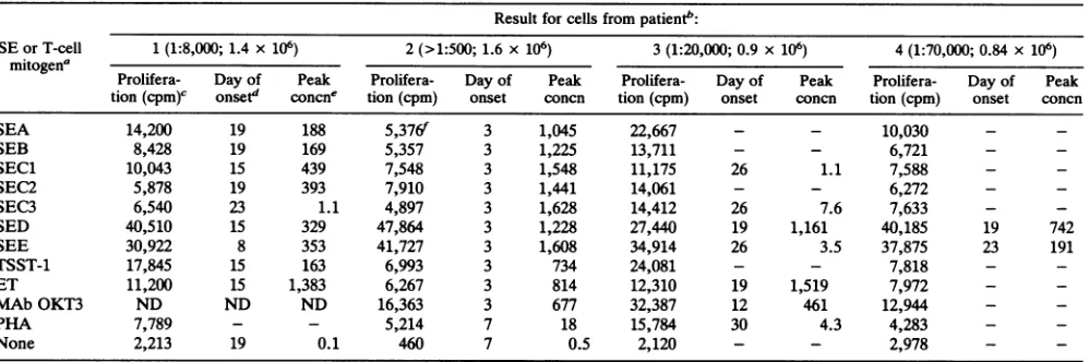
TABLE 1. Proliferationand HIV-1 production by PBMC depleted of CD8+ T cells in response to activation by SE or T-cell mitogens
Resultfor cells from
patientA:
SEorT-cell 1(1:8,000;1.4x 10⁶) 2 (>1:500; 1.6 x 10⁶) 3(1:20,000;0.9 x10⁶) 4 (1:70,000; 0.84x 10⁶)
mitogena
Prolifera- Day of Peak Prolifera- Day of Peak Prolifera- Day of Peak Prolifera- Day of Peaktion(cpm)c onset' concn' tion (cpm) onset concn tion (cpm) onset concn tion (cpm) onset concn
SEA 14,200 19 188 5,376f 3 1,045 22,667 - - 10,030 -
-SEB 8,428 19 169 5,357 3 1,225 13,711 - - 6,721 -
-SEC1 10,043 15 439 7,548 3 1,548 11,175 26 1.1 7,588 -
-SEC2 5,878 19 393 7,910 3 1,441 14,061 - - 6,272 -
-SEC3 6,540 23 1.1 4,897 3 1,628 14,412 26 7.6 7,633 -
-SED 40,510 15 329 47,864 3 1,228 27,440 19 1,161 40,185 19 742
SEE 30,922 8 353 41,727 3 1,608 34,914 26 3.5 37,875 23 191
TSST-1 17,845 15 163 6,993 3 734 24,081 - - 7,818 -
-ET 11,200 15 1,383 6,267 3 814 12,310 19 1,519 7,972 -
-MAb OKT3 ND ND ND 16,363 3 677 32,387 12 461 12,944 -
-PHA 7,789 - - 5,214 7 18 15,784 30 4.3 4,283 -
-None 2,213 19 0.1 460 7 0.5 2,120 - - 2,978 -
-aTSST-1,toxicshock syndrometoxin1; ET,exfoliative toxin;PHA,phytohemagglutinin; none,RPMI medium with fetalcalfserum and notoxin.
bNumbers inparenthesesareratios of infectedtouninfectedCD4+ T cells and numbers of cellsperwell.-,negative; ND,notdone. c Measuredas
[3Hlthymidine
incorporation.dFirst day ofsupernatantHIVp24positivity.
PeaksupernatantHIVp24concentration,innanogramspermilliliter.
fCells were cultured in theabsence ofrIL-2.
anti-HLA DR MAb B8.11 wasused in single-color
fluores-cencestudies. Cellswereanalyzed byflowcytometry
(FAC-Scan; Becton Dickinson, Mountain View, Calif.). The
sig-nals were collected and stored as list mode filesand were
analyzed with the LysisII program.
Cell cultures. Cell subsets were depleted fromperipheral
bloodmononuclear cells(PBMC)by usingimmunomagnetic
beads coated with therelevant MAb, at aratio of 10 beads
percellasdescribedpreviously (9). Following depletion,the
remaining cells always contained less than 2% of the
de-pleted subset. Cells were established as single cultures in
24-well plates (Costar, Cambridge, Mass.) or as triplicate
cultures in 96-well plates in RPMI 1640 (GIBCO, Paisley,
Scotland) containing 10%
(vol/vol)
fetal calfserumsupple-mented with 10 U of recombinant interleukin-2 (rIL-2)
(Amersham International, Amersham, England)perml. The
cellswereactivatedbyanSE(Toxin Technology, Sarasota,
Fla.) at1 ,ug/ml, bythe anti-CD3 MAb OKT3at1
,ig/ml,
orbyphytohemagglutinin at5 ,g/ml.The culturemedium was
changedtwice aweek,and thesupernatantsweretestedfor
the concentration of HIV-1 p24 antigen by means of an
antigen capture enzyme-linked immunosorbent assay (Ab-bott). Thecellswererestimulated bythe original activating
agent atthesameconcentration once aweek. Thecultures,
except those from patient 2, were maintained for 30 days.
SupernatantHIV-1p24inthe cultures of cells frompatient2
peakedondays7through 11,and therefore the cultureswere
terminated onday14. Cell proliferationwas determinedby
transferringvolumescorrespondingto105 cellsaccordingto
the initial cell count to 96-well plates on
day
3,incubating
with
[3H]thymidine
(2 ,uCi) for 24 h, and then determiningthe amount of incorporated radioactivity. In some
experi-ments, separate triplicate cultures containing5 x 104 cells
each were established for the isolation of HIV and for
[3H]thymidine
incorporation.Limiting dilution assays. For the determination of the
frequencyofHIV-1-infectedcells, CD4+ Tcellswere posi-tivelyselectedby using Dynabeadscoated with theanti-CD4
MAb66.1at aratio of four beads per cell
(3).
The cellswereseparated from the beads byincubationwith Fab
anti-serum (DETACHaBEAD; Dynal), washed twice, and left
overnightat37°C.Seriallydilutedpatient CD4+T cellswere
cocultured with CD4+ T cells from HIV-seronegative
do-nors,andthe cellswereactivatedwith beads coated with the
anti-T-cell receptor MAbT10/B9at aratio offour beads per
cell in the presence of 10 U of rIL-2 per ml as described
previously (3). On the basis of the Poisson distribution
formula, the frequency of infected cells is given as the
number of cells per well required to give 63%
HIV-1-producing wells(18).
RESULTSANDDISCUSSION
PBMCweredepleted of CD8+ T cells in ordertoremove
the cellsresponsible for the suppression of HIV-1 replication
whileretainingHLAclassII-expressingcells (4, 22).These
cellswereactivated in culturebySEA,SEB,
SECi
through-3, SED, SEE, toxic shock syndrome toxin 1, exfoliative
toxin,MAbOKT3,orphytohemagglutinin. With the
excep-tionof exfoliativetoxin, afinalconcentration of 1 ,ug of toxin
permlwaswell above thesaturatingconcentrationrequired
for theproliferationofPBMCdepletedofCD8+Tcells(data
not shown). Asummary of the results isgiven in Table 1. Themostimportant determinant for the detection ofHIV-1
p24 in the culturesupernatantswasthefrequency ofHIV-1
provirus-containingCD4+ T cells. Thus, HIV-1 replication
was induced by all the SE in cultures of cells from thetwo
individuals with the highest frequencies of HIV-1-infected
CD4+ T cells (patients 1 and 2). When the proportion of
CD4+ T cells infected
by
HIV-1 waslow,
only
thoseexotoxins which gavehigh proliferativeresponseswereable
toinduce HIV-1expression (patient4).The timerequiredfor
culture supernatants tobecome HIV-1 p24 positivevaried
considerablyand seemedtodependmainlyonthefrequency
of HIV-1 provirus-containing CD4+ T cells and on the
proliferative response toeach of the staphylococcal
super-antigens. However, the timerequiredfor culture
positivity
could also depend on the phenotype of the HIV-1
quasi-species infecting each individual
(17).
Patient2,
whoseexotoxin-stimulated cell culture supernatantswereall HIV-1
on November 9, 2019 by guest
http://jvi.asm.org/
A
CD4+LY 110O
CD4- LY
FORWARDSCATTER
B
FORWARD SCATTER
e
cm
0
l
~c-v
I
-w
w we
FORWARDSCATTER
D
FORWARD SCATTER
Iooo
WU-10
RPMV 0.1 FCS
1.0 10 100 1000
§
0 0
40 X a 1. 40
10
M-Concentration of SEDorSEE(nanogram/ml)
FIG. 2. Cell proliferation and HIV-1 production in response to different concentrations of SED and SEE. PBMCdepleted of CD8+ T cells from patient 2werecultured in the absenceofrIL-2.0 and
[l, proliferationinresponsetoSED andSEE, respectively. * and *,supernatantHIV-1p24 concentrationsonday 7 inresponseto SED andSEE, respectively. Inunstimulatedcells, the p24 concen-trationwas0.2ng/mlandtheproliferativeresponse was235cpm(*). FCS, fetal calfserum.
b100
E
n
CD
0u 1 0
N
01
coI
0 E
.P
c
0 0 0
E
I
A B C D
FIG. 1. Results of flow cytometric studies (a) and HIV-1 p24 concentrations in cell supernatants on day 8 (open bars) and [3H]thymidine incorporation (hatched bars) (allvaluesaremedians
andrangesoftriplicateexperiments) (b).PBMC frompatient2were
depleted of CD8+T cells(A),CD8+andCD4+T cells(B),CD8+T cells and monocytes (C), and CD8+ T cells and HLA class
II-expressingcells(D).The cellswereexaminedbyusinganti-CD4 and anti-CD8 MAb in two-color fluorescence studies, butthe popula-tionsarepresented hereas functionsofanti-CD4MAbstaining (y
axis) and size (forward scatter) (x axis).
p24 positiveonday 3, has been extensivelystudied. Allbulk
virus isolates as well as each of several viral clones have
replicatedincell lines and inducedcytopathologyconsistent
with arapid-high phenotype (la).
In order to identify the cell subsets involved in the
SE-mediated induction of HIV-1 expression from PBMC
depletedofCD8+Tcells, cell subsetdepletion experiments
with cells from patient 2 were performed. The results are
presentedin Fig. 1. WhenCD4+ T cellsweredepleted, cell
proliferation wasconsiderably reduced, and HIV-1
expres-sion was completely abrogated (compare the results from
experimentsB andA). Incontrast, depletion ofmonocytes
left the expression of HIV-1 unchanged and caused only a
slight reduction of [3H]thymidine incorporation (compare
theresults fromexperiments C andA). The results show that
thesourceofexpressedvirus intheseexperiments wasthe
CD4+Tcells.Theoretically, monocytescould have
contrib-uted to the concentration ofHIV p24 in the cell
superna-tants. Monocytes have been shown to contain HIV-1 in
some individuals, and the binding of SEto their HLAclass
II molecules has been shown to cause transcriptional
acti-vation of certain genes (21). Experiment C demonstrated,
however, thatmonocyteswereexpendablebothas asource
of HIV-1 andas asourceof HLA classII.Theimportance of
thepresenceof cellsexpressingHLA class II moleculeswas
demonstrated in experiment D. In this experiment, HLA
class II-expressing cells were depleted, removing all the
monocytesandleavingless than0.2% HLADR-expressing
lymphocytes. Both cell proliferation and the expression of
HIV-1 were completely abrogated, although CD4+ T cells
werepresent at aproportion comparable tothose in
exper-imentsCand A (19versus 17% inexperimentC and 9% in
experimentA).Thus, free solubleSEwereunabletoinduce
[3H]thymidine incorporationand theexpressionof HIV-1 in
theCD4+Tcells.
Long-term T-cell cultures require exogenous IL-2. We
tookadvantageof therapidkinetics of HIV-1productionin
cells from patient 2 to examine whether SE might induce HIV-1expression inthe absence ofexogenouslyaddedcell
growth factors. No differences in HIV-1 p24concentration
between cell cultures established with and without
exoge-nous IL-2 were found (the results of the experiment
per-formed in the absence of rIL-2are presentedin Table 1).
Subsequent experiments involving cells from several more
HIV-infected individuals have shown that theproliferative
responseof PBMCdepletedofCD8+Tcells isnotchanged
by the addition of rIL-2 and that SE-activated PBMC
de-pleted of CD8+ T cells from uninfected donors produce
supersaturatinglevels of IL-2 (5).Thissuggeststhat
endog-enousIL-2produced bytheCD4+T cells inresponsetoSE
is sufficienttosustain these cellsthrough2weeks of
contin-uousHIV-1production invitro.
Dilutions of SED and SEE as low as 100 pg/ml still resulted in cell proliferation and highlevels of HIV-1
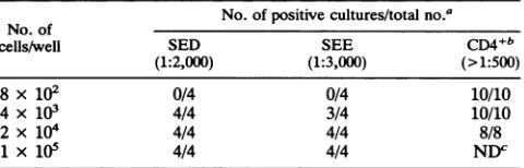
repli-cation(Fig. 2). In celltitrationexperiments, thefrequencies
of HIV-1 provirus-containing PBMC depleted of CD8+ T
cells were found to be 1:2,000 for SED-responsive and
1:3,000 for SEE-responsive cells (Table 2). Assuming that
these exotoxins activate 20% of the CD4+ T cells at most
a
p
.v
on November 9, 2019 by guest
http://jvi.asm.org/
[image:3.612.63.299.55.449.2] [image:3.612.317.558.75.222.2]TABLE 2. Limiting dilution analysis of the occurrence of HIV-1-producing cells in cells from patient 2
No. ofpositive cultures/totalno.a
No. of
cells/well SED SEE CD4+b
(1:2,000) (1:3,000) (>1:500)
8x 10² 0/4 0/4 10/10
4 x 10³ 4/4 3/4 10/10
2 x 10⁴ 4/4 4/4 8/8
1 x 10⁵ 4/4 4/4 NDC
aPBMC depleted of CD8+ T cells wereactivatedby SED or SEE in the
absenceof rIL-2 and allogeneic feeder cells. Numbers inparentheses are
resultsespressed as ratios of infected to uninfected cells.
bFordetails of limiting dilution analysis of HIV-infectedCD4+ Tcells,see
MaterialsandMethods.
cND,notdone.
(12),thisindicates that each exotoxin-binding, HIV-1
provi-rus-containing cell was induced to support active HIV-1
replication. This conclusion is supported by the results of the
experiments using cells from patient 4. PBMC depleted of
CD8+Tcellsusuallycontain less than50% CD4+ T cells (2).
Ifthis was the case also for patient 4, then SED and SEE
induced HIV-1 expression in wells containing fewer than 6
HIV-1provirus-containing CD4+ T cells (Table 1).
Our results contrastwith those in arecent article, which
reportedthat the PBMC depleted of CD8+ T cells from all
asymptomatic HIV-1-infected individuals studied failed to
proliferate and instead showed evidence of death by
apop-tosis in response to activation by SEB (10). That study
involvedalarge number of HIV-infected individuals, and the
methods employed werequitesimilar to the ones used in the
present study. Although this study involved only a small
groupofHIV-1-infected individuals,weshow by
[3H]thymi-dineincorporationthatPBMCdepleted of CD8+ T cells are
capableofproliferation, andbythemeasurement of
super-natantp24 concentrationweshowthat these cellsareableto
continuouslyexpress HIV-1 forat least 3 weeks in culture.
Cell numbers were greatly increased in these long-term
cultures (datanotshown). Insubsequent studies, involving
other HIV-1-infected individuals, we have found that the
SE-induced
proliferation
of PBMCdepletedofCD8+Tcells, measured as [3H]thymidine incorporation (counts perminute)per CD4+ Tcell present in thewell,was as strong
for cells from the HIV-1-infected studyparticipants as for
cells from HLA DR- and age-matched uninfectedcontrols
(2).Thediscrepancyinresults between thetwostudiesmust
beleftunexplainedatthe presenttime.
One unexpected observation made during these
experi-ments was that SED and SEE gave considerably higher proliferativeresponsesthan theother exotoxinsormitogens (Table 1), suggesting binding by SED and SEE to a
high
proportion of T cells in our patients. This contrasted with
resultsobtainedbytestingPBMCdepletedofCD8+ Tcells
obtained from nineHIV-1
seronegative donors,
where SEDand SEE gave proliferative responses similar tothe other
exotoxinsandmitogens
(data
notshown).
Inother studies ofHIV-1 seronegative individuals, neither SED nor SEE has
been showntobindtoa
disproportionately large
fraction ofT-cellreceptor
VP-expressing
cells(6,
16).
Thesignificance
of theseobservations remains tobeestablished.
Certain microbial gene
products,
notably
fromcytomega-lovirus,Epstein-Barr virus,and humanT-cell leukemia virus
type I
(HTLV-I),
have been showntotransactivate HIV-1longterminal repeat(7, 13,
19). Theoretically,
suchtransac-tivating agents might bypass the requirement for cell activa-tion by specific antigen in order for HIV-1 producactiva-tion by infected cells to occur. However, these agents either are rare (HTLV-I) or require coinfection of the same cell for trans-activation of HIV-1 to occur (Epstein-Barr virus and cyto-megalovirus). They are thus likely to be of limited
impor-tance ascofactors for thedevelopment of AIDS. In contrast,
staphylococci are common pathogens, and more than 40% of all strains produce one or more exotoxins (8, 14). Our experiments demonstrate that these exotoxins, at very low concentrations and without exogenously added cell growth factors, can induce virus expression from each
HIV-1-infected,superantigen-responsive CD4+ T cell in vitro. Such
superantigenic T-cell activation may also induce HIV-1 replication in vivo and thus may represent an important cofactor for the development of AIDS.
ACKNOWLEDGMENTS
Weare indebted to M. Sl0rdahl, Section for AIDS Prevention,
Oslo City Department for Health and Environment, and her pa-tients. WethankJ. Hansen, J. S. Thompson, B.Malissen, and S. Funderud for kindgifts of MAbs. We also thank T. Egeland for helping with the flow cytometry studies.
This study wassupported by the Norwegian Cancer Society.
REFERENCES
1. Adams, P. W., E. M. Opremcak, and C. G. Orosz. 1991. Limiting dilution analysisof human, tetanus-reactive helper T
lymphocytes. Arapid method for the enumeration of helper T
lymphocyteswith specificity for solubleantigens. J. Immunol. Methods142:231-241.
la.Asjp,
B., and J. E. Brinchmann. 1991. Unpublished observa-tions.2. Brinchmann,J. E.1992.Unpublished observations.
3. Brinchmann, J. E., J. Albert, and F. Vartdal. 1991. Few infected CD4+ T cells but a high proportion of replication-competent provirus copies in asymptomatichuman immunodeficiency vi-rus type 1infection. J. Virol. 65:2019-2023.
4. Brinchmann, J. E.,G. Gaudernack, and F. Vartdal.1990.CD8+
T cells inhibit HIV replication in naturally infected CD4+ T cells. Evidence for a soluble inhibitor. J. Immunol. 144:2961-2966.
5. Brinchmann, J. E., and T. Lea.Unpublished observations.
6. Choi, Y., B. Kotzin, L. Herron,J. Callahan, P. Marrack, and J.
Kappler. 1989. Interaction of Staphylococcus aureus toxin
"superantigens" with human T cells. Proc. Natl. Acad. Sci.
USA86:8941-8945.
7. Davis, M.G.,S. C. Kenney, J. Kamine, J. S. Pagano, and E.-S. Huang.1987. Immediate-earlygeneregion ofhuman cytomeg-alovirus trans-activates the promoter of human immunodefi-ciency virus.Proc.Natl. Acad. Sci. USA84:8642-8646. 8. Easmon, C. S.F., and M. Goodfellow. 1990.Staphylococcusand
micrococcus, p. 161-186. In M. T. Parker and B. I. Duerden
(ed.), Topley & Wilson's principles of bacteriology, virology
andimmunity,8thed.,vol. 2. Hodder &Stockton, London.
9. Funderud,S.,K.Nustad, T.Lea,F.Vartdal,G.Gaudernack,P. Stenstad, and J. Ugelstad. 1987. Fractionation oflymphocytes byimmunomagnetic beads,p. 55-65. In G. G. B. Klaus (ed.), Lymphocytes.Apractical approach. IRLPress, Oxford.
10. Groux, H., G. Torpier, D.Monte,Y.Mouton,A. Capron,and J. C. Ameisen. 1992.Activation-induced deathby apoptosisin
CD4+ T cells from human immunodeficiency virus-infected
asymptomaticindividuals. J. Exp.Med. 175:331-340.
11. Ho, D. D., T. Moudgil, and M. Alam. 1989. Quantitation of humanimmunodeficiencyvirustype 1in the blood of infected persons. N.Engl. J. Med.321:1621-1625.
12. Janeway,C. A. 1990. Selfsuperantigens? Cell 63:659-661.
13. Kenney,S.,J.Kamine,D.Markovitz,R.Fenrick,andJ. Pagano.
1988. An Epstein-Barr virus immediate-early gene product
trans-activates expression from the human immunodeficiency
on November 9, 2019 by guest
http://jvi.asm.org/
virus long terminalrepeat.Proc.Natl. Acad. Sci. USA 85:1652-1656.
14. Levine, S. J., D. A. White, and A. 0. S. Fels. 1990. The
incidenceandsignificanceofStaphylococcusaureusin respira-torycultures from patients infected with the human immunode-ficiency virus. Am. Rev.Respir. Dis. 141:89-93.
15. Marrack, P., and J. Kappler.1987. TheTcellreceptor.Science 238:1073-1079.
16. Marrack, P., andJ.Kappler.1990. Thestaphylococcal
entero-toxins and their relatives. Science 248:705-711.
17. Meyerhans, A., R. Cheynier, J. Albert, M. Seth, S. Kwok,J. Sninsky, L. Morfeldt-Manson, B. Asjo, and S. Wain-Hobson. 1989.Temporal fluctuations in HIVquasispecies in vivoarenot
reflected bysequentialHIVisolations. Cell 58:901-910. 18. Quintans, J., and I. Lefkovits.1973. Precursorcell frequencyto
sheep red cells in nude mice. Estimation of frequency in the microculturesystem.Eur.J. Immunol. 3:392-397.
19. Sietkevitz, M., S. F. Josephs, M. Dukovich, N. Peffer, F. Wong-Staal, and W. C. Greene. 1987. Activation ofthe HIV-1 LTRby T cell mitogens and the trans-activator protein of HTLV-1. Science 238:1575-1578.
20. Simmonds, P., P.Balfe, J. F. Peutherer, C. A. Ludlam, J. 0.
Bishop, andA. J. L. Brown. 1990. Human immunodeficiency virus-infected individuals contain provirus in small numbers of peripheral mononuclear cells andatlowcopynumbers.J. Virol.
64:864-872.
21. Trede, N.S., R. S. Geha, and T. Chatila. 1991.Transcriptional activation of IL-11 andtumornecrosis factor-agenesbyMHC class II ligands. J. Immunol.146:2310-2315.
22. Wiviott, L. D., C. M. Walker, and J. A. Levy. 1990. CD8+ lymphocytes suppress HIV production by autologous CD4+ cells without eliminating the infectedcells from culture. Cell. Immunol. 128:628-634.