Copyright X 1992, AmericanSocietyforMicrobiology
al/EBP:
a
Leucine
Zipper Protein That Binds
CCAAT/Enhancer Elements in the Avian Leukosis Virus
Long
Terminal
Repeat
Enhancer
W. J.BOWERS ANDA. RUDDELL*
DepartmentofMicrobiologyandImmunologyand CancerCenter, University ofRochester MedicalCenter,
Rochester,
New York 14642 Received 15June 1992/Accepted6August 1992Avianleukosis virus(ALV) induces bursallymphoma inchickensafterintegrationofproviral longterminal repeat(LTR) enhancer sequences next tothe c-myc proto-oncogene. LabileLTR-binding proteinsappeartobe essential for c-mychyperexpression,since both LTR-enhanced transcriptionand the activitiesofLTR-binding proteins are
specifically
decreased after inhibitionofproteinsynthesis (A. Ruddell,M.Linial,W.Schubach,and M. Groudine, J. Virol. 62:2728-2735, 1988). This lability is restricted to hematopoietic cells from ALV-susceptible chickenstrains, suggesting that the labileproteinsplayanimportantroleinlymphomagenesis.Themajor
labileactivity
bindingtothe alLTRregion (A. Ruddell, M. Linial,and M. Groudine,Mol.Cell. Biol. 12:5660-5668, 1989) was purified from bursal lymphoma cells by conventional and oligonucleotideaffinity
chromatography,yielding threeproteinsof35, 40,and 42 kDa. More thanoneof thesespeciesbinds the al LTR region,asjudged by gel shift analysis.Ageneencodinganal-bindingprotein (designated al/EBP)wasclonedby screeningabursallymphomacDNAlibrary for fusion proteinsbindingthe al LTRsite. DNaseIfootprintingand gel shift assays indicate that the al/EBP fusion protein binds multiple LTRCCAAT/enhancer elements ina pattern similartothatof thepurified B-cell protein. DNA sequence analysis shows that this 2.2-kb cDNA encodes a209-amino-acid open reading frame containing carboxy-terminal basic and leucine zipper motifs, indicating thatal/EBPencodesanovel member of theleucinezipper
family
oftranscription factors.Theoncogenic potentialof retrovirusescausingnonacute diseases appears to be regulated by the U3 long terminal repeat(LTR)enhanceraswellasbytheviralenvgene(6, 22, 57). Transcription factorbinding tothe LTR enhancer may regulate the frequencyand tissuespecificityoftumor induc-tion. For example, the oncogenicity of murine leukemia virus strain SL3-3 is influenced by mutations in LTR en-hancerprotein-binding sites (19, 30). Thymic lymphomaor erythroleukemia induction by Moloney or Friend murine leukemiavirus, respectively, is largelydeterminedby differ-encesinmultiple protein-binding sites in the LTR enhancer (16, 56).
Avian leukosis virus(ALV) provides awell-characterized system for analysis ofthe role of transcription factors in tissue-specific transformation. ALV induces B-cell lym-phoma in chickens after therareintegrationofaproviral U3 LTR enhancer next to the c-myc proto-oncogene (39, 42). This LTR deregulation of c-myc expression, giving up to 100-fold increases in c-mycexpression,isanimportant early determinant of lymphomagenesis (12, 40). The LTR en-hancer isrequiredfortumorinduction, asendogenousALV retroviruses lack complete enhancer elements and rarely induce lymphoma (6, 46, 61). Host cell factors also play an important role in lymphomagenesis, as some strains of chickensareresistanttoALVlymphoma (2, 14). Transplan-tationexperiments have demonstrated that the target pre-B cells encode the resistance phenotype (45). The nature of the host factors mediating resistance is not known, as the patterns of ALV infection, integration, and expression are similar insusceptible and resistant strains (2, 14). Moreover, viral expression is similar in many cell types, including
*
Corresponding
author.immature or mature bursal cells (50), even though tumor induction isspecific for immature B cells. Thus, the factors regulatingALVpre-B-cell susceptibilitydonot actsimply by restrictinghigh-level viralproteinoroncogeneexpression to targetpre-B cells.
We have identified one characteristic of ALV LTR en-hancementwhich does correlate withpre-B-cell susceptibil-ityto tumorinduction.LTR-enhanced c-myc and viral gene transcription is specifically decreased 10- to 15-fold after inhibition of protein synthesis in bursal lymphoma cells, while LTR-enhanced transcription is unaffected by protein synthesisinhibition in infected T cellsorembryo fibroblasts (31).Thesefindingssuggestthat labile or short-lived proteins regulate LTR enhancement in B cells. This lability is re-strictedtoimmaturehematopoieticcellsofALV-susceptible chickenstrains, while LTR-enhanced transcription is stable in all tissues of ALV-resistant chicken strains (50). The correlation of labile LTR enhancement with pre-B-cell sus-ceptibility suggests that this lability is important in ALV lymphomagenesis.Labile LTR enhancement could influence c-myc hyperexpression in a manner essential for tumor induction. Forexample, c-myc mRNA and protein are very short-lived (9, 20);consequently, developmental reg-ulation of labile LTR enhancement could transiently down-regulate c-mycexpression, improving the survival of target pre-B cells(50).This labiledown-regulation could reduce the cytotoxicity of hyperexpressed c-myc (11, 57, 59) or could allow bursal differentiation events required for lympho-magenesis (8,45).
We previously analyzed nuclear proteins binding to the ALV LTR enhancer to determine whether labile binding proteinscouldregulate LTR-enhanced transcription in pre-B cells. Five LTRenhancer-binding proteinswereidentified by gel shift and footprinting analyses of nuclear proteins from 6578
on November 9, 2019 by guest
http://jvi.asm.org/
ALV LTR CCAATIENHANCER ELEMENT-BINDING PROTEIN 6579
bursal lymphoma cells (49, 50). Three of the proteins (al, a3, and b*) are specifically labile in pre-B cells, while they are stable or not expressed in other cell types. These findings suggest that binding of the labile proteins is essential for high ratesofLTR-enhanced transcription in B cells. The major al protein-binding site contains multiple CCAAT/enhancer el-ements,which are protected in DNase I footprinting assays with B-cell nuclear extracts or with partially purified B-cell proteins.CCAAT/enhancer elements are found in many viral and cellular gene enhancers (7, 23), and proteins binding to these regions can activate transcription. Several proteins that bind to these elements have been identified. For exam-ple, the C/EBP protein binds many CCAAT/enhancer ele-ments(23, 51) and can activate or repress transcription (13, 44). The Ig/EBP and NF/IL-6 proteins bind CCAAT/en-hancer elements in theimmunoglobulin (Ig) heavy-chain and interleukin-6 (IL-6) gene enhancers, respectively (1, 48). These proteins belong to a family of leucine zipper transcrip-tion factors which feature conserved DNA-binding regions enriched in basic amino acids and conserved leucine heptad repeats which allow formation of homodimers or het-erodimers with other leucine zipper proteins (29, 48). The C/EBP and Ig/EBP proteins also bind CCAAT/enhancer elements in the Rous sarcoma virus (RSV) LTR (48, 51), which is very similar to the ALV LTR (3). Such proteins could beinvolved in labile LTR binding and enhancement if they are in fact expressed in avian pre-B cells.
We have purified the al LTR-binding activity from chicken bursal lymphoma cells. Three proteins of 35 to 42 kDa are enriched after purification, and more than one of these species appear to encodesequence-specific al-binding activity. These could represent distinct proteins or one protein that is differentially modified. To further characterize theseproteins,aXgtll cDNA library from bursal lymphoma cells was screened for cDNAs encoding al LTR-binding activity. One cDNA clone that encodes an al LTR-binding protein closely related to the IglEBP leucine zipper factor (48)was obtained. Analysis of this cloned gene will deter-mine whether it encodes a labile protein regulating LTR enhancement and susceptibility to ALV lymphomagenesis.
MATERIALS AND METHODS
B-cell proteinpurification. S13 bursal lymphoma cells were grownin spinner flasks (Bellco) in RP-9 medium (Dulbecco modified Eagle medium supplemented with 5% calf serum, 1%heat-inactivated chicken serum, and tryptose phosphate broth[GIBCO Laboratories]). Cells were harvested by cen-trifugationat2,000xg,washed inphosphate-buffered saline (140 mM NaCl, 11 mM KCI, 1.5 mM KH2PO4, 6.5 mM Na2HPO4, 0.5mM MgCl2, 0.9 mM CaCl2), andcentrifuged again. Cellpellets wereusedimmediately or were frozen in liquid nitrogen and stored at -70°C. Nuclear extracts were prepared by 0.5 M NaCl treatment of purified nuclei as previously described (50). Extracts were separated by S-300 Sepharose chromatography (Pharmacia) in buffer A (10% glycerol, 50mM KCI, 25 mM N-2-hydroxyethylpiperazine-N'-2-ethanesulfonic acid [HEPES; pH 7.9], 1 mM EDTA, 0.1% Nonidet P40, 1 mM dithiothreitol [DTT], 1 mM sodium metabisulfite, 0.2 ng of leupeptin per ml, 0.2 ng of pepstatin per ml 1 U of aprotinin HCl per ml, 1 mM phenylmethylsulfonyl fluoride [PMSF] [all from Sigma]). Column fractionswere analyzed forprotein content by the Bradford assay(5)and for al LTR-binding activity byagel shift assay (see below). Fractions containing al-binding activity were pooled, heated at 85°C for 10 min, and then
centrifuged for 10 min at 12,000 x g to remove insoluble protein. The heat-purified supematant was applied to a concatenated al oligonucleotide-agarose column (prepared as described by Kadonaga et al. [24]) in 0.075 M KCl-buffer B [50 mM HEPES (pH 7.9), 20% glycerol, 0.1% Nonidet P-40, 1 mM DTT, 1 mM sodium metabisulfite, 10
,ug
of poly(dI-dC) poly(dI-dC) (Pharmacia), 0.2 ng of leupeptin per ml, 0.2 ng of pepstatin per ml, 1 U of aprotinin HCl per ml, 1 mM PMSF], and bound protein was eluted with 0.6 M KCl-buffer B.Gel shift assay. Protein was incubated with 5,000 cpm (approximately 0.1 ng) of the32P-labeled al oligonucleotide probe in gel shift buffer (10 mM Tris HCl, [pH 8.0], 50 mM NaCl, 10%glycerol, 1mM EDTA, 1 mM DTT) and 0.1 mg of poly(dI-dC) poly(dI-dC). The
15-pl
reaction mixtures were incubated at room temperature for 20 min and then electro-phoresed on 4% polyacrylamide gels in TAE buffer (6.7 mM Tris, 3.3 mM sodium acetate, 1 mM EDTA [pH 7.5]) at 30 mA for 1 h as previously described (50). In some assays, unlabeled al or b oligonucleotide was added as a competitor (49). Thedouble-stranded al oligonucleotide probe sequence is 5'-GGGAAATGTAGTClTrATGCAATACTCTAA-3'/5'-TTCCCTlAGAGTATTGCATAAGACTACAT-3'); the b oligonucleotide probe sequence is 5'-AAGGAGAGAAAAAGTACCGTGCATG-3'/5'-CATGCACGGTACT'i-ii-ir'CTCT
CCTT-3'.
SDS-PAGE. Protein samples were diluted to 0.25 ml and were precipitated with 10 ,ug of bovine lactoglobulin carrier (Sigma) in 4 volumes of cold acetone by chilling on dry ice for 30min. Precipitates were centrifuged at 12,000 x g for 20 min, rinsed with cold 80% acetone, recentrifuged, and resus-pended in sodium dodecyl sulfate (SDS) sample buffer (27). Samples were subjected to electrophoresis on 10% poly-acrylamide-SDS-gels (SDS-PAGE) alongside molecular weightmarkers(Amersham) and then subjected to fixation in 50% methanol-0.04% formaldehyde and silver staining (38). Protein renaturation from SDS-polyacrylamidegels. Three hundred micrograms of bursal lymphoma protein (purified by S-300 chromatography and heat treatment) was precip-itated with 80% acetone, resuspended in SDS sample buffer, andseparated by SDS-PAGE as described above. Ten slices wereexcised from the gel lane and were elutedovernight at 22°C in 0.4 ml of buffer C (40
p,g
ofbovine serum albumin [BSA;Miles Biochemical] per ml, 50 mM Tris HCl [pH 7.5], 0.1 mM EDTA, 0.1% SDS, 5 mM DTT, 0.1 M NaCl [21]). The eluate wasprecipitated with 80% acetone as described above and then renatured in 40,ul of buffer D (1.5 mg of BSA per ml, 20% glycerol, 20 mM HEPES, [pH 7.9], 0.05 M NaCl, 0.1% Nonidet P-40, 0.1 mM EDTA, 0.5 mM DTT, 1 mM sodiummetabisulfite, 0.2 ng of leupeptin per ml, 0.2 ng of pepstatin per ml, 1 U ofaprotinin HCl per ml, 0.5 mM PMSF) at 4°Covernight with gentle rocking. Aliquots were analyzed by gel shift assay with or without an unlabeled competitor as described above.DNase I footprinting assay. The 245-bpMstII-EcoRI frag-ment of an ALV LTR subcloned from BK25 bursal lym-phoma cells was 32P end labeled as described previously (49). Gel shiftreactions with32P-labeledALV LTR and 0.01 to 2 ,ug ofal/EBP fusion protein wereincubated with DNase I(bovine pancreatic; Sigma) at 0.01 to 0.05 ,ug/ml in 5 mM CaCl2-5 mMMgCl2 (15). Following a 1-min DNase I treat-ment, the reactions were stopped with TENS (10 mM Tris HCl [pH 8.0], 1 mM EDTA, 0.1 M NaCl, 0.1% SDS) solution, proteinase K treated, and precipitated with 70% ethanol-2 M ammonium acetate. Samples were resolved on VOL.66, 1992
on November 9, 2019 by guest
http://jvi.asm.org/
8 Murea-8% polyacrylamidegelsinparallelwith the corre-spondingA+G sequencereactions (35).
Isolationof a recombinantal/EBP clone. TheXgtll screen-ing techniqueofSinghetal.(54)wasusedtoidentifycDNAs encoding al-binding proteins.
Poly(A)+
RNAwas purified from S13bursal lymphomacells byguanidinium isothiocy-anate-cesium trifluoroacetateultracentrifugation
(41)
and thentworounds ofoligo(dT)-cellulose
chromatography
(Be-thesdaResearchLaboratories).First-strand cDNAwas pre-paredfrom the RNAbyusing murineleukemiavirusreverse transcriptase (Pharmacia), and second-strand cDNA was synthesizedbyusingKlenow DNApolymerase(18);
EcoRI-NotIlinkerswerethenligatedandclonedinto the EcoRIsite ofXgtll
(60). The primaryphage library (1.5 x10⁶
phage) wasplated onEschenchia coli Y1090, and isopropylthioga-lactopyranoside(IPTG)-induced fusion proteinswere trans-ferredtonitrocellulose. Duplicate filterswere screened for binding to the 32P-labeled concatenated al oligonucleotide probe, which wasprepared byligation and nicktranslation. Positive phage were replated andsuccessively
screened twicefor aloligonucleotide-binding activity.
DNA sequence analysis. The al/EBP cDNA insert was isolatedfrom purified Xgtll phage DNAbyNotIdigestion and was subcloned into the NotI site of the Bluescribe plasmid(Stratagene) for DNA sequence analysis. Plasmids werepurifiedby cesiumchloride-ethidiumbromide ultracen-trifugation, and the sequences of both denatured plasmid strandsweredeterminedbydideoxysequencing, using vec-tor or cDNA-specific primers. Deaza-GTP and dITP se-quence reactions were compared to resolve compressed regions (37). DNA sequences were analyzed by using Ge-netics ComputerGroupcomputer programs(10, 43).
Bacterial expression of the al/EBP fusion protein. Phage lysogenswereinduced inE. coli Y1089, and IPTG-induced bacterial lysateswerepreparedas describedby Young and Davis(60)foranalysisingel shift assays.Forexpressionin a plasmidvector, the al/EBP cDNAwas gel purified from theNotI-digested Bluescribe plasmid, treated with Klenow polymerase, andligated intothe SmaI site of the pGEX 2T vector, sothat the cDNA openreadingframewastranslated as a glutathione-S-transferase (GST) fusion protein (55). Fusion protein expression was induced by 1 mM IPTG treatmentfor3h; this procedurewasfollowedby sonication andglutathione-agarose purification oftheal/EBP-GST fu-sionprotein. Proteaseinhibitors (0.2ngofleupeptinperml, 0.2 ng ofpepstatinperml,1 U ofaprotinin HClperml, and 1 mM PMSF) were added immediately after sonication. Purified proteinwasadjustedto8% glycerol-50mMNaCl-1 mMEDTA-1 mMDTT, frozeninliquidnitrogen, and stored inaliquots at -700C.
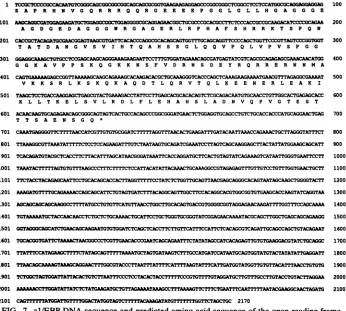
Nucleotide sequence accession number.The sequence data shown in Fig. 7 have been assigned GenBank accession number M95573.
RESULTS
Purification of the al LTR-binding protein. The labile al-binding activity of B cells interacts with regions of the ALV LTR enhancer containing multiple CCAAT/enhancer elements (49, 51). Gel shift analysis of B-cell nuclear pro-teins with an oligonucleotide probe for the al LTR site showsadiffuse ladderofDNA-protein complexes (Fig. 1A). This result could reflect binding to more than, one of the overlapping CCAAT/enhancerelementsin the al oligonucle-otideprobe, multimerizationof complexes, or the binding of severalproteins. The al-binding activitywas purified from
A. 1 2
B.
1 2 mwM
-55
-43
- 3t
- 22
FIG. 1. Analysis of the al oligonucleotide affinity-purified al LTR-binding protein. (A) Gel shift assay of heat-purified B-cell protein(lane 1)and aloligonucleotide affinity-purified protein(lane 2)with the32P-labeled aloligonucleotideprobe.(B)SDS-PAGE and silverstainingofheat-purified protein (lane 1)and oligonucleotide-agarose affinity-purified protein (lane 2).Horizontal lines indicate enriched species. The asterisk indicates the carrierlactoglobulin protein. The migration of molecular weight markers (mwm) is indicated in kilodaltons.
the S13 bursal lymphoma cell line inorder to characterize theproteinorproteins involved in labile LTRenhancement. Nuclear extracts from 80 liters of bursal lymphoma cells wereinitially fractionated by S-300 Sepharose chromatogra-phyasdescribed in Materials and Methods. Fractionswere assayed for al-binding activity by gel shift assaywith the 32P-labeled aloligonucleotide probe.Theal-bindingactivity elutes in theincluded column fractions and is purified two-to threefoldby this chromatography, asestimated by compar-ison ofgel shift binding activity per microgram ofprotein (datanotshown).
Theal-binding activity remains active after heattreatment (49).Therefore, the S-300 fractions were heated at 85°C for 10 min and centrifugedto remove insoluble protein, giving roughly fourfold purification. The heat-purified proteinwas further enriched bybindingto analoligonucleotide-agarose affinity column (24) and elution with 0.6 M KCl. The affinity-purified proteincontinues toproducea diffuseladder of LTR-binding activity in gel shift assays with the 32p_ labeled al oligonucleotide probe (Fig. 1A). This activityis enrichedroughly4,000-fold relativetotheactivityofthe 0.5 M NaCl nuclear extract.
The composition of the oligonucleotide affinity-purified protein was analyzed bySDS-PAGE and silver stainingas described in Materials and Methods. The affinity-purified proteinpreparation is enriched forthree protein species of approximately 35, 40, and 42 kDa relative to the heat-purified protein (Fig. 1B). These specieswere consistently purified in several independent experiments. Proteins of about 32 and 45 kDa are also observed in theaffinity-purified sample, although they are variably present and are not enrichedby aloligonucleotide-agarose affinity chromatogra-phy.
Characterization of theal-binding protein. Oneorall of the 35- to 42-kDa species purified from bursallymphoma cells may bespecifical-binding proteins. Proteinswererenatured from SDS-polyacrylamide gel slices and tested in gel shift assaystoconfirmthatproteins in thismolecularmassrange encode al-binding activity. Heat-purified protein was re-solved on a10% polyacrylamide-SDS gel, and slices of the gelwere cutout, eluted, andrenatured inbuffer containing BSA. Gel shift al-binding activity is detected in two gel
7 X
1. 4g
wo
on November 9, 2019 by guest
http://jvi.asm.org/
[image:3.612.365.503.72.195.2]ALV LTR CCAATIENHANCER ELEMENT-BINDING PROTEIN 6581
B. fraction #1 fraction #2 gel slices
crudel 23456789 10
competitor competitor
al b - - al b
-A.
WT GGGAAATGTAGTCTTATGCAATACTCTAATGCAA
CCCC"TTTACATCAGAATACGTTATGAGATTACGTT
Ml GGGAAATGTAGTCTTATECACTCTAATGCAA
CCCTTTACATCAGAATAd TGAGATTACGTT
-_ w 'Jt4i4
B. C.
D-FIG. 2. Identificationof alLTR-bindingproteins after
renatura-tion from SDS-polyacrylamide gelslices. (A) Gel shiftanalysisof crudeheat-purifiedprotein and of protein renatured from slices ofan
SDS-polyacrylamide gelofheat-purified proteinwith the32P-labeled al oligonucleotide probe. The lanes indicate gel slices in the molecular mass rangeof 20 to200 kDa. The arrow indicatesthe DNA-protein complex. (B) Gelshiftanalysisofprotein ingelslice fractions 1and 2(molecularmass rangesof 20to35 and 35to 45 kDa, respectively)withthe 32P-labeled aloligonucleotide probe.An unlabeled alorboligonucleotide competitor(50-, 100-,or150-fold molarexcess)wasaddedasindicated.
slices which contain 25-to35-kDa(slice 1) and 35-to45kDa (slice 2) proteins (Fig. 2A). The gel shift binding activities from the two slices migrate slightly differently, suggesting that they contain distinct proteins with LTR-binding activ-ity. Both of thebinding activities are sequence specific, as
judgedby gel shift competition with the unlabeled al oligo-nucleotide but not with the unlabeled b oligonucleotide probe(Fig. 2B). The molecularmassesof theseproteinsare
within the range of 35 to 42 kDa observed for purified al proteins. These data alsosupportthe idea thatmorethanone
of these species encodeal-binding activity.
The al-binding proteins recognize multiple LTRCCAAT/ enhancer elements. TheLTRenhancer containsanumber of
consensus CCAAT/enhancer elements [T(T/G)NNG(C/T)
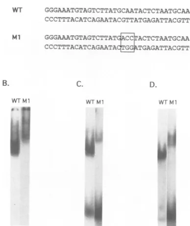
AA(T/G)] recognized by C/EBP (51), and one of these elements is includedinthe al oligonucleotide probe. Three nucleotides in the CCAAT/enhancer element of the wild-type (wt) al oligonucleotide probeweremutated inthe Ml
oligonucleotide probe (Fig. 3A) to determine whether al-binding proteins recognize this element. The diffuse ladder ofcrude nuclearextractbindingingel shiftassaysis altered by this mutation (Fig. 3B). The major rapidly migrating complexesareeliminated, while slowly migrating complexes
appear or are accentuated. These binding activities are
sequence specific, asjudged by gel shift competitionassays
with homologous or heterologous oligonucleotide probes
(datanotshown). Heat-purified and oligonucleotide affinity-purified proteins show thesamebindingpattern(Fig. 3C and D, respectively). These data suggest that the CCAAT/en-hancer element is the major recognition sequence for the rapidly migratingal-binding activity. The Ml gel shift
com-plexes could represent minor or novel binding activities
which are visible after removal of the CCAAT/enhancer
element-binding activity.
The LTR-binding activity of the oligonucleotide affinity-purifiedproteinwasfurtheranalyzed by DNase I
footprint-ing with 2P-labeled ALVLTRsequences(Fig. 4A). A 60-bp
region of the LTR enhancer is protected, extendinginfrom the borderof theU3region (-260 bp fromthetranscription start site; Fig. 4B). Five CCAAT/enhancer elements are
containedinthisregion (Fig.4C), which could be recognized
WTMl
I P
WT Ml WTMl
.4
[image:4.612.66.305.67.186.2].I
FIG. 3. Binding of purified B-cell protein to LTR CCAAT/ enhancer elements.(A) Sequencesofthe WT and Ml oligonucleo-tideprobesused ingelshiftassays.Mutatednucleotidesareboxed.
(B) Gelshiftassayof 0.5Mnuclearextractfrom bursallymphoma cells with the 32P-labeled WT or Ml oligonucleotide probe, as
indicated. (C)Gel shiftassayofheat-purified protein. (D)Gelshift
assayof aloligonucleotide affinity-purified protein.
by the al-binding proteins. AstrongDNaseI-hypersensitive siteonboth strandsseparates twoprotectedregions (al and a2). The al site contains the al oligonucleotide sequence
used foraffinity purification of the protein. This footprinting pattern corresponds to that observed for the heat-purified fraction (49), indicating that the same binding activity is
maintained through the oligonucleotide affinity purification step. This large protected region could represent multiple bindingofone or more proteins.
Identification ofa geneencoding al LTR-binding
activity.
Furtheranalysis would be facilitated by cloning thegene or
genes encoding the al-binding activity. We used a Agtll
screeningtechnique (54)toidentifygenes encoding proteins
bindingto the al LTR site. A cDNAlibrarywas prepared
from S13 bursal lymphoma poly(A)+ RNA andwas cloned
intothe Xgtll vector. Phage expressin lacZ-cDNA fusion proteins were screened for binding to
3P-labeled
concate-nated al oligonucleotide as described in Materials andMethods. Onepositiveclonewasidentifiedina screenof 1.5
x 106 primary phage, which maintained expression of
al-binding activity through three successive screens (datanot shown).
Lysogens of the al/EBP phagewere isolated, and
IPTG-induced lysates were prepared and tested for al LTR-binding activityby gel shiftassays(see Materials and
Meth-ods). The lysate containsasinglebinding activityingel shift
assayswith the 32P-labeled double-stranded al
oligonucleo-tideprobe, which is specifically competed for by the unla-beled al oligonucleotide but not by the b oligonucleotide (Fig. 5A). This al-binding activity was not observed in lysates from control bacteria (datanot shown).
al/EBPbindsmultipleCCAAT/enhancer elements.Theal LTR-binding activity of the al/EBP cDNA clonewasfurther
A.
-1'.
VOL.66, 1992on November 9, 2019 by guest
http://jvi.asm.org/
[image:4.612.344.538.73.304.2]6582 BOWERS AND RUDDELL
A.
non-coding strand
U3 k al
a2
_w
tI *i
It::
as
,m.m.
x '.
<e.
U3
t
A
codingstrand
:U
.:f~
icompetitor
al b
B probe
WT Ml
U-.
.-X.
2
B.
U3
rFO1 a?
-269 hp T -188bp
C.
U-3
Gr;(;A,A-,GGG
AAATGTAGTSTT7ATG'ATATGCTA.ATGCAATACT( AAA AAw'r"T CCTT-7A-A A;AATA>(;TTAT'A.;ATTA2;.TTAT A,GAA 'AT( A;;GA 7:7TA.--AATA :A-T ;.--A-'~.,AAG
-?iA hi, -188 bp
FIG. 4. Binding of purified B-cell proteinto twoLTRenhancer
sites. (A) The LTR probe was 32p labeled on the coding or
noncoding strand, incubated with(+)orwithout(-)oligonucleotide affinity-purified al-binding protein, and digested with DNase I. DNAwas purified andresolved on 8 Murea-8% polyacrylamide gels.The barsindicatestronglyDNaseI-protectedsequences, and thelines indicateweakly protectedsequences.The 5' border of the
U3 LTRregion isindicated. ArrowsindicateDNase
I-hypersensi-tive sites. (B) Map of LTR-binding sites of affinity-purified al-binding protein. The distance from the transcription start site is
shown inbasepairs.(C) Map of LTR CCAAT/enhancer elements.
examined by subcloning the al/EBP cDNA into the pGEX 2T expression vector, so that the al/EBP cDNA open
readingframe is translatedasanal/EBP-GSTfusion protein.
Fusion protein expressionwasinduced by IPTG treatment,
and the fusion protein was purified from the lysate by
glutathione-agarose affinity chromatography (see Materials and Methods). This purified fusion protein also specifically binds tothe32P-labeled al oligonucleotide, asits binding is
[image:5.612.345.535.70.217.2]competed for by the al oligonucleotide but not by the b oligonucleotide in gel shift assays (data not shown). We tested the DNA binding specificity of the al/EBP fusion
FIG. 5. Binding ofal/EBPtoLTRCCAAT/enhancerelements. (A) Lysates ofal/EBPXgtlllysogensweretested ingel shift assays with the 32P-labeled al oligonucleotide probe. Unlabeled al orb competitor (50-, 100-, or 150-fold molar excess) was added as indicated. (B) Purified al/EBP-GST fusion proteinwastestedingel shiftassayswiththe WTorMloligonucleotide probe,asindicated.
protein by gel shift assays with theMl oligonucleotide probe described above (Fig. 3A). Binding of the al/EBP-GST fusion protein is abolished by mutation of the CCAAT/ enhancerelement, indicating that this motif is essential for al/EBP binding (Fig. 5B). The Xgtll
al/EBP-0-galactosi-dasefusionproteinproducesasingle gelshiftcomplex(Fig. 5A), while the al/EBP-GST fusion protein produces two complexes (Fig. SB). The larger ,-galactosidase fusion pro-tein may notbe able to formmultimers, or it may be less susceptible toproteolysis than is the purified al/EBP-GST fusionprotein.
WeperformedDNase Ifootprinting experimentswith the purified al/EBP-GSTfusionproteintodetermine whether it binds to more than one of the LTR CCAAT/enhancer elements. The al/EBP-GST fusion protein protects two regions of the ALV LTR, separated by a strong DNase I-hypersensitive site (Fig. 6). Titration experiments with decreasing amounts ofal/EBP indicate that the al and a2 sitesarebound withequalaffinity (datanotshown). Ourgel shift data (Fig. SB) indicate that the al CCAAT/enhancer element isrequired for al/EBP binding, and it islikely that similar elements are recognized over the a2 site. The pro-tectedregion isnearly identicaltothat observed for purified B-cell protein (Fig. 4), supporting the idea that al/EBP encodes the majoral-binding activityof B cells. This gene could encode any or all of the three purified al-binding species.
DNA sequence analysis of al/EBP cDNA. The protein-coding potentialof the 2.2-kb al/EBP cDNAwas analyzed by primer-directed DNA sequencing (see Materials and Methods). ThecDNAcontains a209-amino-acid open read-ing frame (Fig. 7) which would be translated in the open reading frames expressed in the Agtll andpGEX 2T expres-sion vectors. The al/EBP cDNA also contains a long 3' untranslatedregion.This cDNAprobablydoesnotencode a full-length protein, as the aminoterminus doesnot include an initiator methionine codon. The al/EBP open reading frame could encodea26-kDaprotein.This appearstobe the openreading frameused,astheal/EBP-GST fusion protein expressedin bacteria is 52kDa, containinga26-kDaal/EBP portion in addition to the 26-kDa GST protein (data not shown).
Computer genebanklibrary searches indicate that al/EBP
I
J. VIROL.
4.0 'Ifi.. '.'r
aw dot- de
,.w
GGGAGGGGG AAATGTAGTC T TAT(;C A.ATACTCrAA-, GCAATAC T,- TTG TAGTC TTGCAAC ATGC T T ATG T AACGATG AGC TTC AG
on November 9, 2019 by guest
http://jvi.asm.org/
ALV LTR CCAAT/ENHANCER ELEMENT-BINDING PROTEIN 6583
coding strand
*
is:
:1*
2 _-rl 1!
[image:6.612.71.286.75.407.2]s:s.
FIG. 6. Bindingofal/EBPto two LTRenhancer sites.
32P-end-labeled ALV LTRwasincubated with(+)orwithout(-)
al/EBP-GST fusionproteinand digestedwithDNase I. DNAwaspurified
and resolvedon8 Murea-8%polyacrylamide gels alongwithA+G
sequence reactions (AG). The strongly protected sequences are
indicated by the heavy bar, and weakly protected regions are
indicated by lines. The arrows designate DNase I-hypersensitive
sites. The5'border of theU3regionisshown.
is encodedbyanovelgene, as nocloselyrelatedsequences
arefound in the DNAorprotein data bases. However, the carboxyterminus of theal/EBPopenreadingframe contains amino acid sequence motifs conserved in leucine zipper transcriptionfactors (28). The leucinezipper familyis char-acterized by a carboxy-terminal heptad repeat of leucine residues,which appearstomediate factordimerization(29). The adjacent basicregion is enriched in basic amino acids and isrequiredfor DNAbinding (29).Thesesequencemotifs
wereused toaligntheal/EBPopenreadingframe withthose of other leucinezipper familymembers(Fig. 8).Theal/EBP
openreadingframe is mostcloselyrelated to that ofIg/EBP
(48) and less related to those of C/EBP (28), NF/IL-6 (1),
CELF(25), andCRP-1 (58).Theputativebasic andleucine zipper regions of al/EBP are nearly identical to those of Ig/EBP, showing 94% amino acid identity (Table 1). The al/EBP basic region is about 60% identical with that of C/EBPandNF/IL-6,while their leucinezipper regions are
differentexceptinthe conservedleucines of the fourheptad
repeats (Fig. 8). Interestingly, all of these proteins bind to CCAAT/enhancer elements, supporting the idea that the
conserved basic region specifiesDNAbinding specificity.
The amino termini ofleucinezipperproteins may mediate transcription activation orrepression (13, 44). The amino-terminalregionofa1/EBPisrelatedtothat ofIg/EBPonly in the 45 amino acids adjacent to the basic region; it is very different in the amino-terminal 70 amino acids (data not shown), showing overall 39% identity (Table 1). The corre-sponding amino-terminal regions ofC/EBP and NF/116 are verydifferent in amino acidcompositionfromal/EBP. The amino-terminal region of al/EBP is enriched for basic and acidic residues (Fig. 7),while the corresponding regions of Ig/EBP, C/EBP, and NF/IL-6 are relatively enriched in hydrophobic residues. These data suggestthat al/EBP en-codes a novel transcription factor that contains basic and leucine zipper regions closely related toIg/EBP.
DISCUSSION
Three alLTR-binding proteinsof 35to42 kDa have been purifiedfrombursal lymphoma cells. Gel slice renaturation
experiments
confirm that proteins in this molecular mass range encode al LTR-binding activity. The close size of theseproteins
hasnotallowedus todetermine whether one or all of these species encode al-binding proteins. The al-binding activityislikelytobeassociatedwithmorethan one of these species, as the gel slice renaturation experi-mentssuggestthatatleasttwodifferent-sizeproteinsbindto al LTR sequences in gel shift assays. These multiplespe-cies,
which retain binding activity, could be due toprote-olysis
ofoneal-binding protein.Thispossibilityisdifficultto eliminate(34),
even though protease inhibitorswere usedthroughout
the purification steps. Alternatively, oneal-binding protein
could be modified ina mannerthat affects its apparentmolecularweight,forexample by phosphorylationor
by processing. Finally,
morethanoneal-binding protein could beexpressed
in B cells.We used a Xgtll cDNA library screening technique to
identify
ageneencoding
anal-binding
protein. This cDNA doesnotappeartobe fulllength,
althoughit does encode a 209-amino-acid openreading frame, sufficienttoencodea26 kDaprotein. Cloning
ofthefull-length
al/EBPcDNAwill berequired
todeterminethecomplete protein-codingpotential of this geneandtocomparethe predictedmolecularweight ofal/EBP
withthose of thepurified
al-binding proteinsof B cells. We have obtained genomic clones corresponding to the al cDNA sequence but thus far have not identifiedfull-length coding
sequences. Use of antibodies to al/EBP willdeterminewhetheroneorall of theal-bindingproteinspurified
from B cellsare encodedby
the al/EBPgene. Thepurified
B-cellproteinandthe al/EBP fusion protein both bindtwo LTR enhancersites in DNase Ifootprintingexperiments
andinduceastrongDNaseI-hypersensitivesite between theprotected regions.
Thesesites contain several consensusCCAAT/enhancer
elements, whichappear tobe the motifsrecognized by
these proteins. Similar CCAAT/ enhancer elements which are alsobound by theseproteins arefound in theclosely
related RSV LTR(datanotshown). Nuclear extract activitiesbinding
these sites in the RSV LTR have been observed in avian fibroblasts (53) anderythroblasts (17), suggesting
that al/EBP or relatedCCAAT/enhancer
element-binding proteins
areexpressedin manycell types.The DNase
I-hypersensitive
site inducedby al/EBPandby
B-cellprotein binding
maycorrespondto astrong DNaseI-hypersensitive
site observed in LTR enhancer chromatin from bursallymphoma
cell lines(52).
This chromatin hyper-sensitive sitedisappears
in cells treatedwithprotein
synthe-non-codingstrand
A_
0-_3
.* ._U3flt~ ~ S:
.I
* wX0
Al4 lids
,gm
--
--VOL. 66,1992
.dkw.00-Ift Abft.40
1
.w.
"-a-W4mwa-,.
lw z-0
on November 9, 2019 by guest
http://jvi.asm.org/
1
101
201
301
401
501
601
701 801 901 1001 1101 1201
1301
1401
1501
1601
1701
1801
1901
2001
S A P R H N V G Q R R R Q Q R G B E E E P G .G L G L L H G A G G G E
A G D G E D A G G G V R G A G E R L R P H A F S H R R K T S P Q N
CACCGCTACMAGGGACGAGTAAGCGTGATTCACACCCAGGCGCACAGCATGGTTTGCACAGGTGTTGGTTCCCGTTAGTCCCGGTGGT
T A T D A N G V S V I H T Q A H S S G L Q Q V P Q L V P V S P G G
_1= =TCCGACAAGCAGGGAATTCCTTGTATCG IG ATGG
G G K A V P P S K Q G K K N S F V D R N S D E Y R Q R R E R N N M A CAGTTAAAAAGACCGGTTAAAAAGCAAGCAAAAGCACAAGACACGCTGCAAAGG G G T TTTAGAGGCGAAAAT V K K S R L K S K Q K A Q D T L Q R V T Q L5 E E N E R L E A K I
TAAGCTCCTcCAAGGAGCTGAGCGTACTGAAGACCTATTCCTTGAGCACGCACACAGTCTCGCAGAAATGTGCAACCTGTTGGCACTGAGAGCACC
K L L T K E L S V L K D L F L E H A H S L A D N V Q P V G T E S T ACAACAAGTGCAGAGA GGGCAGTAGCACTGCC CGGGATGAACGGATGCAGCCTGTCTAACCCAGCAGTGAG T T S A E N S G Q *
CAA&TGAGGGTTCTTTTAACCATCGTTGTG1CGGATCTTTTTAGGTTTAA ACTGAAGATTTGATACAATTAAACCAGAAACTGCTTAGGGTATTTCT TTAAAGGCGTTAAATATTTTTCTCCTCCAGAAGATTTGTCTAATAAGTGCAGATCGAA&TCCTTAGTCAGCAAGGAGCTTACTATTATGGAAGCAGCATT TCACAGATGTACGCTCACCTTCTTACATTTAGCATAACGGGATAAATTCACCAGGATGCTTCACTGTAGTATCAGAAAGTCATAATTGGGTGAATTCCTT
TAAATACTTTTTAGTGTGTTTAAGCCCTTTCTTTTCTCCATTACATATTACAAACTGCAAAGGCCGTAGAGAGTTTGTTGTCCTGTTTGGTGAACTGCTT
AAAnATGTTTTGCAGAAAACCAGCAGCATTCTGTAGTGATCTTTACAGGCAGTTGGCTTCCACAGGCACGTGGCGGTGTGAAGCACCAAGTATCAGGTAA
AGaCAGTTTTATGCCTGTGTTCATTTTAACCTGGCTTGCACA CCGTGXGG TGAGAAAAGATTTTGGTTTCCA;CAAAA
TGTAAAAATGCTACCANCAACCTCTGCTCTGCAAAACG CTGCGGGCGGGTATCGGAGAACAAAATACGCAGCTTGGCTGAGCAGCAGAAGG
GGTAGGCAGCATCTGACAGCAAGATGTGTGGATC7CAMCTCACCTTCTTGTTCATTTCCATTCTCACAGCGTCAGATTGCAMCCAGCTGTACAGAAT
TGCACGGTGATT CTAAACTAAGGC PTTGAAACCCGAATCAGCAGAATTTCTATATAGCCATCACAGAGTTGTGTGAAGAGTATCTGCAGC
TTATTTCCATAGAT CTTTTCTATAGCAGTTTTTAAATGCTI ATAAGTCTTTGCCATGATCCATAATGCAGTGGTATGTACTATATATTGAGGATT
AAAACCTTGGAT_ATTATCTCTATAATACCTTTNAG TAACTTTTTACcGTGTTTCTGATGCTTGTCTTTGTACTACCTGTACTTAGGAT
_
ACTTGTTTT1?TGMTcTAGTMAAA!ATANWCTTTAAAMTTCTTTC1%3&TTTCAATTTTTAATACGAAGGCAACTAGATG
100
200
300
400
500
600
700
800
900
1000
1100
1200
1300
1400
1500
1600
1700
1800
1900
2000
2100
[image:7.612.136.482.75.386.2]2101 AAGGGc G- TTTTACAAAGATAGtGTTTTGTTCTAGCTGC 2170
FIG. 7. al/EBP DNA sequence and predicted aminoacidsequence of the open reading frame.
sis inhibitors, concomitant with a large decrease in LTR-enhanced c-myctranscription(31). This findingsupportsthe hypothesis thatbinding of al/EBP to the LTR enhancer, as measuredbyappearanceof theDNaseI-hypersensitive site, is important for labile LTR transcription enhancement. Interestingly, the al/EBP-binding sites are deleted in endog-enousviruses(61),which showlow-level LTR-driven tran-scriptionandrarely promote lymphomagenesis (6, 46).
DNA sequence analysis of the al/EBP cDNA indicates
BASIC REGION
F V D R N S D E Y R Q R R E R N N M A V K K S R L K SK O K A Q D P1N D RNSDE I R Q NNM A V KKC5 L K S1Q KA1 QD S V D K N SN E Y R V R R E R N N I A V RKS R D K A KQR N V E T VDK E Y K IR1R RNNIA VR K R DXA1
that it encodes a leucine zipper factor closely related to Ig/EBP, C/EBP, and NF/IL-6 in the conserved putative DNA-bindingand leucinezipper dimerization domains.The conservation of the basic-region DNA-binding sequence may reflect the fact that these proteins bind to CCAAT/ enhancerelements,includingthosefound in the RSV LTR, whichis closely relatedto the ALVLTR (data notshown) (48, 51). The amino-terminal region of C/EBP encodes transcription-regulatory activities (13, 44).The amino-termi-nal regions arevery different in the various leucine zipper proteins, suggestingthat these proteins have different effects on transcription. Interestingly, the amino terminus of al/ EBP isenriched in acidic and basicresidues, while Ig/EBP and the other leucinezipper proteinsareenrichedin hydro-phobicresidues. The carboxy-terminal tails of theseproteins
LEUCINE ZIPPER
a1/EBP T LQRVTQLK EE N E R L E AK IK L L T K E L SVL K D L F L E H A H S L A D N VQP V Ig/EBP TL QRVNQL K E E N E R L E A K I KLL T N ELSV L K D L F LE H A HSLA D N VQ P I C/EBP T QKVL E LTS D N DRLR KR VEQ LS R E LDTLR G I FRQ L P E S L V K A G N NF/IL6 TQHKVL'L TA E N E R LQK K V EQL S R E L S TLR NL FKQLPE P L L A S S G H C
FIG. 8. Comparison of the al/EBP protein sequence with
se-quences of otherleucine zipper proteins. The putative basic and
leucinezipper regions ofa1/EBPwerealigned withthoseof Ig/EBP,
C/EBP, and NF/IL-6 (1, 28, 48). Conserved leucine residues are
highlighted. Asterisks indicate regions of amino acid identitywith
[image:7.612.63.300.530.692.2]al/EBP.
TABLE 1. Percent amino acididentityof al/EBP with regions of other leucinezipperproteinsa
% Amino acididentity Protein
Aminoterminus(114) Basicregion (33) Leucinezipper(47)
Ig/EBPb 39 94 94
C/EBPC 6 64 28
NF/IL-6d 11 58 36
aAlignmentofputative basic andleucinezipper regions was based on comparison withC/EBP. The number of amino acids compared is given in
parentheses.
bDatafromRoman et al.(48).
CDatafromLandshulzetal.(28).
dData fromAkiraetal.(1).
al1/EBP Ig/EBP
C/LEBP NF/IL6
on November 9, 2019 by guest
http://jvi.asm.org/
[image:7.612.317.557.609.671.2]ALV LTR CCAAT/ENHANCER ELEMENT-BINDING PROTEIN 6585 are also very different, as the 15-amino-acid tail of al/EBP is
only slightly relatedtothe10-amino-acid tail of Ig/EBP.The a1/EBP gene is also different from the Ig/EBP gene in that thea1/EBPproteinappears tobe 35to42kDa, whileIg/EBP is 45 kDa (48). Ig/EBP forms heterodimers with C/EBP in vitro, suggesting that combinations of leucine zipper pro-teins may modulate transcription (48). Further experiments will determine whether an Ig/EBP homolog is expressed in avian B cells and whether itcan dimerizewith al/EBP.
Wehave previously found that the labile al, a3, and/or b* proteins appear to be essential for LTR-enhanced c-myc hyperexpression, and these labile proteins may play an important role in the susceptibility of pre-B cells to ALV lymphomagenesis. Wedo notyet knowwhetherthe protein encoded by the al/EBP geneis labile. However, useof the al/EBP DNAclones will allow us to generate antibodies to al/EBP to determine whether it encodes the labile binding activity of B cells. Computer analysis of the a1/EBP open reading frame indicates several motifs that could regulate labile expression. The amino-terminal region contains a PEST consensus sequence, thought to target rapid protein turnover(47). The al/EBP protein also containsanumber of consensus creatine kinase II,cyclicAMP-dependentkinase, andproteinkinase Cmotifs(26). Labile phosphorylation of a1/EBP could affect its transcription-regulatory activity, as phosphorylation of a number of transcription factors has been shown to affect their DNA-binding activities (4, 32). Further experiments will reveal whether a1/EBP is labile, howthis lability is regulated, and the role of this lability in ALV tumor susceptibilityof immature bursal cells.
ACKNOWLEDGMENTS
WethankMarkGroudineand MaxineLinial for encouragement, advice, andsupportof thiswork, andwethank SuzanneKennedy, PatrickFinnerty, andMary Peretzfor technical assistance.
This work was supported by a Leukemia Society of America Special Fellowship, National Leukemia Association Max Sirlin MemorialFellowship, ACSgrantIRG-18,and NIH grant CA54768
toA.Ruddell.W.J. Bowers was supported by NIH grant AI07362. This work was also supported by NIH grants CA54337 and CA 57156toM. Groudine and CA18282to M.Linial.
REFERENCES
1. Akira, S., H. Isshiki, T. Sugita, 0. Tanabe, S. Kinoshita, Y. Nishnio, T. Nakajima,T. Hirano, and T. Kishimoto. 1990. A nuclear factor for IL-6expression (NF-IL6) is a member of a
CIEBPfamily.EMBOJ. 9:1898-1906.
2. Baba, T. W., and E. H. Humphries. 1984.Differentialresponse
toavian leukosis virusinfection exhibited bytwochickenlines. J. Virol. 51:123-130.
3. Bizub, D., R. A. Katz, and A. M. Skalka. 1984. Nucleotide sequence of noncoding regions in Rous-associated virus 2: comparisonsdelineate conserved regions important in replica-tion andoncogenesis. J. Virol. 49:557-565.
4. Boyle, W. J., T. Sneal, L. H. K. Defize, P. Angel, J. R. Woodgett, M.Karin, and T. Hunter. 1991.Activationof protein kinase C decreases phosphorylation of c-jun at sites that negatively regulate its DNA-binding activity. Cell 64:573-584.
5. Bradford, M. M. 1976. Arapid and sensitive method for the quantitation of microgram quantities of protein utilizing the principleofprotein-dyebinding. Anal.Biochem. 72:248-254. 6. Brown, D. W., B. P. Blais, and H. L. Robinson. 1988. Long
terminalrepeat(LTR)sequences, env, and aregionnearthe 5' LTR influence the pathogenic potential ofrecombinants
be-tween Rous-associatedvirus types 0 and 1. J. Virol.
62:3431-3437.
7. Christy,R.J., V. W. Wang,J. M. Ntanbi, D. Geiman, W. H. Landschulz, A. D. Friedman, Y. Nakabeppu, T. J. Kelly, and M. D. Lane. 1989. Differentiation inducedgene expression in
3T3L1 preadipocytes: CCAAT/enhancer binding protein inter-acts with and activates the promoters of two adipocyte-specific genes. Genes Dev. 3:1323-1335.
8. Coppola, J. A., and M. D. Cole. 1986. Constitutive c-myc oncogene expression blocks mouse erythroleukaemia cell dif-ferentiation but not commitment. Nature (London)320:760-763. 9. Dani, C., J. M. Blanchard, M. Piechaczyk, S. El Sabouty, L. Marty, and P. Jeanteur. 1984. Extreme instability of myc RNA innormaland transformed human cells. Proc. Natl. Acad. Sci. USA 81:7824-7827.
10. Devereu, J., P. Haeberli, and0.Smithies. 1984. A comprehen-sive set of sequence analysis programs for the VAX. Nucleic Acids Res. 12:387-395.
11. Evan, G. I., A. H.Wylie, C. S. Gilbert, T. D. Littlewood, H. Land, M. Brooks, C. M. Waters,L. Z. Penn, and D. C.Hancock. 1992. Induction of apoptosis in fibroblasts by c-myc protein. Cell69:119-128.
12. Ewert, D. L., and G. F. deBoer. 1988. Avianlymphoidleukosis: mechanisms oflymphomagenesis. Adv. Vet. Sci. Comp. Med. 32:37-55.
13. Friedman, A. D., and S. L. McKnight. 1990. Identification of twopolypeptide segments of CCAAT/enhancer-binding protein required for transcriptional activation of the serum albumin gene. Genes Dev. 4:1416-1426.
14. Fung,Y.T., A. M. Fadly, L. M. Crittendon, and H. Kung. 1982. Avianlymphoid leukosis virus infection and DNA integration in preleukotic bursal tissues: a comparative study of susceptible andresistant lines. Virology 119:411-421.
15. Galas, D. J., and A. Schmitz. 1978. DNAase footprinting: a simple methodfor thedetection of protein-DNA binding spec-ificity. NucleicAcids Res. 5:3157-3162.
16. Golemis, E.,Y. Li, T. N. Fredrickson, J. W. Hartley, and N. Hopkins. 1989. Distinct segments within the enhancer region collaborate to specify thetype of leukemia induced by nonde-fective Friend andMoloneyViruses. J. Virol. 63:328-337. 17. Goodwin, G. H. 1988. Identification of three sequence-specific
DNA-binding proteins which interact with the Rous sarcoma virus enhancer and upstream promoter elements. J. Virol. 62:2186-2190.
18. Gubler, and B. J. Hoffman. 1983. Asimple and very efficient method for generating cDNA libraries. Gene 25:263-269. 19. Hallberg, B., J. Schmidt, A. Luz, F. S. Pedersen, and T.
Grundstrom. 1991. SL3-3 enhancer factor 1 transcriptional activators are required for tumor formation by SL3-3 murine leukemia virus. J. Virol. 65:4177-4181.
20. Hann,S. R.,and R. N.Eisenman.1984. Proteins encoded by the human c-myc oncogene: differential expression in neoplastic cells. Mol. Cell. Biol.4:-2486-2497.
21. Hatamochi, A., P. T. Golumbek,E.Van Schaftingen, and B. De Crombrqgghe. 1987. A CCAAT DNA binding factor consisting of two different components that are both required for DNA binding. J. Biol. Chem.263:5940-5947.
22. Holland, C. A., C. Y. Thomas, S. K.Chattopadhyay,C.Koehne, and P. V.O'Donnell.1989. Influence of enhancer sequences on thymotropism and leukenogenicity of mink cell focus-forming viruses. J. Virol.63:1284-1292.
23. Johnson, P. F., W. H. Landschulz, B. Graves, and S. L. McKnight. 1987. Identification of a rat livernuclearprotein that binds to the enhancer core element of three animal viruses. Genes Dev. 1:133-146.
24. Kadonaga, J. T., and R. Tjian. 1986. Affinity purification of sequence-specific DNA binding proteins. Proc. Natl. Acad. Sci. USA83:5889-5893.
25. Kageyama, R., Y. Sasai, and S. Nakanishi. 1991. Molecular characterizationoftranscription factors that bind to the cAMP responsive region of the substance P precursor gene. J. Biol. Chem.266:15525-15531.
26. Kennelly,P.J., and E. G. Krebs. 1991.Consensus sequences as substrate specificity determinants for protein kinases and pro-teinphosphatases. J. Biol. Chem. 266:15555-15558.
27. Laemmli,U.K. 1970. Cleavage ofstructural proteins during the assembly of the head ofbacteriophage T4. Nature (London) 227:680-685.
VOL.66, 1992
on November 9, 2019 by guest
http://jvi.asm.org/
28. Landschulz,W.H., P. F.Johnson, E.Y.Adashi, B. J. Graves, and S. L. McKnight. 1988. The leucine zipper: ahypothetical
structure common to a new class of DNA binding proteins.
Genes Dev.2:786-800.
29. Landschulz, W. H., P. F. Johnson,and S. L.McKnight. 1989.
TheDNA binding domain of theratlivernuclearprotein C/EBP isbipartite. Science243:1681-1699.
30. Lenz, J., D. Celander, R. L. Crowther, R. Patarca, D. W.
Perkins, and W. A. Haseltine. 1984. Determination of the
leukenogenicity ofamurine retrovirus bysequenceswithin the longterminalrepeat.Nature(London) 308:467-470.
31. Linial, M., N. Gunderson, and M. Groudine. 1985. Enhanced transcription ofc-myc inbursal lymphoma cells requires
con-tinuousprotein synthesis. Science 230:1126-1132.
32. Luscher, B., E. Christenson, D. W.Litchfield, E. G. Krebs, and R. N. Eisenman.1990. Myb DNA binding inhibited by phosphor-ylation at a site deleted during oncogenic activation. Nature (London) 344:517-522.
33. Majors,J. 1990. The structure andfunction of retroviral long terminalrepeats. Curr. Top. Microbiol. Immunol. 157:50-84. 34. Mather, E. L. 1988. DNA-binding factors of B lymphoid cells
are susceptible to limited proteolytic cleavage during nuclear
extractpreparation. Mol. Cell. Biol. 8:1812-1815.
35. Maxam, A.M., and W. Gilbert. 1980. Sequencing end-labeled DNAwithbase-specific chemical cleavages. Methods Enzymol. 65:499-560.
36. Metz,R., and E. Ziff. 1991. cAMP stimulates theC/EBP-related transcription factor rNFIL-6totrans-locatetothe nucleus and inducec-fos transcription. Genes Dev.5:1754-1766.
37. Mizusawa, S., S. Nishimura, and F. Seela. 1986. Improvement of
thedideoxy chaintermination method of DNAsequencing by
use ofdeoxy-7-deazaguarosine triphosphate in place of GTP. NucleicAcids Res. 14:1319-1324.
38. Morrissey, J. H. 1981. Silver stain for proteins in polyacryl-amidegels:amodifiedprocedurewithenhanceduniform sensi-tivity. Anal.Biochem. 117:307-310.
39. Neel, B. B., W. S. Hayward, H. L. Robinson, J. Fang, and S. M.
Astrin. 1981. Avian leukosisvirus-induced tumors have
com-mon proviral integration sites and synthesize discrete new
RNAs:oncogenesis bypromotorinsertion.Cell23:323-334.
40. Neiman, P. E., L.Jordan, R. A. Weiss, and L. N. Payne. 1980. Malignant lymphomas of the bursa of Fabricius: analysis of early transformation,p. 519-528. In M. Essex and G. Todaro
(ed.),Viruses innaturally occurringcancers.ColdSpring
Har-borLaboratory, ColdSpring Harbor, N.Y.
41. Okayama, H., M. Kawaichi, M. Brownstein, F. Lee, T. Yokota,
and K.Arai. 1987.High-efficiency cloning of full-length cDNA;
construction and screening of cDNA expression libraries for
mammaliancells. MethodsEnzymol. 154:3-28.
42. Payne, G. S.,S. A. Courtneidge, L. B. Crittenden, A. M. Fadly, J. M. Bishop, and H. E. Varmus. 1981. Analysis of avian leukosis virus DNA and RNA in bursal tumors: viral gene
expressionisnotrequired for maintenance of thetumorstate.
Cell23:311-322.
43. Pearson, W. R., and D. J. Lipman. 1988. Improved tools for biological sequence comparison. Proc. Natl. Acad. Sci. USA 85:2444-2448.
44. Pei, D., and C. Shih. 1990. Transcriptional activation and
repression by cellular DNA-binding protein C/EBP. J. Virol. 64:1517-1522.
45. Purchase, H.G.,P.G.Gilmour, C.H.Romero, and W.Okazaki. 1979.Postinfectiongenetic resistancetoavianlymphoid leuko-sis residesin B targetcells.Nature(London) 270:61-62. 46. Robinson, H. L., B. M. Blais, P. N. Tsichlis, and J. M. Coffin.
1982. At least two regions of the viralgenome determine the oncogenic potentialofavian leukosisviruses. Proc.Natl. Acad. Sci. USA 79:1225-1229.
47. Rogers, S., R. Wells, and M. Rechsteiner. 1986. Amino acid sequences common to rapidly degraded proteins: the PEST hypothesis. Science 234:364-368.
48. Roman, C., J. S. Platero, J. Shumn, and K. Calame. 1990. Ig/EBP-1: a ubiquitously expressedimmunoglobulin enhancer binding protein that is similar to C/EBP and heterodimerizes withC/EBP. GenesDev.4:1404-1415.
49. Ruddell,A., M.Linial,andM. Groudine. 1989.Tissue-specific lability and expression of avian leukosis virus long terminal repeatenhancer-binding proteins. Mol. Cell. Biol. 9:5660-5668. 50. Ruddell, A., M. Linial, W. Shubach, and M. Groudine. 1988. Lability of leukosis virus enhancer-binding proteins in avian hematopoietic cells. J.Virol.62:2728-2735.
51. Ryden, T. A., and K. Beemon. 1989. Avian retroviral long terminal repeatsbind CCAAT/enhancer-binding protein. Mol. Cell.Biol.9:1155-1164.
52. Schubach, W., and M. Groudine. 1984. Alteration of c-myc chromatinstructurebyavianleukosis virusintegration. Nature (London)307:702-707.
53. Sealey,L.,and R.Chalkley.1987. At least twonuclearproteins bindspecificallytotheRoussarcomaviruslong terminalrepeat enhancer. Mol. Cell. Biol. 7:787-798.
54. Singh,H., J. H.LeBowitz,A.S.Baldin, and P. A.Sharp.1988. Molecularcloning ofanenhancerbinding protein: isolationby screening ofanexpression library witharecognitionsite DNA. Cell 52:415-423.
55. Smith, D. B., and K. S. Johnson. 1988.Single-steppurification of polypeptides expressed in Escherichia coli as fusions with glutathione-S-transferase.Gene67:31-40.
56. Speck,N.A., B.Renjifo,E.Golemis, T. N. Fredrickson, J. W. Hartley, and N. Hopkins.1990. Mutation of thecore oradjacent LVbelementsof theMoloney murine leukemia virus enhancer alters diseasespecificity.Genes Dev.4:233-242.
57. Vogt, M., J. Lesley, J.Bogenberger, S. Volkman, and M. Haas. 1986.Coinfection withvirusescarrying thev-Ha-rasandv-myc oncogenesleadstogrowthfactorindependence byanindirect mechanism. Mol. Cell.Biol.6:3545-3549.
58. Williams, S. C., C. A.Cantwell, and P. F.Johnson. 1991. A family of C/EBP related proteins capableofformingcovalently linkedleucinezipper dimersinvitro. GenesDev.5:1553-1567. 59. Wurm,F.M., K. A.Gwinn,and R. E.Kingston. 1986. Inducible overproduction ofthemousec-mycproteininmammaliancells. Proc.Natl. Acad. Sci.USA83:5414-5418.
60. Young, R.A.,and R. W.Davis.1983.Efficientisolation ofgenes byusing antibody probes.Proc.Natl. Acad.Sci. USA 80:1194-1198.
61. Zachow, K. R., and K. F. Conklin. 1992.CArG, CCAAT, and CCAAT-like proteinbindingsites in avianretrovirus long
ter-minalrepeatenhancers. J.Virol. 66:1959-1970.