SHOC2 complex-driven RAF dimerization selectively
contributes to ERK pathway dynamics
Isabel Boned del Ríoa,1, Lucy C. Younga,1, Sibel Saria, Greg G. Jonesa, Benjamin Ringham-Terrya, Nicole Hartiga, Ewa Rejnowicza, Winnie Leia, Amandeep Bhamrab, Silvia Surinovab, and Pablo Rodriguez-Vicianaa,2
aUniversity College London Cancer Institute, University College London, WC1E 6DD London, United Kingdoms; andbProteomics Research Core Facility,
University College London Cancer Institute, WC1E 6DD London, United Kingdom
Edited by Roger J. Davis, Howard Hughes Medical Institute and University of Massachusetts Medical School, Worcester, MA, and approved May 28, 2019 (received for review February 21, 2019)
Despite the crucial role of RAF kinases in cell signaling and disease, we still lack a complete understanding of their regulation. Heterodimerization of RAF kinases as well as dephosphorylation of a conserved“S259”inhibitory site are important steps for RAF ac-tivation but the precise mechanisms and dynamics remain unclear. A ternary complex comprised of SHOC2, MRAS, and PP1 (SHOC2 com-plex) functions as a RAF S259 holophosphatase and gain-of-function mutations in SHOC2, MRAS, and PP1 that promote complex forma-tion are found in Noonan syndrome. Here we show that SHOC2 complex-mediated S259 RAF dephosphorylation is critically required for growth factor-induced RAF heterodimerization as well as for MEK dissociation from BRAF. We also uncover SHOC2-independent mechanisms of RAF and ERK pathway activation that rely on N-region phosphorylation of CRAF. In DLD-1 cells stimulated with EGF, SHOC2 function is essential for a rapid transient phase of ERK activation, but is not required for a slow, sustained phase that is instead driven by palmitoylated H/N-RAS proteins and CRAF. Whereas redundant SHOC2-dependent and -independent mecha-nisms of RAF and ERK activation make SHOC2 dispensable for pro-liferation in 2D, KRAS mutant cells preferentially rely on SHOC2 for ERK signaling under anchorage-independent conditions. Our study highlights a context-dependent contribution of SHOC2 to ERK path-way dynamics that is preferentially engaged by KRAS oncogenic signaling and provides a biochemical framework for selective ERK pathway inhibition by targeting the SHOC2 holophosphatase.
SHOC2
|
RAF|
MRAS|
RAS|
ERKS
ignaling by the RAF-MEK-ERK (ERK-MAPK) pathway is used by many extracellular signals to mediate a vast array of biological responses in a cell-type–dependent manner. The mech-anisms regulating signal specificity remain poorly understood but are known to include modulators, scaffolds, feedbacks, and cross-talk with other signaling pathways that jointly control spatial and temporal dynamics of ERK activation. This in turn regulates phosphorylation of different ERK substrates in a cell-type–, com-partment-, and context-dependent manner (1, 2).Aberrant activation of the ERK pathway is one of the most common defects in human cancer, with oncogenic mutations in RAS and RAF genes found in∼30% and∼8% of cancers, respectively. Up-regulated ERK signaling is also responsible in a family of de-velopmental disorders, referred to as RASopathies (3–5).
ERK pathway inhibitors have shown little clinical benefit against RAS mutant tumors because of resistance and toxicity (5). Strikingly, in both RAS and BRAF mutant cells, most re-sistance mechanisms lead to ERK pathway reactivation, high-lighting a strong“oncogene addiction”of these cancers to ERK signaling. However, the potent pathway suppression required for antitumor activity is limited by the inhibitor doses that can be administered safely because of toxicity (6, 7). ERK activity is essential for normal tissue homeostasis and systemic ablation of MEK1/2 or ERK1/2 genes in adult mice leads to death of the animals from multiple organ failure within 2–3 wk, even under conditions of partial inactivation (8), highlighting the difficulties of inhibiting the ERK pathway with a therapeutic index. To effectively
harness the addiction of RAS mutant cancers to ERK signaling into viable therapies, new strategies to inhibit the pathway with improved therapeutic margins are needed, for example by inhib-iting ERK signaling in a context- or compartment-dependent manner (9, 10).
MEK and ERK kinases are fully activated by phosphorylation in two sites within its kinase domain by RAF and MEK, re-spectively. On the other hand, RAF activation is a complex mul-tistep process that remains incompletely understood (11). A consensus model stipulates that under resting conditions, the three RAF kinases (ARAF, BRAF, and CRAF/RAF1) are kept in the cytosol in an inactive state by an intramolecular interaction me-diated by 14-3-3 dimers binding in a phosphorylation-dependent manner to conserved sites at the N terminus (S214 ARAF, S365 BRAF, S259 CRAF, hereby referred to as the“S259”site) and C-terminal end (S729 in BRAF, S621 in CRAF) (11–13). Upon activation, RAS-GTP binds with high affinity to the RAS binding domain (RBD) of RAF and recruits RAF to the membrane where the cysteine-rich domain (CRD) also plays a role in membrane anchoring. Dephosphorylation of the S259 site is known to provide an additional activating input that releases the 14-3-3 from this site and allows RAF to adopt an open conformation where RAF di-merizes with other RAFs, as well as KSR proteins. Definitive confirmation of this model, however, awaits the crystal structure of full-length RAF with or without bound 14-3-3. Nevertheless, the importance of the S259 dephosphorylation regulatory step is highlighted by RAF1 gain-of-function mutations in Noonan
Significance
The ERK signaling pathway is hyperactivated in a majority of cancers. However, because it mediates myriad physiological re-sponses, the clinical efficacy of current ERK pathway inhibitors has been severely limited by toxicity. This study uncovers both SHOC2 phosphatase complex-dependent and -independent mechanisms of RAF and ERK activation that are differentially
engaged in a context and spatio-temporal–dependent manner.
KRAS oncogenic signaling preferentially depends on SHOC2 dependent-mechanisms, which thus presents a therapeutic op-portunity. This study provides a molecular framework for how targeting the SHOC2-holophosphatase regulatory node of the RAF activation process provides a mechanism for selective in-hibition of ERK signaling.
Author contributions: I.B.R., L.C.Y., and P.R.-V. designed research; I.B.R., L.C.Y., S. Sari, G.G.J., B.R.-T., N.H., E.R., W.L., and A.B. performed research; I.B.R., L.C.Y., A.B., S. Surinova, and P.R.-V. analyzed data; and I.B.R. and P.R.-V. wrote the paper. The authors declare no conflict of interest.
This article is a PNAS Direct Submission.
This open access article is distributed underCreative Commons Attribution-NonCommercial-NoDerivatives License 4.0 (CC BY-NC-ND).
1I.B.R. and L.C.Y. contributed equally to this work.
2To whom correspondence may be addressed. Email: [email protected]. This article contains supporting information online atwww.pnas.org/lookup/suppl/doi:10. 1073/pnas.1902658116/-/DCSupplemental.
syndrome that cluster around S259 to disrupt the interaction with 14-3-3 (14–17). Furthermore, although RAF1 mutations are rare in cancer, they cluster on residues S257 and S259 (cosmic database). The precise dynamics and mechanism of S259 dephosphory-lation remain unclear (11). We have previously shown that MRAS, a closely related member of the RAS family, upon ac-tivation forms a complex with the leucine-rich repeat protein SHOC2 and protein phosphatase 1 (PP1) that functions as a highly specific S259 RAF holophosphatase (18, 19). The im-portance of the SHOC2-MRAS-PP1 complex (SHOC2 complex) in RAF-ERK regulation is validated by gain-of-function muta-tions in Noonan syndrome in all three components—SHOC2, MRAS, and PP1—which promote phosphatase complex forma-tion (20–23). On the other hand, the phosphatase PP2A has also been variously implicated in mediating S259 dephosphorylation (24–27), although this was primarily based on the use of okadaic acid and the misconception that it behaves as a specific PP2A inhibitor (28) (in addition to not discriminating between direct or indirect effects). Furthermore, in contrast to its role as a regu-latory subunit within a phosphatase complex, other studies have suggested that SHOC2 can function as a scaffold that promotes the RAS–RAF interaction (29–33).
RAF proteins also undergo multiple activating phosphoryla-tion events. Among them, phosphorylaphosphoryla-tion within the negative-charge regulatory region (N-region) plays a key divergent role among RAF paralogues (11). In CRAF, S338 and Y341 phos-phorylation within the S338SYY341motif by PAK and SRC family kinases (SFK) plays a crucial role in regulated activation (34). In contrast, the homologous S446SDD motif in BRAF constitutively provides the negative charges required for activity by virtue of acidic D amino acids and constitutive S446phosphorylation (11, 34, 35). This difference in N-region regulation is believed to account for BRAF having higher basal activity, being the most frequent RAF target for mutational activation in cancer and for BRAF being the initial activator in asymmetric RAF hetero-dimers (11, 36).
In this study, we have used RNAi and CRISPR to ablate SHOC2 and RAF function, as well as phosphoproteomics to comprehensively characterize the role of the SHOC2 phospha-tase complex in RAF and ERK pathway regulation. We have uncovered a selective role for SHOC2 in ERK pathway dynamics, and show that although SHOC2 phosphatase-mediated de-phosphorylation of the S259 site is critically required for growth factor-induced RAF heterodimerization, there also exist SHOC2-independent mechanisms of ERK activation, which are dependent on N-region phosphorylation of CRAF. Importantly, KRAS on-cogenic signaling differentially relies on SHOC2-dependent mech-anisms, which provides both a therapeutic opportunity and a molecular framework for selective inhibition of ERK signaling in a compartment and context-dependent manner.
Results
MRAS and SHOC2 Expression Promotes S365 BRAF/S259 CRAF Dephosphorylation, BRAF-MEK Dissociation, and BRAF-CRAF Dimerization. To study the role of the SHOC2 complex in the regulation of RAF kinases, we generated an inducible T-REx-293 cell line (T-17 cells) where addition of the tetracycline analog Doxycycline (Dox) leads to expression of active MRAS-Q71L and SHOC2. In these cells, Dox-induced MRAS/SHOC2 expression led to potent S365 de-phosphorylation of ectopic TAP6-BRAF that is inhibited in a dose-dependent manner by the serine/threonine phosphatase hibitor calyculin A (Fig. 1A). To assess possible RAF regions in-volved in S259 dephosphorylation, transiently transfected BRAF and CRAF mutants were tested for dephosphorylation upon ex-pression of MRAS/SHOC2. Among the mutants tested, only the RBD mutants R188L BRAF and R89L CRAF were defective for MRAS/SHOC2-induced S365/S259 dephosphorylation (Fig. 1B
andC). Interestingly, when the CRAF R89L RBD mutant was
constitutively localized to the membrane by fusion with a RAS membrane-targeting region (CRAF-CAAX R89L), S259 dephos-phorylation was efficiently induced by MRAS/SHOC2 expression
(Fig. 1C). Taken together, these data suggest that membrane re-cruitment through interaction with the RBD is required for effi-cient S259 RAF dephosphorylation.
MRAS/SHOC2 expression levels in T-17 cells did not prove to be tuneable because at the lowest Dox concentration that in-duced expression, there was a maximum effect on MRAS/ SHOC2 protein levels and concomitant S365 dephosphorylation (Fig. 1 D and E). When ectopic T6-BRAF in these cells was purified with streptactin beads, MRAS/SHOC2 expression led to a decrease in the amount of MEK bound to T6-BRAF and a concomitant interaction of T6-BRAF with CRAF (Fig. 1Dand E). To further study the specificity of the role for MRAS/ SHOC2 on RAF–MEK interactions, GST-pulldown assays were performed after cotransfection of myc-MEK1 with GST-tagged CRAF, BRAF, and KSR1 in HEK293T cells. Under basal con-ditions, MEK1 bound most strongly to KSR1 and only weakly to CRAF (KSR1 >BRAF>>CRAF), and Dox-induced MRAS/ SHOC2 expression led to strong dissociation of MEK from BRAF and CRAF but not from KSR1 (Fig. 1F). Taken together, the above data suggest that MRAS/SHOC2-induced S365 BRAF dephosphorylation promotes MEK dissociation from BRAF and BRAF heterodimerization with CRAF.
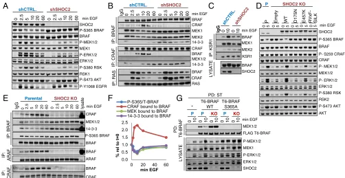
SHOC2 Is Required for EGF-Induced S365/S259 Dephosphorylation, RAF Dimerization, BRAF-MEK Dissociation, and Efficient ERK Pathway Activation.To assess the role of endogenous SHOC2 within the context of growth factor signaling, T-REx-293 cells where SHOC2 expression was stably inhibited by shRNA expression were used to analyze lysates and immunoprecipitates (IPs) of endogenous RAS and RAF proteins in a time course of EGF treatment. EGF-stimulated S365 BRAF dephosphorylation, MEK, ERK, and RSK phosphorylation, but not AKT and EGFR Y1068 phosphoryla-tion, were severely impaired in SHOC2 knockdown (KD) cells, consistent with a selective role of SHOC2 in RAF-ERK pathway activation (Fig. 2A).
When immunoprecipitating RAF, MEK can be readily de-tected in complex with BRAF but not CRAF under basal con-ditions (37), and higher levels of P-S365 BRAF in SHOC2 KD cells correlate with higher levels of MEK and 14-3-3 bound to BRAF (Fig. 2AandB). EGF stimulated MEK and 14-3-3 dis-sociation from BRAF and BRAF binding to CRAF, and this response is strongly inhibited in SHOC2 KD cells (Fig. 2B). EGF-induced BRAF interaction with KSR is also impaired in the absence of SHOC2 (Fig. 2CandSI Appendix, Fig. S1A). In clear contrast, RAF interaction with RAS, as measured on RAS IPs, was not impaired but enhanced in SHOC2 KD cells (Fig. 2B), likely as a result of loss of inhibitory feedbacks (seeDiscussion). To extend these observations to other cell lines, a CRISPR/ CAS9 strategy was used to completely ablate SHOC2 function in DLD-1 KRASG13Dcolon carcinoma cells. EGF-induced dephos-phorylation of P-S365/S259 B/CRAF is impaired in SHOC2 knockout (KO) cells (Fig. 2D). Similarly, EGF-stimulated phos-phorylation of MEK, ERK, and RSK, but not AKT, is strongly inhibited in SHOC2 KO cells and this response is rescued by reexpression of SHOC2 WT but not SHOC2 mutants defective for interaction with MRAS and PP1, such as D175N or RVxF-SILK (18, 19, 23) (Fig. 2D). SHOC2 E457K disrupts MRAS/ PP1 interaction less efficiently (19, 23) and only partially rescues ERK pathway activation by EGF. Therefore, ERK pathway reg-ulation by SHOC2 correlates well with its ability to form a ternary complex with MRAS and PP1.
To analyze RAF interactions in DLD-1 KO cells, endogenous RAF IPs were performed on a time course of EGF stimulation as before. In parental DLD-1 cells, EGF stimulates transient S365 BRAF dephosphorylation with dynamics that mirror MEK and 14-3-3 dissociation from BRAF and BRAF dimerization with ARAF and CRAF (Fig. 2 E andF). As seen in T-REx-293 KD cells, SHOC2 KO DLD-1 have higher basal levels of MEK and 14-3-3–bound BRAF complexes. Moreover, EGF-simulated MEK and 14-3-3 dissociation from BRAF and BRAF heterodimerization
BIO
with CRAF and ARAF are strongly impaired in SHOC2 KO cells (Fig. 2E).
To further validate that the effect of SHOC2 ablation on ERK pathway activation was dependent on its function within an S259 RAF holophosphatase, T6-BRAF WT and S365A mutant (which cannot be phosphorylated and therefore should be insensitive to the phosphatase function of the SHOC2 complex) were stably expressed in parental and SHOC2 KO DLD-1 cells. Expression of BRAF S365A (unlike BRAF WT) leads to higher basal P-MEK and P-ERK levels in both parental and SHOC2 KO cells, consistent with ERK pathway activation by these RAF mutants being insensitive to regulation by SHOC2 (Fig. 2G). When ec-topic T6-BRAF was purified from these cells with streptactin beads, T6-BRAF WT displayed higher basal MEK binding in SHOC2 KO cells, whereas no MEK can be detected in complex with T6-BRAF S365A, consistent with a role for S365 de-phosphorylation in the regulation of the BRAF-MEK interaction (Fig. 2G).
Taken together, the above results strongly suggest that SHOC2 complex-mediated S259 RAF dephosphorylation is re-quired for 14-3-3 dissociation from RAFs, MEK dissociation from BRAF, and BRAF heterodimerization with ARAF, CRAF, and KSR, but not for RAF binding to RAS (SI Appendix, Fig. S2).
SHOC2 Is Selectively Required for Early but Not Late ERK Pathway Activation by EGF in DLD-1 cells. When ERK pathway dynamics were studied in an EGF time course in DLD-1 isogenic cells, MEK, ERK, and RSK phosphorylation was strongly impaired at early time points (2.5–5 min) in SHOC2 KO cells compared with parental cells, whereas little differences were seen between them by 20 min of EGF treatment (Fig. 3AandB). Similar effects were seen on downstream ERK targets sites, such as BRAF T753, CRAF S289/296/301, EGFR T699, and IRS S363/639 feedback sites, as well as RSK targets, such as YB1 S102 (Fig. 3A). No effect was seen in ERK-independent sites on AF6 or RPS6, whereas AKT S473 phosphorylation is enhanced in the absence of SHOC2, consistent with a negative feedback crosstalk upon ERK pathway inhibition (38, 39). This response is reproducibly seen in multiple DLD-1 SHOC2 KO clones tested, ruling out clonal variation (SI Appendix, Fig. S3 A and B) and is completely rescued by reexpression in KO cells of SHOC2 WT but not the MRAS/
PP1 interaction-defective SHOC2 D175N (SI Appendix, Fig. S3 CandD).
When other agonists, such as lysophosphatidic acid and FBS were used to stimulate DLD-1 cells, ERK activation was sim-ilarly impaired preferentially at early time points in the ab-sence of SHOC2. On the other hand, ERK activation by
TNF-α (which is RAS-RAF independent) was completely unaffected (SI Appendix, Fig. S3EandF). Taken together, these results are consistent with an agonist-dependent biphasic ERK activation re-sponse in which a rapid, transient phase requires the SHOC2 complex, whereas a slow, sustained phase is independent of SHOC2 (Fig. 3C).
Phosphoproteomic Analysis of SHOC2’s Contribution to EGF-Regulated Dynamics.To further study the contribution of SHOC2 to ERK pathway dynamics in an unbiased manner, a label-free phospho-proteomic approach was used to compare global EGF-regulated phosphorylation in parental or SHOC2 KO DLD-1 cells. The MEK inhibitor Trametinib was also used in parental cells to compare global pharmacological pathway inhibition to genetic SHOC2 in-hibition (Fig. 4A).
In total, 7,053 phosphosites were quantified, corresponding to 3,091 inferred proteins. In parental cells that were stimulated with EGF, 89 and 78 phosphosites were found to be significantly regulated at 5 and 20 min, respectively (cutoffs: fold-change±2, adjusted P < 0.05) (Fig. 4B and Dataset S1). Functional and phosphorylation motif analysis of the inferred proteins in pa-rental cells are shown inSI Appendix, Fig. S4. Pretreatment with Trametinib dramatically reduced EGF-regulated phosphoryla-tion events with only 5 and 10 phosphosites significantly regu-lated at 5 and 20 min, respectively (94% and 87% inhibition compared with untreated cells) (Fig. 4B). This highlights the crucial role of the ERK pathway in early signaling by EGF either directly or indirectly by providing priming phoshophorylation for other EGF-regulated kinases (40).
In SHOC2 KO cells, inhibition of EGF-regulated phosphor-ylation was significantly more pronounced at 5 min than 20 min of EGF treatment (90% vs. 38.5% inhibition, respectively) (Fig. 4B). When the phosphoproteomes of parental and SHOC2 KO cells were compared at either 5 or 20 min of EGF treatment, only 1 phosphosite was significantly changed at 20 min, whereas 26 phosphosites were differentially regulated by EGF in parental but not SHOC2 KO cells at 5 min (21 down-regulated in SHOC2 0
20 40 60 80 100 120
0 1 2 3 4 5 6 7
% decrease
Fold increase
ng/ml DOX SHOC2 expression CRAF bound to BRAF P-S365BRAF MEK bound to BRAF MRAS expression 33 100 300 900
0 0 11 +DOX
-SHOC2
MRAS Flag (T6-BRAF)
P-ERK1/2
ERK1/2 P-365 BRAF
nM Cal.A
P-S365 BRAF - +
-- + - + - + - + - + - + - + - + - + DOX
T6-BRAF SHOC2 MRAS T6-BRAF
P-S365
0.4
:
D
P
T
S T6-BRAF
2 10 50 250
0 ng/ml DOX
SHOC2
MRAS
E
T
A
S
Y
L
P-ERK1/2
ERK1/2
MEK 1/2 MEK 1/2
CRAF CRAF
A
B
C
D
E
F
GST (baits) + - + - + - + - +
GST MRAS-SHOC2
Myc-MEK1 Myc -MEK1
GST
-PD Myc-MEK1 long
LYSATE Myc-MRAS
Myc-SHOC2 - + - + - + - + - + - + - + - + - +
T6-CRAF P-S259 CRAF
DOX T6-CRAF
SHOC2
MRAS
[image:3.585.44.362.51.287.2]Flag (T6-BRAF)
KO cells, 5 up-regulated) (Fig. 4C). Selected examples of these phosphosites are shown inSI Appendix, Fig. S4C. In conclusion, using phosphoproteomic profiling, we independently determined a selective contribution of SHOC2 to ERK pathway dynamics with a preferential role of SHOC2 at early (5 min) vs. late (20 min) times of EGF treatment.
SHOC2-Independent Late ERK Activation Requires CRAF.To address the contribution of RAF isoforms to early vs. late SHOC2-dependent and -inSHOC2-dependent mechanisms of ERK activation, CRISPR was used to knock out the three RAF paralogues in DLD-1 cells. In contrast to SHOC2 deletion, ablation of one or any two combinations of RAF isoforms had no significant effect on EGF-stimulated ERK activation (Fig. 5AandBandSI Ap-pendix, Fig. S5A–D). However, KD of the remaining CRAF in dual A/B RAF KO cells potently inhibits EGF-stimulated ERK activation (Fig. 5B) and proliferation in colony formation assays (SI Appendix, Fig. S5E). Thus, as observed in other systems (41, 42), there is redundancy among RAF isoforms but RAF function is essential for ERK activation and proliferation of DLD-1 cells. When siRNAs where used to acutely inhibit expression of individual RAF isoforms, transient KD of individual RAF pro-teins in parental DLD-1 cells had no effect on EGF-stimulated ERK phosphorylation, consistent with the complementation observed in RAF KO cells. In clear contrast, however, CRAF KD (but not ARAF or BRAF) strongly inhibited MEK and ERK phosphorylation in SHOC2 KO cells (Fig. 5C andD). Similar results were observed in HEK293T cells, although CRAF KD
has a modest inhibitory effect in control cells as well (SI Appendix, Fig. S5F). Strong ERK pathway inhibition upon combined SHOC2 and CRAF inhibition correlates with a strong inhibition of proliferation in DLD-1 cells (Fig. 5E). Taken together, these data suggest that, whereas there is redundancy among RAF iso-forms in an early phase of SHOC2-dependent ERK pathway ac-tivation, CRAF is the primary RAF kinase driving sustained ERK activation by EGF in the absence of SHOC2.
SHOC2-Independent ERK Activation Requires Palmitoylated HRAS/ NRAS and CRAF N-Region Phosphorylation. Previous studies have shown a biphasic HRAS activation response to EGF with a rapid transient phase occurring at the plasma membrane, followed— with a 10- to 20-min delay—by a sustained phase at the Golgi (43, 44), that is strikingly reminiscent of the ERK response observed in this study. Futhermore, HRAS can differentially activate CRAF in some contexts (45, 46). We thus used siRNAs to investigate the contribution of RAS isoforms to ERK activation by EGF.
KD of any RAS protein had no effect on ERK activity in parental DLD-1 cells, consistent with redundancy as observed for RAF isoforms. However, in SHOC2 KO cells, KD of HRAS and NRAS, but not KRAS, significantly impaired EGF-stimulated ERK activation (Fig. 6A). Furthermore, combined KD of HRAS and NRAS inhibited ERK activity more strongly than NRAS/KRAS or HRAS/KRAS combinations in SHOC2 KO cells (SI Appendix, Fig. S6A). Unlike KRAS, NRAS and HRAS are modified by palmitoylation (47) and pretreatment of DLD-1 cells with the palmitoylation inhibitor 2-bromopalmitate
A
E
D
B
IgG
MEK2 MEK1
14-3-3 BRAF CRAF
IP: BRAF
60
5 10 20 60 0 2.5 5 10 20 0 2.5
shCTRL. shSHOC2
14-3-3 BRAF CRAF
MEK1/2
IP: CRAF
min EGF
RAS BRAF
CRAF
IP: RAS
P-Y1068 EGFR P-MEK1/2 MEK1 SHOC2
BRAF P-S365 BRAF
P-S380 RSK
RSK1 P-S473 AKT P-ERK1/2 ERK1/2
min EGF
60
5 10 20 60 0 2.5 5 10 20 0 2.5
shCTRL. shSHOC2
F
G
P- S259 CRAF 0 0
0 5 5 5 0 5 0 5 SHOC2 WT D175N E457K RVxF- SILK 0 5
P Empty
CRAF
AKT P-S473 AKT ERK1/2 P- ERK1/2
RSK2 P-S380 RSK BRAF P-S365 BRAF
MEK1/2 P- MEK1/2
SHOC2 KO
P
min EGF
0
MEK1/2 Parental SHOC2 KO
5 20
2.5 10 60 0 2.5 5 10 20 60
IgG lysate
BRAF CRAF
14-3-3
BRAF
ARAF
IP: BRAF
IP:
ARAF
CRAF BRAF
IP:
CRAF
P-S365 BRAF min EGF
C
IgG
MEK1 BRAF
MEK2
BRAF
IP: KSR1
LYSATE
min EGF
10
0 0 10
SHOC2 KSR1
P-S365/T-BRAF CRAF bound to BRAF MEK bound to BRAF 14-3-3 bound to BRAF
0.0 0.5 1.0 1.5 2.0 2.5
0 20 40 60
% rel
to
t=
0
min EGF
P
- T6-BRAF WT T6-BRAF
S365A
P KO P KO
PD: ST
FLAG T6-BRAF MEK1/2
SHOC2 ERK1/2 P-ERK1/2 P-MEK1/2
MEK1 0 10 10 0 10 0 10 0
0 10 min EGF
LYSATE
PD:
[image:4.585.39.541.51.310.2]T6-BRAF
Fig. 2. SHOC2 is required for EGF-induced S365/S259 dephosphorylation, RAF dimerization, BRAF-MEK dissociation, and efficient ERK pathway activation. (A) SHOC2 is required for EGF-induced BRAF-S365 dephosphorylation and efficient ERK pathway activation in T-REx-293 cells. Serum-starved cells stably expressing control or SHOC2 shRNA were treated with 25 ng/mL EGF for the indicated times and lysates immunoprobed as indicated. (B) SHOC2 is required for EGF-induced BRAF-CRAF heterodimerization and dissociation of BRAF–MEK complexes but not RAS–RAF interaction. Endogenous BRAF, CRAF, or RAS (238) IPs from lysates used inAwere probed for as indicated. (C) SHOC2 is required for EGF-induced KSR/BRAF dimerization. Endogenous KSR1 IPs and lysates from T-REx-293 cells were immunoprobed as inB. (D) Impaired S365 dephosphorylation and ERK pathway activation by EGF in SHOC2 KO DLD-1 cells is rescued by reexpression of WT but not SHOC2 mutants defective for complex formation with MRAS and PP1. Lysates from DLD-1 parental (P) and SHOC2 KO cells transduced with lentivirures expressing flag-SHOC2 WT, mutants, or empty vector were stimulated with 25 ng/mL EGF for 5 min after serum starvation. (E) SHOC2 is required for EGF-stimulated MEK and 14-3-3 dissociation from BRAF and BRAF dimerization with ARAF and CRAF. Serum-starved DLD-1 cells were stimulated with EGF for the indicated times and endogenous RAF IPs immunoblotted using the Li-COR Odyssey system. (F) Li-COR quantification of CRAF, MEK, 14-3-3 and P-S365 BRAF from BRAF IPs inE, relative to EGF-untreated parental cells. (G) BRAF S365A does not bind MEK and rescues ERK activation in SHOC2 KO cells. DLD-1 cells, nontransduced or stably expressing BRAF WT or S365A, were treated with EGF for 10 min and Streptactin pull-downs of T6-BRAF and lysates probed as indicated.
BIO
(2-BP) selectively reduced ERK activation at 20 min in SHOC2 KO cells (Fig. 6B). These results thus suggest that the SHOC2-independent/CRAF-dependent sustained phase of ERK activity is driven by palmitoylated NRAS/HRAS proteins.
To further investigate additional molecular mechanisms that may be contributing to SHOC2-independent CRAF activation, a panel of kinase inhibitors was tested for their ability to modulate sustained ERK activation. In addition to ERK pathway inhibitors, PAK (FRAX597), FAK (PF-562271), and SRC family (SU6656) kinase inhibitors significantly impaired ERK phosphorylation at 20 min of EGF treatment in SHOC2 KO cells (Fig. 6C andSI Appendix, Fig. S6B). Both PAK and SRC are known to phos-phorylate the CRAF N-region at S338 and Y341, respectively, whereas FAK has been linked to both SRC and RAC/PAK sig-naling. Indeed, FAK inhibitors impaired PAK1 phosphorylation and PAK, FAK, and SFK inhibitors also impaired CRAF S338 phosphorylation (Fig. 6D). Taken together, these results suggest that N-region phosphorylation in CRAF plays an important role in sustained ERK activation by EGF in the absence of SHOC2. A model summarizing all our data is shown in Fig. 6E.
SHOC2 Is Selectively Required for ERK Pathway Activation under Anchorage-Independent Conditions in KRAS Mutant Cells.We have previously shown that SHOC2 is preferentially required for anchorage-independent proliferation in some RAS mutant cell lines (18). We thus set out to use our isogenic DLD-1 system to elucidate a biochemical mechanism for this observation. SHOC2 KO DLD-1 clones had similar growth rates as parental cells in 2D but were impaired in their ability to grow under anchorage-independent conditions in 3D (Fig. 7 A andB). This effect was partially res-cued by reexpression of SHOC2 WT, but not the D175N mutant defective for MRAS/PP1 interaction (Fig. 7B), and is consistent with a selective requirement for the RAF phosphatase function of SHOC2 for tumorigenic properties in some RAS mutant cells.
To study a molecular mechanism for this selective SHOC2 contribution to 3D growth, lysates of parental and SHOC2 KO DLD-1 cells growing in 2D or suspension (poly-HEMA–coated dishes) were compared. In suspension cells, phosphorylation of AKT and its downstream substrate site S1718 AF6 is strongly impaired [consistent with PI3K/AKT signaling being adhesion-dependent in many cell types (48–50)], but this is unaffected in SHOC2 KO cells (Fig. 7 C–F). Similarly, phosphorylation of FAK and PAK kinases, also known to be regulated by integrin-mediated attachment to the extracellular matrix (48), was simi-larly down-regulated in suspension in both parental and SHOC2 KO cells, which correlated with decreased phosphorylation of known PAK sites on CRAF (S338) and MEK (S298) (Fig. 7C). In clear contrast, basal ERK signaling, as determined by phos-phorylation of ERK and ERK substrate sites on BRAF (T753) and CRAF (S289/290/296), was unaffected in parental DLD-1 cells, but significantly decreased in SHOC2 KO clones only in suspension. A selective inhibition of ERK signaling in cells in suspension upon SHOC2 ablation was also seen in other SHOC2 KO KRAS mutant colorectal cell lines, such as HCT116 (Fig. 7D) and SW480 (Fig. 7E) cells, but not in V600E, dimerization-independent BRAF mutant RKO or HT29 cells (Fig. 7F). Thus, SHOC2 is preferentially required for ERK signaling under anchorage-independent conditions in the context of oncogenic KRAS but not BRAF signaling.
An implication of these observations is that SHOC2-independent mechanisms of ERK activation must predominate under 2D basal growth conditions and that a mechanism similar to that observed in the sustained phase of EGF stimulation involving N-region CRAF phosphorylation by FAK/SRC or PAK kinases (Fig. 6) may also independently operate in the context of anchorage-dependent/2D growth. Consistent with this possibility, treatment of DLD-1 cells growing in 2D with PAK, FAK, and SRC family inhibitors led to decreased CRAF S338 phosphorylation in both parental and
0 20 40 60
min EGF
0 20 40 60 0 20 40 60
SHOC2-dependent SHOC2-independent
B
A
EGFR P-T669 EGFR
P-RPS6 RPS6 P-S363/639 IRS1 P-289/296/301 CRAF Parental SHOC2 KO
5 20
0 2.5 10 60 0 2.5 5 10 20 60
CRAF P- S259 CRAF
AKT P-S473 AKT ERK1/2 P- ERK1/2 SHOC2
P-S380 RSK BRAF P-S365 BRAF
MEK1/2 P- MEK1/2
YB1 P- S102 YB1 RSK2 P-S359 RSK P-T753 BRAF
AF6 P- S1718 AF6 P-S43 CRAF P- S338CRAF min EGF
C
0.0 0.2 0.4 0.6 0.8 1.00 20 40 60
P/ T-ERK
min EGF P-ERK1/2 0
50 100 150
0 20 40 60
P-S365/ T-BRAF
min EGF P-S365 BRAF
0 50 100 150
0 20 40 60
P-S259/ T-CRAF
min EGF P-S259 CRAF
0.0 0.2 0.4 0.6 0.8 1.0
0 20 40 60
P-S380/ T-RSK
min EGF P-S380 RSK
0.0 0.2 0.4 0.6 0.8 1.0 1.2
0 20 40 60
P-S473/ T-AKT
min EGF P-S473 AKT
Integrated P-ERK 0.0 0.2 0.4 0.6 0.8 1.0
0 20 40 60
P/ T-MEK
min EGF P-MEK1/2
[image:5.585.136.457.53.331.2]P KO
SHOC2 KO cells, but more potently inhibited ERK phosphoryla-tion in the absence of SHOC2 (Fig. 7G).
Taken together, our observations suggest that SHOC2-dependent and CRAF/N-region–dependent mechanism of RAF activation differentially contribute to ERK activation in a context-dependent manner: whereas redundancy makes SHOC2 dispensable for ERK activity under anchorage-dependent 2D growth conditions, in the
absence of attachment to the extracellular matrix KRAS-mutant cells preferentially rely on SHOC2-dependent mechanism for ERK signaling (DiscussionandSI Appendix, Fig. S8).
Discussion
This study highlights a key role for S259 RAF dephosphorylation by the SHOC2 phosphatase complex in regulating the dissociation 7053 quantified phosphosites
3091 inferred proteins
A
B
89
5 9
78
10 48
0 20 40 60 80 100
No EGF
-regulated
phosphopeptides
5 min 20 min +EGF
Cell lysis, reduction, alkylation, proteolytic digestion DLD-1
0 min 5 min 20 min
Enrichment of phosphopeptides with TiO2
LC-MS/MS analysis on a Q-Exactive Plus
Label-free quantification in MaxQuant and MSstats
KO vs P at 20 min
Log2 fold change
-2.5 0 2.5
SHOC2 KO
P MEKiP +
C
KO vs P at 5 minLog2 fold change
)
e
ul
a
v
−
p
d
et
s
uj
d
a(
0
1
g
o
L
−
-5 0 5
2
1
0
[image:6.585.37.356.51.379.2]downregulated upregulated
Fig. 4. Phosphoproteomic analysis of SHOC2’s con-tribution to EGF-regulated dynamics. (A) Experimental strategy used for quantitative phosphoproteomics. DLD-1 parental or SHOC2 KO cells were serum-starved and left untreated or stimulated with 25 ng/mL EGF for 5 or 20 min. Parental cells were also pretreated with the MEK inhibitor Trametinib (100 nM) for 20 min. (B) Sig-nificantly regulated phosphorylated sites (cutoffs: fold-change±2, adjustedP<0.05) at 5 and 20 min of EGF stimulation. Representative ofn=3 experiments. (C) Volcano plots of quantified phosphosites regulated by EGF at 5 and 20 min in parental but not SHOC2 KO cells. Blue up-regulated, red down-regulated upon EGF stimulation; gray no regulation. SeeSI Appendix, Fig. S4Cfor representative phosphosite plots.
ERK1/2 P-ERK1/2
RSK2 P-S380 RSK MEK1/2 P-MEK1/2 ARAF BRAF
CRAF Parental ARAF KO
5 20
0 2.5 10 60 0 2.5 5 10 20 60 0 2.5 5 10 20 60 0 2.5 5 10 20 60 BRAF KO CRAF KO
min EGF
A
B
C
Parental
siRNA: ARAF BRAF CRAF
SHOC2 KO 1
SHOC2 KO 2
SCR
D
E
siSCR siARAF siBRAF siCRAF
0.0 0.5 1.0
0 20 40 60
P /
T
-ERK
min EGF P-ERK1/2
BRAF
ARAF CRAF
ERK1/2 P-ERK1/2 MEK1 P-MEK1/2 DLD-1 SHOC2 KO
siSCR siARAF
5 20
0 2.5 10 60 0 2.5 5 10 20 60 0 2.5 5 10 20 60 0 2.5 5 10 20 60 siBRAF siCRAF
min EGF
ERK1/2 P- ERK1/2
P-T669 EGFR
EGFR
RPS6 P- RPS6 CRAF BRAF
P-S380 RSK ARAF KO BRAF KO ARAF/
5 20
0 2.5 10 60 0 2.5 5 10 20 60 0 2.5 5 10 20 60 0 2.5 5 10 20 60 ARAF/
CRAF KOKO + siCRAFARAF/BRAF min EGF
0.0 0.5 1.0
0 20 40 60
P /T-MEK
min EGF P-MEK1/2
Fig. 5. SHOC2-independent ERK activation requires CRAF. (A) KO of individual RAF isoforms does not affect ERK pathway activation by EGF. Serum-starved DLD-1 parental (P) or ARAF, BRAF, and CRAF KO cells generated by CRISPR were stimulated with 25 ng/mL EGF. (B) ERK pathway activation by EGF in CRAF-only or BRAF-only double KO DLD-1 cells (generated by second round CRISPR of BRAF or CRAF respectively in ARAF KO cells) is normal. However, KD of CRAF in ARAF/BRAF KO (CRAF-only) cells inhibits ERK activa-tion. DLD-1 cells transfected with SCR or CRAF siRNAs were stimulated with EGF as before. (C) CRAF KD (but not ARAF or BRAF) inhibits delayed ERK activa-tion in SHOC2 KO cells. DLD-1 parental and SHOC2 KO cells transfected with ARAF, BRAF, or CRAF siRNAs, stimulated with 25 ng/mL EGF and lysates probed by Li-COR. (D) Li-COR quantification of P-MEK and P-ERK inC(mean±SD) (n =2). (E) CRAF KD inhibits growth in SHOC2 KO cells. Parental and two SHOC2 KO clones transfected with SCR or A/B/CRAF siRNAs were used for colony formation assays.
BIO
[image:6.585.38.356.504.727.2]of 14-3-3 from the N-terminal RAF regulatory region and RAF dimerization. In the absence of SHOC2, EGF-stimulated BRAF-ARAF, BRAF-CRAF, and BRAF-KSR heterodimerization are strongly impaired, whereas RAF interaction with RAS is actually increased (Fig. 2B). This result shows that the RAS–RAF interaction can be uncoupled from RAF dimerization in some contexts and is consistent with a model where coordinate inputs from RAS and the SHOC2 holophosphatase are required for RAF hetero-dimerization and activation. Increased RAS–RAF interaction in the absence of SHOC2 is incompatible with a role for SHOC2 as a scaffold promoting RAS–RAF interaction as suggested by some overexpression studies (29, 30). Instead, it is consistent with decreased ERK activity in the absence of SHOC2, leading to relief of ERK inhibitory feedbacks, both upstream of RAS and at the level of RAF, such as CRAF S289/296/301 and BRAF T753 that disrupt RAF–RAS interaction (51, 52). Similarly, in-hibitory ERK feedback sites on EGFR (T699) and IRS-1 (S636/ 639) are also inhibited in the absence of SHOC2 and likely
contribute to increased AKT phosphorylation upon SHOC2 and ERK pathway inhibition (Fig. 3AandB) (38, 39, 53).
There is controversy around the precise order of the initial steps in the RAF activation cycle and whether S259 dephosphorylation precedes or follows RAS-GTP binding (11). S259A mutation in CRAF promotes association with RAS (54), which can be inter-preted to suggest that S259 dephosphorylation may precede RAS binding, possibly by 14-3-3 dissociation facilitating access of the RAF RBD to RAS. However, our studies support an alternative model (SI Appendix, Fig. S2) where RAS-GTP binding to RAF and recruitment to the membrane is independent of, and precedes S259 dephosphorylation by the SHOC2 complex: in a time course of EGF stimulation, RAF binding to RAS peaks at 2.5 min and precedes S259 dephosphorylation, which peaks at 5–10 min (Fig. 2B). Additionally, in the absence of SHOC2, under conditions where levels of P-S259 RAF and RAF–14-3-3 complexes are high, RAF readily interacts with RAS in response to EGF (in fact there is increased RAS–RAF interaction; see discussion above and GRB2
B
A
min EGF
P-S380 RSK siSCR siNRAS
5 20
0 2.5 10 60 0 2.5 5 10 20 60 0 2.5 5 10 20 60 0 2.5 5 10 20 60
siHRAS siKRAS
DLD-1 SHOC2 KO
ERK1/2 P- ERK1/2
RSK2
NRAS KRAS
HRAS
E
D
P-S144/141 PAK PAK + FAK + SRC PAK + FAK + SRC
PAKs (FRAX597)
0
FAK (PF-562271) SRC (SU6656) PAK + FAK PAK + SRC FAK + SRC
5 20 20 20 20 20 20 20 20 0 5 20 20 20 20 20 20 20 20 DMSO
min EGF
ERK1/2
RSK2
P-S473 AKT AKT
AF6 P- S1718 AF6
Parental SHOC2 KO
CRAF P-S338 CRAF
FAK P-Y397 FAK DMSO
PAKs (FRAX597) FAK (PF-562271) SRC (SU6656) PAK + FAK PAK + SRC FAK + SRC
P-S380 RSK P- ERK1/2
0 5 20 20 20 20 0 5 20 20 min EGF
ERK1/2 P-S380 RSK RSK2
AKT
P-S636/639 IRS1
AF6 P- S1718 AF6 pre-treatment time SHOC2 KO
DMSO
0.5h 1h 2h
20 20 0.5h 1h 2h Parental
P- ERK1/2
P-S473 AKT
2-BP DMSO 2-BP
C
0.25
0.01 0.42
0.39 0.51
1.04 0.83
0.45 0.75
1.01 1.25
0.99 0.93
1.43 1.32
1.53
0.0 0.2 0.4 0.6 0.8 1.0 1.2 1.4 1.6
DMSO
RAFi (L
Y300912
0)
MEKi (Trametinib) PAK
i (FRAX597
)
FAKi (PF-56227
1)
SFKi (SU6656) AKTi (MK2206) PI3Ki (Alpel
isib)
Palm.i (
2-BP)
PKA
i (H-89
)
CK1i (D4476)
CK2i (CX-4945
)
ROCKi (GSK4
29286A)
GSK
-3i (SB 216763
)
PDK1i (GS
K
2334470
)
JNK
i (JNK-IN-8)
JAKi (R
uxo
litin
ib)
P/
T
-ERK
DLD-1 SHOC2 KO
*** ***
*** **
* ***
***
0 20 40 60
min EGF
P P P
0 20 40 60
min EGF S341
MEK
P
KRAS4B
-GTP
SHOC2 PP1
MRAS
-GTP
MEK P H/NRAS
-GTP
Depalmitoylation
Secretory pathway
P
De/reacylation cycle
Rapid/transient P-ERK: SHOC2-dependent
Slow, sustained P-ERK: SHOC2-independent
H/N-RAS & CRAF N-region-dependent PAK
SOS EGFR
P P P
FAK SFK SFK
ERK
P
PAK
ERK P endocytosis
H/NRAS -GTP
SFK
Integrated P-ERK
P
S341
PS338
PP
S338
S341
PPS338
0 20 40 60
min EGF EGF
H/NRAS -GTP H/NRAS
[image:7.585.44.538.55.389.2]-GTP
Fig. 2B). Furthermore, we have previously shown that S259 phos-phorylation can be readily detected on the RAS-bound RAF (19). Taken together, these observations suggest that S259-phosphorylated RAF is able to bind to RAS and that the RAF RBD is likely to be accessible for interaction with RAS within the closed RAF conformation.
Our proposed model also allows for the observations that S259A CRAF promotes RAS binding or that SHOC2 can ac-celerate the RAS–RAF interaction (31, 54–56) when the tandem arrangement of RBD and CRD and their cooperation in RAF membrane localization is considered: the CRD can interact with RAS as well as phospholipids, and helps anchor RAF at the membrane (57–60). The CRD hydrophobic loops are likely to be buried in the closed/inactive RAF conformation and may only be exposed for membrane interaction in the open/active confor-mation upon release of 14-3-3 from the regulatory domain in a mechanism analogous to that proposed for KSR (61). According to this possibility, CRD exposure upon S259 dephosphorylation or experimentally in S259A RAF mutants, would increase mem-brane avidity and stabilize RAS binding to the RBD (59, 60). We also note that our model is consistent with the observation that a CRAF-CAAX mutant that is constitutively localized at the membrane, is independent of RAS but can still be further acti-vated by EGF (62, 63) as well as by S259 dephosphorylation (SI Appendix, Fig. S1B).
Our study suggests a role for SHOC2-mediated BRAF S365 dephosphorylation in the regulation of the BRAF–MEK in-teraction, which inversely correlates with BRAF dimerization. Because under resting conditions MEK interacts with BRAF much more strongly than ARAF or CRAF, we speculate that the unique N-terminal BRAF-specific (BRS) domain of BRAF may mediate an additional interaction with MEK in the inactive BRAF conformation. The BRAF BRS domain also interacts with KSR1 (64), suggesting a mechanism for competitive displacement upon growth factor stimulated BRAF-KSR dimerization (SI Appendix, Fig. S2). A definitive answer awaits determination of the crystal structure of full-length BRAF in complex with 14-3-3 and MEK. Our study has uncovered a selective contribution of the SHOC2 phosphatase complex to ERK pathway dynamics. In
DLD-1 cells EGF stimulates ERK pathway activation in a pat-tern consistent with a biphasic response in which SHOC2 is re-quired for a rapid, transient phase, but not a slower, sustained phase that instead depends on palmitoylated HRAS/NRAS and CRAF signaling. SHOC2 complex formation is driven by MRAS-GTP and thus its cellular location is likely to be determined pri-marily by the membrane localization signals within the carboxyl-terminal hypervariable region (HVR) of MRAS. The HVR of RAS proteins directs their differential spatial segregation, with palmitoylated HRAS and NRAS being able to signal from the plasma membrane as well as endomembrane compartments, whereas the polybasic-motif–containing KRAS-4B is thought to signal exclusively from the plasma membrane (47). The MRAS HVR contains a polybasic motif as a second membrane targeting signal and is thus expected to closely mirror KRAS-4B in its plasma membrane localization, while being refractory to the in-tracellular trafficking mechanisms of palmitoylated proteins. In-deed, overexpression of YFP/mCherry-fusion proteins in human mammary epithelial cells supports this scenario, as in addition to the plasma membrane, HRAS and NRAS (but not KRAS-4B or MRAS) can be readily detected to colocalize with CRAF at the Golgi and/or other intracellular compartments (SI Appendix, Fig. S7).
We propose a model (Fig. 6E) where upon EGF stimulation, the rapid phase of SHOC2-dependent ERK activation occurs at the plasma membrane, where SHOC2 complex formation upon MRAS activation leads to S259 dephosphorylation on proximal A/B/C-RAF proteins recruited by H/N/K-RAS proteins. In this phase there is redundancy among RAS and RAF isoforms for ERK pathway activation, whereas SHOC2 appears to play an essential, nonredundant role (Fig. 3C). The slow, sustained phase of ERK activation may be driven by internalization of palmitoylated RAS proteins that thereby become spatially seg-regated from the SHOC2 complex that remains anchored at the plasma membrane by MRAS, alongside KRAS-4B. Internaliza-tion may result from intracellular trafficking by the constitutive acylation cycle of palmitoylated proteins and/or receptor-mediated endocytosis and/or other mechanisms operating in a nonmutually exclusive manner (44, 65, 66). From these intracellular 0
20 40 60 80 100 120
0 24 48 72 96 120
% e
c
n
e
ulf
n
o
C
Hours 2D Growth
Parental SHOC2 KO 1 SHOC2 KO 2 SHOC2 KO 3 SHOC2 KO + WT SHOC2 KO + D175N
P KO 1 KO 2 KO 3 P KO 1 KO 2 KO 3
ERK1/2 P- ERK1/2
CRAF
P- S289/290/296 CRAF SHOC2
P-S473 AKT AKT 2D
AF6 P- S1718 AF6 3D
BRAF P-T753 BRAF
P-S338 CRAF
FAK P-Y397 FAK P-S298 MEK MEK1/2 P-S144/141 PAK1/2
C
B
A
E
*** ** *
SHOC2 KO Re-expression 100
39 31 33 75
41
0 20 40 60 80 100
Parental clone 1 clone 2 clone 3
KO + WT
KO + D175N
% 3D
growth
3D Growth
P-S338 CRAF
P-Y397 FAK
PAKs (FRAX597) FAK
(PF-562271)
SRC (
SU6656)
PAK + FAK PAK + FAK
+ SRC
PAK + SRC FAK +
SRC
DMSO
ERK1/2 P- ERK1/2
Parental SHOC2 KO
CRAF
FAK
P-S144/141 PAK1/2
DMSO PAKs (FRAX597) FAK (PF-562271) SRC (
SU6656)
PAK + FAK PAK + SRC FAK + PAK + FAK + SRC
SRC
DMSO DMSO
G
D
P KO 1 KO 1 +Empty KO 1 KO 1
+
Empty
KO 1
+
WT
KO 1
+
WT
KO 2 KO 1
+
D175N
KO 2 KO 1
+
D175N
P
2D 3D
HCT116 (KRASG13D)
ERK1/2 P- ERK1/2
P-S473 AKT AKT SHOC2
P- ERK1/2
P-S473 AKT AKT
P KO P KO P KO P KO
2D 3D RKO
2D HT-29
3D
ERK1/2 SHOC2 (BRAFV600E)
P KO 1 KO 2 KO 3 P KO 1 KO 2 KO 3
2D 3D
SW480 (KRASG12V)
AKT ERK1/2 P- ERK1/2
P-S473 AKT SHOC2
[image:8.585.37.359.53.336.2]F
Fig. 7. SHOC2 is selectively required for ERK path-way activation under anchorage-independent conditions in KRAS mutant cells. (A) SHOC2 is dis-pensable for anchorage-dependent/2D growth. Incucyte growth curves of DLD-1 parental and three independent SHOC2 KO clones stably expressing WT and D175N SHOC2 were generated using the Incu-Cyte Live Cell imaging system. Representative ofn= 2 experiments. (B) SHOC2 KO impairs growth in 3D. Cells inAwere seeded in low attachment plates and growth at day 5 measured by Alamar blue staining (mean ±SD) (n =2–4). Significance is determined using a two tailedttest *P<0.05, **P<0.01, or ***P<0.001. (C) SHOC2 is preferentially required for ERK pathway activation in 3D in 1 cells. DLD-1 cells were grown for 24 h on regular or poly-HEMA–coated plates and lysates immunoprobed as indicated. (D) As in C but with HCT116 KRASG13D
cells. (E) As inCandDwith SW480 KRASG12Vcells. (F)
SHOC2 is dispensable for ERK phosphorylation in 2D and 3D in the BRAF mutant (V600E) RKO and HT-29 cell lines. (G) PAK (FRAX597), SRC (SU6656), and FAK (PF-562271) family inhibitors inhibit basal ERK signaling more potently in the absence of SHOC2. DLD-1 cells growing in log phase in the presence of 10% FBS were incubated with 10-μM inhibitors for 1 h and lysates immunoblotted as indicated.
BIO
compartments, H/N-RAS proteins signal primarily through CRAF, which is now uncoupled from regulation by the SHOC2 complex but dependent on N-region phosphorylation by kinases such as PAK, SRC, and FAK (directly or indirectly).
A biphasic HRAS activation by EGF with a slow sustained phase at the Golgi dependent on the acylation cycle (44, 65, 67), as well as differential CRAF activation by HRAS (but not KRAS) dependent on endocytosis (45), are both consistent with this model. A similar biphasic ERK response upon G protein-coupled receptor internalization has been linked to phosphorylation of different ERK substrates from spatially distinct signaling platforms (1, 68) and it is likely that SHOC2-dependent and -independent phases of ERK activation are also associated with phosphorylation of ERK substrates at distinct spatial compartments. We also note that similar biphasic kinetics linked to compartment-specific RAS-ERK signaling have been observed during the process of thymo-cyte selection (66, 69) and future studies should address the role of the SHOC2 complex in immune tolerance.
The contribution of CRAF S259 dephosphorylation and/or dimerization to the slow ERK activation phase remains unclear. We were unable to detect significant S259 dephosphorylation or RAF heterodimerization in the absence of SHOC2, but low levels below the sensitivity of our experimental conditions cannot be ruled out. On the other hand, we note that experimental con-straints when analyzing endogenous proteins have not allowed us to measure homodimerization, and S259-independent CRAF homodimerization during the slow, sustained phase remains a distinct possibility. Reports of N-region CRAF phosphorylation promoting relief from autoinhibition and dimerization (70, 71) and of high levels of S338 phosphorylation activating CRAF in the presence of high levels of inhibitory phosphorylation at S43 and S259 (72) support this scenario. It is also worth noting that both SFK and PAK activators, such as RAC and CDC42, are palmi-toylated and expected to travel with H/N-RAS during both en-docytosis and acylation cycle scenarios of intracellular trafficking, which would thus facilitate N-region phosphorylation of the H/N-RAS bound CRAF at these compartments.
The biochemical mechanisms of SHOC2-independent, CRAF N-region–dependent ERK activation observed in the sustained phase of EGF stimulation in DLD-1 cells appear to operate as well in the context of anchorage-dependent proliferation in 2D (SI Ap-pendix, Fig. S8). Integrin signaling regulates both FAK-SRC and PAK activation and cooperates with RTKs to regulate sustained ERK activation in multiple contexts (73–79). Thus, integrins are well poised to mediate, at least in part, SHOC2-independent ERK activation from sites of attachment to the extracellular matrix.
Redundant SHOC2-dependent and SHOC2-independent/ CRAF-dependent mechanisms of ERK activation under basal 2D conditions are likely to account for the observation that both SHOC2 and CRAF ablation alone are well tolerated, whereas combined inhibition potently inhibits growth (Fig. 5E), as com-plete inhibition of the ERK response is incompatible with pro-liferation (8, 41, 42). In clear contrast however, in the absence of adhesion to the extracellular matrix, a key contribution of SHOC2 to ERK activity in KRAS mutant cells is uncovered in 3D (Fig. 7). Basal PI3K/AKT and FAK/PAK activation is strongly impaired in the absence of matrix-dependent attachment, which is likely to enhance the dependency on SHOC2-dependent ERK signaling for anchorage-independent growth in RAS mutant cells
(18). Taken together, our results thus provide a molecular mech-anism for a selective RAS oncogene addiction to SHOC2 that has also been observed in other studies (80, 81) (https://depmap.org) and presents a therapeutic opportunity.
Current ERK pathway inhibitors have failed in the clinic against RAS-driven cancers primarily because toxicity precludes a therapeutic index. Our study suggests the SHOC2 phosphatase complex functions as a regulatory node for only a subset of the ERK signaling response. Thus, in contrast to targeting RAF/ MEK/ERK core pathway components that inhibit global ERK signaling, targeting the SHOC2 complex may provide a mecha-nism for selective ERK pathway inhibition that may provide better therapeutic margins against RAS-driven tumors. PP1 holophophosphatases remain underexplored targets of pharma-cological inhibition (82–84) and future efforts should drive de-velopment of inhibitors of the SHOC2 holophosphatase.
In summary, this study highlights a selective contribution of the SHOC2 phosphatase complex to RAF regulation and ERK pathway spatiotemporal dynamics that is differentially engaged by KRAS oncogenic signaling and that may allow for context and compartment-specific inhibition of ERK signaling.
Materials and Methods
Cell Proliferation in Anchorage-Dependent and Independent Assays.Growth curves in 24-well plates were generated using the IncuCyte system (Essen BioScience). Pictures were taken every 2 h, with each data point a composite of four different images from the same well. Growth medium was replaced every 2 d.
Anchorage-independent growth (or growth in 3D) was assessed by seeding 1,000 cells in 384-well ultralow attachment plates (Greiner). After 5 d, Alamar blue was added to cells and fluorescence measured using a plate reader.
Colony assays were performed 2 d after the siRNA transfection by seeding 2,000 cells in 24-well plates or 30,000 cells in 6-well plates. Cells were grown for 10 d replacing media every 2 d, stained with 0.5% Crystal violet, and photographed using a digital scanner.
Cell Lysis and IP Assays.Cells were lysed in PBS with 1% Triton-X-100, protease inhibitor mixture (Roche), phosphatase inhibitor mixture, and either 1 mM EDTA or 5 mM MgCl2. Tagged proteins were immunoprecipitated/pulled
down from cleared lysates using either FLAG (M2) agarose (Millipore Sigma), glutathione Sepharose, or Streptactin beads (GE Healthcare). Endogenous proteins were immunoprecipitated using antibodies (SI Appendix, Supple-mentary Materials and Methods) and protein A/G beads (GE Healthcare). After 2-h rotating incubation at 4 °C, beads were extensively washed with PBS-E or PBS-M lysis buffer, drained, and resuspended in NuPAGE LDS sample buffer (Life Technologies). Samples were analyzed by Western blot with HRP (GE Healthcare) and DyLight (Thermo Scientific) conjugated sec-ondary antibodies. Membranes were visualized using an Odyssey scanner (Li-COR) or Image Quant system (GE Healthcare).
Statistical Analysis.Data are presented as mean±SD. Significance was de-termined with GraphPad Prism 7 software using the Student’sttest, where *P<0.05, **P<0.01, or ***P<0.001.
ACKNOWLEDGMENTS. We thank Miriam Molina, Benoit Bilanges, Aditya Shroff, and Andrew Wolfe for critical reading of this manuscript. The contribution of G.G.J. and I.B.R. was supported by Cancer Research UK grants. S. Sari is sponsored by The Republic of Turkey Ministry of National Education. Rosetrees Trust and Stoneygate Trust Award M190-F2 supported this work.
1. B. N. Kholodenko, J. F. Hancock, W. Kolch, Signalling ballet in space and time.Nat. Rev. Mol. Cell Biol.11, 414–426 (2010).
2. Y. D. Shaul, R. Seger, The MEK/ERK cascade: From signaling specificity to diverse functions.Biochim. Biophys. Acta1773, 1213–1226 (2007).
3. K. A. Rauen, The RASopathies.Annu. Rev. Genomics Hum. Genet.14, 355–369 (2013). 4. D. K. Simanshu, D. V. Nissley, F. McCormick, RAS proteins and their regulators in
human disease.Cell170, 17–33 (2017).
5. A. A. Samatar, P. I. Poulikakos, Targeting RAS-ERK signalling in cancer: Promises and challenges.Nat. Rev. Drug Discov.13, 928–942 (2014).
6. C. J. Caunt, M. J. Sale, P. D. Smith, S. J. Cook, MEK1 and MEK2 inhibitors and cancer therapy: The long and winding road.Nat. Rev. Cancer 15, 577–592 (2015).
7. G. Bollaget al., Clinical efficacy of a RAF inhibitor needs broad target blockade in BRAF-mutant melanoma.Nature467, 596–599 (2010).
8. R. B. Blascoet al., c-Raf, but not B-Raf, is essential for development of K-Ras oncogene-driven non-small cell lung carcinoma.Cancer Cell19, 652–663 (2011). 9. K. L. Jamesonet al., IQGAP1 scaffold-kinase interaction blockade selectively targets
RAS-MAP kinase-driven tumors.Nat. Med.19, 626–630 (2013).
10. A. Herreroet al., Small molecule inhibition of ERK dimerization prevents tumori-genesis by RAS-ERK pathway oncogenes.Cancer Cell28, 170–182 (2015). 11. H. Lavoie, M. Therrien, Regulation of RAF protein kinases in ERK signalling.Nat. Rev.
Mol. Cell Biol.16, 281–298 (2015).
13. M. M. McKay, D. K. Morrison, Integrating signals from RTKs to ERK/MAPK.Oncogene 26, 3113–3121 (2007).
14. B. Panditet al., Gain-of-function RAF1 mutations cause Noonan and LEOPARD syn-dromes with hypertrophic cardiomyopathy.Nat. Genet.39, 1007–1012 (2007). 15. M. A. Razzaqueet al., Germline gain-of-function mutations in RAF1 cause Noonan
syndrome.Nat. Genet.39, 1013–1017 (2007).
16. T. Kobayashiet al., Molecular and clinical analysis of RAF1 in Noonan syndrome and related disorders: Dephosphorylation of serine 259 as the essential mechanism for mutant activation.Hum. Mutat.31, 284–294 (2010).
17. M. Molzanet al., Impaired binding of 14-3-3 to C-RAF in Noonan syndrome suggests new approaches in diseases with increased Ras signaling.Mol. Cell. Biol.30, 4698– 4711 (2010).
18. L. C. Younget al., An MRAS, SHOC2, and SCRIB complex coordinates ERK pathway activation with polarity and tumorigenic growth.Mol. Cell52, 679–692 (2013). 19. P. Rodriguez-Viciana, J. Oses-Prieto, A. Burlingame, M. Fried, F. McCormick, A
phos-phatase holoenzyme comprised of Shoc2/Sur8 and the catalytic subunit of PP1 functions as an M-Ras effector to modulate Raf activity.Mol. Cell22, 217–230 (2006). 20. V. Cordedduet al., Mutation of SHOC2 promotes aberrant protein N-myristoylation and
causes Noonan-like syndrome with loose anagen hair.Nat. Genet.41, 1022–1026 (2009). 21. E. M. Higginset al., Elucidation ofMRAS-mediated Noonan syndrome with cardiac
hypertrophy.JCI Insight2, e91225 (2017).
22. R. M. Zambranoet al., Further evidence that variants in PPP1CB cause a rasopathy similar to Noonan syndrome with loose anagen hair.Am. J. Med. Genet. A.173, 565–567 (2017). 23. L. C. Younget al., SHOC2-MRAS-PP1 complex positively regulates RAF activity and contributes to Noonan syndrome pathogenesis.Proc. Natl. Acad. Sci. U.S.A.115, E10576–E10585 (2018).
24. S. Ory, M. Zhou, T. P. Conrads, T. D. Veenstra, D. K. Morrison, Protein phosphatase 2A positively regulates Ras signaling by dephosphorylating KSR1 and Raf-1 on critical 14-3-3 binding sites.Curr. Biol.13, 1356–1364 (2003).
25. D. Abrahamet al., Raf-1-associated protein phosphatase 2A as a positive regulator of kinase activation.J. Biol. Chem.275, 22300–22304 (2000).
26. M. Kubiceket al., Dephosphorylation of Ser-259 regulates Raf-1 membrane associa-tion.J. Biol. Chem.277, 7913–7919 (2002).
27. M. Jaumot, J. F. Hancock, Protein phosphatases 1 and 2A promote Raf-1 activation by regulating 14-3-3 interactions.Oncogene20, 3949–3958 (2001).
28. M. Swingle, L. Ni, R. E. Honkanen, Small-molecule inhibitors of ser/thr protein phospha-tases: Specificity, use and common forms of abuse.Methods Mol. Biol.365, 23–38 (2007). 29. W. Li, M. Han, K. L. Guan, The leucine-rich repeat protein SUR-8 enhances MAP kinase activation and forms a complex with Ras and Raf.Genes Dev.14, 895–900 (2000). 30. E. R. Jang, E. Galperin, The function of Shoc2: A scaffold and beyond.Commun.
In-tegr. Biol.9, e1188241 (2016).
31. R. Matsunaga-Udagawaet al., The scaffold protein Shoc2/SUR-8 accelerates the in-teraction of Ras and Raf.J. Biol. Chem.285, 7818–7826 (2010).
32. E. R. Janget al., HUWE1 is a molecular link controlling RAF-1 activity supported by the Shoc2 scaffold.Mol. Cell. Biol.34, 3579–3593 (2014).
33. P. Dai, W. C. Xiong, L. Mei, Erbin inhibits RAF activation by disrupting the sur-8-Ras-Raf complex.J. Biol. Chem.281, 927–933 (2006).
34. C. S. Masonet al., Serine and tyrosine phosphorylations cooperate in Raf-1, but not B-Raf activation.EMBO J.18, 2137–2148 (1999).
35. D. A. Rittet al., CK2 is a component of the KSR1 scaffold complex that contributes to Raf kinase activation.Curr. Biol.17, 179–184 (2007).
36. J. Huet al., Allosteric activation of functionally asymmetric RAF kinase dimers.Cell 154, 1036–1046 (2013).
37. J. R. Halinget al., Structure of the BRAF-MEK complex reveals a kinase activity in-dependent role for BRAF in MAPK signaling.Cancer Cell26, 402–413 (2014). 38. A. B. Turkeet al., MEK inhibition leads to PI3K/AKT activation by relieving a negative
feedback on ERBB receptors.Cancer Res.72, 3228–3237 (2012).
39. Y. Ganet al., Differential roles of ERK and Akt pathways in regulation of EGFR-mediated signaling and motility in prostate cancer cells.Oncogene29, 4947–4958 (2010). 40. C. Pan, J. V. Olsen, H. Daub, M. Mann, Global effects of kinase inhibitors on signaling
networks revealed by quantitative phosphoproteomics.Mol. Cell. Proteomics8, 2796– 2808 (2009).
41. C. Dorardet al., RAF proteins exert both specific and compensatory functions during tumour progression of NRAS-driven melanoma.Nat. Commun.8, 15262 (2017). 42. W. Guo, B. Hao, Q. Wang, Y. Lu, J. Yue, Requirement of B-Raf, C-Raf, and A-Raf for the
growth and survival of mouse embryonic stem cells.Exp. Cell Res.319, 2801–2811 (2013). 43. V. K. Chiuet al., Ras signalling on the endoplasmic reticulum and the Golgi.Nat. Cell
Biol.4, 343–350 (2002).
44. O. Rockset al., An acylation cycle regulates localization and activity of palmitoylated Ras isoforms.Science307, 1746–1752 (2005).
45. S. Roy, B. Wyse, J. F. Hancock, H-Ras signaling and K-Ras signaling are differentially dependent on endocytosis.Mol. Cell. Biol.22, 5128–5140 (2002).
46. S. Royet al., Dominant-negative caveolin inhibits H-Ras function by disrupting cholesterol-rich plasma membrane domains.Nat. Cell Biol.1, 98–105 (1999). 47. O. Rocks, A. Peyker, P. I. Bastiaens, Spatio-temporal segregation of Ras signals: One
ship, three anchors, many harbors.Curr. Opin. Cell Biol.18, 351–357 (2006). 48. P. Moreno-Layseca, C. H. Streuli, Signalling pathways linking integrins with cell cycle
progression.Matrix Biol.34, 144–153 (2014).
49. A. Riedlet al., Comparison of cancer cells in 2D vs 3D culture reveals differences in AKT-mTOR-S6K signaling and drug responses.J. Cell Sci.130, 203–218 (2017). 50. T. Muranenet al., Inhibition of PI3K/mTOR leads to adaptive resistance in
matrix-attached cancer cells.Cancer Cell21, 227–239 (2012).
51. D. A. Ritt, D. M. Monson, S. I. Specht, D. K. Morrison, Impact of feedback phosphor-ylation and Raf heterodimerization on normal and mutant B-Raf signaling.Mol. Cell. Biol.30, 806–819 (2010).
52. M. K. Doughertyet al., Regulation of Raf-1 by direct feedback phosphorylation.Mol. Cell17, 215–224 (2005).
53. K. Bouzakriet al., Reduced activation of phosphatidylinositol-3 kinase and increased serine 636 phosphorylation of insulin receptor substrate-1 in primary culture of skeletal muscle cells from patients with type 2 diabetes.Diabetes52, 1319–1325 (2003). 54. A. S. Dhillon, S. Meikle, Z. Yazici, M. Eulitz, W. Kolch, Regulation of Raf-1 activation
and signalling by dephosphorylation.EMBO J.21, 64–71 (2002).
55. Y. Li, M. Takahashi, P. J. Stork, Ras-mutant cancer cells display B-Raf binding to Ras that activates extracellular signal-regulated kinase and is inhibited by protein kinase A phosphorylation.J. Biol. Chem.288, 27646–27657 (2013).
56. N. Dumaz, R. Marais, Protein kinase A blocks Raf-1 activity by stimulating 14-3-3 binding and blocking Raf-1 interaction with Ras.J. Biol. Chem.278, 29819–29823 (2003).
57. T. R. Brtvaet al., Two distinct Raf domains mediate interaction with Ras.J. Biol. Chem. 270, 9809–9812 (1995).
58. C. D. Huet al., Cysteine-rich region of Raf-1 interacts with activator domain of post-translationally modified Ha-Ras.J. Biol. Chem.270, 30274–30277 (1995).
59. S. Li, H. Jang, J. Zhang, R. Nussinov, Raf-1 cysteine-rich domain increases the affinity of K-Ras/Raf at the membrane, promoting MAPK signaling.Structure26, 513–525.e2 (2018).
60. T. Traverset al., Molecular recognition of RAS/RAF complex at the membrane: Role of RAF cysteine-rich domain.Sci. Rep.8, 8461 (2018).
61. W. Kolch, Coordinating ERK/MAPK signalling through scaffolds and inhibitors.Nat. Rev. Mol. Cell Biol.6, 827–837 (2005).
62. S. J. Leevers, H. F. Paterson, C. J. Marshall, Requirement for Ras in Raf activation is overcome by targeting Raf to the plasma membrane.Nature369, 411–414 (1994). 63. D. Stokoe, S. G. Macdonald, K. Cadwallader, M. Symons, J. F. Hancock, Activation of
Raf as a result of recruitment to the plasma membrane.Science264, 1463–1467 (1994).
64. H. Lavoieet al., MEK drives BRAF activation through allosteric control of KSR proteins. Nature554, 549–553 (2018).
65. E. Choyet al., Endomembrane trafficking of ras: The CAAX motif targets proteins to the ER and Golgi.Cell98, 69–80 (1999).
66. M. A. Danielset al., Thymic selection threshold defined by compartmentalization of Ras/MAPK signalling.Nature444, 724–729 (2006).
67. J. S. Goodwinet al., Depalmitoylated Ras traffics to and from the Golgi complex via a nonvesicular pathway.J. Cell Biol.170, 261–272 (2005).
68. L. M. Luttrell, Composition and function of g protein-coupled receptor signalsomes controlling mitogen-activated protein kinase activity.J. Mol. Neurosci.26, 253–264 (2005).
69. I. Perez de Castro, T. G. Bivona, M. R. Philips, A. Pellicer, Ras activation in Jurkat T cells following low-grade stimulation of the T-cell receptor is specific to N-Ras and occurs only on the Golgi apparatus.Mol. Cell. Biol.24, 3485–3496 (2004).
70. M. Takahashi, Y. Li, T. J. Dillon, Y. Kariya, P. J. S. Stork, Phosphorylation of the C-Raf N-region promotes Raf dimerization.Mol. Cell Biol.37, e00132-17 (2017).
71. N. H. Tran, J. A. Frost, Phosphorylation of Raf-1 by p21-activated kinase 1 and Src regulates Raf-1 autoinhibition.J. Biol. Chem.278, 11221–11226 (2003).
72. M. L. Edin, R. L. Juliano, Raf-1 serine 338 phosphorylation plays a key role in adhesion-dependent activation of extracellular signal-regulated kinase by epidermal growth factor.Mol. Cell. Biol.25, 4466–4475 (2005).
73. B. P. Eliceiri, R. Klemke, S. Strömblad, D. A. Cheresh, Integrin alphavbeta3 requirement for sustained mitogen-activated protein kinase activity during angio-genesis.J. Cell Biol.140, 1255–1263 (1998).
74. A. K. Howe, R. L. Juliano, Regulation of anchorage-dependent signal transduction by protein kinase A and p21-activated kinase.Nat. Cell Biol.2, 593–600 (2000). 75. T. H. Lin, Q. Chen, A. Howe, R. L. Juliano, Cell anchorage permits efficient signal
transduction between ras and its downstream kinases.J. Biol. Chem.272, 8849–8852 (1997).
76. K. Roovers, G. Davey, X. Zhu, M. E. Bottazzi, R. K. Assoian, Alpha5beta1 integrin controls cyclin D1 expression by sustaining mitogen-activated protein kinase activity in growth factor-treated cells.Mol. Biol. Cell10, 3197–3204 (1999).
77. M. A. del Pozo, L. S. Price, N. B. Alderson, X. D. Ren, M. A. Schwartz, Adhesion to the extracellular matrix regulates the coupling of the small GTPase Rac to its effector PAK.EMBO J.19, 2008–2014 (2000).
78. A. J. Kinget al., The protein kinase Pak3 positively regulates Raf-1 activity through phosphorylation of serine 338.Nature396, 180–183 (1998).
79. A. Chaudharyet al., Phosphatidylinositol 3-kinase regulates Raf1 through Pak phos-phorylation of serine 338.Curr. Biol.10, 551–554 (2000).
80. E. R. McDonald, , 3rd,et al., Project DRIVE: A compendium of cancer dependencies and synthetic lethal relationships uncovered by large-scale, deep RNAi screening.Cell 170, 577–592.e10 (2017).
81. T. Wanget al., Gene essentiality profiling reveals gene networks and synthetic lethal interactions with oncogenic Ras.Cell168, 890–903.e15 (2017).
82. I. Daset al., Preventing proteostasis diseases by selective inhibition of a phosphatase regulatory subunit.Science348, 239–242 (2015).
83. W. Peti, R. Page, Strategies to make protein serine/threonine (PP1, calcineurin) and tyrosine phosphatases (PTP1B) druggable: Achieving specificity by targeting substrate and regulatory protein interaction sites.Bioorg. Med. Chem.23, 2781–2785 (2015). 84. S. De Munter, M. Köhn, M. Bollen, Challenges and opportunities in the development
of protein phosphatase-directed therapeutics.ACS Chem. Biol.8, 36–45 (2013).
BIO