AN
IN VITRO
STUDY OF DENDRITIC CELL
DEVELOPMENT FROM PRECURSORS
By Heather Wilson
A thesis submitted for the degree of
Doctor of Philosophy
STATEMENT
The experiments presented in this thesis represent the results of my own work, except for technical assistance provided by staff which has been acknowledged. This
manuscript has not been previously submitted for a degree at this or any other university.
~ap
Heather L. WilsonSchool of Biochemistry and Molecular Biology Faculty of Science
The Australian National University April, 2002
ACKNOWLEDGMENTS
I wish to express my deepest thanks to my supervisor Dr. Helen O'Neill for her advice, guidance and support throughout experimentation and for her assistance in the
preparation of this manuscript. I would also like to thank the other members of the lab, Keping Ni, Janice Abbey and Ben Quah, for providing lively discussion of issues
relating to this project and a friendly and helpful lab atmosphere to work in.
Thank.you to my family and friends for their love, support and encouragement. I would like to say a special thankyou to Mum and Dad for always being there for me and for making Tuesday night a time to relax and enjoy good food and wine. And to Paul who always listens and probably feels like he has been doing a PhD himself.
My thanks also to Geoff Osborne and Sabine Gruninger for assistance and advice in FACS-sorting and analysis; to CSIRO Divisions of Entomology and Plant Industry for providing radiation sources; to Cameron McCrae for sequencing of subtracted clones; and to Dr. Joanne Banyer for technical advice and reagents for the subtraction
procedure.
ABSTRACT
Dendritic cells (DC) are the most effective antigen presenting cells since they have the ability to stimulate naive T cells and induce a primary immune response. They also interact with a range of other immune cells and play an important role in the
maintenance of immune tolerance. Within this lab, DC are produced continuously by a unique spleen-derived long-term culture (LTC) system in which haemopoiesis is
supported by a splenic stromal layer in the absence of added growth factors. The non-adherent cell population of LTC comprises two major cell subsets of small and large LTC-DC. The consistent presence of these subsets over five years indicates their importance to the structure and continuation of the L TC system. It was predicted that small LTC-DC were precursors of large LTC-DC. This study has characterised the small and large LTC-DC subsets by analysis of surface marker expression, gene expression, function and developmental capacity. The role of the stroma in L TC-DC development and survival has also been examined. All data suggest that the LTC system represents an in vitro model highly suitable for the study of development of DC from committed precursors.
Large LTC-DC represent a homogeneous population of CDl lc+CDl lb+MHCII1 °CD8a-DC. They also express DC markers including CD205, 33Dl and CD86, and display properties characteristic of both immature and activated DC. Like immature DC, large LTC-DC were found to be highly endocytic, displayed short dendritic projections,
expressed low surface MHC Class II and CD40 and were responsive to stimulation with lipopolysaccharide (LPS). However, they were also shown to be potent stimulators of both syngeneic and allogeneic MLR and expressed the costimulatory markers CD80 and CD86, properties which reflect activated DC.
The subset of small L TC cells represents a heterogeneous population containing
committed DC precursors. They lack dendrites and display reduced functional capacity and DC marker expression. Most small LTC-DC express CDllb, CD16/32, MHC Class I and CD80, but only subsets of cells stain weakly for CDl lc, CD86 and MHC Class II. This study provides direct evidence that cells within the small subset differentiate into large LTC-DC. Upon transfer to a stromal cell layer small cells proliferated and
developed large LTC-DC characteristics. Unlike large LTC-DC, small LTC-DC development is dependent on cell-to-cell contact with the stroma. Proliferation and differentiation were prevented when cells were physically separated from the stromal monolayer and a subpopulation of apoptotic cells unique to the small cell subset
increased in frequency when small LTC-DC were cultured without stroma. Expression of lineage markers and function by some small cells, as well as loss of self-renewing capacity places small L TC-DC late in the DC developmental pathway. This information has been used to build a model for cell development within L TC.
Subtracted cDNA libraries were generated containing sequences differentially expressed in either small or large L TC-DC. This is the first time that the gene
expression of DC precursors and progeny has been studied in the absence of cytokines. Known genes isolated from subtracted libraries were found to reflect stages in LTC-DC development, and supported and extended findings regarding the function of small and large LTC-DC. Large LTC-DC expressed higher levels of a number of immunologically important genes including CD86, MIP-1
a
receptor, osteopontin and lysozyme. Small LTC-DC expressed higher levels of genes relating to the organisation of thecytoskeleton, regulation of antigen processing and a number of mitochondrial and ribosomal proteins. A number of novel transcripts were isolated from small and large DC subtracted libraries which encode potentially novel proteins important in LTC-DC development and function.
TABLE OF CONTENTS
ST A TEMENT ... .... ... i
ACKNOWLEDGMENTS ... ... ii
ABSTRACT ... iii
TABLE OF CONTENTS ... ... ... V ABBREVIATIONS ... X CHAPTER 1 General Introduction . ... 1
1.1 UNIQUE PROPERTIES OF DENDRITIC CELLS (DC) ... ... 1
1.2 FUNCTIONAL MATURATION OF DC ... 2
1.3 DIFFERENT DC POPULATIONS ... 5
1.3.1 Mature DC subsets ... 5
1.3.2 DC progenitors ... ... ... 9
1.4 USE OF CYTOKINES TO CULTURE DC IN VITRO ... 9
1.5 ANALYSIS OF GENE EXPRESSION IN DC ... ... 12
1.6 ANALYSIS OF DC PRODUCED IN LONG-TERM CULTURE (LTC) ... 13
1. 6 .1 L TC of haemopoietic cells in vitro . ... 13
1.6.2 Long-term cytokine-dependent DC lines ... 14
1.6.3 LTC which support DC development in vitro ... 14
1.7 OBJECTIVES OF THIS STUDY ... .. ... 16
CHAPTER 2 Materials and Methods . ... ... 18
2.1 PRODUCTION OF DC IN LTC ... ... 18
2.1.1 Establishment of LTC-DC from B10.A(2R) mice ... .. 18
2.1.2 Collection of non-adherent DC from LTC ... ... ... 18
2.1.3 Cultivation of the ST-X3 stromal cell line ... 18
2.2 FLOW CYTOMETRIC ANALYSIS OF CELL SURFACE MARKER EXPRESSION ... .. ... ... ... 19
2.2.1 Crystal Violet (CV) treatment of LTC-DC ... ... ... 19
2.2.2 Blocking Fey receptors ... 19
2.2.3 Antibody staining of cells in suspension - single-colour staining .. ... 19
2.2.4 Antibody staining of cells in suspension - two-colour staining ... 20
2.2.5 Antibody staining of small numbers of cells in micro titre plates ... 20
2.2.6 Flow cytometry settings ... 21
2.3 CELL FUNCTION ASSAYS ... 21
2.3.1 Flow cytometric analysis of endocytosis ... 21
2.3.2 Sorting LTC-DC into small and large cell subsets ... 22
2.3.3 Antigen presentation by LTC-DC in the MLR ... 22
2.3.4 Statistical analysis ... 23
2.4 ANALYSIS OF DC DEVELOPMENT WITHIN LTC ... 23
2.4.1 Detection of apoptotic and necrotic cells using an Annexin-V assay ... 23
2.4.2 Cell cycle analysis using PI staining ... 24
2.4.3 Bromodeoxyuridine (BrdU) incorporation as a measure of cell proliferation and turnover ... 24
2.4.4 Development of isolated small and large LTC-DC on ST-X3 stromal cells. 25 2.4.5 Colony assays ... 25
2.4.6 Trans well assays ... 26
2.5 PREPARATION AND AMPLIFICATION OF cDNA FROM CELL SUBSETS IN LTC ... 26
2.5.1 Isolation of total RNA from subsets of LTC-DC ... 26
2.5.2 Analysis of RNA/cDNA quality and yield ... ... 27
2.5.3 cDNA synthesis and amplification ... 28
2.6 PREPARATION OF cDNA FOR SUBTRACTION ... 29
2.6.1 Endonuclease restriction digest of cDNA with Rsal. ... 29
2.6.2 Nucleotrap purification of RsaI-digested cDNA ... 29
2.6.3 Ligation of adaptors to tester cDNA ... 30
2.7 SUBTRACTIVE HYBRIDISATION AND SUPPRESSIVE PCR ... 31
2.7 .1 Subtractive hybridisation reactions ... 32
2.7.2 Suppressive PCR amplification of hybridisation products ... 32
2.7.3 PCR analysis of subtraction efficiency ... 33
2.7.4 Use of control RNA to monitor subtraction procedure ... 33
2.8 GENERATION OF SUBTRACTED cDNA LIBRARIES ... 34
2.8.1 Second round PCR of subtracted cDNA using UDG primers ... 34
2.8.2 Purification of PCR products using the Clontech Nucleospin Extraction Kit ... 35
2.8.3 Cloning of subtracted cDNA using the CloneAmp pAMPl0 system ... 35
2.9 DIFFERENTIAL SCREENING OF SUBTRACTED CLONES ... ... 35
2.9.1 Preparation of cDNA arrays of subtracted clones ... 36
2.9.2 Hybridisation of random primer-labelled cDNA probes with subtracted cDNA clones ... 37
2.10 SEQUENCING AND ANALYSIS OF SELECTED CLONES ... 38
2.10.1 Mini prep of clones ... 38
2.10.2 Purification of mini preps ... 38
2.10.3 PCR amplification and purification ... 39
2.10.4 Sequencing using Big Dye Terminator. ... 39
2.10.5 Database searches for homologous sequences ... 39
CHAPTER 3 Non-adherent cells produced in LTC represent two subsets of small and large L TC-DC ... 41
3.1 INTRODUCTION ... 41
3.2 RESULTS ... 42
3.2.1 Production of DC in LTC ... 42
3.2.2 Non-adherent cells produced in LTC represent two major subsets ... 43
3.2.3 Apoptosis and cell death within small and large LTC-DC subsets ... 44
3.2.4 Optimisation of conditions for sorting small and large LTC-DC ... 45
3.2.5 The rate of cell division within small and large LTC-DC subsets ... 45
3.2.6 Expression of DC markers on viable small and large cell subsets ... 47
3.3 DISCUSSION ... 48
CHAPTER 4 Large LTC-DC represent immature CDllc+CDllb+MHCII1°CD8a: DC ... 51
4.1 INTRODUCTION ... 51
4.2 RESULTS ... 52
4.2.1 Small and large LTC-DC display a myeloid-like cell surface phenotype ... 52
4.2.2 Small and large LTC-DC differ in their expression of maturation markers .. 53
4.2.3 Small and large L TC-DC subsets maintain consistent cell surface marker expression ... 54
4.2.4 Small and large LTC-DC differ in their endocytic and immunostimulatory capacity ... : ... 54
4.2.5 Both small and large LTC-DC become activated in response to LPS ... 55
4.3 DISCUSSION ... ... 57
..
CHAPTER 5 Small L TC-DC represent committed precursors of large
LTC-DC . ... 61
5.1 INTRODUCTION ... 61
5.2 RESULTS ... 62
5.2.1 The small LTC-DC population contains precursors of large LTC-DC ... 62
5.2.2 Small LTC-DC show limited proliferative capacity in colony assays ... 63
5.2.3 Cell-to-cell contact with the stromal cell layer is required for small LTC-DC development ... 65
5.3 DISCUSSION ... 66
CHAPTER 6 Generation of small and large LTC-DC subtracted cDNA libraries . ... 68
6.1 INTRODUCTION ... 68
6.2 PROCEDURE ... 68
6.3 RESULTS ... 70
6.3.1 Extraction of total RNA from FACS-sorted small and large LTC-DC ... 70
6.3.2 Amplification of cDNA as starting material for subtractive hybridisation ... 71
6.3.3 Endonuclease restriction digest of cDNA generates short, blunt-ended molecules ... ... 72
6.3.4 Ligation of adaptor 1 and adaptor 2R to the ends of tester cDNA ... 72
6.3.5 Generation of subtracted cDNA using a combination of subtractive hybridisation and suppressive PCR ... 73
6.3.6 Confirmation of subtraction procedure using control cDNA ... 75
6.3.7 Second round PCR of subtracted cDNA using UDG primers and subsequent cloning into pAMPl0 ... 76
6.4 DISCUSSION ... 76
CHAPTER 7 Identification of differentially expressed genes in small and large
LTC-DC subsets ... 79
7.1 INTRODUCTION ... 79
7.2 RESULTS ... 80
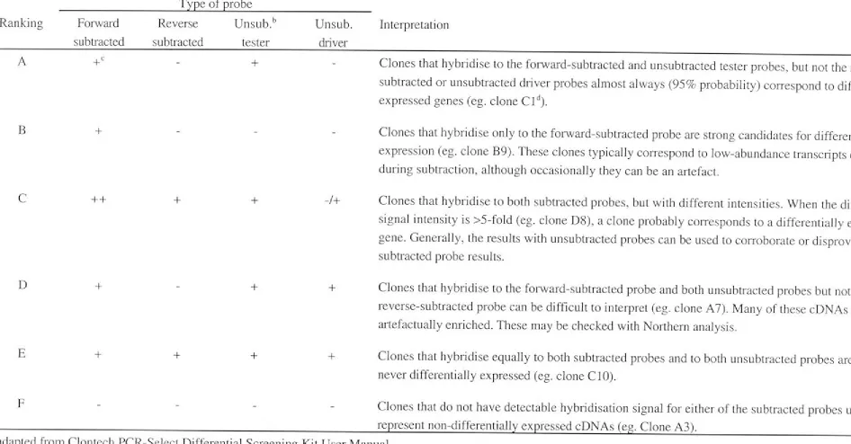
7 .2.1 Differential screening of subtracted clones ... 80
7 .2.2 Interpretation of screening results ... 81
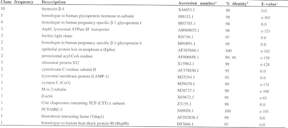
7 .2.3 Analysis of differentially expressed sequences ... 82
7 .2.4 Identity of differentially expressed genes in small LTC-DC ... 83
7.2.5 Identity of differentially expressed genes in large LTC-DC ... 85
7.3 DISCUSSION ... 87
CHAPTER 8 General Discussion . ... 92
8.1 THE LTC SYSTEM PRODUCES CDl lc+CDl lb+MHCII1°CD8cx- DC ... 92
8.2 LARGE LTC-DC AS IMMATURE DC ... 94
8.3 DC DEVELOPMENT FROM SMALL LTC-DC PRECURSORS ... 97
8.4 A MODEL FOR DC DEVELOPMENT WITHIN LTC ... 100
APPENDIX A ... 104
APPENDIX B ... 107
REFERENCES ... 109
PUBLICATIONS ... 134
3 HT ATP ANU BM BrdU CLP CMP CSIRO CTL CV DC DTT ddH20 EST EtBr PACS PCS PITC GM-CSP HBSS HPC HSA HSC Hsp IPN
lg
IL JCSMR LC Lin-LN LPS LTC LTC-DC LTC-CSN ABBREVIATIONS thymidine adenosine triphosphateAustralian National University bone marrow
bromodeoxyuridine
committed lymphoid progenitor committed myeloid progenitor
Commonwealth Scientific and Industrial Research Organisation cytotoxic T lymphocyte
crystal violet dendritic cell dithiothreitol
double distilled water expressed sequence tag ethidium bromide
fluorescence activated cell sorter fetal calf serum
fluorescein isothiocyanate
granulocyte/macrophage colony stimulating factor Hanks balanced salt solution
haemopoietic progenitor cell heat stable antigen
haemopoietic stem cell heat shock protein interferon
immunoglobulin interleukin
John Curtin School of Medical Research Langerhans cells
lineage marker negative lymph node
lipopolysaccharide long-term culture
LTC-derived dendritic cell
L TC-derived culture supernatant ( or conditioned medium)
M-CSF MHC MIIC
MIP-la
MLR NCBI
NK
OV-FITC PBS
PCR PE PI SCF sDMEM TCR
TGF-~ Th TLR TNF-a UDG UV
macrophage colony stimulating factor major histocompatibility complex
MHC Class II-rich compartment
macrophage inflammatory protein- I a mixed 1 ymphocyte reaction
National Center for Biotechnology Information natural killer
flurorescein isothiocyanate conjugated ovalbumin phosphate buffered saline
polymerase chain reaction phycoerythrin
propidium iodide stem cell factor
supplemented Dulbecco' s Modified Eagle's Medium T cell receptor
transforming growth factor-~ T helper cell
toll-like receptor
tumour necrosis factor-a uracil DNA glycosylase ultraviolet
CHAPTER 1
General introduction
1.1 UNIQUE PROPERTIES OF DENDRITIC CELLS (DC).
DC were discovered in 1973 when Steinman and Cohn (1973), observing murine spleen cells, 'noticed a large stellate cell with distinct properties'. Almost thirty years on, DC are one of the most studied cells in immunology, partly because of their
immunotherapeutic importance for malignancies (Boczkowski et al., 2000; Fields et al., 1998; Mayordomo et al., 1995; Nestle et al., 1998; Reichardt et al., 1999), infectious agents (Bhardwaj et al., 1994; Takahashi et al., 1993) and autoimmune disease (Clare-Salzler et al., 1992). A major role of DC is to acquire exogenous antigen in the
peripheral tissues and to present antigenic peptide via major histocompatibility complex (MHC) Class I and II molecules to CD4 and CD8 T cells in secondary lymphoid organs. DC are the only antigen presenting cells that can stimulate naive T cells and induce a primary immune response (Levin et al., _1993).
Dendritic cells are located in low numbers throughout the body (Hart, 1997). They are present in non-lymphoid sites like the blood and tissues such as liver, heart, mucosa and skin. Langerhans cells (LC) of the skin are distinguished by cell surface markers
including E-cadherin, CD207 (langerin) and CD la (Borkowski et al., 1994; De Panfilis et al., 1988; Valladeau et al., 1999), and by their possession of a distinct organelle
called the Birbeck granule (Birbeck et al., 1961). DC also exist in secondary lymphoid tissues such as lymph nodes (LN) and spleen. These DC can be divided into subsets that differ in phenotype and functional capacity (Henri et al., 2001; Maldonado-Lopez et al., 1999b; Pooley et al., 2001; Vremec et al., 2000). DC within the thymus play an
essential role in central tolerance by tolerising T lymphocytes to self-antigen (Brocker et al., 1997; Carlow et al., 1992).
The DC lineage includes populations of cells with different properties and at this time, a specific DC marker remains elusive. As a result, DC are defined using a combination of properties. These include (1) morphology, (2) phenotype and (3) the ability to
efficiently take up and present antigen to naive T cells. DC display cytoplasmic
extensions that can be short or veil-like when cells are immature, but increase in length upon maturation (Nijman et al., 1995). Electron microscopic features include prominent mitochondria and the presence of endosomes and lysosomes essential for antigen
processing (Hart, 1997). Murine DC are known to express a range of cell surface
markers including CDllc (Metlay et al.,1990), CDllb (Leenen et al., 1998; Pulendran et al., 1997), CD205 (Inaba et al., 1995; Kraal et al., 1986), MHC Class II (Coates et al., 1996; Pierre et al., 1997) and 33Dl (Nussenzweig et al., 1982).
DC interact with a wide range of immune cells. They induce a cell-mediated immune response by presenting specific antigen to naive and memory CD4 and CD8 T cells (Ingulli et al., 1997; Levin et al., 1993; Roth and Spiegelberg, 1996; Albert et al., 2001; Norbury et al., 2002). Immature and mature monocyte-derived DC can induce the
proliferation and activation of resting natural killer (NK) cells in vitro (Ferlazzo et al., 2002; Gerosa et al., 2002) and DC also have the capacity to cluster and interact directly with B cells (Dubois et al., 1997; Kushnir et al., 1998). They can present unprocessed antigen to na1ve B cells and modulate naive B cell proliferation and differentiation into antibody secreting cells in a T cell-dependent manner (Dubois et al., 1997; Wykes et al., 1998). DC can also provide signals for isotype switching to immunoglobulin (Ig)G and IgA (Fayette et al., 1997; Wykes et al., 1998), suggesting that they can regulate the type of humoral response induced.
It has recently been shown in vivo and in vitro that DC are the antigen presenting cell responsible for the phenomenon of cross-presentation whereby exogenous antigens gain access to the MHC Class I presentation pathway (Albert et al., 1998; Kurts et al., 2001). Immature· DC phagocytose apoptotic cells and upon maturation, present the processed peptides to CD8 T cells in the context of MHC Class I molecules (Albert et al., 2001; Albert et al., 1998; Kurts et al., 2001). Cross-priming results in the activation of
responding CD8 T cells and generation of a cytotoxic lymphocyte (CTL) response. This has been demonstrated when DC present antigen acquired from tumour cells (Huang et al., 1994) and apoptotic influenza-infected cells (Albert et al., 1998). Cross-presentation is also thought to represent a means by which peripheral tolerance can be maintained in vivo. The cellular features that distinguish cross-priming and cross-tolerance remain unclear. Cross tolerance may be the result of presentation of self-antigen by immature DC lacking a second signal for T cell activation (Green and Beere, 2000; Mellman and Steinman, 2001), or presentation of self-antigen by mature DC in the absence of CD4 T cell help (Albert et al., 2001).
1.2 FUNCTIONAL MATURATION OF DC.
In fulfilling their function as professional antigen presenting cells, DC move through various stages of maturation. The terms 'immature' and 'mature' have been used, albeit loosely, to describe these stages. The term 'immature' was originally coined in the
literature to describe DC precursors which were usually blood monocytes or bone marrow (BM) precursors. For the purpose of this study, immature DC are defined as fully differentiated DC. DC which are immature take up and process antigen into peptides that are then loaded on to MHC molecules. Immature DC represent the
immune steady-state, endocytic but not activated by exposure to antigen. Maturation or activation of DC occurs after exposure to pathogen or disease, when DC begin to
express molecules that enable them to interact with T cells to initiate an immune
response. Mature or activated DC are end-stage cells that cannot revert to an immature state. They become apoptotic after interaction with T cells (Matsue et al., 1999).
Immature DC are located in the periphery or non-lymphoid organs. They capture antigen by three endocytic mechanisms. Constitutive, high level fluid uptake via
macropinocytosis allows them to continually internalise large volumes of fluid (Sallusto et al., 1995). Phagocytosis of particles (Matsuno et al., 1996) leads to presentation of antigen in the context of MHC Class II (Inaba et al., 1998) while phagocytosis of apoptotic cells leads to cross-presentation of viral, tumour and self-antigens in the context of MHC Class I (Albert et al., 1998). Receptor-mediated endocytosis involves mannose receptors (Sallusto et al., 1995), CD205 (Jiang et al., 1995; Mahnke et al., 2000) and Fey receptors (FcyR) (Sallusto and Lanzavecchia, 1994 ). Mannose receptors and CD205 release their captured ligands into acidified endosomes and then recycle to the cell surface. This allows them to deliver ligands in consecutive rounds and to
capture antigen in amounts that far exceed the number of receptors (Mahnke et al., 2000; Sallusto et al., 1995). FcyR may direct antigen to MHC Class I molecules and so contribute to cross-presentation (Regnault et al., 1999; Rodriguez et al., 1999).
Macropinocytosis allows the continuous internalisation of soluble antigen, while endocytic receptors may give some degree of selectivity for non-self molecules.
Freshly isolated non-lymphoid 'immature' DC express only low levels of MHC Class II/peptide complexes on their cell surface. However, abundant intracellular MHC Class II is present within specialised MHC Class II-rich endocytic compartments (MIIC)
similar to those seen in B cells (Kleijmeer et al., 1995; Nijman et al., 1995; Pierre et al., 1997). MIIC are positioned at the intersection of the biosynthetic route of MHC Class II and the endocytic route and are the site of antigen degradation and peptide loading of MHC Class II molecules (Kleijmeer et al., 1995). These compartments are
multilaminar, acidic, LAMP-I+ and contain invariant chain (Ii), HLA-DM and cathepsin S (Nijman et al., 1995; Turley et al., 2000). Following endocytosis, antigen can reach MIIC within 30 minutes of contact (Kleijmeer et al., 1995). However, immature DC do not immediately demonstrate increased antigen presentation capacity upon contact with
antigen. In the absence of a signal to initiate maturation or activation of DC a majority of new MHC Class II/peptide complexes accumulate within MIIC where most are
degraded and some stored for later presentation (Pierre et al., 1997; Turley et al., 2000). This mechanism appears to safeguard against an immune response to self antigen.
Immature DC express inflammatory chemokine receptors such as CCRl, CCR2, CCR5, CCR6 and CXCR4 (Carramolino et al., 1999; Vecchi et al., 1999). These receptors account for the chemotactic response to the inflammatory chemokines CCL3 (MIP-1 ex), CCL4 (MIP-1~), CCL5 (RANTES), CCL7 (MARC), CCL20 (MIP-3cx) and CXCL12 (SDF-1) (Sozzani et al., 1999; Vecchi et al., 1999), guiding DC to the site of
inflammation where they can be triggered to mature by a number of different stimuli. DC maturation can be achieved in vivo and in vitro by exposing immature cells to
soluble antigen (Nijman et al., 1995), necrotic cells (Gallucci et al., 1999; Sauter et al., 2000), cytokines such as granulocyte/macrophage colony stimulating factor (GM-CSF) and tumour necrosis factor (TNF)-cx, microbial products such as lipopolysaccharide (LPS) and T cell signals such as CD40L (De Smedt et al., 1996; Hochrein et al., 2001; Pie1Te et al., 1997; Sallusto et al., 1995; Vecchi et al., 1999). In response to these
maturation stimuli, DC in vivo transiently produce large amounts of inflammatory
chemokines themselves (Sallusto et al., 1999), recruiting other antigen presenting cells and effector cells to the site of infection. Inflammatory chemokine receptors are then downregulated and constitutive chemokine receptors such as CCR7 are upregulated (Sallusto et al., 1999; Vecchi et al., 1999), allowing maturing DC to exit the
inflammatory site and migrate to secondary lymphoid organs.
The homing of DC to T cell areas in lymphoid tissues involves cell surface adhesins and chemotactic factors released by these tissues. CCR 7 allows DC to migrate from
peripheral sites to lymphatic vessels where CCL21 (6Ckine, SLC) (Chan et al., 1999) is produced, and then to the T cell areas of secondary lymphoid organs where CCLI 9 (MIP-3~, ELC) is produced by resident mature DC (Sallusto et al., 1999). The
importance of CCR7 in immune regulation is demonstrated in CCR7-/- mice which have impaired migration of B cells, T cells and DC and lack secondary lymphoid organ structure (Forster et al., 1999). Within lymphoid organs, mature DC also produce a range of chemokines to attract naive or activated T cells to enhance interaction between antigen-bearing DC and antigen-specific T cells. These chemokines include CCL22 (MDC), CCL 17 (T ARC) and DC-CK 1 (Adema et al., 1997; Lucy Tang and Cyster, 1999; Sallusto et al., 1999).
Inflammatory signals not only initiate migration of DC, but also trigger irreversible phenotypic and functional changes. Maturing DC lose attributes associated with antigen capture and processing. Mannose receptors and FcyR are downregulated and
biosynthesis of Ii and MHC Class II molecules ceases (Austyn, 1996; Sallusto et al., 1995). MHC Class II/peptide complexes are transferred to LAMP-, non-lysosomal compartments termed CIIV (Turley et al., 2000) that also contain MHC Class I
molecules and CD86. CIIV are intermediates in the transfer of MHC Class II/peptide from lysosomes to the cell surface. Once on the plasma membrane, MHC Class
II/peptide complexes, MHC Class I and CD86 continue to co-localise (Turley et al., 2000). These could form an immunological synapse for activating naive T cells.
Costimulatory molecules such as CD80 (Larsen et al., 1992), CD86 (Inaba et al., 1994 ), CD40 (Caux et al., 1994; McLellan et al., 1996) and adhesion molecules like CD58 (LFA-3) and CD54 (ICAM-1) are upregulated on the cell surface of mature DC
(Sallusto et al., 1995). Costimulatory molecules provide signals to T cells in addition to signals through T cell receptor (TCR)-MHC/peptide engagement (McLellan et al.,
1996; Mondino and Jenkins, 1994). They may also influence the commitment of T cells to the T helper type (Th)l or Th2 pathways (Cella et al., 1996; Gause et al., 1997;
Kuchroo et al., 1995). For example, CD40:CD40L interaction results in production of interleukin (IL)-12 which mediates development of Thl responses (Cella et al., 1996). It can also induce differentiation of naive CD40-activated B cells into IgM-secreting
plasma cells (Dubois et al., 1998).
Large clusters of naive CD4 or CD8 T cells form around individual antigen-presenting DC in vivo (Ingulli et al., 1997; Norbury et al., 2002), allowing small numbers of DC to stimulate many T cells in a short period of time. Few T cells are observed surrounding DC in the absence of relevant antigen (Ingulli et al., 1997; Norbury et al., 2002). This suggests that the interaction is only stabilised if the DC display appropriate
MHC/peptide complexes, perhaps upregulating expression of adhesion molecules, such as CD1 la/CD18 (LFA-1) and CD58 (Mondino and Jenkins, 1994; Steinman, 1991) on the T cells. Cluster formation coincides with IL-2 production by T cells, followed by proliferation and differentiation into effector cells (Ingulli et al., 1997).
1.3 DIFFERENT DC POPULATIONS. 1.3.1 Mature DC subsets.
A number of DC subsets or classes have been defined on the basis of cell surface marker expression and function. Cells commonly studied ex vivo are MHC Class Ir+
mature DC, and subsets of these have been defined on the basis of surface marker expression of CD8cx, CD4, CD205 and CD 11 b. To date, three different murine DC subsets have been identified in spleen, two in thymus (Vremec et al., 2000) and five in LN, one of which is thought to represent mature LC (Henri et al., 2001). The
relationship between these DC subsets remains unclear. The resemblance of CD8cx+ DC to thymic DC led to the proposal that CD8cx- and CD8cx+ DC represented separate
myeloid and lymphoid lineages, respectively (Pulendran et al., 1997; Vremec and Shortman, 1997). Recent studies indicate that this is not the case (Manz et al., 2001; Traver et al., 2000). Current evidence suggests that DC subsets may either reflect different stages in maturation or may regulate different arms of the immune response (Hochrein et al., 2001; Maldonado-Lopez et al., 1999b; Martinez del Hoyo et al., 2002; Merad et al., 2000; Pooley et al., 2001). The function and origin of DC subsets is one of the most controversial areas of DC biology. Information generated is highly dependent on the way in which DC subsets are produced in vitro and handled during
experimentation. In addition, selective techniques used to isolate DC ex vivo on the basis of marker expression sometimes excluded subsets of DC (Vremec and Shortman,
1997; Vremec et al., 1992).
Mature splenic DC can be divided into three CDI lc+MHCII+ subsets: CD4+CD8cx-CD205-cD 11 b+ DC, CD4-CD8cx+CD205+CD 11 b- DC and CD4-CD8cx-CD205-CD 11 b+
DC (Vremec et al., 2000). Both the CD4+CD8cx-CD205-CD1 lb+ DC and CD4-CD8cx-CD205-CD1 lb+ DC resemble myeloid-lineage cells since they express myeloid markers
like CD 11 b, 33D 1, F4/80 and lack CD8cx (Pulendran et al., 1997; Vremec et al., 2000). They are located in the marginal zone between white and red pulp (Crowley et al.,
1989; Leenen et al., 1998; Pulendran et al., 1997; Vremec et al., 1992), but can migrate to T cell-dependent areas of spleen upon stimulation (De Smedt et al., 1996).
CD4+CD8cx- differ from CD4-CD8cx- DC in the expression of CD4 itself, greater
adhesion capacity, higher levels of F4/80 expression (Vremec et al., 2000) and lower cytokine production following stimulation (Hochrein et al., 2001). However, both
CD8cx- DC subsets are efficient stimulators of CD4 and CD8 T cells and show efficient MHC Class II presentation to antigen-specific CD4 T cells (Kronin et al., 1996; Pooley et al., 2001; Suss and Shortman, 1996). Hereafter, both subsets will be referred to as CD8cx- DC. Other reports have shown that CD8cx- DC direct Th2 type immune
responses since they stimulate cells that produce IL-4 and little IFN-y (Maldonado-Lopez et al., 1999b).
The CD4-CD8cx+CD205+CD1 lb- subset (hereafter referred to as CD8cx+ DC) resemble the lymphoid-like thymic DC and occupy the T cell areas of the spleen (Crowley et al.,
1989; Leenen et al., 1998; Pulendran et al., 1997; Vremec and Shortman, 1997). CD8a+ DC exhibit very different functional capacity in comparison to CD8a- DC. Ex vivo
CD8a+ DC have been shown to have regulatory effects on T cells. They activate both CD4 and CD8 T cells, but induce apoptosis in CD4 T cells (Suss and Shortman, 1996) and limit CD8 T cell proliferation by reducing IL-2 production (Kronin et al., 1996). In contrast, under conditions consistent with activation, such as prior exposure to GM-CSP (Maldonado-Lopez et al., 1999b; Pulendran et al., 1999) or the presence of IL-2 (Pooley
et al., 2001), CD8a- DC exert different effects on CD8 T cells. They efficiently present antigen to CD8 T cells in the context of MHC Class I ( den Haan et al., 2000; Pooley et al., 2001) and so have capacity to cross-present exogenous soluble antigen for
subsequent generation of cytotoxic T cell responses. Other studies have shown that
CD8a+ DC induce Thl type immune responses in vivo, consistent with activated CD8a+ DC being the major producers of IL-12 (Hochrein et al., 2001; Maldonado-Lopez et al.,
1999a; Pulendran et al., 1999). This suggests that in the immune steady state, CD8a+ DC exert regulatory effects over T cells. However, once stimulated they can induce a Th 1 type immune response.
It was initially proposed that CD8a- and CD8a+ DC represented cells of myeloid and lymphoid lineage, respectively. This concept relied heavily on the phenotype of ex vivo isolated DC and reports that precluded one subset from being the precursor of another (Karnath et al., 2000; Pulendran et al., 1997). CD8a+ DC were also defined as lymphoid on the basis of experiments showing that 'CD4 low' thymic precursors (Wu et al.,
1991) gave rise to only CD8a+ DC upon intrathymic transfer (Ardavin et al., 1993) and develop into DC independently of GM-CSP in vitro (Saunders et al., 1996). However, more recently it was demonstrated that 'CD4 low' precursors can generate both CD8a+ and CD8a- DC in vivo (Martin et al., 2000). The analysis of mice deficient for
transcription factors affecting haemopoeisis was also used to assess the lineage
relationship of CD8a- and CD8a+ DC. The conflicting nature of these reports did not allow strong conclusions to be drawn. For example, one report suggesting that RelB and PU.I selectively regulate development of CD8a- myeloid-like DC (Wu et al., 1998) was contradicted by another showing that PU.I deficient mice lack both CD8a- and CD8a+ DC (Anderson et al., 2000). It has also been suggested that the environmental defects in these knockout mice could contribute to the altered DC phenotypes observed (Ardavin et al., 2001; Traver et al., 2000).
A more direct and definitive test of lineage relationships requires studies on cells
developing from lineage-restricted progenitors. Committed lymphoid progenitors (CLP) from mouse BM of the phenotype lineage marker negative
Sca-l1°CD1 l 7(c-kit)10 produce T cells, B cells and NK cells (Kondo et al., 1997) while committed myeloid progenitors (CMP) or lL"-7R-CD34+FcyR10 cells produce
monocytes/macrophages, granulocytes, megakaryocytes and erythrocytes (Akashi et al., 2000). Both CMP and CLP can generate both CD8cx- and CD8cx+ DC subsets in vivo and in vitro (Manz et al., 2001; Traver et al., 2000; Wu et al., 2001). Both CMP and CLP give rise to mature CD8cx- and CD8cx+ splenic DC and CD8cx+ thymic DC in vivo.
Furthermore, all DC subsets express RelB and PU.I while only CD8cx+ DC express IL-12 (Manz et al., 2001; Traver et al., 2000). By examining the number of DC produced from single progenitor cells and the abundance of CMP and CLP within BM, it has been calculated that a majority (up to 90%) of splenic DC and approximately half of thymic DC are derived from the myeloid lineage (Manz et al., 2001).
Clonogenic studies indicate that cell surface phenotypes alone cannot be used to define lineage. The role of the CD8cx molecule itse_lf remains unclear. It is not a marker of the lymphoid lineage and does not appear to have a functional role in DC (Kronin et al.,
1997). Some evidence suggests that CD8cx may be a marker of DC maturation since maturing LC en route to the LN upregulate CD8cx (Merad et al., 2000), as do BM-derived DC activated with LPS or IFN-cx (Brasel et al., 2000). Recently it was shown that a majority of purified CD8cx- DC upregulate CD8cx, CD205 and CD24 following
intravenous injection into mice (Martinez del Hoyo et al., 2002). Furthermore, most splenic CD8cx- but not CD8cx+ DC express CCR6 (Kucharzik et al., 2002), a chemokine receptor associated with immature DC (Carramolino et al., 1999). These results suggest that CD8cx+ DC could represent a final stage in DC differentiation, playing an important role in the induction of immune responses through T cell stimulation, cross-priming and production of cytokines including IL-12.
Recent evidence now suggests that CD8cx- DC and CD8cx+ DC do not represent separate lineages of DC, but do represent DC of distinct functional capacity. The presence of functionally distinct DC subsets would help to explain the wide range of DC activities described in vivo. The question remains as to the triggers which induce formation of these distinct subsets. It is possible that DC phenotype and function reflects different maturational states. Another possibility is that distinct DC subsets are involved in the immune response to pathogens and the maintenance of self tolerance. Development of these subsets could be dictated by specific cytokines and cell types present in tissue microenvironments.
1.3.2 DC progenitors.
DC can be generated from a number of different progenitor or precursor cells. These include Lin-CDl 17+ BM and fetal liver cells (Zhang, Y. et al., 1998; Zhang et al. , 2000), peripheral blood monocytes (Schreurs et al., 1999) and 'CD4 low' cells in the thymus (Ardavin et al., 1993). Progeny DC have been derived from purified, primitive self-renewing haemopoietic stem cells (HSC) as well as lineage-restricted CLP, CMP, granulocyte/macrophage-restricted precursors, and pro-T cells (Manz et al., 2001). These studies have not yet revealed a DC-committed progenitor. Most cytokine supplemented in vitro cultures drive progenitors quickly through to mature DC, preventing the characterisation of intermediate precursors . A CD1
lc+MHCir-B220+CD19- DC precursor population was recently isolated from mouse blood (del Hoyo et al., 2002). These cells appear to represent committed precursors since they can reconstitute all splenic DC subsets, but not other lymphomyeloid compartments.
CD1 lc+MHCifB220+CD19- precursors may represent cells downstream in development from CMP and CLP. Resident DC progenitors in spleen have not yet been reported.
Most research has focused on the study of mature MHC Class II+ splenic DC,
conforming with the concept that maturing DC migrate to this secondary lymphoid organ from the periphery. However, DC progenitors must exist in spleen since Lu et al. demonstrated that DC can be generated from proliferating MHC Class If splenic
progenitors when cultured in the presence of GM-CSF (Lu et al., 1995).
1.4 USE OF CYTOKINES TO CULTURE DC
IN VITRO.
DC are commonly generated in vitro from progenitor cells using cocktails of cytokines since they represent a trace population of cells in vivo. GM-CSF has been the most co1mnonly used cytokine for in vitro culture of DC. Initially it was demonstrated that GM-CSF could enhance survival and differentiation of MHC Class n+ blood DC for up to 6 weeks (Markowicz and Engleman, 1990). Since mature DC cannot divide (Bowers and Berkowitz, 1986), amplification of DC numbers was achieved by culture of
replicating MHC Class If precursors in mouse blood (Inaba et al., 1992b) and BM (Inaba et al., 1992a) in medium supplemented with GM-CSF. These cultures generated granulocytes and macrophages in addition to mature DC and early stages featured
aggregates of DC affixed to a stromal monolayer comprising macrophages and
fibroblasts (Inaba et al., 1992a; Inaba et al. , 1992b ). It is highly likely that in addition to GM-CSF, stromal-derived cytokines were important in the generation of DC.
GM-CSF supplemented cultures (Inaba et al., 1992a; Inaba et al., 1992b) do not produce a pure population of DC. More recently, greater numbers of DC have been
generated from in vitro culture systems that use defined progenitor starting populations and combinations of cytokines. Some of these culture systems use a set of specific cytokines to support an initial phase of proliferation producing 'immature DC',
followed by a second phase of culture with a different set of cytokines to induce DC 'maturation' (Hausser et al., 1997; Zhang, Y. et al., 1998; Zhang et al., 1999).
However, most in vitro cultured cells resemble mature DC on the basis of MHC Class II and costimulatory molecule expression as well as T cell stimulatory capacity,
suggesting that the immature DC stage has been bypassed in culture. Alternatively, some intermediate 'immature DC' retain capacity to form macrophages when exposed to mediators such as macrophage colony-stimulating factor (M-CSF). These bipotential cells appear to represent intermediate precursors rather than fully differentiated
immature DC. Figure 1.1 summarises the in vitro culture systems used to generate DC from various progenitor populations. Intermediate cell types in Figure 1.1 are indicated
'precursor' or 'mature' based on the above rationale.
Culture of monocytes or BM cells with GM-CSF, TNF-a and IL-4 or stem cell factor (SCF) represent the most common procedures used to derive DC in vitro. Mouse
peripheral blood mononuclear cells and Lin- BM cells can be induced to become MHC Class Ir+ mature DC if cultured with a combination of GM-CSF and IL-4 (Schreurs et al., 1999) (Fig I. IA). In some cultures, TNF-a is added to induce further activation (Hausser et al., 1997). TNF-a alone provides no growth promotion for haemopoeitic progenitor cells (HPC), but assists in the develop1nent of mature DC (Caux et al., 1992). By comparison, IL-4 maintains DC in an immature state, capable of antigen capture and processing (Sallusto and Lanzavecchia, 1994 ). SCF was incorporated into in vitro
culture systems after it was shown to increase DC yield from human CD34+ HPC
(Young et al., 1995). When cultured with SCF in combination with GM-CSF and TNF-a, Lin-CD 117+ BM cells proliferate and develop into two distinct dendritic precursor
populations (Zhang, Y. et al., 1998) (Fig 1. IB). In a second period of non-proliferative differentiation, GM-CSF and TNF-a are used to generate mature LC from
DC-committed precurso!s (CDI lb1°CDI lc+) or mature DC from bipotent myeloid
precursors (CDI lbhiCDl lc+). Similar cells are generated from human CD34+ HPC using GM-CSF and TNF-a (Caux et al., 1996).
Transforming growth factor (TGF)-~ has a specific role in LC development. It is
essential for the differentiation of LC in vivo (Borkowski et al., 1996) and has been used in combination with GM-CSF, SCF and TNF-a to generate LC in vitro from
Lin-CDI 17+ BM cells (Zhang 99) (Fig I.IC). TGF-~ appears to be effective in DC culture only when TNF-a is also present, otherwise it inhibits DC production (Riedl et al.,
Figure 1.1. Cytokine-dependent cultures used to generate DC from precursors in
vitro . Starting precursor populations, intem1ediate cells and end-stage cells are indicated in
black text. Starting populations include lineage 1narker negative (Lin-) bone marrow (BM), peripheral blood 1nononuclear cells (PBMC), Lin-CD 117+ BM, thyn1ic 'CD4 low'
precursors, corrunon lymphoid progenitors (CLP) and common myeloid progenitors
(A)
(B)
Lin- BM; PBMC
Lin-CD 117+ BM
GM-CSF IL-4 GM-CSP SCP TNF-a GM-CSP SCP Mature DC
CD1 lb1°CD1 lc+ precursor
CDl lbhiCDl le+ precursor
I
GM-CSF TNF-a GM-CSP TNF-a GM-CSP TNP-a M-CSP Activated DC Mature Langerhans-like DC Mature DC Macrophage
(C) Lin-CD 117+ BM TGP-~ Monocyte/macrophage
precursor
GM-CSP TNP-a
Langerhans-like DC
(D) Lin- BM
(E) CMP
(F) Thymic 'CD4 low' precursor
(G)
CLPPlt3L
IPN-a or LPS
CDl lbhiCDl le+
mature DC - - - - CD8a- activated DC
IPN-a or LPS
CD1 lb1°CD1 le+
mature DC - - - - CD8a+ activated DC
IL-3 SCP TNP-a IL-4 Flt3L GM-CSP
IL-1~ IL-3
IL-7 SCP
TNP-a Plt3L
1997). Flt3L can be used to expand DC numbers in vivo (Maraskovsky et al., 1996), although it does not act specifically on DC. When injected into mice, it expands many haemopoietic cell types, including lymphocytes, NK cells, erythroid cells, monocytes and granulocytes (Brasel et al., 1996; Shaw et al., 1998). Flt3L can generate both CD8a- and CD8a+ DC in vitro from Lin- BM when activated with IFN-a or LPS (Brasel et al., 2000) (Fig 1. lD). However, Flt3L is not solely responsible for the
development of DC from Lin- BM cells because DC grow from aggregates associated with adherent macrophages and endogenously produced IL-6 is also important in their development (Brasel et al., 2000).
There is now evidence to suggest that GM-CSF is essential only for the development of myeloid DC. CMP in BM, like other lin- BM populations require GM-CSP for DC
production (Manz et al., 2001) (Fig 1.lE). Lymphoid DC have different cytokine requirements. Colonies of thymic DC have been generated by culture of 'CD4 low' thymic precursor cells in a cocktail of seven growth factors including IL-1 ~' TNF-a, IL-3, IL-7, SCP, Flt3L and anti-CD40 monoclonal antibody (Saunders et al., 1996) (Fig 1.lF). IL-7 and not GM-CSP is the essential factor for development of thymic DC. Cells produced are functional, mature DC. However, in contrast to normal thymic DC, they do not express CD8a and BP-1, suggesting that this culture may not mimic in vivo
conditions for thymic DC development (Saunders et al., 1996). Consistent with this, IL-7 is essential for the generation of DC from CLP in BM (Manz et al., 2001) (Fig 1.lG). These studies confirm that myeloid and lymphoid progenitors have different cytokine requirements for the generation of DC.
Cytokine supplementation can be used to generate high numbers of DC in vitro for experimentation. These studies have defined stages in DC development and maturation as well as lineage. However, a number of limitations are associated with the use of cytokines. They are used in concentrations higher than would be expected in vivo and could direct development along pathways that are not physiologically normal. In vitro generated DC have been shown to have different marker expression, in comparison with their ex vivo counterparts. DC generated in vitro survive for only a limited period of time and the purity of DC in these cultures is also questionable. Furthermore, DC
produced from monocytes and BM or cord blood HPC vary in their functional capacity (Herbst et al., 1997; Schreurs et al., 1999; Triozzi and Aldrich, 1997). Different sources of precursor cells and methods of culture can generate different DC. This is an
important consideration when preparing a DC population for study or for use as an immunotherapeutic tool.
1.5 ANALYSIS OF GENE EXPRESSION IN DC.
DC gene expression analysis offers the opportunity to study genetic changes that underlie DC development from progenitors. It offers enormous potential for
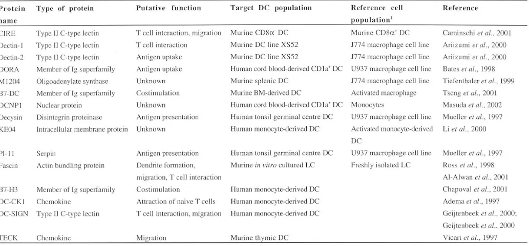
investigation of DC lineage and function through the generation of gene expression profiles and identification of novel genes. The identification of novel DC-associated genes has led to the characterisation of novel proteins that contribute to the unique function of DC. A variety of procedures have been employed to detect DC-associated genes, including the random sequencing of cDNA libraries (Adema et al., 1997; Vicari et al., 1997), screening cDNA libraries for sequences homologous to known molecules (Chapoval et al., 2001) and the generation and screening of subtracted libraries
(Diatchenko et al., 1996; Tiefenthaler et al., 1999). Subtracted libraries have been used to isolate genes specifically expressed in DC and not macrophages. They have also been used to isolate genes associated with DC activation. One study comparing CD8cx- and CD8a+ DC has led to the isolation of the novel C-type lectin CIRE (Caminschi et al., 2001). To date, novel genes have been isolated from both human and mouse DC
populations including spleen and germinal centre DC, as well as BM-, cord blood- and monocyte-derived DC and DC lines like XS52 (Table 1. 1).
A list of novel DC-associated proteins is presented in Table 1.1. It includes proteins involved in antigen capture such as Dectin-2 (Ariizumi et al., 2000) and DORA (Bates et al., 1998), chemokines such as DC-CKl (Adema et al., 1997) and TECK (Vicari et al., 1997), costimulatory molecules like B7-DC (Tseng et al., 2001) and molecules that assist antigen presentation including PI-11 (Mueller et al., 1997 a) and Decysin (Mueller et al., 1997b). Proteins essential to DC function like DC-CKl, Fascin and DC-SIGN were isolated after identification of unique genes (Adema et al., 1997; Geijtenbeek et al., 2000b; Ross et al., 1998). DC-CKl is a chemokine produced by mature DC in
secondary lymphoid organs which functions by attracting naive CD4 and CDS T cells to facilitate antigen presentation (Adema et al., 1997). The expression of Fascin, an actin bundling protein, coincides with the formation of dendritic projections in LC (Ross et al., 1998). DC-SIGN is expressed on the surface of DC and mediates DC migration through interaction with CD102 (ICAM-2) (Geijtenbeek et al., 2000a) and transient clustering between DC and T cells through interaction with CD50 (ICAM-3)
(Geijtenbeek et al., 2000b ).
Gene expression studies using serial analysis of gene expression (Hashimoto et al. , 1999; Hashimoto et al., 2000) or oligonucleotide microarrays (Granucci et al., 2001; Huang et al. , 2001; Le N aour et al., 2001) have generated profiles of genes unique to different DC populations. By identifying clusters of genes that identify a particular cell
Table 1.1 Examples of DC-associated proteins. Protein name CIRE Dectin-1 Dectin-2 DORA Ml204 B7-DC DCNPl Decysin KE04 PI-11 Fascin
Type of protein
Type II C-type lectin
Type II C-type lectin
Type II C-type lectin
Member of Ig superlamily
Oligoadenylate synthase
Member of Ig supe1family
Nuclear protein
Disintegrin proteinase
Intracellular membrane protein
Serpin
Actin bundling protein
B7-H3 Member of Ig superfamily
DC-CKl Chemokine
DC-SIGN Type II C-type lectin
TECK Chemokine
Putative function
T cell interaction, migration
T cell interaction
Antigen uptake
Antigen uptake
Unknown
Costimulation
Unknown
Antigen presentation
Unknown
Antigen presentation
Dendrite formation,
migration, T cell interaction
Costimulation
Attraction of naive T cells
T cell interaction, migration
Migration
Target DC population
Murine CD8a- DC
Murine DC line XS52
Murine DC line XS52
Human cord blood-derived CD la+ DC
Murine splenic DC
Mmine BM-derived DC
Human cord blood-derived CD la+ DC
Human tonsil germinal centre DC
Human monocyte-derived DC
Human tonsil germinal centre DC
Murine in vitro cultured LC
Human monocyte-derived DC
Human monocyte-derived DC
Human monocyte-derived DC
Murine thymic DC
1
The production of subtracted libraries involves the subtraction of reference cDNA from the target cDNA population.
Reference cell
population1
Murine CD8a+ DC
1774 macrophage cell line
177 4 macrophage cell line
U937 macrophage cell line
1774 macrophage cell line
Activated macrophage
Monocytes
U93 7 macrophage cell line
Activated monocyte-de1i ved
DC
Reference
Caminschi et al., 2001
Ariizumi et al., 2000
Ariizumi et al., 2000
Bates et al., 1998
Tiefenthaler et al., 1999
Tseng et al., 2001
Masuda et al., 2002
Mueller et al., 1997
Li et al., 2000
U937 macrophage cell line Mueller et al., 1997
Freshly isolated LC Ross et al., 1998
Al-Alwan et al., 2001
Chapoval et al., 2001
Adema et al., 1997
Geijtenbeek et al., 2000;
Geijtenbeek et al., 2000
[image:27.1180.86.1119.114.596.2]population, comparison can be made of gene expression changes upon differentiation, under altered conditions or between different cell types. Two studies have profiled changes in gene expression during DC differentiation from monocytes (Hashimoto et al., 1999; Le Naour et al., 2001) to identify genes unique to monocyte-derived DC. Genes identified are expressed or upregulated as DC differentiate. These genes encode proteins associated with cell structure such as vinculin and chemokines including T ARC and MDC (Hashimoto et al., 1999). At this stage, the only studies on DC development from progenitors involve DC derived from monocytes after culture with cytokines. It will be important to identify gene expression changes as DC develop from other
progenitors, such as BM or thymic precursors. However, the difficulty of cell isolation has so far precluded these studies. Phenotypic and functional analyses indicate that
pathways for DC development are numerous, leading to production of DC with different functions and properties. Gene profiling studies could be used to test this hypothesis and define the genetic changes that underlie different pathways of development.
Some studies have attempted to profile gene expression upon DC maturation or
activation. DC lines or monocyte- and BM-derived DC have been activated in various ways by exposure to LPS (Chen et al., 2002; Hashimoto et al., 2000), inflammatory cytokines (Dietz et al., 2000; Granucci et al., 2001) or whole microbes (Huang et al., 2001). Many of the genetic changes that occur with activation are consistent with those predicted from functional studies. For example, the CCR 7 gene is upregulated in LPS-activated BM-derived DC, while CCRl is downregulated during activation (Chen et al., 2002).
1.6 ANALYSIS OF DC PRODUCED IN LONG-TERM CULTURE (LTC). 1.6.1 L TC of haemopoietic cells in vitro.
LTC 1nethods are useful for studying haemopoiesis in vitro. In LTC, haemopoiesis is dependent on the establishment of an adherent layer of stromal cells which provide an appropriate environment for stem cell survival, self-renewal and differentiation (Dexter,
1979; Dexter and Testa, 1980). Haemopoietic cell development occurs through cell-to-cell interactions and the secretion of cytokines (Whetton and Dexter, 1993). Typically, LTC simulate development under conditions closer to normal physiological conditions than do cultures supplemented with cytokines. They also allow observation of cell
behaviour and function in vitro. Dexter and Lajtha (1974) first established haemopoiesis in LTC in vitro, in a BM culture system in which granulopoiesis predominated. Stem cells were induced to proliferate in cultures containing an adherent layer of BM stromal cells and haemopoiesis was maintained for several months (Dexter and Testa, 1980).
Whitlock and Witte developed a LTC along Dexter's model which supported
production of cells representing early stages in B cell development (Whitlock et al., 1984). LTC systems provide the opportunity to study early stages in the development of haemopoietic cells.
1.6.2 Long-term cytokine-dependent DC lines.
A long-term spleen-derived culture system has been established by culturing
splenocytes in medium supplemented with GM-CSF and conditioned medium from fibroblasts (Winzler et al., 1997). Cell growth is maintained for more than 12 months, but is strictly dependent on cytokines. The homogeneous population of DC produced in these cultures, named D 1, display an immature DC phenotype. Cells express moderate levels of MHC Class II and CD86, but have inefficient allostimulatory activity.
However, they can be activated by microbes, LPS and cytokines like TNF-a or IL-1~ to display mature or activated DC characteristics. D 1 cells have been used as a model for studying DC activation (Granucci et al., 1999; Granucci et al., 2001).
Two 'immature' DC lines have been established from fetal or newborn mouse skin. The first of these is a GM-CSP-dependent MHC Class I+/If cell line that can stimulate
allogeneic CD8 T cells (Elbe et al., 1994 ). The second DC line, named XS, requires GM-CSF, supernatant from the Pam 212 keratinocyte cell line and supernatant from the NS fibroblast cell line for optimum growth (Xu et al., 1995). The homogeneous
population of cells produced resembles freshly isolated LC and weakly activates naive T cells. Both DC lines are phenotypically stable, retaining immature DC characteristics after one (Xu et al., 1995) or two years (Elbe et al., 1994) of in vitro culture.
Furthermore, MHC Class I+/If cells do not upregulate MHC Class II following
exposure to GM-CSF or IFN-y, or after co-culture with allogeneic T cells (Elbe et al., 1994). Like short-term cytokine-dependent cultures, these cytokine-dependent LTC and DC lines are established and maintained in a very selective and artificial environment. In addition, these systems do not offer the potential to study stages in DC development from progenitors.
1.6.3 L TC which support DC development in vitro.
DC have been produced continuously in a unique spleen-derived LTC system in which haemopoiesis is supported by an adherent stromal cell layer in the absence of exogenous growth factors (Ni and O'Neill, 1997). The confluent growth of a stromal layer occurs within 4 weeks and this comprises endothelial cells, fibroblasts and some isolated fixed macrophages (Ni and O'Neill, 1998). After a further 2 weeks, small non-adherent cells can be observed in LTC and within a further 2-3 weeks, larger non-adherent cells with
cytoplasmic extensions are produced (Ni and O'Neill, 1997). These non-adherent cells are easily collected for experimentation by removal of medium. Before release into
medium, the non-adherent cells cluster above foci of endothelial cells that are connected to networks of fibroblasts and macrophages. Stable productive cultures are associated with foci of replicating cells and the maintenance of low numbers of small-sized cells above the stromal cell layer (Ni and O'Neill, 1998).
The non-adherent cells produced by LTC have been characterised as DC. Established LTC do not produce T or B lymphocytes, macrophages, granulocytes or mast cells (Ni and O'Neill, 1999; Ni and O'Neill, 1997; Ni and O'Neill, 1998). This has been
demonstrated by electron microscopy, cell surface marker expression, histochemistry analysis and functional studies. Non-adherent cells possess dendritic-like morphology exemplified by the presence of cytoplasmic projections. They also possess large
numbers of mitochondria and vacuoles or endosomes in the cytoplasm (Ni and O'Neill, 1997). Non-adherent LTC cells express markers associated with DC, including CDI lc, CD 11 b, CD205 and 33D 1. Histochemical analysis has confirmed that non-adherent LTC cells are not mast cells and are distinct from monocytes, macrophages and
granulocytes (Ni and O'Neill, 1997). Non-adherent LTC cells have also demonstrated a potent stimulatory capacity in mixed lymphocyte reaction (MLR) with both syngeneic or allogeneic spleen responders. They are also able to present antigen to the I-Ak
restricted conalbumin-specific Th cell clone D 1 0.G4.1 (Ni and O'Neill, 1997; O'Neill et
al., 1999b). Production of cells has continued for 9 years for some LTC (O'Neill et al., 1999b).
Maintenance of a stromal layer within L TC is essential for production of non-adherent DC. New cultures can only be established when a mixture of stromal cells and non-adherent cells are transferred to a new flask. Non-non-adherent DC do not survive past 7 days if cultured in medium only (Wilson et al., 2000). Stromal cells or LTC-conditioned medium (LTC-CSN) are required for cluster formation from single LTC-derived DC (LTC-DC) in colony assays. Combinations of recombinant cytokines including GM-CSF, IL-I, IL-3, IL-6, IL-7, SCF, TNF-cx and Flt3L (Wilson et al., 2000) do not substitute for stroma or LTC-CSN in colony assays. IL-3 and IL-6 are the only
cytokines detected so far in LTC-CSN (Ni and O'Neill, 1997) and these alone do not support LTC-DC proliferation. LTC-CSN may contain undefined growth factor (s), or the stroma may provide critical cell-to-cell signalling and a stable environment for the production of progenitor cells.
The long-term nature of this culture system implies that long-term self-renewing
progenitors are maintained within it. To date, no HSC have been identified amongst the non-adherent cell population. This population contains replicating precursors that
appear to be committed to the DC lineage since only clusters of undifferentiated cells or differentiated DC develop in in vitro colony assays (Wilson et al., 2000). LTC provides a unique model system for investigating DC development since it produces DC without the addition of exogenous cytokines. Development of DC in contact with stromal cells more closely mimics in vivo development. Many different LTC have been established from a number of inbred mouse strains, including B 10.A(2R) and C57BL/6J, and have consistently generated non-adherent cells with dendritic morphology, phenotype and function (O'Neill et al., 1999b). The LTC system is a reliable source of DC and an efficient system for production of cells for experimentation. Finally, preliminary experiments have shown that L TC-DC can migrate and show antigen presenting capacity when adoptively transferred into animals (O'Neill et al., 1999a).
1.7 OBJECTIVES OF THIS STUDY.
DC are the most effective antigen presenting cells. Many studies generate DC from the in vitro culture of BM or monocyte precursors in the presence of combinations of
reco1nbinant cytokines. However, these cultures do not sustain DC production for long periods and do not allow the study of intermediate DC progenitors. DC lines are also limited in that they are cytokine-dependent and generate homogeneous, differentiated DC populations. The spleen-derived LTC system is unique in that it continuously produces DC in the absence of exogenous cytokines. Two major cell subsets are
produced and can be collected as non-adherent cell populations. These have been named 'small' and 'large' LTC-DC. Cells of the large LTC-DC subset are large in size and possess distinct cytoplasmic projections and intracellular bodies. Small LTC-DC have an average diameter three-fold smaller and display undifferentiated morphology. They lack cytoplasmic projections and intracellular bodies. These cell subsets are consistently produced over time and in many individual LTC. Stable productive cultures are
associated with maintenance of small numbers of small-sized cells above the stromal cell layer.
It
is hypothesised that cells of the small L TC-DC subset are precursors of the large subset. This represents the first time that DC and their precursors have been identified within an in vitro culture system both simultaneously and in the absence of added cytokines. Additionally, little is known about DC differentiation from precursors derived from spleen. This system represents a unique opportunity to study thedevelopment of DC from precursors as well as the role that stromal cells play in this process. To date, there is no reported study of gene expression relating to DC
development from precursors in the absence of cytokine-induced activation. A
comparison of the gene expression of small and large L TC-DC could identify genes related to DC development and function. This system represents an opportunity to identify these genes in distinct cell populations maintained within the same culture, eliminating gene expression relating to background or common cell functions.
The aims of this study are:
1. To further define the two L TC-DC subsets by surface marker expression, function and developmental potential.
2. To delineate the role of stroma in development and survival of cells within LTC.
3. To isolate differentially expressed genes which identify developmental stage and function of small and large LTC-DC.
4. To identify novel genes and proteins related to DC development.
CHAPTER2
Materials and methods
The compositions of commonly used solutions are included in Appendix A. Details of suppliers of antibodies, reagents and equipment are listed in Appendix B.
2.1 PRODUCTION OF DC IN LTC.
2.1.1 Establishment of LTC-DC from B10.A(2R) mice.
Cells were cultured in supplemented Dulbecco's Modified Eagle's Medium (sDMEM). The composition of sDMEM is given in Appendix A. Prior to use sDMEM was
con1pleted with Hepes buffer (CSL Biosciences), fetal calf serum (FCS; CSL
Biosciences), 2-mercaptoethanol (BDY Chemicals) and antibiotics as described in Appendix A. Procedures for the establishment of stroma-dependent LTC from
B 10 .A(2R) mice have been described previously (Ni and O'Neill, 1997; O'Neill et al., 1999a). Cells were obtained from the spleens of 6-8 week old female mice. Spleens were asceptically removed, the tissue homogenised in sterile culture medium and cells filtered through a fine wire sieve into 5ml sterile medium. Cell clumps were dispersed or removed by pipetting and filtration through a 200G nylon filter. Filtered cells (2xl06 cells/ml) were cultured in 25cm2 or 75cm2 tissue culture flasks (TPP) and maintained at 37 °C in 5% CO2 in air. Culture medium was changed once every 2-4 days or when cultures became acidic.
2.1.2 Collection of non-adherent DC from LTC.
LTC-DC were collected from culture flasks without disturbing the stroma by removing supernatant by pipetting. Fresh culture medium was added back and the flask gently rocked to collect any loosely adherent cells. Medium was again removed by pipetting. After collection of cells by centrifugation (300g, 5 minutes) (Beckman TJ6), the cell suspension was filter~d through a 200G nylon filter to remove cell aggregates and resuspended at an appropriate concentration.
2.1.3 Cultivation of the ST-X3 stromal cell line.
ST-X3 is a Bl0.A(2R) spleen stromal cell line derived from a LTC which had ceased production of DC (Ni and O'Neill, unpublished data). Stromal ST-X3 were established through a se1ies of passages from a DC-producing LTC, established as described in Section 2.1.1. Those LTC that developed a confluent stromal cell layer but were
unproductive in terms of non-adherent cell production were passaged by scraping a
free section of stroma and transferring cells to a new flask of fresh medium. These stromal cell cultures were observed and passaged again in the same manner. This procedure was repeated several times until stromal cultures were established that produced few, if any, non-adherent DC. Maintenance of spleen stromal cell cultures required a medium change every 5-7 days which involved washing away any non-adherent cells from the stroma.
2.2 FLOW CYTOMETRIC ANALYSIS OF CELL SURFACE MARKER EXPRESSION.
2.2.1 Crystal Violet (CV) treatment of LTC-DC.
Prior to staining with fluorescent antibodies, L TC-DC were incubated with CV (Sigma) to quench background autofluorescence. This procedure, developed specifically for LTC-DC, has been shown to assist marker analysis by lowering fluorescent background levels by 10-fold (Ni and O'Neill, 2000). Non-adherent LTC-DC were collected as
described in Section 2.1.2 and pelleted by centrifugation at 300g for 5 minutes. Excess supernatant was removed and the cell pellet resuspended in the remaining small volume of fluid. CV solution (500µ1 of 2mg/ml in saline) was added to cells at room
temperature. After incubation for 7 minutes on ice, cells were washed three times by resuspension in 1.5ml incomplete sDMEM/1 % FCS/0.1 % NaN3 (staining medium) followed by centrifugation at 300g for 5 minutes. Cells were then resuspended in staining mediu1n in preparation for fluorescent antibody staining.
2.2.2 Blocking Fey receptors.
In some antibody staining experiments, cells were incubated with 1 0µl of FcyR block (anti-CD16/32) for 15 minutes on ice prior to addition of primary antibody. FcyR block was not used with goat anti-rat or goat anti-hamster second stage antibody since these preparations bound to FcyR block.
2.2.3 Antibody staii:iing of cells in suspension - single-colour staining.
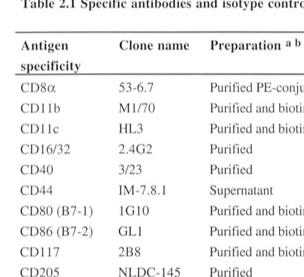
Specific antibody was absorbed to cells (2-5xl05) in a 100µ1 volume in 1.5ml
microcentrifuge tubes (Sarstedt). Details of the specific antibodies used are shown in Table 2.1. After incubation for 30 minutes on ice, cells were diluted with 250µ1 of staining n1edium and washed once using an underlay of 600µ1 FCS followed by
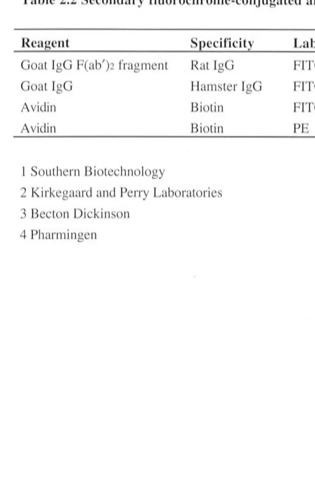
centrifugation at 300g for 10 minutes. Cells were then washed twice by resuspension in 1ml of staining medium and centrifugation at 300g for 5 minutes. The same incubation and washing procedure was repeated for addition of 100µ1 of the diluted fluorochrome-conjugated second stage reagent. Details of fluorochrome-fluorochrome-conjugated second stage