0022-538X/97/$04.0010
Copyrightq1997, American Society for Microbiology
Characterization of Two Temperature-Sensitive Mutants of
Coronavirus Mouse Hepatitis Virus Strain A59 with Maturation
Defects in the Spike Protein
WILLEM LUYTJES,* HELEEN GERRITSMA, EVELYNE BOS,ANDWILLY SPAAN
Department of Virology, Leiden University, 2333 AA Leiden, The Netherlands
Received 26 August 1996/Accepted 25 October 1996
Two temperature-sensitive (ts) mutants of mouse hepatitis virus strain A59, ts43 and ts379, have been described previously to be tsin infectivity but unaffected in RNA synthesis (M. J. M. Koolen, A. D. M. E. Osterhaus, G. van Steenis, M. C. Horzinek, and B. A. M. van der Zeijst, Virology 125:393–402, 1983). We present a detailed analysis of the protein synthesis of the mutant viruses at the permissive (31&C) and nonpermissive (39.5&C) temperatures. It was found that synthesis of the nucleocapsid protein N and the membrane protein M of both viruses was insensitive to temperature. However, the surface protein S of both viruses was retained in the endoplasmic reticulum at the nonpermissive temperature. This was shown first by analysis of endoglycosidase H-treated and immunoprecipitated labeled S proteins. The mature Golgi form of S was not present at the nonpermissive temperature for the tsviruses, in contrast to wild-type (wt) virus. Second, gradient purification of immunoprecipitated S after pulse-chase labeling showed that only wt virus S was oligomerized. We conclude that the lack of oligomerization causes the retention of thetsS proteins in the endoplasmic reticulum. As a result,tsvirus particles that were devoid of S were produced at the nonpermissive temperature. This result could be confirmed by biochemical analysis of purified virus particles and by electron microscopy.
Coronaviruses (31) are enveloped viruses with a large single-stranded RNA genome of approximately 30,000 nucleotides. They infect a range of mammals and birds, but the individual members of the Coronaviridae usually have narrow host spec-ificity (18). The best-studied coronavirus is mouse hepatitis virus (MHV), which serves as a model for the coronavirus replication cycle, including such aspects as entry, protein syn-thesis, RNA replication, and virus assembly.
Virus-specific components of MHV virions are four struc-tural proteins and RNA; the envelope is derived from the intermediate compartment (IC), which is the site of budding (13). The largest of the structural proteins is the surface pro-tein S, a membrane propro-tein that forms the characteristic coro-navirus spikes. S is responsible for attachment of virions to the host cell and plays a role in fusion of the viral envelope with the host cell membrane (33). Two other membrane proteins are important for virus particle formation and play a role in bud-ding: the membrane protein M and the small membrane pro-tein E. The fourth structural propro-tein, N, encapsidates the viral RNA. Except E, the structural proteins of MHV have been studied extensively. Much is known about the synthesis and maturation of particularly S and M, initially from studies of cells infected with wild-type (wt) or variant viruses. By using a range of different expression systems in the absence of virus, these proteins were studied in greater detail by mutagenesis of their cloned genes (reviewed in references 5 and 28). This approach was also taken to determine the functions of the structural proteins, but these experiments have the disadvan-tage of not being in the context of natural infections, and much remains to be elucidated. Only the nucleocapsid gene is
cur-rently subjectable to mutagenesis on the genomic RNA by a targeted RNA recombination protocol (24, 25).
Our current state of knowledge of the synthesis of the MHV structural proteins can be summarized as follows (for recent reviews, see references 4, 15, and 28). Membrane proteins S and M (probably also E, but no data are available) are synthe-sized on the rough endoplasmic reticulum (ER). Cotransla-tional N-glycosylation of the 120,000-molecular weight (120K) precursor of S yields a 150K form. M is posttranslationally O-glycosylated to an approximately 25K M protein. The 150K S protein folds with a half-time of approximately 20 min to its native state (22) and then associates with M, most likely before it oligomerizes (20). S oligomers are probably trimers, in ac-cordance with the properties reported for the surface protein of transmissible gastroenteritis virus (6). Oligomeric S-M com-plexes are transported out of the ER into the IC. Monomeric, misfolded S is retained in the ER. In the IC, nucleocapsids interact with the S-M complexes, and budding into the IC results in virions (13, 34). It is unknown whether nucleocapsids are preformed or associate during budding. Also, it should be noted that the E protein plays a crucial but to date unknown role in the budding process (2, 36). Virions are transported from the IC to the Golgi compartment, where S and M are further glycosylated to the 180K mature form of the spike protein and a range of M proteins. At the trans Golgi, virions are encapsulated into vesicles of the constitutive pathway, transported to the plasma membrane, and subsequently re-leased. During transport, a portion of S180 (depending on the MHV strain) is cleaved into two 90K forms: S2, which remains membrane bound; and S1, which is associated to S2.
Our laboratory is interested in the functions of coronavirus MHV-A59 S in attachment to the receptor and fusion of the virus envelope with the host cell plasma membrane. In a pre-vious investigation, we studied maturation and cleavage of expressed mutants of S, altered in the cleavage site. In addi-tion, S proteins with mutations in the transmembrane se-* Corresponding author. Mailing address: Department of Virology,
Leiden University, Albinusdreef 2, 2333 AA Leiden, The Netherlands. Phone: 71-526 1657/1652. Fax: 71-526 6761. E-mail: luytjes@virology .azl.nl.
949
on November 9, 2019 by guest
http://jvi.asm.org/
MHV-A59 ts mutants ts43 and ts379 were prepared as described previously (12). MHV-A59 wt, ts43, and ts379 were diluted in phosphate-buffered saline– DEAE–2% FCS and subsequently used to infect L cells at a multiplicity of infection (MOI) of 0.1. After 2 days of growth at 318C, virus was harvested. Virus titers were determined at 31 and 39.58C on L cells. MHV-A59 wt was prepared as described previously (17).
Isolation and analysis of viral RNAs.Dishes (10-cm2diameter) with L cells
were infected in duplicate with MHV-A59 wt, ts43, or ts379 at an MOI of 10 for 1 h at 318C. At t51, the inoculum was replaced by 1.5 ml of DMEM–3% FCS. One dish was placed at 318C, and the duplicate was placed at 39.58C. Intracel-lular RNA was isolated at t517 (318C) or t58 (39.58C) as described previously (32). RNA was separated on a denaturing 1% agarose gel as described previously (35). The gel was dried and hybridized to 100 ng of oligonucleotide 048 (35) which was 59-end labeled with [g-32P]ATP (NEN-Dupont) and T4
polynucle-otide kinase.
Isolation and analysis of viral proteins.Dishes (10-cm2diameter) with L cells
were infected in duplicate with MHV-A59 wt, ts43, or ts379 at an MOI of 10 for 1 h at 318C. At t51, the inoculum was replaced by 1.5 ml of DMEM–3% FCS; one dish was placed at 318C, and one was placed at 39.58C. At t54 (39.58C) or
t59 (318C), the medium was replaced by 500ml of DMEM lacking both cysteine and methionine. One hour later, 8ml of Expres35S label (10mCi/ml; NEN) was
added. Intracellular proteins were obtained by lysing the cells at t513 (318C) or
t57 (39.58C) in 300ml of radioimmunoprecipitation assay (RIPA) buffer (50 mM Tris [pH 7.5], 150 mM NaCl, 1% Nonidet P-40, 0.5% sodium deoxycholate, 0.1% sodium dodecyl sulfate). The lysates were cleared from nuclei and mem-brane fragments by centrifugation, and the proteins in supernatants were immu-noprecipitated in the presence of Pansorbin (Calbiochem) with polyclonal anti-body k134, directed against MHV-A59 whole virus, and separated on a 12.5% polyacrylamide gel as described previously (1).
For the analyses of the S protein, a portion of the cell lysates was immuno-precipitated with a mixture of two monoclonal antibodies (MAbs) against the spike protein of MHV-A59: A2.3 and A1.4, kindly provided by J. Fleming. Immunoprecipitation pellets were washed three times in RIPA buffer and dis-solved in 50ml of 50 mM Tris (pH 6.8)–0.25% sodium dodecyl sulfate. The samples were incubated at 958C for 5 min and cleared by centrifugation for 2 min. Ten-microliter aliquots of the supernatants were mixed with 15ml of 30 mM sodium acetate and incubated in the presence or absence of endoglycosidase H (endo-H; 1 mU/ml; Boehringer) for 16 h at 378C. The samples were separated on a 12.5% polyacrylamide gel as described above.
Sucrose gradient purification of virus.Dishes (10-cm2diameter) with L cells
were infected in duplicate with MHV-A59 wt, ts43, or ts379 at an MOI of 10 for 1 h at 318C. At t51, the inoculum was replaced by 1.5 ml of DMEM–3% FCS; one dish was placed at 318C, and the other was placed at 39.58C. At t53.5 (39.58C) or t511 (318C), the medium was replaced by 500ml of DMEM lacking cysteine and methionine. Thirty minutes later, 8ml of35S-Trans-label (10mCi/ml)
was added. At t514.5 (318C), the medium was replaced by 500ml of DMEM containing 400 mM methionine and 400 mM cysteine. Virions released into the medium were finally collected at t57 (39.58C) or t516.5 (318C) and mixed with 500ml of 20% sucrose in TESV (20 mM Tris [pH 7.4], 1 mM EDTA [pH 8.0], 100 mM NaCl). This mixture was loaded on top of a 20 to 50% linear sucrose gradient. The gradient was centrifuged for 16 h at 16,000 rpm at 48C in an SW40Ti rotor, and 16 fractions of 690ml were collected. The virus-containing fractions (particles migrated around a density of 1.18 g/ml) were located; three peak fractions were combined, and their volume was adjusted to 5 ml with TESV. Virus was pelleted by centrifugation in an SW55Ti rotor for 3 h at 35,000 rpm at 48C. The resulting pellet was dissolved in 30ml of Laemmli sample buffer and loaded directly on a 12.5% polyacrylamide gel.
Electron microscopy.Virus preparations were obtained by infection of 145-cm2dishes of L cells with an MOI of 10 at 318C for 1 h. At t51, the inoculum
was replaced by 15 ml of DMEM–3% FCS, and incubation continued at 39.58C. At t57, the medium was harvested and diluted 1:1 with 45 ml of TESV–20%
linear sucrose gradients (5 to 20% in MNT buffer with 0.1% Triton X-100) in SW55Ti tubes and centrifuged for 10 h at 45,000 rpm at 48C. Fractions of 300ml were collected from the gradients. Then 150ml of each fraction was mixed with 10ml of antispike MAb A2.3, incubated overnight at 48C, precipitated in the presence of 50ml of Pansorbin, and washed three times in RIPA buffer. The pellets were dissolved in 50ml of Laemmli sample buffer and loaded on a 12.5% polyacrylamide gel.
Thermal inactivation.Thermal inactivation of virus grown at the permissive temperature was done as described previously (11). Viruses were incubated in 50 mM Tris-HCl (pH 6.5)–100 mM NaCl–1 mM EDTA–10% FCS at 39.58C for different periods and then titered by plaque assay at 318C.
RESULTS
Our studies into the role of the coronavirus MHV-A59 sur-face protein S in the replication cycle have to date been limited to systems in which a set of S-gene mutants was expressed transiently in the absence of the virus (1). Awaiting a reverse genetics approach to study S on virions, we decided to set up a system in which our mutant S proteins would complement viruses with defective S genes. To this end, we have reexam-ined a panel of ts mutants of MHV-A59, isolated in 1983 by Koolen et al. (12), and characterized two viruses unaffected in RNA synthesis at the nonpermissive temperature.
Virus titers.The two selected ts viruses, ts43 and ts379, and wt virus were grown on L cells at the nonpermissive tempera-ture (39.58C), harvested after 8 h, and subsequently plaqued on L cells at the permissive temperature (318C). The virus titers were essentially the same as reported previously (12) and were at the nonpermissive temperature 1.23104(ts43), 2.83105 (ts379), and 108(wt) and at the permissive temperature 23108 (ts43), 1.83108(ts379), and 53108, (wt).
RNA synthesis.Next, the ability of each virus to produce RNA at the nonpermissive temperature was determined. L cells were infected with each virus at 318C and shifted to 39.58C after 1 h or kept at 318C. RNA was then isolated at 19 h postinfection (p.i.) (318C) or 7 h p.i. (39.58C) and separated on an agarose gel. The RNA was visualized by hybridizing the dried gel to oligonucleotide 048, which recognizes the nested set of MHV-A59 RNAs. Figure 1 shows that there is no major difference in accumulation of RNAs between the permissive and nonpermissive temperatures for each virus, confirming the RNA1phenotype. Also, it can be concluded that there are no apparent differences in RNA accumulation between the three viruses, which is also true for earlier time points (data not shown).
Analysis of virion proteins.We first analyzed whether virus particles were being formed at the nonpermissive temperature. To find these particles, L cells were infected for 1 h with ts43,
ts379, or wt virus at 318C and then grown at either 31 or 39.58C.
on November 9, 2019 by guest
At 11 h p.i. (318C) or 3.5 h p.i. (39.58C), the cells were labeled with [35S]methionine for 3.5 h. Virus particles from the me-dium of the cells kept at 39.58C were harvested directly, while the cells kept at 318C were chased for 2 more h and then virus particles were harvested. Virus particles were purified on su-crose gradients. Figure 2 shows the result of polyacrylamide gel electrophoresis (PAGE) of virus directly loaded on a gel. At the permissive temperature, the three major structural pro-teins, the membrane protein M, the nucleocapsid protein N,
and the surface protein S plus its 90K cleavage product, were easily detectable on wt and ts mutant virions, indicating normal virus production. In contrast, at the nonpermissive tempera-ture, only wt virions contained these proteins. The ts mutants lacked both forms of the spike protein yet contained large amounts of the nucleocapsid and membrane proteins. This result shows that the ts mutants produce virus particles in quantities comparable to wt virus, but these particles lack spike proteins.
Synthesis of the structural proteins.To determine at what stage of ts virus replication the addition of S was blocked, we continued to study the synthesis and maturation of the viral structural proteins at the nonpermissive and permissive tem-peratures. Even when RNA synthesis is at a maximum, at 318C (the permissive temperature), wt and ts mutant viruses each replicate at slightly different rates, which are approximately half the respective rates at 39.58C (the nonpermissive temper-ature [reference 12 and data not shown]). This thwarts a direct comparison of the levels of the proteins produced by these viruses at the time point of analysis but does not prevent us from ascertaining whether the viral proteins are correctly ma-tured. Virus-specific proteins from L cells infected with ts43,
ts379, or wt MHV-A59 were labeled from 9 to 13 h p.i.
(per-missive temperature) or 4 to 7 h p.i. (nonper(per-missive tempera-ture), and the lysates were immunoprecipitated.
Figure 3 shows the accumulation of the S, M, and N proteins immunoprecipitated with the polyclonal antibody k134. This antiserum will recognize all structural proteins of MHV-A59, including the 150K precursor of S180, but has a low affinity for the mature 180K form of the S protein. Expression of E is too low to be detectable by our antibodies. In contrast to the data presented by Koolen et al. (12), there was no significant dif-ference in accumulation of the structural proteins either be-tween the different viruses or bebe-tween the permissive and non-permissive temperatures in the lysates from infected cells. All three proteins accumulated to high levels. However, this
[image:3.612.381.489.69.275.2]anal-FIG. 1. Synthesis of viral RNA at the permissive and nonpermissive temper-atures. RNA from L cells infected with MHV-A59 or ts mutant viruses was harvested 17 (318C [permissive temperature]) or 8 (39.58C [nonpermissive tem-perature]) h p.i. and separated on a polyacrylamide gel. Dried gels were hybrid-ized to oligonucleotide 048, which binds to the 39end of positive-stranded viral RNA and recognizes the nested set of MHV RNA. At the top are shown the viruses used and the temperature at which they were allowed to replicate. The viral RNAs are indicated to the right.
FIG. 2. Analysis of virion proteins of wt and ts mutant viruses grown at the permissive and nonpermissive temperatures. Viral proteins from L cells infected with wt and ts mutant viruses were labeled from 11.5 to 14.5 h p.i. (318C), and virions were isolated from the medium at 16.5 h p.i. or labeled from 4 to 7 h p.i. (39.58C) and then isolated from the medium at 7 h p.i. Virions were purified on linear sucrose gradients and directly subjected to PAGE. Viruses and the growth conditions are indicated at the top of the gel; protein species are indicated to the right.
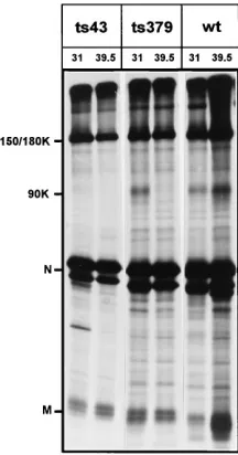
FIG. 3. Virus-specific proteins immunoprecipitated with polyclonal anti-serum k134. Viral proteins from L cells infected with wt and ts mutant viruses were labeled from 10 to 13 (318C) or 5 to 7 (39.58C) h p.i., isolated, immuno-precipitated with polyclonal antibody k134, and subjected to PAGE. Viruses and the growth conditions are shown at the top of the gel; protein species are indicated to the left. 150/180K, 150K ER form of S protein/180K Golgi form of S protein; 90K, 90K cleavage product of 180K S protein; N, nucleocapsid protein; M, membrane protein.
on November 9, 2019 by guest
http://jvi.asm.org/
ysis did not allow us to determine which form of S was accu-mulating, although interestingly, for ts379 and ts43, the 90K protein was present at the permissive temperature only. This cleavage product of 180K S can be present only when the mature 180K S protein is present. The 90K protein was clearly detectable for wt virus at both temperatures. Several additional bands (those not indicated by the markers) which may repre-sent breakdown products or aggregated proteins are detect-able. They are often observed when the polyclonal serum is used for immunoprecipitation.
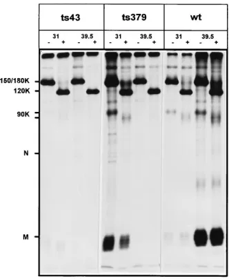
Maturation of the spike protein.In the previous experiment, accumulation of the spike protein was high at both the per-missive and nonperper-missive temperatures in the infected cell lysates. As mentioned, the polyclonal serum did not allow determination of the maturation species of the S protein. To study the fate of S in more detail, we used endo-H treatment, followed by immunoprecipitation with a mixture of anti-S MAbs A2.3 and A1.4, which are equally reactive to the differ-ent maturation forms of the S protein. Mature 180K spike proteins, on transport through the Golgi compartment, be-come endo-H resistant, whereas the ER, IC, and early Golgi 150K forms are endo-H sensitive. Since S150 and S180 are not resolved by PAGE, treatment with endo-H is necessary to detect the 180K form of S.
At the permissive temperature, all three viruses produced high levels of the 150K endo-H-sensitive form of the spike protein (Fig. 4). After endo-H treatment, most of the protein was shifted to the 120K deglycosylated form, and low amounts of 180K mature spike became detectable for each virus.
At the nonpermissive temperature, the differences in matu-ration of the S protein became evident. The 150K S and the 120K endo-H form of S were produced at the same levels as at the permissive temperature for each virus. However, only in the wt virus-infected cells was the mature 180K form of the spike protein detectable. No mature 180K S protein in the ts mutant virus-infected cells was detectable at the nonpermissive temperature. Also, the 90K cleavage product of 180K S was
Oligomerization of the spike protein.The difference in mat-uration of the S protein between wt and ts virus at the non-permissive temperature observed in the previous experiment suggested a transport defect for the ts mutant spikes. Several studies have suggested that an important prerequisite for transport of membrane proteins is their correct folding and oligomerization in the ER (7, 9). Therefore, we went on to study the oligomerization of the spike proteins at the nonper-missive temperature.
Proteins from L cells infected with wt and ts mutant viruses were pulse-labeled for 15 min and isolated or chased for 2 hours and then isolated. The proteins were separated on linear sucrose gradients, and 15 fractions were collected and sub-jected to PAGE. Figure 5A shows that the S monomers, la-beled during the short pulse, accumulated in fractions 5 to 7 for each virus. Two hours later, only the wt virus S proteins were oligomerized, as proven by a shift in migration from fractions 5 to 7 to the bottom of the gradient (Fig. 5B). Clearly, the ts mutant spike proteins remained in the same fractions of the gradient and thus did not form oligomers. The shifted S bands in the wt chase panel of Fig. 5B are double bands (only visible on a short exposure), representing 150K and 180K S. No 180K S was seen in any fraction of the ts mutant gradients. The chase experiment also revealed the presence of the 90K cleav-age products of 180K S in the wt fractions. They were absent in the ts mutant virus fractions. Additional bands were observed above and below the position of 150/180K S. As can be seen from the wt chase panel of Fig. 5B, most of these band did not shift in the gradient. This may indicate that they represent folding intermediates or aggregates of misfolded S proteins.
Electron microscopy.To determine the morphology of virus particles produced at the nonpermissive temperature from wt and ts mutant virus-infected L cells, sucrose-purified virus preparations were studied by electron microscopy (Fig. 6). L cells infected with either ts mutant produced exclusively virus particles devoid of the characteristic spike projections, con-firming the spikeless nature of the virus particles produced at 39.58C. wt virus-infected L cells produced normal virus parti-cles containing the spike corona, but these virions were in general highly pleomorphic, and up to 60% spikeless virus particles were seen in some wt preparations also (not shown).
Thermostability oftsvirions.Conditionally lethal mutations affecting conformations of surface proteins often render these proteins thermolabile. We tested whether this was the case for
ts43 and ts379 by incubating the viruses grown at the permissive
temperature at 39.58C for periods of up to 24 h. Surprisingly,
ts43 was unaffected by this treatment. Its titers dropped only by
a factor of 6, which was the same as for the control wt virus. However, ts379 appeared increasingly sensitive to
high-tem-FIG. 4. Endo-H sensitivity of S-protein species. Viral proteins from L cells infected with wt and ts mutant viruses were labeled from 10 to 13 (318C) or 5 to 7 (39.58C) h p.i., isolated, immunoprecipitated with a mix of two MAbs directed at the S protein (A2.3 and A1.4), treated with endo-H, and subjected to PAGE. Viruses and the growth conditions are shown at the top of the gel; protein species are indicated to the left.2, without endo-H treatment;1, with endo-H treat-ment; 120K, 120K endo-H-deglycosylated form of S.
on November 9, 2019 by guest
[image:4.612.95.261.72.274.2]perature incubation, resulting in at least a 5-log drop in infec-tivity after 6 h (Table 1). This drop in titer was not due to physiological conditions, because the titer of ts379 was un-changed when the virus was incubated at 08C (data not shown). These data indicate that the ts lesion in the two mutant viruses is essentially of a different nature.
DISCUSSION
[image:5.612.101.517.70.341.2]In this report, we describe two ts mutants of coronavirus MHV-A59, ts43 and ts379. The mutants are not impaired in RNA synthesis at the nonpermissive temperature (39.58C), nor is the synthesis of the membrane and nucleocapsid protein
FIG. 5. Oligomerization of the S proteins of the wt and ts mutant viruses at the nonpermissive temperature. S proteins from L cells infected with wt and ts mutant viruses at 39.58C were labeled for 15 min at 4 h p.i. and isolated (A) or additionally chased for 2 h and then isolated (B). Proteins were separated on a linear 5 to 20% sucrose gradient, and 15 fractions were immunoprecipitated with S-specific MAb A2.3 and subjected to PAGE (12.5% gel). Fraction numbers are indicated at the top (bottom of gradient51); protein species are indicated to the left.
FIG. 6. Electron micrographs of wt and ts mutant virions produced at the nonpermissive temperature. Virus was grown on L cells at 39.58C, harvested from the medium at 7 h p.i., and prepared for electron microscopy as described in Materials and Methods. The viruses are indicated at the top; the scale is shown at the bottom right.
on November 9, 2019 by guest
http://jvi.asm.org/
[image:5.612.90.530.489.698.2]affected. However, ts mutant virions produced at the nonper-missive temperature are devoid of the surface protein, because the S proteins expressed by these viruses fail to exit from the ER.
The S protein of wt MHV-A59 is synthesized and cotrans-lationally N-glycosylated in the ER to a 150K protein. Folding and oligomerization take place with a half-time of approxi-mately 1 h (21, 37), a process that likely occurs in the ER (6). Oligomerization of S is most likely required for the protein to be transported out of the ER, as it is for many other viral membrane proteins (7, 9, 27). Likewise, oligomerization itself is dependent on correct folding of the protein monomers (7). The membrane protein M forms a complex with S, probably with correctly folded S monomers before oligomerization oc-curs, since this interaction is detectable within 20 min of the onset of S synthesis (23). However, the M-S interaction is not required for S to be correctly oligomerized and transported to the cell surface. Studies in which S was transiently expressed by itself have indicated that its characteristics cannot be distin-guished from those of S proteins produced in viral infections (1, 5, 37). The S-M oligomeric complexes are incorporated into virions budding into the IC (13). S is further glycosylated to the mature 180K form in the Golgi compartment. The ts mutant S proteins at the nonpermissive temperature are lacking from virions and are unable to mature to the 180K Golgi state. The earliest event in the S protein synthesis pathway that we could show to be different from wt is the oligomerization step. Since this step is dependent on correct monomer folding, the ts defect of the S proteins at the nonpermissive temperature is most likely incorrect folding of the spike monomers. In many cases, misfolded proteins form aggregates, destined to enter the degradation pathway (9). There is no clear evidence for aggregated S proteins, as no significant additional bands are present in the cell lysates analyzed at the nonpermissive tem-perature in Fig. 3 and 4 and no additional bands were observed in the ts gradient fractions from Fig. 5. However, it is possible that these aggregates are not recognized by the antibodies used in the immunoprecipitations under the experimental condi-tions. A better indication for correct folding of MHV-A59 spikes is their interaction with M (21), detected by coprecipi-tation of the membrane protein from cell lysates when anti-S MAbs are used. In the ts mutant-infected cells, no such copre-cipitation is detectable at the nonpermissive temperature. Thus, we conclude that these S proteins are in a folding state that prevents interaction with M.
The fact that the two ts mutant viruses are differently sensi-tive to incubation at the nonpermissive temperature suggests that they contain different ts lesions. Preliminary data
corrob-stabilizing interactions with other viral proteins, particularly the membrane protein. There is a difference in affinity for M between the two ts mutant S proteins, as can be judged from the coprecipitation data. Experiments are under way in our lab to study ts S-M interactions in more detail.
Recently, a ts mutant of MHV-A59, ts18, with a mutation in the spike gene that was sensitive to incubation at 408C and that also produced spikeless virus particles was described (26). The surface protein was not characterized in detail; therefore, its phenotype cannot be linked to specific maturation defects. Fu and Baric (8) described several MHV-A59 ts mutants with mutations in the spike gene. For these mutants, only the loca-tion of the mutaloca-tion was mapped; the S phenotypes were not investigated.
The ts mutant viruses that we have characterized again show that coronaviruses can produce particles without spike pro-teins, as was observed previously (10, 29). Similar findings have recently been reported for rabies virus (19). Recent experi-ments have shown that coronavirus-like particles can be pro-duced from cells in which the viral structural proteins are expressed in the absence of the spike protein gene (2, 36). These findings, together with our observation that the ts S protein is retained in the ER, argue against the possibility that a membrane-embedded domain of ts S is left behind after an unspecified degradation process in the ER, to be incorporated in virions, as was shown for the surface protein of VSV tsO45 (20). Our analysis of virion proteins showed that there was no apparent difference in particle production at the nonpermis-sive temperature between wt and ts viruses when judged by the levels of the membrane and nucleocapsid proteins. This finding indicates that S plays no significant stimulatory role in virion production.
We are currently cloning, expressing, and sequencing both ts mutant S genes to locate the ts mutations. We plan to use the
ts mutant viruses in complementation studies with our panel of
mutant S genes (1). Such an approach has been used success-fully for other viruses, such as VSV (38, 39) and influenza virus (30), and will produce new insights in the role of the spike protein in the coronavirus life cycle.
ACKNOWLEDGMENTS
We thank J. Fleming for a kind gift of MAbs A2.3 and 1.4, Hans van der Meulen of the Laboratory for Electron Microscopy of the Leiden University for help with the electron microscope, and Guido van Marle, Richard Molenkamp, Jessika Dobbe, Jan Carette, and Linong Zhang for help and stimulating discussions.
W.L. is a fellow of the Royal Dutch Academy of Sciences. E.B. is supported by grant 901-02-148 from the Dutch Organization for Sci-entific Research.
on November 9, 2019 by guest
[image:6.612.58.300.99.190.2]REFERENCES
1. Bos, E. C. W., L. Heijnen, W. Luytjes, and W. J. M. Spaan. 1995. Mutational analysis of the murine coronavirus spike protein: effect on cell-to-cell fusion. Virology 214:453–463.
2. Bos, E. C. W., W. Luytjes, H. Van der Meulen, H. K. Koerten, and W. J. M.
Spaan.1996. The production of recombinant infectious DI-particles of a murine coronavirus in the absence of helper virus. Virology 218:52–60. 3. Carleton, M., and D. T. Brown. 1996. Events in the endoplasmic reticulum
abrogate the temperature sensitivity of Sindbis virus mutant ts23. J. Virol.
70:952–959.
4. Cavanagh, D. 1995. The coronavirus surface glycoprotein, p. 73–113. In S. G. Siddell (ed.), The Coronaviridae. Plenum Press, New York, N.Y. 5. De Groot, R. J., R. W. Van Leen, M. J. M. Dalderup, H. Vennema, M. C.
Horzinek, and W. J. M. Spaan.1989. Stably expressed FIPV peplomer protein induces cell fusion and elicits neutralizing antibodies in mice. Virol-ogy 171:493–502.
6. Delmas, B., and H. Laude. 1990. Assembly of coronavirus spike protein into trimers and its role in epitope expression. J. Virol. 64:5367–5375. 7. Doms, R. W., R. A. Lamb, J. K. Rose, and A. Helenius. 1993. Folding and
assembly of viral membrane proteins. Virology 193:545–562.
8. Fu, K., and R. S. Baric. 1994. Map locations of mouse hepatitis virus tem-perature-sensitive mutants: conformation of variable rates of recombination. J. Virol. 68:7458–7466.
9. Helenius, A., T. Marquardt, and I. Braakman. 1992. The endoplasmic re-ticulum as a protein-folding compartment. Trends Cell Biol. 2:227–231. 10. Holmes, K. V., E. W. Doller, and L. S. Sturman. 1981. Tunicamycin resistant
glycosylation of coronavirus glycoprotein: demonstration of a novel type of viral glycoprotein. Virology 115:334–344.
11. Koetzner, C. A., M. M. Parker, C. S. Ricard, L. S. Sturman, and P. S.
Masters.1992. Repair and mutagenesis of the genome of a deletion mutant of the coronavirus mouse hepatitis virus by targeted RNA recombination. J. Virol. 66:1841–1848.
12. Koolen, M. J., A. D. Osterhaus, G. Van Steenis, M. C. Horzinek, and B. A.
Van der Zeijst.1983. Temperature-sensitive mutants of mouse hepatitis virus strain A59: isolation, characterization and neuropathogenic properties. Vi-rology 125:393–402.
13. Krijnse Locker, J., M. Ericsson, P. J. Rottier, and G. Griffiths. 1994. Char-acterization of the budding compartment of mouse hepatitis virus: evidence that transport from the RER to the Golgi complex requires only one vesic-ular transport step. J. Cell Biol. 124:55–70.
14. LaFay, F. 1974. Envelope proteins of vesicular stomatitis virus: effect of temperature-sensitive mutations in complementation groups II and V. J. Vi-rol. 14:1220–1228.
15. Laude, H., and P. S. Masters. 1995. The coronavirus nucleocapsid protein, p. 141–179. In S. G. Siddell (ed.), The Coronaviridae. Plenum Press, New York, N.Y.
16. Lefkowitz, E. J., Pattnaik, A. K., and L. A. Ball. 1990. Complementation of a vesicular stomatitis virus glycoprotein G mutant with wild-type protein expressed from either a bovine papilloma virus or a vaccinia virus vector system. Virology 178:373–383.
17. Luytjes, W., H. Gerritsma, and W. J. M. Spaan. 1996. Replication of syn-thetic defective interfering RNAs derived from coronavirus mouse hepatitis virus-A59. Virology 216:174–183.
18. McIntosh, K. 1996. Coronaviruses, p. 1095–1133. In B. N. Fields, D. M. Knipe, P. M. Howley, et al. (ed.), Fields virology. Lippincott-Raven, Phila-delphia, Pa.
19. Mebatsion, T., M. Ko¨nig, and K.-K. Conzelmann.1996. Budding of rabies
virus particles in the absence of the spike glycoprotein. Cell 84:941–951. 20. Metsikko¨, K., and K. Simons.1986. The budding mechanism of spikeless
vesicular stomatitis virus particles. EMBO J. 5:1913–1920.
21. Opstelten, D. J. 1995. Envelope glycoprotein interactions in coronavirus assembly. Thesis. State University Utrecht, Utrecht, The Netherlands. 22. Opstelten, D. J., P. De Groote, M. C. Horzinek, H. Vennema, and P. J.
Rottier.1993. Disulfide bonds in folding and transport of mouse hepatitis coronavirus glycoproteins. J. Virol. 67:7394–7401.
23. Opstelten, D. J., M. J. Raamsman, K. Wolfs, M. C. Horzinek, and P. J.
Rottier.1995. Envelope glycoprotein interactions in coronavirus assembly. J. Cell Biol. 131:339–349.
24. Peng, D., C. A. Koetzner, and P. S. Masters. 1995. Analysis of second-site revertants of a murine coronavirus nucleocapsid protein deletion mutant and construction of nucleocapsid protein mutants by targeted RNA recombina-tion. J. Virol. 69:3449–3457.
25. Peng, D., C. A. Koetzner, T. McMahon, Y. Zhu, and P. S. Masters. 1995. Construction of murine coronavirus mutants containing interspecies chi-meric nucleocapsid proteins. J. Virol. 69:5475–5484.
26. Ricard, C. S., C. A. Koetzner, L. S. Sturman, and P. S. Masters. 1996. A conditional-lethal murine coronavirus mutant that fails to incorporate the spike glycoprotein into assembled virions. Virus Res. 39:261–276. 27. Rothman, J. E., and F. T. Wieland. 1996. Protein sorting by transport
vesi-cles. Science 272:227–234.
28. Rottier, P. J. M. 1995. The coronavirus membrane protein, p. 115–139. In S. G. Siddell (ed.), The Coronaviridae. Plenum Press, New York, N.Y. 29. Rottier, P. J., M. C. Horzinek, and B. A. Van der Zeijst. 1981. Viral protein
synthesis in mouse hepatitis virus strain A59-infected cells: effect of tunica-mycin. J. Virol. 40:350–357.
30. Sambrook, J., L. Rodgers, J. White, and M. J. Gething. 1985. Lines of BPV-transformed murine cells that constitutively express influenza virus hemagglutinin. EMBO J. 4:91–103.
31. Siddell, S. G. 1995. The small-membrane protein, p. 181–189. In S. G. Siddell (ed.), The Coronaviridae. Plenum Press, New York, N.Y.
32. Spaan, W. J. M., P. J. Rottier, M. C. Horzinek, and B. A. M. Van der Zeijst. 1981. Isolation and identification of virus-specific mRNAs in cells infected with mouse hepatitis virus (MHV-A59). Virology 108:424–434.
33. Sturman, L. S., and K. V. Holmes. 1985. The novel glycoproteins of coro-naviruses. Trends Biochem. Sci. 10:17–20.
34. Tooze, J., S. Tooze, and G. Warren. 1984. Replication of coronavirus MHV-A59 in sac-cells: determination of the first site of budding of progeny virions. Eur. J. Cell Biol. 33:281–293.
35. Van der Most, R. G., P. J. Bredenbeek, and W. J. Spaan. 1991. A domain at the 39end of the polymerase gene is essential for encapsidation of corona-virus defective interfering RNAs. J. Virol. 65:3219–3226.
36. Vennema, H., G. J. Godeke, J. W. A. Rossen, W. F. Voorhout, M. C.
Hor-zinek, D. J. E. Opstelten, and P. J. M. Rottier.1996. Nucleocapsid-indepen-dent assembly of coronavirus-like particles by co-expression of viral envelope protein genes. EMBO J. 15:2020–2028.
37. Vennema, H., P. J. M. Rottier, L. Heijnen, G. J. Godeke, M. C. Horzinek, and
W. J. M. Spaan.1990. Biosynthesis and function of the coronavirus spike protein. Adv. Exp. Med. Biol. 276:9–19.
38. Whitt, M. A., P. Zagouras, B. Crise, and J. K. Rose. 1990. A fusion-defective mutant of the vesicular stomatitis virus glycoprotein. J. Virol. 64:4907–4913. 39. Whitt, M. A., L. Chong, and J. K. Rose. 1989. Glycoprotein cytoplasmic domain sequences required for rescue of a vesicular stomatitis virus glyco-protein mutant. J. Virol. 63:3569–3578.