0022-538X/97/$04.0010
Copyrightq1997, American Society for Microbiology
Altered Rous Sarcoma Virus Gag Polyprotein Processing and
Its Effects on Particle Formation
YAN XIANG,1TODD W. RIDKY,1NEEL K. KRISHNA,2ANDJONATHAN LEIS1*
Department of Biochemistry, Case Western Reserve University School of Medicine, Cleveland, Ohio 44106,1and
Department of Microbiology and Immunology, Pennsylvania State University College of Medicine,
Hershey, Pennsylvania 170332
Received 10 July 1996/Accepted 26 November 1996
Proteolytic processing of the Rous sarcoma virus (RSV) Gag precursor was altered in vivo through the introduction of amino acid substitutions into either the polyprotein cleavage junctions or the PR coding sequence. Single amino acid substitutions (VP2S and PP4G), which are predicted from in vitro peptide substrate
cleavage data to decrease the rate of release of PR from the Gag polyprotein, were placed in the NC portion of the NC-PR junction. These substitutions do not affect the efficiency of release of virus-like particles from COS cells even though recovered particles contain significant amounts of uncleaved Pr76gagin addition to mature viral proteins. Single amino acid substitutions (AP3F and SP1Y), which increase the rate of PR release
from Gag, also do not affect budding of virus-like particles from cells. Substitution of the inefficiently cleaved MA-p2 junction sequence in Gag by eight amino acids from the rapidly cleaved NC-PR sequence resulted in a significant increase in cleavage at the new MA-p2 junction, but again without an effect on budding. However, decreased budding was observed when the AP3F or SP1Y substitution was included in the NC-PR junction
sequence between the MA and p2 proteins. A budding defect was also caused by substitution into Gag of a PR subunit containing three amino acid substitutions (R105P, G106V, and S107N) in the substrate binding pocket that increase the catalytic activity of PR. The defect appears to be the result of premature proteolytic processing that could be rescued by inactivating PR through substitution of a serine for the catalytic aspartic acid residue. This budding defect was also rescued by single amino acid substitutions in the NC-PR cleavage site which decrease the rate of release of PR from Gag. A similar budding defect was caused by replacing the Gag PR with two PR subunits covalently linked by four glycine residues. In contrast to the defect caused by the triply substituted PR, the budding defect observed with the linked PR dimer could not be rescued by NC-PR cleavage site mutations, suggesting that PR dimerization is a limiting step in the maturation process. Overall, these results are consistent with a model in which viral protein maturation occurs after PR subunits are released from the Gag polyprotein.
Translation of retroviral genomic RNA on free ribosomes in the cell cytoplasm leads to synthesis of two polyproteins, Gag and Gag-Pol, which migrate to the plasma membrane. This transport is directed by a signal located at the amino terminus of Gag referred to as the membrane binding (M) domain. In mammalian viruses, this signal includes an N-terminal myristic acid (4). In avian viruses, the signal is contained within MA
(32). The viralenvgene products are translated concomitantly
on the rough endoplasmic reticulum-associated ribosomes. They are modified posttranslationally during transport through the reticulum to the plasma membrane, where particles bud from the cell surface (18). Aggregation of Gag polyproteins (Gag and Gag-Pol) is necessary to form particles. This process is driven in part by the M domain concentrating Gag on the membrane but is assisted by an internal Gag interaction (I) domain that in Rous sarcoma virus (RSV) maps to the NC protein (28, 32). Efficient budding is also dependent on a domain required late in the budding process, referred to as the L domain (33). In RSV, the L domain maps to a proline-rich sequence, PPPPYV, in p2 of Gag. This sequence probably interacts with a cell protein containing a WW domain (35), a conserved motif that has two tryptophan residues separated by 20 to 22 amino acids. The formation and release of immature
virus particles from cells do not require proteolytic processing,
incorporation of viral RNA, orpolorenvgene products.
How-ever, the presence of the latter normal components alters the characteristics of particles released.
During or after the budding process, polyproteins are cleaved by a virus-encoded protease (PR) to release mature proteins (18). Concomitant with processing, there are morpho-logical changes in the appearance of particles released from cells, and virions become infectious (11, 15, 19, 26). This is the rationale for the development and use of antiprotease drugs in the treatment of AIDS (12, 17, 23). An active PR is composed of a dimer of two identical subunits, and Gag and Gag-Pol polyprotein processing requires PR to recognize at least nine unique cleavage sites of eight amino acids each (7, 9, 16, 23, 24, 27, 34). The molecular basis for this remarkable specificity is dependent primarily on optimization of van der Waals inter-actions between amino acid side chains in the polyprotein cleavage site and key amino acid residues in the enzyme’s substrate binding pocket (7, 23, 24).
In avian retroviruses, the PR subunit is found at the carboxyl terminus of Gag (Fig. 1). Cleavage at the NC-PR boundary is believed to be catalyzed by PR itself (3, 5) and seems to be required before processing of other viral protein junctions in Gag occurs (6). The timing of this activation is tightly regulated and appears to coincide with the process of virus budding. When budding is prevented by disruption of the Gag M do-main, the level of polyprotein processing is greatly diminished (2, 28). Particle formation is also aborted if PR is activated
* Corresponding author. Mailing address: Department of Biochem-istry, Case Western Reserve University, 2119 Abington Rd., Cleveland, OH 44106. Phone: (216) 368-3360. Fax: (216) 368-4544. E-mail: jx18 @biochemistry.cwru.edu.
2083
on November 9, 2019 by guest
http://jvi.asm.org/
prematurely, before Gag reaches the plasma membrane (2, 5). This is most likely because premature cleavage separates the Gag proteins from the M-domain transport signal. Similar re-sults were obtained with processing of human immunodefi-ciency virus type 1 (HIV-1) Gag polyproteins (20).
Mechanisms which delay PR activation until budding are not yet understood. To investigate this process of virus maturation, we have introduced a series of amino acid changes into Gag polyprotein cleavage sites that are predicted from cleavage data derived from peptide substrates to either increase or decrease their cleavage rates. Despite the fact that cleavage of the NC-PR site in vivo might be influenced by accessibility and/or conformation of the site in the polyprotein, we find that there is a correlation between the steady-state kinetic cleavage data of the NC-PR peptides in vitro and cleavage of the altered sites in vivo. Analysis of these sites, coupled with mutations
that change the intrinsic enzymatic properties of PR, has re-vealed that there is also a correlation between the rate of viral polyprotein processing and particle budding. Moreover, these data further support a model in which PR must be released from the Gag polyprotein before processing of other Gag cleavage sites occurs.
MATERIALS AND METHODS
Reagents.All reagents were as previously described (33). Oligodeoxynucleo-tides were purchased from Midland Certified Reagent Company (Midland, Tex.) and used directly for mutagenesis. The wild-type RSVgaggene is from pATV-8, an infectious molecular clone of the RSV Prague C strain. The plasmid pSV.Myr0 is a simian virus 40-based mammalian expression vector carrying a wild-type copy of the RSVgagallele, the product of which efficiently directs production of virus-like particles from COS-1 cells (2, 28, 30, 32, 33, 35).
Oligodeoxynucleotide-directed mutagenesis.Site-directed mutagenesis of the RSVgaggene was carried out by overlap extension mutagenesis as described by FIG. 1. Substitutions in the RSV Gag polyprotein. The RSV Gag polyproteins are represented by rectangular boxes. The vertical lines inside the boxes represent cleavage sites between the different proteins. A series of substitutions that change PR76gagare listed below the boxes and were expressed from mutant plasmids constructed by overlap extension mutagenesis (1) using oligodeoxynucleotides listed in Table 1. (A) Single amino acid substitutions in the NC-PR cleavage site. The eight-amino-acid sequence representing the NC-PR cleavage site is expanded, and the positions relative to the scissile bond are indicated byP4toP49. The full-length black line represents the wild-type (WT) NC-PR cleavage site in Gag. Amino acid substitutions introduced into theP4toP1positions are as indicated and are aligned with the expanded NC-PR cleavage site sequence. (B) Substitutions that change the sequence of the MA-p2 cleavage site to that of the NC-PR cleavage site. The amino acid sequence of the MA-p2 cleavage site is expanded, and the amino acid substitutions are indicated below. The one-amino-acid difference between MA-p23NC-PR and MA-p23NC(PP4G)-PR, MA-p23NC (AP3F)-PR, or MA-p23NC(SP1Y)-PR is indicated in boldface. (C) Substitutions in PR which alter its activity. Part of the amino acid sequence of RSV PR is expanded, and the numbers below indicate positions relative to the amino terminus of mature PR. Amino acid substitutions are as indicated. (D) Constructs that combine the RSV PR(S105-7) and NC-PR cleavage site substitutions. The altered PR is shown as PR* in Gag. All the other representations are as in panel A. (E) Deletions in Gag. The shaded areas represent deleted sequence. In theDNC construct, most of NC has been removed with the exception of seven amino acids from the amino and carboxyl termini. The positions of two copies of the I domain are also indicated. In theD(GSGL) construct, the p2-p10 cleavage junction is expanded. The shaded amino acids have been deleted; this region includes the junction between p2 and p10 (reverse arrow). Other notations are as in panel A. (F) Constructs that combine a linked PR dimer with NC-PR cleavage site substitutions. The four-glycine linker is shown between two PR subunits. All the other representations are as in panel A.
on November 9, 2019 by guest
http://jvi.asm.org/
Aiyar et al. (1). A list of mutations made is presented in Fig. 1. In most cases, the mutations create a new restriction enzyme site to facilitate identification of clones containing the desired mutation (Table 1). The mutantDNC was created by oligodeoxynucleotide-directed mutagenesis using single-stranded MGAG template DNA, containing the RSVgaggene (32), and a synthetic oligonucleo-tide having the sequence 59-GCAGTAGTCAATAGAGAGAGGCCTGAGCC ACCTGCCGTC-39. The deletion removes all of the NC coding sequence except that for the first and last seven residues. The mutation was inserted into the mammalian expression plasmid by transferring theBglII-BssHII fragment ofgag. The presence of the mutation was confirmed by DNA sequencing, and multiple clones of the mutant were characterized in transfection experiments.
Transfection of mammalian cells.COS-1 cells grown in Dulbecco’s modified Eagle’s medium supplemented with 3% fetal bovine serum and 7% calf serum (HyClone Inc.) were transfected by the DEAE-dextran-chloroquine method as described previously (30). Plasmid DNAs, at a concentration of 25mg/ml, were digested withXbaI and incubated with T4 DNA ligase before transfection. This removes the bacterial plasmid sequence and joins the 39end of thegaggene with the simian virus 40 late polyadenylation signal (30).
Metabolic labeling and immunoprecipitations.In most experiments, cells in 35-mm-diameter dishes were labeled 48 h after transfection withL-[35 S]methi-onine (1,000 Ci/mmol, 75mCi/ml of tissue culture medium) for 2.5 h at 378C as described previously (30). The cells or growth medium from each labeled culture was mixed with lysis buffer containing protease inhibitors. Rabbit antiserum directed against whole RSV (reactive with MA, CA, NC, and PR Gag proteins) was added, and immunoprecipitation was carried out for 2 h at 48C. Precipitates were collected by using protein A-agarose (Gibco-BRL), and the proteins were subjected to electrophoresis (35). Pulse-chase experiments were carried out with sets of identically transfected cells incubated in methionine-free medium for 30 min, pulse-labeled with [35S]methionine for 15 min, and chased with a 1,000-fold excess of cold methionine in serum-free medium for the indicated times. Den-sitometric quantitation of the fluorograms was carried out by using the Bioanaly-sis program on a Sci-Scan 5,000 (U.S. Biochemicals).
Isopycnic sucrose gradient analysis.COS-1 cells expressing theDNC mutant were radiolabeled, and the particles released into the medium were subjected to isopycnic sucrose gradient analysis as previously described (2). Unlabeled Molo-ney murine leukemia virus (MoMLV) was added to the gradient as an internal control and detected by reverse transcriptase activity.
SDS-polyacrylamide gel electrophoresis and fluorography. Immunoprecipi-tated proteins were separated by electrophoresis in sodium dodecyl sulfate (SDS)-polyacrylamide gels containing acrylamide monomer andN,N9 -methyl-enebisacrylamide at a ratio of 30:1.2. Resolving gels contained 12% acrylamide, 0.1% SDS, and 400 mM Tris-HCl (pH 8.8), while the stacking gels contained 3% acrylamide, 0.1% SDS, and 60 mM Tris-HCl (pH 6.8). After electrophoresis, gels were fixed in a solution of 7.5% methanol and 7% acetic acid. Radiolabeled proteins were detected by fluorography using Kodak BIO MAX MR film at room temperature. Overnight exposures were typically required.
In vitro protease assay.As described previously (23), RSV protease activity was assayed in a volume of 25ml of 100 mM sodium phosphate (pH 5.9), 2.4 M sodium chloride, 100mM peptide substrate, and 50 ng of PR. Reactions were initiated by the addition of protease, incubated at 378C for 3 to 15 min, and stopped by the addition of 300ml of 0.5 M sodium borate (pH 8.5). Twenty microliters of 0.05% (wt/vol) fluorescamine was then added. Relative fluores-cence intensity was measured on a Perkin-Elmer LS-50B luminesfluores-cence spectro-photometer, using an excitation wavelength of 386 nm and an emission wave-length of 477 nm. Each activity measurement represents the mean of at least
three independent experiments. In each case, the standard error for all experi-ments did not exceed 20% of the value reported. Kinetic constants were deter-mined by using the assay described above. Initial rate data from substrate satu-ration curves were fit to the Michaelis-Menten equation by using the NFIT program. Correlation coefficients of the fit were greater than 0.98, and the standard deviation of the constants reported was less than 20%.
Peptides.Peptide substrates, 9 to 12 amino acids in length and representing various RSV Gag polyprotein cleavage sites, were prepared as described previ-ously (24). These peptides include the eight amino acids, P4 to P49, required for efficient and specific cleavage by the retroviral protease. Peptides were solubi-lized in 1 mM dithioerythritol, and their concentrations were determined by quantitative amino acid composition analysis. The presence of Pro on the amino terminus renders the substrate nonreactive to fluorescamine; the inclusion of Arg residues on the carboxyl terminus improves solubility of the peptides (24).
Purification of soluble bacterially expressed mutant RSV proteases. Polyhis-tidine-tagged RSV protease was expressed inEscherichia coliM15 pDM1.1 as previously described (24). The active RSV protease is greater than 95% pure as judged by SDS-polyacrylamide gel electrophoresis and was obtained in a final yield of about 2 mg. The RSV PR(S105-107) and the covalently linked dimer protease were prepared as described for wild-type PR.
RESULTS
Single amino acid substitutions in the NC-PR cleavage site alter overall Gag processing.It was reported previously that a small deletion introduced into the RSV NC-PR junction of Gag blocked processing of all Gag cleavage sites (6). While this result suggests that processing of Gag cleavage sites is depen-dent on the release of PR from the polyprotein, this experi-ment could not exclude the possibility that the deletion pre-vented dimerization and activation of the PR subunits (27, 36). To examine more closely the relationship between cleavage of the NC-PR junction and initiation of viral polyprotein process-ing, we introduced single amino acid substitutions into the NC side of the NC-PR cleavage junction. These mutations are predicted from in vitro cleavage data (Table 2) to either in-crease or dein-crease the ability of the sequence to be cleaved by PR. The effects of these substitutions were analyzed in vivo by expressing the Gag alleles in COS-1 cells in the presence of
[35S]methionine, immunoprecipitating the viral proteins from
both the cell lysate and cell media, and fractionating the pre-cipitated protein by SDS-polyacrylamide gel electrophoresis (see Materials and Methods). The viral proteins detected in the media are contained in virus-like particles released from the cell surface.
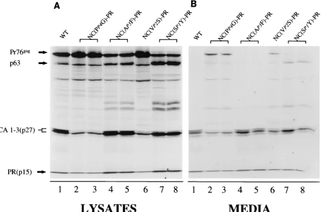
[image:3.612.62.555.82.239.2]Substitution of Gly for Pro in the P4 or Ser for Val in the P2 position of the NC-PR cleavage site results in an order of magnitude decrease in the ability of wild-type PR to cleave this
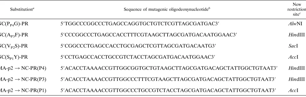
TABLE 1. Mutagenic oligodeoxynucleotides
Substitutiona Sequence of mutagenic oligodeoxynucleotideb restrictionNew
sitec
NC(PP4G)-PR 59TGGCCCGGCCCTGAGCCAGGTGCTGTCTCGTTAGCGATGAC39 AlwNI
NC(AP3F)-PR 59CCCGGCCCTGAGCCACCTTTCGTAAGCTTAGCGATGACAATGGAAC39 HindIII
NC(VP2S)-PR 59CGGCCCTGAGCCACCTGCGAGCTCGTTAGCGATGACAATG39 SacI
NC(SP1Y)-PR 59CCTGAGCCACCTGCCGTCTACCTAGCGATGACAATGGAAC39 AccI
MA-p23NC-PR(P4) 59ACACCTAAAACCGTTGGCGGTGCTGTAAGCTTAGCGATGACAGCTATTGGCTGTAAT39 HindIII
MA-p23NC-PR(P3) 59ACACCTAAAACCGTTGGCCCTTTCGTAAGCTTAGCGATGACAGCTATTGGCTGTAAT39 HindIII
MA-p23NC-PR(P1) 59ACACCTAAAACCGTTGGCCCTGCCGTCTACCTAGCGATGACAGCTATTGGCTGTAAT39 AccI
aSubstitutions in the Gag NC-PR and MA-p2 cleavage junctions are defined as in Fig. 1.
bOligodeoxynucleotides used to introduce substitutions into the RSV Gag NC-PR and MA-p2 junctions. cNew restriction sites were introduced to facilitate identification of mutated clones.
on November 9, 2019 by guest
http://jvi.asm.org/
sequence in vitro (Table 2). Introduction of either of these
substitutions into thegag allele resulted in a decrease in the
processing of all Gag polyprotein cleavage sites. This is de-tected most readily in the medium fraction (Fig. 2B), as seen by
the accumulation of Pr76gagand the concomitant decrease in
mature CA and PR bands (Fig. 2B; compare lanes 2, 3, and 6
with lane 1). This is also observed in the lysate fractions. While Gag processing was decreased, the overall rate of particle release from cells into the medium fraction was not affected. This is expected, because an active PR is not required for virus budding (31; see also Fig. 4B, lane 7). In a separate set of experiments, the ability to cleave the NC-PR junction was increased by substitution of Phe for Ala in P3 or Tyr for Ser in
P1 (Table 2). The AP3F substitution resulted in slightly faster
processing of Pr76gagin the lysate but again had little effect on
the overall particle release. In the case of the SP1Y
substitu-tion, there was a significant increase in processing of Pr76gagin
the lysate. This is seen by the dark band migrating with a molecular mass of 63 kDa (Pr63). This band corresponds to that expected for a Gag polyprotein lacking the PR subunit (Fig. 2A, lanes 7 and 8). Despite the fact that this substitution increased the ability to cleave the NC-PR cleavage site as a peptide substrate, the overall amount of particles released from cells was again similar to that of the wild type. Particles released from cells under these conditions, however, contained Pr63 in addition to fully processed proteins. These results support a model in which PR that dimerizes within the struc-ture of the polyprotein cleaves initially at the NC-PR junction. PR subunits must be released from the viral polyprotein before processing occurs at other polyprotein sites.
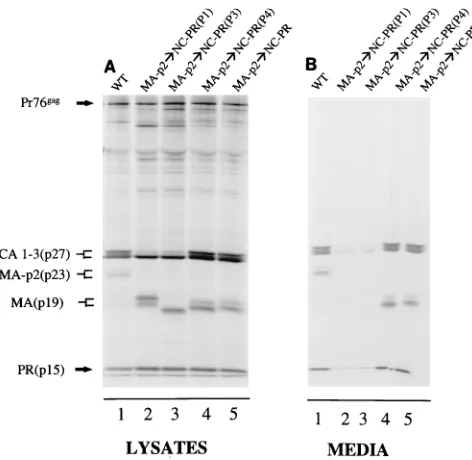
Replacing the MA-p2 cleavage junction in Gag with the amino acid sequence from the NC-PR cleavage junction. To determine whether cleavage of the NC-PR site and subsequent PR subunit release from Gag play a unique role in processing, we substituted the same set of NC-PR cleavage site sequences for the natural site between the MA and p2 proteins (Fig. 1B). The MA-p2 cleavage site is one of the slowest-cleaved sites in Gag and Pol analyzed both in vitro and in vivo (8). As a consequence, the MA protein is usually detected in virus-like particles released from cells as an MA-p2 fusion of approxi-mately 23 kDa (Fig. 3B, lane 1). When the more efficiently cleaved NC-PR is substituted for the MA-p2 cleavage junction, processing at this site is significantly increased in vivo. This is seen by the appearance of mature MA and the concomitant
[image:4.612.149.467.479.689.2]FIG. 2. Single amino acid substitutions in the NC side of the NC-PR cleavage site alter the overall processing of Gag. All labeling and protein detection were done as described in Materials and Methods. The positions of migration of Pr76gagand its cleavage products are indicated on the left. Substitutions in the NC-PR cleavage site of Gag are indicated above the lanes and are defined in Fig. 1A. In almost every case, two independent clones for each mutation were constructed and analyzed. WT, wild type.
TABLE 2. Steady-state kinetic activities of various RSV PRs on peptide substratesa
Protease Peptideb Name Km (mM)
kcat (min21)
kcat/Km (mM21 min21)
Wild type PAVS-LAMT NC-PR 56 16.4 0.3
GAVS-LAMTc P
P4G 64 2.9 0.04
PFVS-LAMT AP3F 5.4 14.9 2.8
PASS-LAMT VP2S 2,400 13 0.005
PAVY-LAMT SP1Y 24.5 244 5.73
TSCY-HCGTc MA-p2 427 8.8 0.02
RSV PR (S105-107)
PAVS-LAMT NC-PR 9 36.8 4.1
GAVS-LAMT PP4G 11 21.6 1.92
PFVS-LAMT AP3F 4 26.2 6.6
PASS-LAMT VP2S 6 4.3 0.7
TSCY-HCGT MA-p2 72 74 1
RSV PR-GGGG-PR
PAVS-LAMT NC-PR 43 7.7 0.18
GAVS-LAMT PP4G 56 0.72 0.01
PFVS-LAMT AP3F 8.2 5.5 0.67
aSteady-state kinetic parameters were determined with the wild-type, RSV
PR(S105-107), and PR-GGGG-PR covalently linked proteases, using peptide substrates as described in Materials and Methods.
bAll peptides contain two Arg residues not shown on the carboxyl terminus.
PAVS-LAMT and TSCY-HCGT represent the NC-PR and MA-p2 cleavage sites, respectively. Lines under residues indicate changes from wild-type se-quence.
cThese peptides contain an additional Pro residue on the amino terminus (not
shown).
on November 9, 2019 by guest
http://jvi.asm.org/
loss of the MA-p2 fusion band in the virus-like particles (Fig. 3B, lane 5). The doublets observed with MA-containing bands are due to partial phosphorylation (14, 21) of the protein in COS cells. While this site is processed more efficiently, there is
no detectable loss of particles released from cells. Moreover, when the mutation was cloned into the virus genome and analyzed in vivo, infectious virus particles, which spread throughout the cells in culture, were obtained (29).
The same single amino acid substitutions in NC that in-creased or dein-creased the rate of cleavage of NC-PR were then introduced into the substituted site at the MA-p2 junction. In contrast to the data shown in Fig. 2B (lanes 2 and 3), substi-tution of Gly in P4 did not prevent the processing of Gag (Fig. 3B, lane 4), nor did it affect the release of virus-like particles from cells. Thus, it is the substitution of Gly in P4 of NC-PR and the release of the PR subunits from the polyprotein that were responsible for the change in processing observed in Fig. 2. The combination of replacing MA-p2 with the NC-PR se-quence and modifying its P1 or P3 position to increase further PR cleavage resulted in a significant decrease in virus-like particles released from cells (Fig. 3B, lanes 2 and 3) compared to the wild type (lane 1). The loss of budding with the substi-tutions in P1 and P3 may be the result of the premature cleavage of the Gag polyprotein. Alternatively, it is possible that the P1 or P3 substitution directly influenced the budding process even though neither did so in the NC-PR junction at the wild-type position. Also, the wild-type MA-p2 junction sequence is not required for budding since it can be replaced by the NC-PR sequence (Fig. 3B, lane 5).
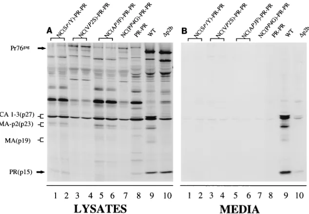
Gag polyproteins with modified PR subunits cause defects in budding.The relationship between Gag processing and for-mation of virus-like particles was further studied by introduc-ing an altered PR subunit containintroduc-ing substitutions R105P, G106V, and S107N into the Gag allele (Fig. 1C). This modified PR, termed RSV PR(S105-107), cleaves the RSV NC-PR and MA-p2 peptide substrates 9- and 50-fold faster, respectively, than the wild-type PR (Table 2). The PR(S105-107), like the wild type, must dimerize to form an active enzyme. When the PR(S105-107) substitution was introduced into Gag, a
de-FIG. 3. Moving the NC-PR cleavage site sequence to the junction between the MA and p2 proteins in Gag. The NC-PR wild-type (WT) and altered cleav-age site sequences have been moved to the junction between the MA and p2 proteins in Pr76gagas shown in Fig. 1B. Specific changes are indicated above the lanes. Viral proteins from the cell lysate and media were analyzed as described in the legend to Fig. 2. Migration positions of viral proteins are indicated on the left.
FIG. 4. Gag polyproteins with modified PR subunits have budding defects resulting from increased Gag processing inside the cell. Substitutions that alter the sequence of the PR subunit in Pr76gagare shown in Fig. 1C and are indicated above the lanes. A four-amino-acid deletion (GSGL), which disrupts the p2-p10 junction in Gag, is as in Fig. 1E. Viral proteins from the cell lysate and media were analyzed as described in the legend to Fig. 2. Migration positions of viral proteins are indicated on the left. WT, wild type.
on November 9, 2019 by guest
http://jvi.asm.org/
[image:5.612.60.296.67.297.2]crease in the level of virus-like particles released from cells was observed (Fig. 4B; compare lane 5 to lane 4). The defect can be rescued by inactivating the PR(S105-107) by substitution of a serine for the catalytic aspartic acid residue (Fig. 4B, lane 6). In addition to the defect in particle release, maturation of the carboxyl terminus of CA was incomplete. This is seen by the predominant CA1 band (22), representing CA plus a nine-amino-acid spacer sequence (Fig. 4A, lane 4).
The budding defect caused by the PR(S105-107) substitu-tion can be rescued by slowing the rate of cleavage at the
NC-PR junction.If cleavage of the NC-PR site is a prerequisite for PR-dependent processing of Gag as indicated in Fig. 2, then introduction of amino acid substitutions at the NC-PR cleavage site which slow down the release of the PR subunit are predicted to rescue defects observed with the RSV PR(S105-107) (Fig. 1D). When the RSV PR(S105-107)
substi-tution was combined with either the NC (VP2S)-PR or the
NC(PP4G)-PR substitution, wild-type levels of virus-like
parti-cles were released from transfected cells (Fig. 5B; compare lanes 1, 2, 7, and 8 to lane 9). Both of these substitutions are
[image:6.612.131.482.68.293.2]FIG. 5. The defect in budding observed with PR(S105-7) can be rescued by slowing down the rate of cleavage of the NC-PR junction in Gag. Constructs that combine the PR and NC-PR cleavage site substitutions are shown in Fig. 1D and are indicated above the lanes.D(p2b) deletes the 11 amino acids defined as p2b in Fig. 1A. This deletion disrupts the L assembly domain as previously described (34). Viral proteins from the cell lysate and media were analyzed as described in the legend to Fig. 2. Migration positions of viral proteins are indicated on the left. WT, wild type.
FIG. 6. Defects in particle release observed with a covalently linked PR dimer in Gag cannot be rescued by substitutions to the NC-PR cleavage site. Constructs that combine the linked PR dimer with the NC-PR cleavage site substitutions are as shown in Fig. 1F and are indicated above the lanes. Viral proteins from the cell lysate and media were analyzed as described in the legend to Fig. 2. Migration positions of viral proteins are indicated to the left of the panels. WT, wild type.
on November 9, 2019 by guest
http://jvi.asm.org/
[image:6.612.149.466.478.698.2]predicted to lower the rate of cleavage of the NC-PR junction (Table 2). Interestingly, while release of virus-like particles from cells was normal, removal of the spacer peptide from the end of CA was still defective (Fig. 5B; compare lanes 1, 2, 7, and 8 to lane 9). When the rate of cleavage of the NC-PR junction was increased by the introduction of either the
NC(SP1Y)-PR or the NC(AP3F)-PR substitution, a budding
defect that was more severe than that observed with RSV PR(S105-107) by itself was seen (Fig. 5B, lanes 3 to 6). As a control, we analyzed the ability of a small deletion mutation that inactivates the p2-p10 cleavage junction to rescue the budding defect observed with the RSV PR(S105-107) enzyme
(Fig. 1E). TheD(GSGL) deletion interferes with cleavage at
the p2-p10 junction (Fig. 4B, lane 3), resulting in a p33 band representing the MA-p2-p10 proteins detected in released
par-ticles from Gag alleles with wild-type PR. TheD(GSGL)
de-letion does not disturb the biological function of the adjacent L domain, a conserved amino acid sequence (PPPPYV) re-quired for budding of virus particles from cells (33, 35). Thus, wild-type levels of virus-like particles are released from cells
(Fig. 4B, lane 3). When theD(GSGL) deletion was combined
with the RSV PR(S105-107) substitution, there was no rescue of the particle defect (Fig. 4B, lane 1). Thus, altering a Gag polyprotein junction other than NC-PR has little effect on PR activation. Taken together, these results indicate that the bud-ding defect caused by PR(S105-107) is dependent on release of modified PR subunits from the Gag polyprotein.
Defective particle release observed with a covalently linked PR dimer cannot be rescued by substitutions in the NC-PR cleavage site.A budding defect is observed when a PR dimer linked by four glycine residues is introduced into the Gag allele (5). This enzyme, purified from the soluble fraction of bacterial cell lysates, has a specific activity with the reference NC-PR peptide substrate similar to that seen with wild-type PR (Table 2). However, in contrast to the results shown with RSV PR(S105-107) in Fig. 5, decreasing the cleavage rate of the
NC-PR site by introduction of either the NC(VP2S)-PR or the
NC(PP4G)-PR substitution (Fig. 1F) does not rescue the
as-sembly defect observed with the linked dimer (Fig. 6B, lanes 1 to 4). Increasing the cleavage rate of the NC-PR junction also had little detectable effect (Fig. 6B, lanes 5 to 8). These results suggest strongly that initial cleavage at the NC-PR junction, which is a requisite for PR dimerization, is the rate-limiting step in PR activation and subsequent polyprotein processing.
Combining RSV PR(S105-107) and covalently linked PRs with deletions of the Gag I domain. The I domain in Gag promotes aggregation of polyproteins during particle assembly (2, 10, 28) and is therefore predicted to influence the process of PR dimerization in the polyprotein. To test this hypothesis,
we made use of mutantDNC (Fig. 1E), which lacks Gag
resi-dues 496 to 570, including both copies of the I domain. Cells
FIG. 7. The I domains of Gag can facilitate dimerization of viral PR. (A) Expression ofDNC. Cells transfected with the indicated DNAs were metaboli-cally labeled as described in Materials and Methods. WT, wild type. (B) Particle density analysis ofDNC. Forty-eight hours posttransfection, COS-1 cells were labeled for 8 h in 1 ml with leucine-free, serum-free Dulbecco’s medium
sup-plemented with 500mCi ofL-[4,5-3
H(N)]leucine. The sample was spiked with unlabeled MoMLV (as an internal control [2, 28] for density of wild-type retro-virus particles) and loaded onto a 10 to 50% sucrose gradient, which was spun in an ultracentrifuge at 70,0003gfor 16 h. Fractions containing MoMLV (å) were identified by assaying for reverse transcriptase (RT) activity.DNC proteins (F) from each fraction were immunoprecipitated by using anti-RSV serum, subjected to electrophoresis on an SDS–12% polyacrylamide gel, and visualized by fluo-rography. The optical density (O.D.) of each band was determined by densitom-etry. The arrow indicates the direction of sedimentation. (C) Pulse-chase exper-iments with [35
S]methionine were carried out as described in Materials and Methods in cells expressing Gag polyproteins containing PR(S105-7) (h), PR-PR (E), PR(S105-7) plus theDNC mutation (■), or PR-PR plus theDNC mutation (F). TheDNC mutation is shown in Fig. 1E. The percentage of detectable labeled Gag was graphed as a function of chase time.
on November 9, 2019 by guest
http://jvi.asm.org/
transfected with this mutant released particles less efficiently than the wild type (Fig. 7A). Unlike previously described
I-do-main mutants of RSV (2, 28),DNC contains complete CA and
PR sequences. Since these proteins participate in interactions
within the virion, we determined whether theDNC clone
pro-duced particles of low rather than high density, the criterion used to define the effects of I-domain mutations (2, 28).
Anal-ysis ofDNC particles in sucrose density gradients revealed that
the particles were of low density and thus behaved as expected for I-domain mutants (Fig. 7B).
We then introduced theDNC deletion into Gag constructs
containing either RSV PR(S105-107) or covalently linked PR. Pulse-chase experiments were conducted to characterize the stability of Gag in whole-cell extracts, including released par-ticles (Fig. 7C). Under these conditions, Gag containing RSV PR(S105-107) or any of the covalently linked enzymes had an estimated half-life of 0.75 h. Introduction of the I-domain deletion into Gag containing the RSV PR(S105-107) enzyme
increased the half-life of Pr76gag about twofold, to 1.5 h,
whereas there was little effect when the deletion was intro-duced in combination with the covalently linked PR. While the differences observed are small, the same magnitude of effect was observed in two separate pulse-chase experiments. These results are consistent with the postulated role of the I domain in promoting Gag polyprotein interactions which facilitate viral PR dimerization.
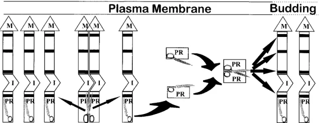
DISCUSSION
Processing of viral polyproteins by retroviral PR is a late event in replication which is required for production of infec-tious virus. If processing occurs too early, Gag proteins are separated prematurely from the amino-terminal M-domain transport signal. This prevents them from reaching the site of virus assembly and results in failure to release particles. The exact mechanism by which PR activation is delayed is not fully understood. A model presented in Fig. 8, which depicts this process, is based on previous as well as present studies. Pre-cursor processing appears to be initiated by release of PR subunits from the polyprotein. While a RSV PR dimer, as part of the Gag polyprotein, is capable of cleaving the NC-PR junction, processing of all other Gag cleavage sites requires release of PR subunits from the polyprotein. This is shown by the results of Burstein et al. (6) and findings reported here where inefficient cleavage of mutant NC-PR junctions delays subsequent processing of all other Gag cleavage sites. These mutations were placed in the NC side of the NC-PR junction and do not alter the catalytic properties of PR. Furthermore,
introduction of a catalytically enhanced PR [RSV PR(S105-107)], which has to dimerize, or a covalently linked PR dimer, which does not, results in premature processing of the Gag polyprotein. However, the budding defect created by the RSV PR(S105-107) is rescued by slowing release of the altered PR from Gag, whereas the defect caused by a covalently linked PR is not rescued. Similarly, it is reported that in HIV, precursor forms of PR are less efficient in Gag processing (37). Precursor processing is dependent on release of the PR subunit, probably because of the requirement for PR dimerization. This process occurs slowly in the polyprotein but rapidly with free PR sub-units. This observation is supported by recent studies of Sellos-Moura and Vogt (25) which demonstrate that recombinant RSV NC-PR fusion and PR proteins migrate on gels as mono-mer and dimono-mer, respectively.
In the processing model depicted in Fig. 8, a small amount of PR as part of the polyprotein has to dimerize to initiate the autoproteolytic processing. Whether the initial PR cleavage
occurs incis, with the PR dimer cleaving itself out of its own
polyproteins, or intrans, with the PR dimer cleaving PR
sub-units from different Gag polyproteins, is not known. However,
structural considerations make theciscleavage seem unlikely.
The initial dimerization process would be facilitated both by aggregation of the precursor polyproteins at the site of budding and through protein-protein interactions involving the I do-main (2, 28). It has been shown that in HIV, NC and p6 can facilitate PR dimerization (38). In this report, we find that an I-domain deletion, which interferes with Gag aggregation, can decrease the processing rate of Gag containing RSV PR(S105-107) but not that of Gag containing the covalently linked dimer. This suggests that I-domain-mediated interactions among Gag molecules facilitate the formation of PR dimers at the level of the polyprotein. The budding defect caused by premature PR activation is experimentally similar to the bud-ding defect observed by deletion of the Gag L domain in p2 (Fig. 5B, lane 10). It differs, however, in that the budding defect caused by premature PR activation can be rescued by inactivating PR (Fig. 4B, lane 6) whereas that caused by the L-domain mutation cannot (33).
[image:8.612.149.467.71.194.2]Is there a preferred cleavage order for the processing sites in Gag? The NC-PR site must be cleaved first, because cleavage of all other sites in the Gag polyprotein is dependent on re-lease of PR and assembly of active homodimers. In contrast, removal of the spacer peptide between the CA and NC pro-teins and cleavage at the MA-p2 junction appear to be late events, as polyprotein intermediates of MA-p2 and CA-spacer can be detected in newly budded particles from cells. Removal of the spacer peptide between the CA and NC proteins is
FIG. 8. Model for proteolytic processing of viral polyproteins. Gag polyproteins are represented by gray boxes. The PR cleavage sites in Gag are represented by thick black lines. The M domain and Gag-Gag I domain are indicated as M and I, respectively. The PR subunit is shown as a half scissors, while a dimerized PR is shown as a full scissors.
on November 9, 2019 by guest
http://jvi.asm.org/
correlated with the rate of particle budding. When the budding process is delayed, removal of the spacer is slowed concomi-tantly (22, 35), suggesting that the two processes are linked closely.
Substitutions introduced in the Gag p2-p10 cleavage site, which decreased its predicted cleavage rate, resulted in MA-p2-p10 fusion proteins being accumulated in particles (33). This occurred without an observable effect on overall Gag processing or particle release (Fig. 4, lanes 3). Changing the cleavage rate of the MA-p2 junction by more than 10-fold also results in little effect on particle budding except that mature MA is released more rapidly. That cleavage rates of Gag polyprotein sites can be altered by more than 10-fold without causing a biological defect in assembly underscores the re-markable flexibility built into the viral replication process. This wide tolerance allows for production of infectious virus even when polyprotein processing is altered by protease mutations conferring resistance to PR-directed drugs used to treat pa-tients with AIDS. This replication allows further mutations to develop at the cleavage sites that can partially compensate for mutated PR. One such case has recently been reported (13).
ACKNOWLEDGMENTS
We thank Terry Copeland, NCI-Frederick, for the preparation of peptide substrates, Beth Morrison for construction of the MA-p23NC-PR clones depicted in Fig. 1B, and A. M. Skalka for critical reading of the manuscript.
N.K.K. is supported by Public Health Service grant CA-47482 and American Cancer Society grant FRA-427 awarded to John W. Wills at Pennsylvania State University, in whose laboratory theDNC clone was constructed and analyzed. This work was supported in part by Public Health Service grants CA38046 and CA52047 from the National Can-cer Institute.
REFERENCES
1.Aiyar, A., Y. Xiang, and J. Leis.1995. Site-directed mutagenesis using over-lap extension PCR. Methods Mol. Biol.57:177–191.
2.Bennett, R. P., T. D. Nelle, and J. W. Wills.1993. Functional chimeras of Rous sarcoma virus and human immunodeficiency virus Gag proteins. J. Vi-rol.67:6487–6498.
3.Bizub, D., I. T. Weber, C. E. Cameron, J. P. Leis, and A. M. Skalka.1991. A range of catalytic efficiencies with avian retroviral protease subunits geneti-cally linked to form single polypeptide chains. J. Biol. Chem.266:4951–4958. 4.Bryant, M., and L. Ratner.1990. Myristoylation-dependent replication and assembly of human immunodeficiency virus 1. Proc. Natl. Acad. Sci. USA 87:523–527.
5.Burstein, H., D. Bizub, and A. M. Skalka.1991. Assembly and processing of avian retroviral Gag polyproteins containing linked protease dimers. J. Virol. 65:6165–6172.
6.Burstein, H., D. Bizub, M. Kotler, G. Schatz, V. M. Vogt, and A. M. Skalka. 1992. Processing of avian retroviral Gag polyprotein precursors is blocked by a mutation at the NC-PR cleavage site. J. Virol.66:1781–1785.
7.Cameron, C., B. Grinde, P. Jacques, J. Jentoft, J. Leis, A. Wlodawer, and I. Weber.1993. Comparison of the substrate-binding pockets of the Rous sarcoma virus and human immunodeficiency virus type 1 proteases. J. Biol. Chem.268:11711–11720.
8.Cameron, C. E., B. Grinde, J. Jentoft, and J. Leis.1992. Mechanism of inhibition of retroviral protease by a Rous sarcoma virus peptide substrate representing the cleavage site between Gag p2 and p10 proteins. J. Biol. Chem.267:23735–23741.
9.Cameron, C. E., T. W. Ridky, S. Shulenin, J. Leis, I. T. Weber, T. Copeland, A. Wlodawer, H. Burstein, D. Bizub-Bender, and A. M. Skalka.1994. Mu-tational analysis of the substrate binding pockets of the Rous sarcoma virus and human immunodeficiency virus-1 proteases. J. Biol. Chem.269:11170– 11177.
10. Campbell, S., and V. M. Vogt.1995. Self-assembly in vitro of purified CA-NC proteins from Rous sarcoma virus and human immunodeficiency virus type 1. J. Virol.69:6487–6497.
11. Crawford, S., and S. P. Goff.1985. A deletion mutation in the 59part of the
polgene of Moloney murine leukemia virus blocks proteolytic processing of thegagandpolpolyproteins. J. Virol.53:899–907.
12. Debouck, C.1992. The HIV-1 protease as a therapeutic target for AIDS. AIDS Res. Hum. Retroviruses8:153–164.
13. Doyon, L., G. Croteau, D. Thibeault, F. Polin, L. Pilote, and D. Lamarre. 1996. Second locus involved in human immunodeficiency virus type 1 resis-tance to protease inhibitors. J. Virol.70:3763–3769.
14. Erikson, E., J. Brugge, and R. Erikson.1977. Phosphorylated and nonphos-phorylated forms of avian sarcoma virus polypeptide p19. Virology80:177– 185.
15. Gelderblom, H. 1990. Morphogenesis, maturation, and fine structure of lentiviruses, p. 159–180.InL. H. Pearl (ed.), Retroviral proteases: control of maturation and morphogenesis. Stockton Press, New York, N.Y. 16. Grinde, B., C. Cameron, J. Leis, I. Weber, A. Wlodawer, H. Burstein, D.
Bizub, and A. M. Skalka.1992. Mutations that alter the activity of the Rous sarcoma virus protease. J. Biol. Chem.267:9481–9490.
17. Ho, D. D., A. U. Neumann, A. S. Perelson, W. Chen, J. M. Leonard, and M. Markowitz.1995. Rapid turnover of plasma virions and CD4 lymphocytes in HIV-1 infection. Nature373:123–126.
18. Katz, R. A., and A. M. Skalka.1994. The retroviral enzymes. Annu. Rev. Biochem.63:133–173.
19. Kohl, N. E., E. A. Emini, W. A. Schleif, L. J. Davis, J. C. Heimbach, R. A. Dixon, E. M. Scolnick, and I. S. Sigal.1988. Active human immunodeficiency virus protease is required for viral infectivity. Proc. Natl. Acad. Sci. USA 85:4686–4690.
20. Krausslich, H. G.1991. Human immunodeficiency virus proteinase dimer as component of the viral polyprotein prevents particle assembly and viral infectivity. Proc. Natl. Acad. Sci. USA88:3213–3217.
21. Leis, J., N. Phillips, X.-D. Fu, P. T. Tuazon, and J. A. Traugh.1989. Phos-phorylation of avian retrovirus matrix protein by Ca11 /phospholipid-depen-dent protein kinase. Eur. J. Biochem.179:415–422.
22. Pepinsky, R., J. A. Papayannopoulos, E. P. Chow, N. K. Krishna, R. C. Craven, and V. M. Vogt.1995. Differential proteolytic processing leads to multiple forms of the CA protein in avian sarcoma and leukemia viruses. J. Virol.69:6430–6438.
23. Ridky, T., and J. Leis.1995. Development of drug resistance to HIV-1 protease inhibitors. J. Biol. Chem.270:29621–29623.
24. Ridky, T. W., D. Bizub-Bender, C. E. Cameron, I. T. Weber, A. Wlodawer, T. Copeland, A. M. Skalka, and J. Leis.1996. Programming the Rous sarcoma virus protease to cleave new substrate sequences. J. Biol. Chem.271:10538– 10544.
25. Sellos-Moura, M., and V. M. Vogt.1996. Proteolytic activity of purified avian sarcoma and leukemia virus NC-PR protein expressed inEscherichia coli. Virology221:335–345.
26. Stewart, L., G. Schatz, and V. M. Vogt.1990. Properties of avian retrovirus particles defective in viral protease. J. Virol.64:5076–5092.
27. Weber, I. T.1990. Comparison of the crystal structures and inter subunit interactions of human immunodeficiency and Rous sarcoma virus proteases. J. Biol. Chem.265:10492–10496.
28. Weldon, R. A., Jr., and J. W. Wills.1993. Characterization of a small (25-kilodalton) derivative of the Rous sarcoma virus Gag protein competent for particle release. J. Virol.67:5550–5561.
29. Wills, J. W.Personal communication.
30. Wills, J. W., R. C. Craven, and J. A. Achacoso.1989. Creation and expression of myristylated forms of Rous sarcoma virusgagprotein in mammalian cells. J. Virol.63:4331–4343.
31. Wills, J. W., and R. C. Craven.1991. Form, function, and use of retroviralgag
proteins. AIDS5:639–654.
32. Wills, J. W., R. C. Craven, R. A. Weldon, Jr., T. D. Nelle, and C. R. Erdie. 1991. Suppression of retroviral MA deletions by the amino-terminal mem-brane-binding domain of p60src. J. Virol.65:3804–3812.
33. Wills, J. W., C. E. Cameron, C. B. Wilson, Y. Xiang, R. P. Bennett, and J. Leis.1994. An assembly domain of the Rous sarcoma virus Gag protein required late in budding. J. Virol.68:6605–6618.
34. Wlodawer, A., and J. W. Erickson.1993. Structure-based inhibitors of HIV-1 protease. Annu. Rev. Biochem.62:543–585.
35. Xiang, Y., C. E. Cameron, J. W. Wills, and J. Leis.1996. Fine mapping and characterization of the Rous sarcoma virus PR76gaglate assembly domain. J. Virol.70:5695–5700.
36. Zhang, Z. Y., R. A. Poorman, L. L. Maggiora, R. L. Heinrikson, and F. J. Kezdy.1991. Dissociative inhibition of dimeric enzymes. Kinetic character-ization of the inhibition of HIV-1 protease by its COOH-terminal tetrapep-tide. J. Biol. Chem.266:15591–15594.
37. Zybarth, G., H. G. Krausslich, K. Partin, and C. Carter.1994. Proteolytic activity of novel human immunodeficiency virus type 1 proteinase proteins from a precursor with a blocking mutation at the N terminus of the PR domain. J. Virol.68:240–250.
38. Zybarth, G., and C. Carter.1995. Domains upstream of the protease (PR) in human immunodeficiency virus type 1 Gag-Pol influence PR auto process-ing. J. Virol.69:3878–3884.