A STUDY ON CORRELATION
BETWEEN HYSTEROSCOPY AND
TRANSVAGINAL ULTRASONOGRAPHY
IN EVALUATION OF ABNORMAL
UTERINE BLEEDING
THIS DISSERTATION IS SUBMITTED FOR
MD DEGREE EXAMINATION
BRANCH II
OBSTETRICS AND GYNAECOLOGY
STANLEY MEDICAL COLLEGE
CHENNAI – 1.
Submitted to the Tamil Nadu
Dr. MGR MEDICAL UNIVERSITY CHENNAI
1.2 ACKNOWLEDGEMENT
I gratefully acknowledge and sincerely thank Dr.A.PRIYA, M.S., D.O. Dean Govt. Stanley Medical College and Hospital and Dr. S.CHITRA, M.D., PhD., Dean Govt. Stanley Medical College, Chennai for granting me permission to utilize the facilities of the institution for my study.
I am extremely grateful to our Superintendent, Prof Dr. C.VENI, M.D., D.G.O., D.N.B, Govt. R.S.R.M. Lying in Hospital, Chennai for the guidance and encouragement given in fulfilling my work.
I am also extremely grateful to Prof. Dr. Dr. S.CHITRA, M.D., PhD., H.O.D. Dept. of Pathology, Govt. Stanley Medical College & Hospital, Chennai for giving me permission to do the study.
I express my sincere thanks to Prof Dr. A.KALAICHELVI, M.D.D.G.O., D.N.B., Dr. N.HEPHZIBAH KIRUBAMANI, MD.D.G.O., Ph.D., and Dr. T.RUCKMANI, M.D.D.G.O., Professors of Obstetrics and Gynaecology Govt. R.S.R.M. Lying in Hospital for their guidance in carrying out this study.
I am also extremely grateful to all the Professors, and Assistants and my colleagues for their help in research work.
1.3 CERTIFICATE
This is to certify that the dissertation entitled “CORRELATION BETWEEN HYSTEROSCOPY AND TRANSVAGINAL ULTRASONOGRAPHY IN EVALUATION OF ABNROMAL UTERINE BLEEDING” is the bonafide original work of Dr. M. RAMYA, under the guidance of Dr. C.VENI, M.D., D.G.O., DNB,
Professor of Department of Obstetrics and Gynaecology, Stanley Medical College, Chennai in partial fulfillment of the requirements for MD (Obstetrics and Gynaecology) branch II examination of The Tamilnadu Dr. M.G.R. Medical University to be held in March 2010. The period of post graduate study and training was from May 2007 to February 2010.
Prof. Dr. C.VENI, M.D., D.G.O., D.N.B
Professor and Head
Department of Obstetrics and Gynaecology Stanley Medical College,
Chennai – 600 001.
The DEAN
1.4 CONTENTS
1.4.1 Sl.No Title Page No.
1. 1.5 INTRODUCTION 1
2. 1.6 REVIEW OF LITERATURE 3
3. 1.7 AIM OF THE STUDY 32
4. 1.8 MATERIALS & METHODS 33
5. 1.9 RESULTS & ANALYSIS 61
6. 1.10 DISCUSSION 67
7. 1.11 SUMMARY 70
8. 1.12 CONCLUSION 72
9. 1.13 PROFORMA
10. 1.14 BIBLIOGRAPHY
INTRODUCTION
Abnormal uterine bleeding (AUB) is defined as the changes in frequency of menstruation, duration of flow or amount of blood loss. It can occur in women of adolescent, reproductive, perimenopausal and postmenopausal age groups. In the reproductive women, regular menstruation is the norm and any departure is likely to be due to pregnancy, birth control methods, or hormone imbalance . During perimenopause, bleeding patterns are usually irregular. After menopause, the significance of bleeding depends to some degree on whether a woman is taking hormone replacement therapy (HRT), but postmenopausal bleeding is always a concern.
INCIDENCE:
REVIEW OF LITERATURE
Introduction
Various literatures have been reviewed to find out the best method for diagnosing the cause of AUB, the commonest gynaecological problem in women1.
Causes of AUB
The significant causes of AUB are endometrial abnormalities which run across the entire age spectrum. Various investigatory methods are available to find out the causes of AUB in these age groups. The terms used to describe patterns of AUB are based on periodicity and quantity of flow.
Terminologies of AUB 8
Term Definition
Menorrhagia Prolonged or excessive bleeding at regular intervals
Metrorrhagia Irregular, prolonged bleeding of varying amounts but not excessive
Menometrorrhagia Prolonged or excessive bleeding at irregular intervals Polymenorrhoea Regular bleeding at intervals of less than 21 days
Oligomenorrhoea Infrequent / Irregular bleeding, variable in amount, scanty in duration greater than 35 days
Amenorrhoea No uterine bleeding for at least 90 days.
AUB in adolescent age6
• Anovulation (90%): 80% of cycles are anovulatory in first year after menarche.
• Pregnancy related bleeding like spontaneous abortions, ectopic pregnancy.
• Hematological up to 20% presenting with menorrhagia have a bleeding disorder
e.g., Von Willebrands disease, Idiopathic Thrombocytopenic Purpura.
• Medical illnesses like hyperthyroidism (initial stages) hypothyroidism, Diabetes
mellitus, Tuberculosis etc.
• Pelvic inflammatory disease.
AUB in reproductive Age10
• Exogenous hormone use, pregnancy related bleeding, polyp, fibroids
adenomyosis, pelvic inflammatory disease, genital tuberculosis, dysfunctional uterine bleeding, bleeding disorders, endocrine disorders like thyroid dysfunction.
Perimenopausal AUB
Etiology: Anovulation, Anatomic (Fibroids, adenomyosis, polyps), Endometrial hyperplasia, endometrial cancer & functioning ovarian tumors
Post –menopausal AUB (30-50% are due to malignancy of genital tracts)8
Etiology: Endometrial carcinoma (5-10%), endometrial hyperplasia, fibroids, HRT, feminizing ovarian tumors, Hypertension, bleeding disorders.
Dysfunctional Uterine Bleeding (DUB)
reproductive age, but remains a diagnosis of exclusion9.
An understanding of normal menstruation is essential in investigating the complaint of AUB. The interval between the menstrual cycle, duration of flow and volume of flow remain relatively constant during woman’s reproductive years. In the first part of the cycle, estrogen halts menstrual flow and promotes endometrial proliferation. After ovulation, progesterone stops endometrial growth, and then promotes differentiation. If pregnancy doesn’t occur the corpus luteum regresses, progesterone production falls, the endometrium sheds its lining and menstrual bleeding follows.
The causes of DUB are related to one of three hormonal imbalance conditions, estrogen break through bleeding, estrogen withdrawal bleeding and progesterone breakthrough bleeding.
Peri-menopausal & post menopausal
AUB:-Erratic bleeding will be distressing; whatever may be its cause. Nearly 70% of peri and post menopausal women seek gynaecological care for the problem. Many women undergo hysterectomy to eliminate abnormal bleeding but better understanding has led to less invasive, more individualized approaches 9.
Perimenopause
During perimenopause, intervals between menstrual cycles may become longer and the amount of blood flow decreases and the duration of flow shorter. The main reason for this is fewer ovulations. Any deviation from the above pattern is abnormal and should be investigated.
Bleeding in menopausal women:-
without menstrual periods and women receiving HRT often have bleeding. Bleeding in any menopausal woman is considered abnormal and should be evaluated as the risk for endometrial cancer is 5-10%.
Endometrial abnormalities seen in
AUB:-Normal Endometrium
SECRETORY ENDOMETRIUM
PROLIFERATIVE ENDOMETRIUM
COMPLEX ATYPICAL HYPERPLASIA
ADENOCARCINOMA
Uterine leiomyoma
many women, it is estimated to be clinically significant in at least 25%.
Endometrial polyp:-
It is usually associated with endometrial hyperplasia. They are seen in 10% of post-menopausal bleeding. It is present as intermenstrual bleeding or menometrorrhagia. Diagnosis is based on either visualization with hysteroscopy or sonohysterography or microscopic assessment of tissue obtained by biopsy.
Endometrial hyperplasia
It represents an exaggerated, proliferative response of the endometrial glands and stroma due to prolonged, unopposed estrogen stimulation. In most cases, benign
endometrial hyperplasia is resolved with D&C or progestin therapy. This accounts for 5-10% of AUB in perimenopausal and postmenopausal women19.
Kurman et al classification
Classification type Progression to cancer
Simple(cystic without atypia) 1% Complex (adenomatous without atypia) 3% Atypical(cystic with atypia) 8% Atypical complex(adenomatous with atypia) 29%
Endometrial carcinoma:-
ENDOMETRIAL CARCINOMA
Methods of endometrial assessment in AUB Endometrial cytology
• Pap smear (only 30-50% of patients with endometrial cancer have abnormal pap test)
• Aspiration cytology
Endometrial Biopsy
• Novak curette
• Pipelle or vabra aspirator
Dilatation & curettage Imaging technique
1. Trans abdominal ultrasonography 2. Endovaginal ultrasonography 3. Hysterosalpingography 4. Hydrosonography 5. Hysteroscopy
6. Other procedures like color flow Doppler of endometrium and MRI
Endometrial
Biopsy:-This is an outpatient procedure. Devices like Pipelle or Vabra aspirator can be used. They yield adequate samples in about 90% of the cases.
Dilatation & Curettage
Even though dilatation and curettage has been employed to sample and study endometrium in women with dysfunctional uterine bleeding, it is proved that only 50-60% of the endometrium can be studied10.
Hysterosalpingography (HSG)
It has been traditionally used to diagnose uterine abnormalities but the
disadvantages of using these methods include less than optimal sensitivity, radiation exposure, inability to differentiate submucous fibroid from polyp and inability to visualize myometrial and adnexal abnormalities12.
Hysteroscopy
History & development
The hysteroscopy that is being used today has evolved over the past 175 years. In 1805 Bozzini constructed a device called a ‘light conductor’ to inspect various passages and body cavities. The first hysteroscopy was described in 1869 by PANTALEONI24. He used reflected candle light from a concave mirror to illuminate the uterine cavity. The hysteroscopy used by Pantaleoni was similar to an instrument developed by Desormeaux in 1865 for examination of urinary bladder. Now hysteroscopy has developed into an important minimal invasive procedure to diagnose and treat intracavitary abnormalities.
The uterine cavity is distended with ringer lactate. TYPES:
• MICRO HYSTEROSCOPE
• CONTACT HYSTEROSCOPE
HYSTEROSCOPIC VIEW OF NORMAL ENDOMETRIUM
VARIOUS HYSTEROSCOPIC VIEWS OF UTERINE CAVITY
ENDOMETRIUM-POLYP
ENDOMETRIOTIC-NODULE
ENDOMETRIUM - HYPERPLASIA
L – Left cornua S – Septum
TRANSVAGINAL ULTRASOUND – SUBMUCOUS FIBROID
As stated by VALLE, hysteroscopy is indicated in any situation in which intrauterine visualization will enhance diagnostic accuracy and define therapy.
INDICATIONS FOR DIAGNOSTIC HYSTEROSCOPY24
1) Unexplained abnormal uterine bleeding
• Reproductive • Premenopausal • Post menopausal
2) Selected infertility cases
• Abnormal hysterography or transvaginal ultrasonography • Unexplained infertility.
3) Recurrent Spontaneous abortions.
INDICATIONS FOR THERAPEUTIC HYSTEROSCOPY
• Adhesiolysis
• Division of uterine septum
• Resection of Myomas
• Endometrial destruction through Nd: YAG Laser vaporization or radio
• Removal of foreign bodies.
• To position occluding devices in the fallopian tube for sterlization.
Only diagnostic hysteroscopy is dealt in this study.
CONTRAINDICATION OF HYSTEROSCOPY
i. Pregnancy
ii. Active or recent endometrial infections iii. Menstruation is a relative contra indication iv. Patient with cardiac and pulmonary disease
TIMINGS OF HYSTEROSCOPY
Studies should be preferably done in postmenstrual phase of menstrual cycle.
COMPLICATIONS OF HYSTEROSCOPY
Adverse effects resulting from diagnostic hysteroscopy are few compared to those following its therapeutic use. Faulty technique and selection of inappropriate patients are most frequent causes of untoward sequelae.
1. TRAUMA
Cervical laceration can result from rough manipulation by holding the cervix with tenacula. Forceful dilatation provokes unnecessary bleeding. Uterine perforation and thermo intestinal accidents can occur.
2) INTRAVASATION
Endometrial tuberculosis, sub mucous tumors, hypo plastic uterus and proximal tubal obstruction-are predisposing factors to venous intravasation. The risk of pulmonary embolism is very minimal.
3) INFECTION
4) MORTALITY
Due to faulty technique of co2 insufflation. It is a very rare complication.
ENDOMETRIAL STUDY BY HYSTEROSCOPE AT RSRM HOSPITAL
PROLIFERATIVE ENDOMETRIUM
• The surface is smooth and the color is white or yellow
• Height of endometrium is 2-5mm
• Pores of endometrial glands are seen and are situated regularly
• Superficial vascularisation forms are relatively poor and are seen as
interrupted and punctuate lines • Tubal ostia are normal
SECRETORY ENDOMETRIUM
The surface is smooth or slightly rough. The color varies from yellow to pink.
Height of the endometrium is 4-5mm
Superficial vessels have typical geometrical pattern mimicking a net.
Tubal ostia are normal
NATURAL ATROPHY
• Surface is smooth and appears as white or yellow
• Height of the endometrium is less than 1mm
• Visible glandular openings are absent
• There is complete absence of superficial vessels though deeper vessels of stroma
can be seen
INDUCED ATROPHY
• The surface is rough and the color is ochre • Height is 1 to 2 mm
• Visible glandular pore are absent
• Superficial vessels are inadequate but deeper stromal vessels are seen • Tubal ostia show characteristic atrophy
COMPLEX HYPERPLASIA
• This presents with a rough surface and the color is variable from white to yellow or even pink
• The height of the endometrium is uneven and usually very thick
• Rich superficial vascularisation with no specific pattern is seen Endoscopic
examination easily provokes hemorrhage
• Though some glandular orifice can be seen, they are no longer well delineated and
the regular disposition is lost • Tubal ostia are normal
SIMPLE HYPERPLASIA
• As with complex hyperplasia , the surface is rough and the colour varies from
white to yellow or even pink
• The height of endometrium is uneven and thick. Rich superficial
vascularisation with network appearance are seen
• Trapped in the meshes of the net are several transparent cysts which often
PSEUDO DECIDUALISATION
• The surface is rough
• Height of endometrium is variable and has pseudopolypoidal appearance
• Rich congestive vascularisation is noted as in secretory phase
• Visible glandular pores are absent
• Tubal ostia are absent
ENDOVAGINAL SONOGRAPHY
Normally the uterine cavity is empty and hence it is represented by the two layers of the endometrium in apposition and the sonographic appearance is that of a clear echogenic line.
MENSTRUAL ENDOMETRIUM
The endometrial cavity is filled with small echo free spaces suggesting the presence of blood clots. The endometrial lining will be thin and less echoic.
PROLIFERATIVE ENDOMETRIUM
The growth of the endometrial glands and stromal edema is identified in the form of gradually increasing thickness of the endometrium. During the mid-proliferative phase the hypo-echoic area surrounded by echogenic line widens due to increasing stromal edema. In the impending pre-ovulatory period the stromal edema is maximal. This results in significant widening of echo free zone and intense echogenicity of the endometrial lining. The endometrial thickness increases gradually from 2-3 mm in early proliferative phase to upto 8-10 mm in late proliferative phase.
SECRETORY ENDOMETRIUM
During the luteal phase, the secretions of glands are discharged into the lumen of the
endometrial cavity. This is recognized at transvaginal scan in the form of an echo-free space being lined by thick and relatively echogenic endometrium. During mid-secretory phase there is regression of endometrial glandular elements and prominence of stromal cells. This leads to increased thickness of the endometrium which becomes more echogenic. During the late secretory phase there is prominence of stromal cells which give a strong echogenic fluffy appearance to the endometrium and also adds to the thickness of the endometrium. The endometrial thickness can increase to 10-14 mm during the secretory phase.
HYPERPLASTIC ENDOMETRIUM
Hyperplastic endometrium has characteristic picture by sonography. The intensely echogenic and thick endometrium surrounded by echo free periphery is quite diagnostic. The endometrium
thickens and become pseudopolypoid in configuration. In these patients, thickening of the endometrium beyond what is expected for women of comparable age is detected. In perimenopausal women
AIM OF THE STUDY
• To compare the efficacy of hysteroscopy and transvaginal ultrasound in diagnosing endometrial pathology in AUB .
• To correlate the hysteroscopy and sonography findings with histopathological
MATERIALS AND METHODS
This is a prospective study conducted at Government RSRM Hospital. 150 patients were taken up randomly from those who are attending the gynaecological outpatient department in the age group 25-50 years with following complaints
Menorrhagia, polymenorrhoea, amenorrhoea, metrorrhagia and oligomenorrhoea
INCLUSION CRITERIA
• Only parous women of age groups 25-50 years
• Patients without medical illness and patients with diabetes and hypertension
under control
• Fair, general condition
• In postmenstrual phase of menstrual cycle
EXCLUSION CRITERIA
• Hemoglobin level <8 g/dl
• Pelvic infection
• Profuse bleeding per vaginum
Thorough history taking (as per proforma) was done, general examination, systemic examination, P/A, P/S and bimanual pelvic examination were done. Baseline investigations like hemoglobin, Urine-albumin, Sugar, Deposits, electro cardiography.
• Informed consent for the procedure & anaesthetic assessment for
hysteroscopy and D& C obtained.
• Patients are advised to have light dinner before 10 PM on night prior to
hysteroscopy
• Preparation done as for other surgical procedures
• The next day an endovaginal USG is performed on all patients prior to
hysteroscopy
ENDOVAGINAL USG
Done with 5 mega hertz curvilinear probe. After emptying the bladder the patient lies down in the dorsal position, the probe covered by condom was inserted into the introitus. After gentle separation of labia majora by examiners fingers and gentle downward pressure on posterior commisure of labia minora saggital and coronal with transverse sections of uterus were viewed and findings noted.
FINDINGS
1) Thickness of endometrium
3) Other uterine or adnexal pathology which could not be detected clinically. After endovaginal USG patient is wheeled to theatre where hysteroscopy and Dilatation and Curettage were performed
HYSTEROSCOPY
In this study hysteroscopy is performed under intravenous ketamine anesthesia.
ANAESTHESIA
ROUTE
• Intravenous
DRUGS USED
1. Ketamine hydrochloride (2mg/kg) 2. Diazepam 10mg
3. Atropine 0.6mg
DURATION
15-20 minutes
INSTRUMENTS
• Telescope –eye piece and object lens
Diagnostic sheath required to deliver distending media inside uterine cavity. Ringer lactate is used as distending media. Speculum, Valsellum, Sponge holding forceps
Under anaesthesia after catheterizing the bladder a bimanual pelvic examination was done. After introducing Sim’s speculum the anterior lip of the cervix was caught with valsellum. After measuring length of the uterine cavity the internal os is dilated upto 7 Hegars dilator which was sufficient in most of the patients. Hysteroscope introduced into cervical canal under vision. The inflatable cuff surrounding the medium was inflated to 100 mm of Hg & maintained between 80-100 mm Hg. The drip set attached to the distending medium was attached to the inflow channel of the hysteroscope. The uterine cavity is examined and the following findings are
noted:-• Endometrial surface, color
• Vascularity
• Glandular pores
Any blood clots present in the cavity will be rinsed off by the fluid used as
distending medium. The time taken for the procedure and amount of fluid used are noted carefully at the end of the procedure.
Abnormalities like
• Endometrial hyperplasia
• Sub mucous fibroid
• Endometrial atrophy
• Neoplasia
• Synechiae
• AV malformations
• Polyp
• Hemangioma are visualized.
Advantages
• Direct visualization of pathology
• Accurate localization of lesion
• To take biopsy from lesion (good volume of tissue obtained)
Dilatation and Curettage
After the procedure was over the patients were shifted to post operative ward and kept under observation for 24 hours.
[image:35.612.115.420.244.361.2]TABLES AND CHARTS
Table 1
DISTRIBUTION OF AUB IN RELATION TO AGE Age No. of patients Percentage %
25-29 15 10
30-34 45 30
35-39 37 24.6
40-44 27 18
45-50 26 17.4
Total 150 100
Majority of patients with AUB were 35 years & above (>60%) Figure 1
25-29 30-34 35-39 40-44 45-50 0
5 10 15 20 25 30
10
30
24.6
18 17.4
Percentage %
Table 2
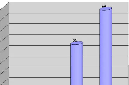
DISTRIBUTION OF AUB IN RELATION TO PARITY No. of patients Percentage %
Nullipara 6 4
Para1 9 6
Para2 42 28
Para3 66 44
Para4 & above
27 18
Total 150 100
[image:36.612.100.516.101.369.2]Figure 2
DISTRIBUTION OF AUB IN RELATION TO PARITY
Nullipara Para1 Para2 Para3 Para4 & above 0
5 10 15 20 25 30 35 40 45
4
6
28
44
18
Table 3
DISTRIBUTION OF AUB IN RELATION TO SOCIO-ECONOMIC CLASS(SE CLASS)
No. of patients Percentage%
SE class iii 6 4
SE class iv 18 12
SE class v 126 84
Total 150 100
84% of the patients belong to class V socio-economic status. Figure 3
DISTRIBUTION OF AUB IN RELATION TO SOCIO-ECONOMIC CLASS(SE CLASS)
SE class iii SE class iv SE class v Total
[image:38.612.107.536.459.731.2]Table 4
TYPE OF BLEEDING
No. of patients Percentage
Menorrhagia 96 64
Metrorrhagia 16 10.67
Menometrorrhagia 18 12
Polymenorrhoea 20 13.33
TOTAL 150 100
Menorrhagia was the commonest complaint found in 64% of study group. Figure 4
Menorrhagia Metrorrhagia Menometrorrhagia Polymenorrhoea 0
10 20 30 40 50 60
70 64
10.67 12
13.33
[image:40.612.75.548.84.650.2]Percentage
Table 5
DURATION OF COMPLAINT
Duration No. of patients Percentage
<3 months 27 18
3-5 months 27 18
6-9 months 54 36
1-2 years 30 20
>2 years 12 8
150 100
Figure 5
DURATION OF COMPLAINT
<3 months 3-5 months 6 - 9 months 1-2 years >2 years 0
5 10 15 20 25 30 35 40
18 18
36
20
8
Table 6
ASSOCIATED MEDICAL CONDITIONS
[image:42.612.58.511.304.580.2]Anaemia and Diabetes was seen in 18 % of the study group, Hypertension was seen in 20% of study group.
Figure 6
ASSOCIATED MEDICAL CONDITIONS
Anaemia Hypertension Diabetes mellitus Asthma Hypothyroidism 0
2 4 6 8 10 12 14 16 18 20
Table 7
FINDINGS IN HISTOPATHOLOGIC EXAMINATION
Age Proliferative Secretory Simple Complex
25-29 10 (6.6%) 5 (3.3%) -
-30-34 12 (8%) 32 (21.3%) 1 (0.7%)
-35-39 15 (10%) 20 (13.3%) 2 (1.3%)
-40-44 3 (2%) 2 (1.4%) 22 (14.7%)
-45-50 - 1 (0.7%) 24 (16%) 1 (0.7%)
Total 40 (26.6%) 60 (40%) 49 (32.7%) 1
• Among 26.6% of proliferative endometrium reported by Histo Pathologic
Examination, 24.6% was present within 39 years of age. Only 2% of patients in age group 40-50 years had proliferative endomerium.
• Among 40% of secretory Endometrium reported by histopathologic examination
37.9% was present within 39 years of age, only 2.1% in age group 40-50 years had secretory endometrium.
• 30.7% patients above 40 years had hyperplastic endometrium
[image:43.612.52.508.153.356.2]Figure 7
FINDINGS IN HISTOPATHOLOGIC EXAMINATION
25-29 30-34 35-39 40-44 45-50 0 5 10 15 20 25 30 35 10 12 15 3 0 5 32 20 2 1 0 1 2 22 24
0 0 0 0
1
Table -8
FINDINGS IN TRANSVAGINAL SONOGRAPHY
Endometrial thickness
Type of endometrium
Number of patients
Percentage
5-10 mm Proliferative 50 33.3
11-14mm Secretory 60 40
>14mm Hyperplastic 40 26.7
Total 150 100
Figure -8
FINDINGS IN TRANSVAGINAL SONOGRAPHY
5-10 mm 11-14mm >14mm
0 5 10 15 20 25 30 35 40
33.3
40
26.7
Table -9
FINDINGS IN HYSTEROSCOPY
Colour of Endometrium
Pattern of Endometrium Type of Endometrium No. of patients Percentage White or Yellow
Smooth surface, Poor superficial vascularisation seen as interrupted and puncuate lines
Proliferative 42 28
Yellow to Pink
Smooth or slightly rough surface. Geometric pattern of superficial vessles (Net Pattern)
Secretory 60 40
White to Yellow or even pink
Rough surface, Rich superficial vascularisition with network appearance
Simple 47 31.40
Pink Rich superficial
vascularistion with no specific pattern
hemorrhage is easily provokable
Complex 1 0.6
TOTAL 150 100
Figure -9
FINDINGS IN HYSTEROSCOPY
Percentage 0
5 10 15 20 25 30 35
40 28
40
31.4
0.6
White or Yellow
Table -10
COMPARISION OF TRANSVAGINAL ULTRASOUND AND HYSTEROSCOPY FINDINGS WITH HISTOPATHOLOGIC REPORT
Type of Endometrium
Finding in Histopathology
Findings in Hysteroscopy
Findings in Transvaginal
ultrasound
Proliferative 40 42 50
Secretory 60 60 60
Simple 49 47 40
Complex 1 1
-Total 150 150 150
Figure -10
COMPARISION OF TRANSVAGINAL ULTRASOUND AND HYSTEROSCOPY FINDINGS WITH HISTOPATHOLOGIC REPORT
Finding in Histopathology Findings in Hysteroscopy Findings in Transvaginal ultrasound
0 10 20 30 40 50 60
40 42
50
60 60 60
49
47
40
1 1
0
RESULTS & ANALYSIS
TOTAL NUMBER OF CASES = 150
The secretory and proliferative endometrium are taken as normal. The hyperplastic endometrium is considered as abnormal. Both transvaginal ultrasound and hysteroscopically diagnosed patterns are correlated with histopathology of endometrium.
INCIDENCE OF ENDOMETRIAL PATHOLOGY BY HISTOPATHOLOGIC EXAMINATION
Type of Endometrium Number of Cases Percentages
Normal Endometrium (Both Proliferative and Secretory)
100 66.7%
Hyperplastic endometrium Simple - 49
Complex - 1
32.7%
0.6%
Total 150 100%
Here simple hyperplasia includes simple cystic hyperplasia and cystic glandular hyperplasia. Complex hyperplasia includes complex hyperplasia without atypia. In the present studies, there was no atypical hyperplasia or malignancies.
SENSITIVITY AND SPECIFICITY OF HYSTEROSCOPY IN DIAGNOSIS OF ABNORMAL ENDOMETRIUM
Number of hyperplastic endometrium diagnosed by hysteroscope = 48
Out of these 48 cases, 40 were confirmed to be hyperplastic by histopathologic examination
Number of normal endometrium diagnosed by hysteroscope=102
Out of these 102 cases, 94 were proved to have normal endometrium by histopathology examination
Hence No. of true negatives=94 No. of false negatives=8
Sensitivity =a/a+c x 100= 40/48 x 100 =83.3% Specificity = d/d+b x 100= 94/102 x 100 = 92.1%
Positive predictive value = a/a+b x 100 = 40/48 x 100 = 83.3% Negative predictive value = d/c+d x 100 = 94/102 x 100 = 92.1%
SENSITIVITY & SPECIFICITY OF TRANSVAGINAL SONOGRAPHY IN DIAGNOSIS OF ABNORMAL ENDOMETRIUM
No. of hyperplastic endometrium
diagnosed by transvaginal sonography = 40
Out of these 40 cases 28 were confirmed to be hyperplastic by histopathology.
Hence, no. of true positives = 28
No. of false positives = 12
diagnosed by transvaginal sonography = 110
Out of these 110 cases 90 were proved to have normal endometrium by histopathology. Hence, No. of true negatives = 90
No. of false negatives = 20
Sensitivity = a/ a+c *100 = 28/48 x100=58.3% Specificity = d/d+b *100 = 90/120 x 100 = 88.2%
Positive predictive value = a/a+b * 100 =28/40 x 100 = 70% Negative predictive value = d/c+d * 100 = 90/110 x 100 =81.9%
FINAL RESULTS OF THE STUDY
Hysteroscopy TVS
Sensitivity 83.3% 58.3%
Specificity 92.1% 88.2%
Positive predictive value 83.3% 70% Negative predictive
value
Significance (Chi-square test)
In this study, 50 patients had hyperplasia out of 150 patients. Using transvagnial ultrasound only 40 cases were diagnosed, hysteroscopy on the other hand diagnosed 48 cases.
Number of Hyperplasia
detected
Number of Hyperplasia not
detected
P-value
Hysteroscopy 48 2
0.014* Transvaginal
ultrasound
40 10
* Significant at 5% level
DISCUSSION
This study was conducted at Government RSRM Lying in hospital, Royapuram , Chennai from September 2008 to September 2009. This study compares the efficacy of Hysteroscopy & Transvaginal Ultrasound in diagnosis of endometrial pathology in AUB.
A study conducted by European society of Human reproduction & embryology 2002 concludes that hysteroscopy with endometrial biopsy is the “Gold standard”
investigation for AUB20.
Study Hysteroscopy Transvaginal Ultrasound
Sensi Speci PPV NPV Sensi Speci PPV NPV
Caserta D et al
(2009) 98% 96% 99% 98% 77% 94% 84% 91.6%
Gviazda Im et al
(2006, June) 79% 93% 80% 93% 54% 90% 74% 83%
Sajdak. S et al
(2005) 86% 89% 88% 93% 60% 93% 68% 84%
Terrask et al (2001)
86.6 %
92.3
% 89% 94%
67.3 %
87.5
% 72% 90%
In this study 83.3
% 92.1 % 83.3 % 92.1 % 58.3 % 88.2
% 70% 81.9%
Sensi – Sensitivity PPV – Positive Predictive Value Speci – Specificity NPV – Negative Predictive Value
Another study in August 2000 at University teaching hospital based- Outpatient clinic evaluated diagnostic hysteroscopy efficacy in identifying endometrial pathology concludes that hysteroscopy with biopsy has the highest sensitivity and positive predictive value in diagnosing endometrial hyperplasia.52
A study conducted at Institute of Gynecologic Oncology – poznanio in 1993 evaluated the usefulness of Transvaginal ultrasound and Hysteroscopy in diagnosing endometrial hyperplasia. Ultrasound diagnosed 44% cases & hysteroscopy diagnosed 84% cases40.
Another study conducted at University of Winconsin, Madison showed hysteroscope with biopsy allows visualization of endometrial cavity and is regarded as gold standard for endometrial assessment 1.
SUMMARY
This study conducted at Government RSRM Lying in Hospital Royapuram, Chennai compares the efficacy of Hysteroscopy and Transvaginal Ultrasound in the diagnosis of endometrial pathology in patients with Abnormal uterine bleeding.
150 patients with abnormal uterine bleeding were selected from gynaecology out patient department. They were subjected to Endovaginal ultrasound and hysteroscopy after preliminary investigations. The results were correlated with histopathology report.
Endovaginal ultrasound could diagnose 80% of abnormalities whereas
hysteroscopy was able to diagnose 96% of abnormalities. In hysteroscopy the sensitivity & specificity in diagnosing hyperplastic endometrium were 83.3% and 92.1%
respectively. The sensitivity of transvaginal ultrasound in diagnosing the same was 58.3% and the specificity about 88.2%.
The results were correlated with the histopathologic examination of endometrial tissue obtained by dilatation & curettage. This procedure requires anaesthesia & hospitalization for three days.
Hysteroscopy can also be done as an office procedure. Under direct visualization endometrial biopsy can be obtained. When hysteroscopy with a small diameter (<4mm) is used it doesn’t require dilatation. Hence the time required for hysteroscopy is relatively less.
Routine hysteroscopy in all cases of abnormal uterine bleeding during endometrial biopsy will help in identifying any other hitherto unknown intra-uterine pathology which might be the cause of uterine bleeding.
It will also assist both the gynecologist & pathologist to obtain a good volume of endometrium from any suspicious areas. With increase in regular use of endometrial sampling under vision , the need for repeat biopsies for lack of sufficient material will no longer be encountered.
CONCLUSION
In this study hysteroscopy correlated more with Histopathologic findings and also identified associated pathology like polyps and submucous fibroids.
It is both accurate and feasible when compared to Transvaginal Ultrasound in identifying intracavitary abnormalities and has additional advantage of taking visually directed biopsy.
PROFORMA
Name : Age :
In patient Number : Socio-Economic status : Literacy :
Occupation : Place :
Married since : Parity :
Time since Last child birth : Sterilization :
Last Menstrual Period :
Presenting Complaints of : Pattern of bleeding : Number of diapers/day : Last menstrual Period :
Past menstrual History :
Prior treatment with hormones : Prior Dilatation and Curettage : Other presenting complaints :
H/o white discharge per vaginum : Scanty or profuse :
Blood stained :
Itching/ foul smelling : H/o post coital bleeding :
H/o pain abdomen in relation to menses: H/o burning micturition :
H/o drug intake :
H/o endocrine disorders : Menstrual History
H/o Regularity of menstrual period : How many pads/day?
Marital and obstetric history Married since :
Parity, Live, Abortion (spontaneous/ induced): Contraception History
Temporary methods –
Oral contraceptive pills : Barrier methods :
Intra Uterine Contraceptive Device: Natural methods:
Permanent methods :
Puerperal Sterilization:
Medical termination of pregnancy with trans abdominal tubectomy: Medical termination of pregnancy with Laparoscopy sterilization : Interval Laparoscopy sterilization
No contraception:
Past medical/ surgical History :
General examination • Weight
• Built/Nourishment
• Anaemia
• Pedal edema
• Thyroid ,spine, breast
Vital signs pulse rate
blood pressure temperature respiratory rate
Cardio vascular system Respiratory system Per abdomen
Per speculum Per vaginum
INVESTIGATIONS Urine Routine
Blood urea
Chest x-ray (Patient greater than 35 years for anesthesia) Electrocardiography (Patient greater than 35 years) Informed consent for hysteroscopy
Trans vaginal sonography findings Hysteroscopy findings
BIBLIOGRAPHY
1. Abnormal uterine bleeding: Kathleen A. orier, Sarina Schrager University of Winconsin, Madison. http://www.aafp.org/afp/991001/ap/137-html availed on 23rd November 2009.
2. Adstran K,Femtral J:The diagnostic possibilities of a modified hysteroscopic technique.Acta Obstet Gynecol scand 49:327,1970
3. Albers JR, Hull Sk, Wesley RM. Department of Family and Community Medicine, Southern Illinois university school of medicine, Springfield, llinois.
4. Barbero M ,Enria R,Pagliano M,Canni M et al:Comparative study of diagnostic hysteroscopy and transvaginal ultrasonography in patient with abnormal uterine hemorrhage during the peri-and post-menopausal period. Minerva Ginecol.1997 Nov;49(11):491-7.
5. Behrman SJ:Historical aspects of hysteroscopy Fertil Steril 24 : 243, 1973.
6. Behrman SJ:Hysteroscopy : An overview, clin. Obstet Gynecol 19 : 307,1976
7. Campo V,Campo S:Hysteroscopy requirements and complications Minerva Gynecol 2007 Aug;59(4):451-7.
251-3
9. Cochrane database of systematic Reviews 2007, issue 3. Art No:CD 006604, DOI: 10.1002/14651858 CD 006604 uterine distention media for outpatient hysteroscopy.
10. Cohen M.A:Anaemia and menstrual loss.J Reprod .med,1994,39.755-759
11. Comparing Transvaginal ultrasound, sonohysterography
hysterosalpingography & operative hysteroscopy in predicting endometrial hyperplasia by M Nabil El Tabbakh & Peter slamka. Study taken from internet http://www.obgyn.net/ hysteroscopy// hysteroscopy.asp?page=/hysteroscopy/articles/tv-sh-hyst-eltabbakh
availed on November 15, 2009
12. Davidson KG, Dubinsky TG : Ultrasonographic evaluation of endometrium in postmenopausal vaginal bleeding
13. Deckardt R ,Lueken RP,Gallinat A ,Nugent et al:Comparision of transvaginal ultrasound ,hysteroscopy,and dilatation and curettage in the diagnosis of abnormal uterine bleeding and intra uterine pathology in perimenopausal and postmenopausal women. J Am Assoc Gynecol Laparosc.2002 Aug;9(3):277-82
15. Emanuel MH, Verdel MJ, Wamsteker K, Lammes FB et al. A prospective comparision of transvaginal ultrasonography and diagnostic hysteroscopy in the evaluation of patients with abnormal uterine bleeding: clinical implications Am J Obstet Gynecol.1995 Aug;173(2):675: Am J Obstet Gynecol.1995 oct;173(4):1351-2 Am J Obstet Gynecol.1995oct;173(4):1353-4
16. Ewoka Okara et al : The use of ultrasound in the management of gynecological conditions : Progress in obstetrics and gynecology, Churchill Livingston,spain,2003,15,273-297
17. George.A.Vilos :Guidelines for the management of abnormal uterine bleeding.SOGC Clinical practice guidelines,2001,No.106
18. Gunjan Sabberwal et al:Role of Transvaginal sonography,Diagnostic Hysteroscopy and Dilatation and Curettage in cases of menorrhagia in the perimenopausal age group.J. of obst and Gyn of index,2002;52-48-51
19. Hysteroscopic view in atypical endometrial Hyperplasias :A correlation with pathologic findings on hysterectomy specimens. Garuti G, Mirra M, Luerti M.
20. Investigation of infertile couple / Hysteroscopy with Endometrial
biopsy is the gold standard investigation for AUB. Human Reproduction. Vol. 17, No. 8, 1947-1949, August 2002 taken from internet
http://www.humrep.oxfordjournals.org/cgi/content/abstract/17/18/1947
21. Johnson JE :Hysteroscopy and Diagnostic Curettage. Acta Radiol 320:1973
22. Jonathan.s. Berek : Novak’s gynecology,Lippincott, Williams and Wilkins, Philadelphia, 2002.
23. Kathleen.A.sarina schrager:Abnormal uterine bleeding,university of Wisconsin,Dept.of family medicine,problem oriented diagnosis,1999.
24. Krampl E,Bourne T,Istre o, Hurlen-Solbakken H et al : Transvaginal
ultrasonography sonohysterography and operative hysteroscopy for the evaluation of abnormal uterine bleeding; Acta Obstet Gynecol Scand.2001Jul;80(7):616-22.
25. Krolikowski A ,Gupta R, Mathew M:Role of transvaginal ultrasonography and diagnostic hysteroscopy in the evaluation of patients with abnormal uterine bleeding Int J Gynaecol Obstet.2000 Dec;71(3):251-3
26. Kudela M,pilka R :The importance of sonography and hysteroscopy at suspected findings on endometrium
27. Lee RS:Hysteroscopy J . AM Obstet Assoc 77 :118,1977
28. Lopes TM ,Arraiano MB:Diagnostic hysteroscopy Acta Med Port .1997 Oct;10(10):669-75
29. Luo QD:Evaluation of hysteroscopy in diagnosis of abnormal uterine bleeding Zhonghia Fu Chan Ka Za Zhi ,1986 may :21(3):152-4
of hysteroscopy. Saudi Med J.2001 Feb;22(2):153-6
31. Makris N, Skartados N, Kalmantis K et al : Evaluation of Abnormal uterine bleeding by transvaginal 3-D hysterosonography & diagnostic hysteroscopy. Eur J Gynaecol Oncol . 2007;28(1) :39-42
32. Mortakis AE, Mavrelos K. TVS and Hysteroscopy in diagnosis of endometrial abnormalities. J AM Assoc Gynecol Laparosoc. 1997 Aug; 4 (4): 449-52.
33. Mukhopadhayays, Bhattacharyya SK:Comparitive evaluation of perimenopausal bleeding by transvaginal ultrasound, hysteroscopy and endometrial biopsy J Indian Med Assoc.2007 Nov; 105(11) : 624, 626, 628.
34. Newwirth RS : Operative Hysteroscopy, In Albano J : Cittidini E(Eds):1981
35. Norment WB:The hysteroscopy.Am J Obstet Gynecol 71:426,1956
36. Obstet Gynecol Clin North Am. 1995 Sep 22(3):457-71 Office Hysteroscopy Siegler AM
37. Pal L, Lapenesee L, Toth TL, Isaacson KB. et al. Comparision of office hysteroscopy, transvaginal ultrasonography and endometrial biopsy in evaluvation of abnormal uterine bleeding. JSLS. 1997 Apr - Jun ; 1 (2) : 125-30
38. Patel S.R.Dysfunctional uterine bleeding.place of hysterectomy and its management.J.postgrad.med, 1986, 32 :150-3
postmenopausal women with abnormal uterine bleeding Reprod Med.1987;32(80):577-82
40. Randolph J et al : Comparision of real time transonography, hysterosalpingography, and Laparoscopy / Hysteroscopy in the Evaluation of uterine abnormalities and tubal patency,Fertil Steril 1986,46:828-832. The IGSE News;Dec 2001,8(2)
41. Sajdak S, Michalska M, Kedzia H, Spaczynskini et al: usefulness of TVS and hysteroscopy in diagnosing endometrial hyperplasia. Ginekol pol. 1993 Sep : 64 (9): 431-7.
42. Sciarra JJ,Valle RF:Hysteroscopy:A Clinical experience with 320 patients.Am J Obstet Gynecol 127:340,1977.
43. Siengler AM:A comparision of gas and liquid for hysteroscopy.J Reprod.Med 15;73,1975
44. Speroff K.L.,Glan R.H,Kase N.G et al : Abnormal uterine bleeding. Clinical Gynaecologic Endocrinology and Infertility, Baltimore William and Wiekins co 1978.
45. Steiner RA,fink D:Abnormal menstrual bleeding. J minim invasive gynaecol. 2006 Jun. Aug; 13(4); 325-30.
46. Sugimoto O : Hysteroscopy normal endometrial findings.Obstet Gynecol Ther Jpn 23:585,1971
47. Tessher et al :Transabdominal versus endovaginal pelvic sonography prospective study ,Radiology,1989,170;553-556.
48. The role of outpatient diagnostic hysteroscopy in identifying anatomic
Minimally Invasive Gynecology Vol7, Issue3, pages 381-385(August 2000).
49. Treatment of menorrhagia: study taken from internet
http://www.aafp.org/afp/ 20070615/1813.html taken on November 23rd
2009.
50. Valeute s;Diagnostic significance of hysteroscopy in gynecologic pathology.Acta-Medico 681,1979
51. Van Dongen H :Diagnostic hysteroscopy in abnormal uterine bleeding Bjog.2007 J UN:114(6):664-75
52. Van Trotsenburg M,Wieser F,Nagele F:Diagnostic hysteroscopy for the
investigation of abnormal uterine bleeding in premenopausal patients. Contrib Gynecol Obstet .2000;20:21-6.
53. Vercellini P, Cortesi I, Oldani S, Moschetta M et al The role of Transvaginal sonography and outpatient diagnostic hysteroscopy in evaluation of patients with menorrhagia. Hum reprod.1997 Aug;12(8) : 1768-71.