PREGNANCY OUTCOME IN FIRST TRIMESTER
BLEED
Dissertation Submitted To
THE TAMILNADU DR.M.G.R. MEDICAL UNIVERSITY
In partial fulfillment of the regulations For the award of the degree of
M.D.DEGREE BRANCH-II
OBSTETRICS AND GYNAECOLOGY
MADRAS MEDICAL COLLEGE
THE TAMILNADU DR.M.G.R. MEDICAL UNIVERSITY CHENNAI, INDIA.
BONAFIDE CERTIFICATE
This is to certify that the dissertation titled “PREGNANCY OUTCOME IN FIRST TRIMESTER BLEED ” is the original work done by Dr. M.SUGANYA, postgraduate in the Department of
Obstetrics and Gynaecology, Institute of Social Obstetrics and Government Kasturiba Gandhi Hospital, Madras Medical College, Chennai to be submitted to The Tamilnadu Dr. M.G.R. Medical University, Chennai-600032, towards the partial fulfillment of the requirement for the award of M.D. Degree in Obstetrics and Gynaecology, March 2010. The period of study is from July 2008 to October 2009.
DEAN
DIRECTOR
Madras Medical College, Institute of Social Obstetrics Government Chennai Kasturiba Gandhi Hospital
ACKNOWLEDGEMENT
I am thankful to the Dean, DR.J.MOHANASUNDARAM M.D.,DNB.,Phd.,
Madras Medical College, Chennai for allowing to use the facilities and clinical materials available in the hospital.
It is my pleasure to express my thanks to Prof. Dr. MOHANAMBAL MD.,D.G.O., Director, Institute of Social Obstetrics Government Kasturiba Gandhi Hospital for her valuable guidance, interest and encouragement in this study.
I take this opportunity to express my deep sense of gratitude and humble regards to my beloved teacher Dr. RATHNA KUMAR M.D., D.G.O. for his timely guidance suggestions and constant inspiration enabled me to complete this dissertation.
I thank Dr.KALAI SELVI, M.D., D.M.(Onco) Medical oncologist, Institute of Obstetrics and Gynaceology, Egmore for her valuable guidance, encouragement and help rendered during the study period.
I thank all my Professors, Assistant Professors and Paramedical Staffs of this Institute and Institute of Obstetrics and Gynaceology , Egmore.
I thank all my patients for their cooperation and hence for the success of study.
contents
S.NO TITLE PAGE No.
1. INTRODUCTION 1
2. AIM AND OBJECTIVE 28
3. REVIEW OF LITERATURE 19
4. MATERIALS AND METHODS 29
5. RESULTS AND ANALYSIS 31
6. DISCUSSION 44
7. SUMMARY AND CONCLUSION 47
8. PROFORMA 48
9. MASTER CHART
INTRODUCTION
The urge for Motherhood is unique. The term ‘Safe- Motherhood’ is nowadays a
slogan, not only in relation to mother but also in relation to fetus.
Uterine Bleed in early pregnancy represents a definite threat to developing embryo and constitutes a source of Anxiety to both the patient and the clinician.
Vaginal Bleed during first trimester has been estimated to occur in 16% of all pregnant women. A spectrum of causes for first trimester Bleed has been identified ranging from Threatened Abortion, Complete Abortion. Incomplete Abortion, Missed Abortion, Gestational Trophoblastic disease, Ectopic Gestation.
In first trimester pregnancies Complicated by bleed less than 50% progress normally beyond 20 weeks of Gestation, 10-15% will be ectopic pregnancy, 0.2% will be a hydatidiform mole and 30% miscarry Approximately 5% of Women elect to
terminate the pregnancy.
About 15% of Pregnancies are complicated by Threatened miscarriage.
Threatened Abortion is a clinically descriptive term that applies to women who are
It has been shown to be associated with an increased risk of poor obstetric outcomes such as preterm labour, Low birth weight and premature rupture of
Membranes.
Moreover when pregnant - women have bleeding, it may cause stress and anxiety for the Mother to be about the outcome of pregnancy. This can be a difficult time for women because of uncertainty of outcome, lack of preventive measures and emotional
significance of early pregnancy loss.
Although few studies have evaluate outcomes other than viability at term. Most agree that adverse pregnancy outcome is associated with first trimester vaginal Bleed.
The outcome of ongoing pregnancies after first firmest bleeding is of relevance to women and obstetricians for planning antenatal care and clinical interventions in pregnancy.The Prognosis of threatened Abortion is very unpredictable whatever method
treatment is employed either in hospital or at home. Threatened Abortion is such an event during pregnancy which needs meticulous attention to fulfil the Purpose.Since the knowledge of increased risks associated with first trimester Bleed may facilitate decision making regarding management and decisions regarding mode, place and timing of
AIM AND OBJECTIVE
AIM OF STUDY
To assess the Pregnancy outcome in women with Threatened miscarriage in First Trimester.
OBJECTIVE
To compare the pregnancy outcome in women with or without threatened first
REVIEW OF LITERATURE
Threatened Miscarriage is a common complication of pregnancy occurring in
15-20% of ongoing pregnancies. When pregnant women have Bleed, it may cause stress and anxiety for the mother to be about the outcome of pregnancy. So it is necessary to be diagnosed and Managed to prevent maternal or fetal Morbidities and Mortalities.
The clinical diagnosis of threatened abortion is presumed when a Bloody vaginal discharge or bleeding appears through a closed cervical os during the first half of
pregnancy. Occuring commonly Vaginal spotting or heavier bleeding during early gestation may persist for days or weeks and may affect one out of four or five pregnant women. (Over all, approximately half of these pregnancies will abort, although the risk is substantially lower if fetal cardiac activity can be documented (Tongston and Colleagues 1995) The Scientific literature regarding threatened abortion is relatively limited on the subject of outcomes and viability at term. Many studies suggest that first trimester Vaginal Bleed is associated with a worse outcome. However there have been
few studies that evaluated the outcomes other than viability at term after documentation of a living embryo.
their colleagues) Importantly the risk of a malformed infant does not appear to be increased.
Batzofin JH (1984) reported that threatened miscarriage doubled the risk of delivery before 37 weeks.
Hertz JB. Heisenberg (1985) reported that Retention of placenta was associated with threatened miscarriage and the rate of Manual Removal was 14%. They postulated that adhesive scarring between the uterine wall and the placenta at the site of bleeding
might be responsible for the increased incidence of retention of placenta in women with threatened miscarriage.
Williams MA, Mittendorf R., Liebermon E, Monson RR (1991) found a 2.5 fold increase in the risk of neonatal death in women with threatened miscarriage.
Das AG, Gopalan S, Dhaliwal LK (1996) reported and increased risk for a low lying placenta among women with threatened miscarriage but found no difference in placental location compared with control subjects by 36 weeks of Gestation.
Mulik V, Bethel J, Bhalk 2004 found a significantly higher risk of placenta previa at 37 weeks in women who experienced a first trimester vaginal Bleed.
P. Bowe and H. Murphy (1987) reported that half these pregnancies will continue though numbers will be influenced by individual admission policy. The earlier the bleed
the greater the risk of loss but this is only 9.3% if the fetal heart can be shown on ultrasonography. Comparative studies showed that should pregnancy continue to viability there is a higher incidence of preterm births, small for gestation age babies,
retained placenta and prenatal deaths.
Weiss L, Fergal D. Malone 2003 reported that patients with vaginal bleeding, light or heavy were more likely to experience a spontaneous loss before 24 weeks of Gestation and caesarean delivery.
John vidaver, Robert H. Ball, Christine H 2003 reported that Light bleeding subjects were more likely to have preeclampsia, preterm delivery and placental abruption. Heavy vaginal bleeding subjects were more likely to have intrauterine growth restriction, preterm delivery, preterm premature rupture of membranes and
Patients with symptoms of a threatened abortion and living embryo that was documented after 10 weeks of gestation are extremely likely to reach viability. The
likelihood of maintaining a viable pregnancy was 98% through 24 weeks of gestation. These confirm the works of Farrell and owen P (1996), Uer Pairojkit B (2001) and Williams MA (1991).
Weiss JL, Malone FD, Vidaver J (2004) reported that first trimester vaginal bleed may indicate underlying placental dysfunction which may manifest on late pregnancy by a variety of adverse outcomes that have also been related to placental dysfunction.
Verma (1993) reported that pregnancy induced hypertension was significantly more common in subjects without vaginal bleed.
Both Batzofin and Williams reported that patients with bleeding had double the risk of preterm delivery compared with patients without bleed.
The study of Iams JD 2003 was limited to first trimester bleed. Batzofin included patients with bleeding upto 20 weeks. Strobino Band Pantel Silverman J (1989) failed to show an association between preterm delivery before 36 weeks of Gestation with light
Haddow JE, knight GJ, KlozaEM (1986) reported an increased risk for low birth weight (<2500g) in pregnancy that was complicated by vaginal bleed. This risk was
increased if the maternal serum Alpha Fetoprotein level was elevated.
Weiss L, Vidaver John, Christine H. Comstock 2003 suggested an association between threatened abortion and PROM. Although the cause is unclear, The disruption
of chorionic amniotic plane by adjacent hemorrhage make the membranes more susceptible to rupture was the hypothesis.
Alternatively, the prolonged presence of blood may act as nidus for intrauterine infection. Persistent or recurrent placental hemorrhage could also stimulate uterine contractions that result in cervical change with eventual rupture of membranes.
Placenta previa is a common cause of obstetric vaginal bleed. It is possible that first trimester bleeding could be a reflection of placenta previa in some patients. Das AG, Gopalan S (1996) observed a higher rate of placenta previa among patients with heavy vaginal bleed during the first trimester.
giving conservative Management. They concluded that first trimester Vaginal bleed is an independent risk factor for adverse obstetric outcome that is directly proportional to
the amount of Bleeding.
Jemma Johns and Eric Jauniaux (2006) reported that women presenting with threatened miscarriage were more likely to deliver prematurely 5.6% and this was most
likely to be between 34 and 37 weeks. They were also more likely to have preterm prelabor rupture of membranes.
Overall there was no difference in mean birth weight and in the incidence of other obstetric complications. Women in threatened miscarriage group were more likely to deliver neonates between 1.501 g and 2000g.
They concluded that women with threatened miscarriage in the first trimester are at increased risk of premature delivery and this risk factor should be taken into consideration when deciding upon antenatal surveillance and management of their
pregnancies.
Several studies have reported an association between first trimester bleed and
The largest study was conducted by Weiss JL (2004) and concluded that first trimester bleeding was an independent risk factor for adverse obstetric outcome. The Spontaneous pregnancy loss rates before 24 weeks were extremely low.
Jemma Johns and Eric Jauniaux (2006) suggest that bleeding between the chorionic membrance and the uterine wall can result in a spectrum of effects on pregnancy development and outcome. At one end, direct pressure and disruption of the placental bed results in miscarriage. At the other end of the spectrum is PPROM, Where
there is minimal or no disruption to uteroplacental development but a chronic inflammatory reaction within the dedicua and placental membranes, with weakening and eventual rupture of the membranes.
Yank J, Savitz DA, Dole N (2005) found a correlation between passive smoking and second trimester bleeding. This finding was supported by Mulik V (2005).
Wijesiriwardana A, Bhattacharya S, Shetty A (2006) reported that women with threatened miscarriage were more likely to have antepartum hemorrhage of unknown origin Elective Cesarean and manual removal of placenta were performed more
miscarriage are at a slightly higher risk obstetric complications and interventions.
Duff GB (1975) assessed cases of threatened abortion by means of sonar, urinary estrogen, Pregnanediol and human chorionic gonadotrophin assays and clinical examination. Assay of estrogen excretion was the most accurate 85% in predicting the ultimate outcome of pregnancy but did not give as much information as sonar which
gave an accurate prognosis in 84% cases. Assay of urinary pregnanediol excretion gave an accurate indication of outcome in 74% of cases and 24 hour urinary HCG in 70% of cases.
Garoff L, Seppnil M (1975) found that abnormally, low human placental lactogen (HPL) or high alpha fetoprotein (AFP) levels in maternal serum are unfavourable
prognostic signs in women with threatened abortion but normal levels cannot be used to discriminate between viable and nonviable pregnancies. HPL and AFP levels provide complementary information as to the fetal outcome in threatened abortion.
Mark Deutchman; Amy Tanner Tubcy; David K Turok (2009) suggest that a normal pregnancy should exhibit a gestational sac by Transvaginal ultrasonography when beta HCG levels are 1500 – 2000 mIU / ml. When the gestational sac is greater then 10 mm diameter a yolk sac must be present. A live embryo must exhibit cardiac
Mbugua Gitau G, Liversedge H, Goffey D (2009) Suggested that Age over 35 years was significantly associated with reduced live-birth and increased miscarriage rates. Women over 35 years of age had higher cesarean section and pregnancy loss rates than the younger women. The combination of bleeding in early pregnancy and advanced age increase risk of pregnancy loss even after ultrasound has confirmed FH
pulsation.
Eskild A, Vatten LJ (2009) suggested that factors associated with preeclampsia are inverse proportional to vaginal bleeding early in pregnancy, but positively associated with excess bleeding after delivery.
Aziz S, Cho RC, Bater DB (2008) concluded that In pregnant with Vaginal bleeding, embryos of 5 mm and smaller without a heartbeat all resulted in pregnancy failure.
Sony YL, Zhu LP (2007) concluded that the effect of Zhixue Baotai deco ction is superior to progesterone in treating women of early first trimester bleed.
associated plasma protein A and follistatin could be useful in predicting adverse pregnancy outcome in Women presenting with threatened miscarriage. Inhibin A was
best at predicting the likelihood of subsequent miscarriage.
Mezzesimi A, Florio P, Reis FM (2006) concluded that the detection of anti beta – 2 glycoprotein 1 antibodies is associated with a increased risk of pregnancy loss in
women with threatened abortion in the first trimester.
Yin A, Ng EH, Zhang X (2007) found the correction of matermal plasma total cell-free DNA and fetal DNA levels with short term outcome of first trimester vaginal Bleed.
They concluded that matermal plasma fetal and total DNA Concentration
increased throughout the first trimester . Significantly high levels of fetal and total DNA were found in those who miscarried.
Poulose T, Richardson R, Ewings P (2006) showed the probability of early pregnancy loss in women with vaginal bleed and a singleton live fetus at ultrasound scan.
than twice the rate of miscarriage compared with those with light bleed.
A total of 14% of women had an intrauterine hematoma and those were 2.6 times more likely to miscarry than those without.
Leite J, Ross P, Rossi Ac (2006) found that very large hematomas were associated with adverse outcome in 46% of the pregnancies. When the hematoma was diagnosed at an early gestational age, the outcomes were worse.
De sutter P, Bontirick J (2006) found that first trimester bleeding in an ongoing singleton pregnancy following ART increases the risk for pregnancy complications.
Omar MH, Mashita MK, Lim PS (2005) Concluded that Corpus luteal support with dydrogesterone has been shown to reduce the incidence of pregnancy loss in threatened abortion during the first trimester in women without a history of recurrent abortion.
Basama FM, Crosofil F (2004) found that parity, previous miscarriage, the amount and number of episodes of vaginal bleed seem to have no influence in the rate of
Harville EW, Wilcox AJ, Baird DD (2003) found that very early bleed in clinical pregnancies is generally light and not likely to be mistaken for LMP. Thus early bleed is unlikely to contribute to errors in LMP based gestational age. Nearly all women with bleed went on to have successful pregnancies.
Johns J, Hyett J, Jauniaux E (2003) studied the obstetric outcome after threatened miscarriage with and without a hematoma on ultrasound.
They concluded that threatened miscarriage in the first trimester is associated with an increased incidence of adverse pregnancy outcome, independently of the presence of an intrauterine hematoma. Higher MSAFP (Maternal Serum Alpha Fetoprotein) in
threatened miscarriage suggests a direct placental injury even in the absence of hematoma.
After sonography of pregnancies with first trimester bleed and a small intrauterine
gestational sac without a demonstrable embryo Falco P, Zagonari S, Gabrielli S (2003)
found that in cases with threatened abortion demonstration by Transvaginal sonography of an intrauterine gestational sec< 16mm without an embryo may be compatible with a
They suggested that this finding was associated with a poor outcome, with miscarriage occurring in two thirds of patients.
Dukovski A (2002) Studied about treatment with human chorinic gonadotropin (Pregnyl) of patients with vaginal bleed in the first trimester and found that the treatment with pregnyl is very good in patients with vaginal bleed in early pregnancy.
AlzarJL, Ruiz – Perez ML (2000) studied about uteroplacental circulation in patients with first – trimester threatened abortion using Transitional color Doppler
ultrasound measurement of peak systolic velocity and pulsatility of uterine arteries.
There was no apparent alteration in the early uteroplacental circulation in patients with threatened abortion with a living embryo, The use of transvaginal color Doppler is
not useful for predicting pregnancy outcome.
Tongsong T, Srisomboon J, Wanapirak (1995) concluded that first trimester bleed with visible fetal heart appears to associate significantly with higher subsequent
spontaneous abortion rate.
low folate levels do not appear to be associated with an increased risk of pregnancy loss and adverse outcome.
Sipil P. Hartikainen - Sorri AL, Oja H, Von Wendt L (1992) made a study on perinatal outcome of pregnancies complicated by vaginal bleeding. They concluded that bleeding was most frequent in women of more advanced age (>35 years) with previous
miscarriages, with infertility problems or using an IUCD prior to the pregnancy. Caesarean section rate and placental complications during the third trimester and at delivery were more common among the bleeders than in the reference group. The Low Birth Weight rate was three fold among bleeders and the preterm birth rate was two fold.
No association existed between bleeding and perinatal mortality. So Bleeding duing the first trimester indicates a poor pregnancy outcome and an increased risk of low Birth weight and preterm birth and Congenital malformation.
Dickey RP, Olar TT, Curole DN, Taylor, SN (1992) made a study on the relationship of first trimester subchorionic bleed detected by color Doppler ultrasound to subchorionic fluid, clinical bleeding and pregnancy
outcome. They found that Embryonic deaths were increased only in patients with large amounts of subchorionic fluid observed on abdominal ultrasound. Embryonic deaths occurred equally often in women with no fluid and in those with subchorionic fluid, with and without subchorionic
cases with subchorionic bleeding , subchorionic fluid and no fluid. These findings indicate that subchorionic fluid and subchorionic bleed are
common findings in early pregnancy and are not associated with embryonic death unless they are accompanied by clinical bleeding.
Ruge S. Srensen S. Pedersen JF, Lange A.P. Bohn H (1991) made a study on the role of Secretory endometrial protein PP14 in women with early pregnancy bleeding.
They concluded that no difference was found in the serum level of Secretory Endometrial protein PP14 compared to that in normal
pregnancies but women with Vaginal bleeding and decreased PP14 levels appeared to have a 5 fold higher risk of preterm delivery than women with bleeding and normal PP14 levels.
Hill LM, Guzick D. Fries J, Hixson J (1991) studied about the fetal loss rate after ultrasonically documented cardiac activity between 6 and 14 weeks of menstrual age and reported that the miscarriage rate was 4.2% in
patients without Vaginal bleed as compared to12.7% in patients with vaginal bleeding. First trimester bleed was a significant covariate in the determination of the spontaneous miscarriage rate after fetal cardiac
activity has been confirmed.
ultrasound monitoring of the placenta in patients with bleeding during first trimester concluded that subchorionic hematoma and marginal separation of
placenta may be important causes of bleeding during pregnancy, monitoring of the placenta and fetus by ultrasound was used to obtain precise information in order to manage patients with bleeding. It might be speculated that extrachorial placenta results from subchorionic hematoma
or its absorption.
Rosati P, Exacoustos C, Masini L, Mancuso S (1989) studied about the usefulness and limits of the ultrasonic examination in the diagnosis and
prognosis of early pregnancies with vaginal bleeding and concluded that there are no significant differences between normal or abnormal ultrasonic examinations. The ultrasound scanning in patients with early pregnancy
bleeding is able to differentiate between live gestation or abortion but cannot predict the future.
Bloch C, Altchek A, Levy Ravetch M (1989) made a study on the significance of subchorionic hemorrhage by using sonography in pregnancy with first trimester bleed and they concluded that the prognosis of pregnancy in the group of women with bleed and sonographic evidence of subchorionic hemorrhage and fetal cardiac activity was 80% favourable.
glycoprotein in the serum of women with a complicated early pregnancy and concluded that the examination of pregnancy specific beta I
glycoprotein in women with threatened early pregnancy is of prognostic significance for the outcome of pregnancy.
Adelusi B, Dava OA (1980) studied on the prognosis of pregnancy after threatened abortion and concluded that uterine bleed as a symptom of abortion appears to be of greater risk if this occurs in the second trimester of pregnancy and the predictive value of circulating progesterone levels in Threatened abortion is significant. It is probable that uterine bleed in the
second trimester especially when this is associated with low circulating levels of progesterone may be pathognomonic of imminent abortion.
MATERIALS AND METHOD
In the present study, cases of threatened miscarriage have been examined from early in the first trimester and followed up prospectively until the end of pregnancy.
Pregnant women who seek hospital assessment for Vaginal bleed less
than 12 weeks of Gestation are the subjects for study with a view to evaluate the outcome of pregnancy following close antenatal and intranatal and postnatal supervision.
This prospective cohort study was done in the Dept of Obstetrics and Gynecology, Institute of social obstetrics, Govt. Kasthurba Gandhi Hospital, Triplicane, Chennai in the year 2008-2009.
The cases were selected from the inpatient department. Participants with significant vaginal bleed in the first trimester were recruited. In one year of period 200 cases were selected as study group. After informed consent, women were recruited into the study.
ultrasound database as having attended for routine first trimester screening. Control cases were excluded if they had attended the Early pregnancy Unit
with threatened miscarriage in the first trimester or if they gave any history of first trimester bleeding.
The sample size was 400, 200 pregnant women in the threatened
miscarriage group and 200 controls.
All women in the study group were followed prospectively from their first appointment until delivery. The Characteristics of all the patients related to their age, gravidity,. period of Gestation, Ultrasonic results,
duration of Bleed, duration of hospital stay, treatment modalities and outcome were determined and data were collected through self administered structured questionnaire.
Outcome data were obtained from the hospital notes and confirmed by telephone follow up wherever necessary.
Potential confounding factors were identified and adjustment was made
in the statistical models. The potential confounding factors included maternal age ,. gravidity and previous recurrent abortion, previous preterm delivery, previous induced abortion, previous term delivery, previous pregnancy with a chromosomal abnormality previous pregnancy with a
The sample population was limited to primigravida and second gravida to minimise the potential confounding effect of parity.
Women presenting with complete and incomplete Abortions were excluded. Women presenting with missed miscarriages were excluded. Women opting for termination and Women with multiple pregnancies
were excluded from the study group.
Women with fetal malformations or hydatidiform Moles were excluded. Women who had a second trimester miscarriage were also excluded. Women with Congenital uterine Anomaly, large leiomyomata
distorting the uterine cavity or a known thrombophilia were excluded from the study group.
Women seeking hospital assessment for vaginal Bleed <12 wks of
Gestation were included. Those women should not be on any drugs for hematological problems. There should be no history of similar complaints in previous pregnancy.
There should not be any known hematological disorders.
PREGNANCY COMPLICATIONS
Antepartum hemorrhage
a) Abruptio Placenta (Premature
Separation of a normally implanted Placenta)
b) Placenta Previa (Placenta
completely or partially covering the internal os)
Low Lying placenta (Placental
edge actually does not reach the internal os
but is in close promixity to it)
Preeclampsia (Blood
pressure > 140/90 mm Hg on atleast two
occasions > 6 hours apart with significant proteinuria)
Eclampsia
DELIVERY COMPLICATIONS
1) Induction of Labour
2) Instrumental delivery by forceps or Vaccuum
4) Caesarean Delivery
5) Manual Removal of Placenta
6) Post Partum Hemorrhage
NEONATAL COMPLICATIONS
1) Preterm Delivery
2) Still Birth
3) Early Neonatal Death
4) Low Birth Weight
5) Apgar Score <7 at 5 Mts
6) Admission to NNU
For all these patients, a detailed clinical study was undertaken including investigations according to the proforma given later on. They
RESULTS AND ANALYSIS
In this study 200 cases were taken into consideration which were collected from
inpatient department of Govt. Kasturba Gandhi Hospital, Chennai.
Pregnant Women seeking hospital assessment for Vaginal bleed less than 12
weeks of Gestation are considered for Study. Patients who are enrolled in the case study had no significant past Medical illness. Patients are not on drugs for hematological problems and no history of similar complaints in previous pregnancy. Patients do not
have known hematological disorders and had no evidence of congenital anomalies.
Pregnant women with Incomplete and complete abortion or missed miscarriages are excluded from the study. Women opting for termination and women with multiple
pregnancies are excluded. Women with fetal malformations or hydatidiform mole are excluded. Women with second trimester miscarriages are also excluded from the study group.
Patients are divided according to the outcome measures selected.
a) Patients with signs and symptoms of Preeclampsia
b) Patients with Eclampisa
c) Patients with signs and symptoms of Abruptioplacenta
d) Patients with placenta previa.
2. Patients with Delivery Complications
a) Patients with Caesarean Delivery
b) Patients who had manual Removal of Placenta done after delivery. c) Patients who developed significant post partum hemorrhage. d) Patients who went in for preterm Labour
3. Patients with Neonatal Complications
a) Patients with babies weight less than 2500 g
b) Patients with babies delivered less than 37 completed weeks of Gestation but after 24 weeks.
c) Patients with babies who went in for Early Neonatal death.
d) Patients with babies delivered with an APGAR score less than 7 at 5 minutes and admission to the neonatal unit.
TABLE – I
Total number of cases and control subjects studied in the present study.
Frequency Percent Valid
Percent
Cumulative
Percent Valid Case
Control Total
200 200 400
50.0 50.0 100.0
50.0 50.00 100.0
50.0 100.0
Table – I shows the total number of cases and control subjects in the present study to be
TABLE – II
Group Statistics
Group N Mean Std
Deviation
STDError
Mean
AGE Case 200 23.37 3.20 .23
Control 200 24.22 3.95 .28
Table – III
TABLE – IV
Case Processing Summary
TABLE – IV Independent Samples Test
13.930 .000 -2.379 398 .018 -.86 .36 -1.56 -.15
-2.379 381.486 .018 -.86 .36 -1.56 -.15 Equal variances assumed Equal variances not assumed AGE F Sig. Levene's Test for Equality of Variances
t df Sig. (2-tailed)
Mean Difference
Std. Error
Difference Lower Upper 95% Confidence
Interval of the Difference t-test for Equality of Means
Case Processing Summary
400 100.0% 0 .0% 400 100.0%
400 100.0% 0 .0% 400 100.0%
400 100.0% 0 .0% 400 100.0%
400 100.0% 0 .0% 400 100.0%
400 100.0% 0 .0% 400 100.0%
400 100.0% 0 .0% 400 100.0%
400 100.0% 0 .0% 400 100.0%
400 100.0% 0 .0% 400 100.0%
400 100.0% 0 .0% 400 100.0%
400 100.0% 0 .0% 400 100.0%
400 100.0% 0 .0% 400 100.0%
400 100.0% 0 .0% 400 100.0%
GROUP * TY_GRAV GROUP * PRE ABORTION GROUP * PRE ECLAMPSIA
GROUP * ECLAMPSIA GROUP * PROM GROUP * ABR PLACENTA GROUP * LLP
GROUP * Preg outcome GROUP * MOD OF DEL GROUP * MAN REMOVAL GROUP * PPH
GROUP * B_WT
N Percent N Percent N Percent
Valid Missing Total
TABLE – V
Type of Gravida of Patients
Table – V shows the type of Gravida of the patients. If has been found that 159 patients (79.5%) in the case group and 150 patients (75%) in the control Group have been
Crosstab
159 41 200
79.5% 20.5% 100.0%
150 50 200
75.0% 25.0% 100.0%
309 91 400
77.3% 22.8% 100.0%
Count
% within GROUP Count
% within GROUP Count
% within GROUP Case Control GROUP Total Primi Multi TY_GRAV Total Chi-Square Tests
1.152b 1 .283
.910 1 .340
1.154 1 .283
.340 .170
1.149 1 .284
400 Pearson Chi-Square
Continuity Correctiona
Likelihood Ratio Fisher's Exact Test Linear-by-Linear Association N of Valid Cases
Value df Asymp. Sig. (2-sided) Exact Sig. (2-sided) Exact Sig. (1-sided)
Computed only for a 2x2 table a.
0 cells (.0%) have expected count less than 5. The minimum expected count is 45.50.
primigravida and it has been 41 patients (20.5%) in the case group and 50 patients (25.0%) in the control group have been Multigravida. It has been found that large
TABLE –V I
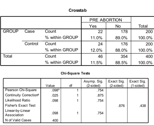
The number of previous abortions.
Number of Previous abortions
Table VI shows the number of previous abortions in the study group. If has been found that 22 patients (11.0%) in the study group and 24 patients (12%) in the control Group
have history of previous abortions. 89% of patients in the case Group and 88% of patients in the Control Group had no history of previous abortion. No significant difference in the study group [P.875].was noted regarding the incidence of previous
abortions.
Crosstab
22 178 200
11.0% 89.0% 100.0%
24 176 200
12.0% 88.0% 100.0%
46 354 400
11.5% 88.5% 100.0%
Count
% within GROUP Count
% within GROUP Count
% within GROUP Case Control GROUP Total Yes No PRE ABORTION Total Chi-Square Tests
.098b 1 .754
.025 1 .875
.098 1 .754
.876 .438
.098 1 .754
400 Pearson Chi-Square
Continuity Correctiona
Likelihood Ratio Fisher's Exact Test Linear-by-Linear Association N of Valid Cases
Value df Asymp. Sig. (2-sided) Exact Sig. (2-sided) Exact Sig. (1-sided)
Computed only for a 2x2 table a.
0 cells (.0%) have expected count less than 5. The minimum expected count is 23.00.
TABLE – VII
Incidence of Preeclampsia
Table VII shows the incidence of preeclampsia in the study group. It has been found that 10 patients (5.0%) in the study group and 15 patients (7.5%) in the control group had signs and symptoms of preeclampsia in the antenatal period. No Statistically
significant difference was detected regarding the incidence of preeclampsia [P.409] in this study.
Crosstab
10 190 200
5.0% 95.0% 100.0%
15 185 200
7.5% 92.5% 100.0%
25 375 400
6.3% 93.8% 100.0%
Count
% within GROUP Count
% within GROUP Count
% within GROUP Case Control GROUP Total Yes No PRE ECLAMPSIA Total Chi-Square Tests
1.067b 1 .302
.683 1 .409
1.073 1 .300
.409 .205
1.064 1 .302
400 Pearson Chi-Square
Continuity Correctiona
Likelihood Ratio Fisher's Exact Test Linear-by-Linear Association N of Valid Cases
Value df Asymp. Sig. (2-sided) Exact Sig. (2-sided) Exact Sig. (1-sided)
Computed only for a 2x2 table a.
0 cells (.0%) have expected count less than 5. The minimum expected count is 12.50.
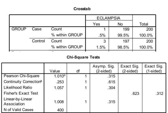
TABLE VIII
Incidence of Eclampsia
Table VIII shows the incidence of Eclampsia in the study group. It has been observed that 1 patient[.5%] in the case group and 3 patients[1.55%] in the control group had
Eclampsia in the antenatal period. No statistically significant difference [P.615]was observed regarding the incidence of Eclampsia in this study.
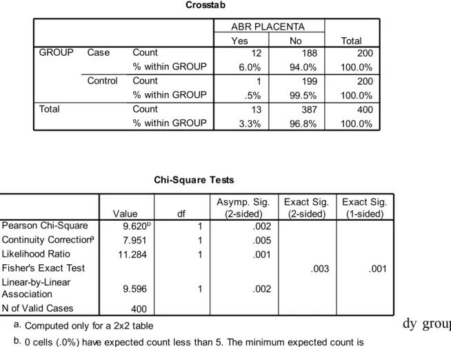
TABLE – IX
Incidence of Abruptio Placenta
Crosstab
1 199 200
.5% 99.5% 100.0%
3 197 200
1.5% 98.5% 100.0%
4 396 400
1.0% 99.0% 100.0% Count
% within GROUP Count
% within GROUP Count
% within GROUP Case Control GROUP Total Yes No ECLAMPSIA Total Chi-Square Tests
1.010b 1 .315
.253 1 .615
1.057 1 .304
.623 .312
1.008 1 .315
400 Pearson Chi-Square
Continuity Correctiona
Likelihood Ratio Fisher's Exact Test Linear-by-Linear Association N of Valid Cases
Value df Asymp. Sig. (2-sided) Exact Sig. (2-sided) Exact Sig. (1-sided)
Computed only for a 2x2 table a.
2 cells (50.0%) have expected count less than 5. The minimum expected count is 2.00.
Table IX shows the incidence of Abruptio Placenta in the study group. It has been observed 12 patients (6.0%) in the case group and 1 patient in the control Group
( .5%) has Abruptio placenta. A statistically significant difference (P.005)was noted between the study and Control Group in this study.
TABLE – X
Incidence of PROM (Premature Rupture of Membranes) Crosstab
12 188 200
6.0% 94.0% 100.0%
1 199 200
.5% 99.5% 100.0%
13 387 400
3.3% 96.8% 100.0% Count
% within GROUP Count
% within GROUP Count
% within GROUP Case Control GROUP Total Yes No ABR PLACENTA Total Chi-Square Tests
9.620b 1 .002
7.951 1 .005
11.284 1 .001
.003 .001
9.596 1 .002
400 Pearson Chi-Square
Continuity Correctiona
Likelihood Ratio Fisher's Exact Test Linear-by-Linear Association N of Valid Cases
Value df Asymp. Sig. (2-sided) Exact Sig. (2-sided) Exact Sig. (1-sided)
Computed only for a 2x2 table a.
0 cells (.0%) have expected count less than 5. The minimum expected count is 6.50.
b.
Crosstab
13 187 200
6.5% 93.5% 100.0%
2 198 200
1.0% 99.0% 100.0%
15 385 400
3.8% 96.3% 100.0% Count
% within GROUP Count
% within GROUP Count
Table X shows the incidence of Premature Rupture of Membranes in the study group. It
has been observed 13 Patients ( 6.5% ) in the case group and2 Patients (1% ) in the control Group had PROM. A statistically significant difference was (p.008)noted between the study and control Group in the study.
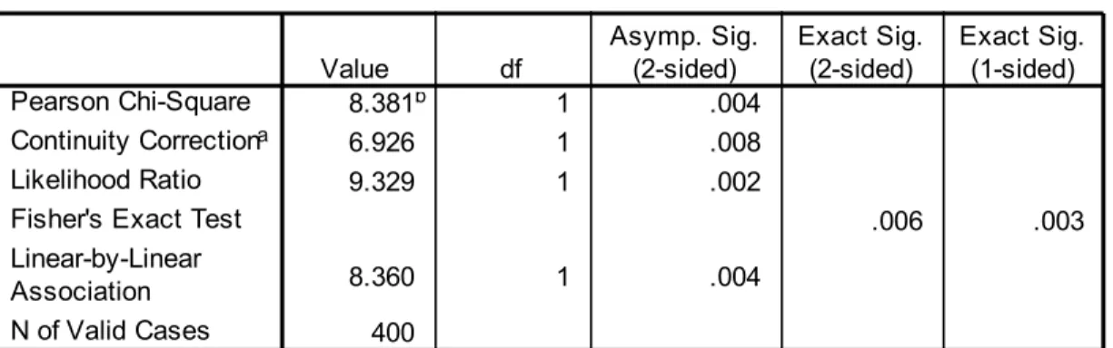
TABLE XI
Incidence of Low lying placenta
Chi-Square Tests
8.381b 1 .004
6.926 1 .008
9.329 1 .002
.006 .003
8.360 1 .004
400 Pearson Chi-Square
Continuity Correctiona
Likelihood Ratio Fisher's Exact Test Linear-by-Linear Association N of Valid Cases
Value df Asymp. Sig. (2-sided) Exact Sig. (2-sided) Exact Sig. (1-sided)
Computed only for a 2x2 table a.
0 cells (.0%) have expected count less than 5. The minimum expected count is 7.50.
b.
Crosstab
6 194 200
3.0% 97.0% 100.0%
2 198 200
1.0% 99.0% 100.0%
8 392 400
2.0% 98.0% 100.0% Count
% within GROUP Count
% within GROUP Count
% within GROUP Case Control GROUP Total Yes No LLP Total Chi-Square Tests
2.041b 1 .153
1.148 1 .284
2.134 1 .144
.284 .142
2.036 1 .154
400 Pearson Chi-Square
Continuity Correctiona
Likelihood Ratio Fisher's Exact Test Linear-by-Linear Association N of Valid Cases
Value df Asymp. Sig. (2-sided) Exact Sig. (2-sided) Exact Sig. (1-sided)
Computed only for a 2x2 table a.
2 cells (50.0%) have expected count less than 5. The minimum expected count is 4.00.
Table XI Shows the incidence of Low lying Placenta in the study group. It has been observed 6 patients (3.0%) in the case group and 2 patients (1.0%) in the control Group had Low lying placenta. No Statistically significant difference(p.284) was noted
between the study and control Group in this study.
TABLE –XII
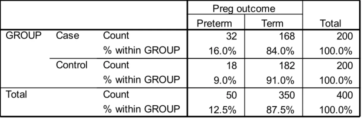
Pregnancy outcome in the study group
Crosstab
32 168 200
16.0% 84.0% 100.0%
18 182 200
9.0% 91.0% 100.0%
50 350 400
12.5% 87.5% 100.0%
Count
% within GROUP Count
% within GROUP Count
% within GROUP Case Control GROUP Total Preterm Term Preg outcome Total Chi-Square Tests
4.480b 1 .034
3.863 1 .049
4.533 1 .033
.049 .024
4.469 1 .035
400 Pearson Chi-Square
Continuity Correctiona
Likelihood Ratio Fisher's Exact Test Linear-by-Linear Association N of Valid Cases
Value df Asymp. Sig. (2-sided) Exact Sig. (2-sided) Exact Sig. (1-sided)
Computed only for a 2x2 table a.
0 cells (.0%) have expected count less than 5. The minimum expected count is 25.00.
Table XII shows the incidence of pregnancy outcome in the study group. It has been found that 32 patients (16.0%) in the case group and 18 patients (9.0%) in the
control Group had Preterm Delivery. It has been observed that 168 patients (84.0%) in the case group and 182 patients (91.0%) in the control group had Term Deliveries. A statistically difference was noted between the study and Control in the number of preterm deliveries in the present study.
TABLE - XIII
Mode of delivery in the study group
Table XIII compares the incidence of Mode of Delivery in the study group. It has been observed that 72 patients 36% in the case group and 88 patients (44%) in the
Crosstab
72 128 200
36.0% 64.0% 100.0%
88 112 200
44.0% 56.0% 100.0%
160 240 400
40.0% 60.0% 100.0% Count
% within GROUP Count
% within GROUP Count
% within GROUP Case
Control GROUP
Total
LSCS Normal MOD OF DEL
Total
Chi-Square Tests
2.667b 1 .102
2.344 1 .126
2.670 1 .102
.126 .063
2.660 1 .103
400 Pearson Chi-Square
Continuity Correctiona
Likelihood Ratio Fisher's Exact Test Linear-by-Linear Association N of Valid Cases
Value df Asymp. Sig. (2-sided) Exact Sig. (2-sided) Exact Sig. (1-sided)
Computed only for a 2x2 table a.
0 cells (.0%) have expected count less than 5. The minimum expected count is 80.00.
Control group got delivered by Caesarean section and 128 patients (64.0%) in the case group and 112 patients (56%) in the control Group delivered by Labour Natural. No
Significant difference(P.1260) in the incidence of type of delivery was noted in this study.
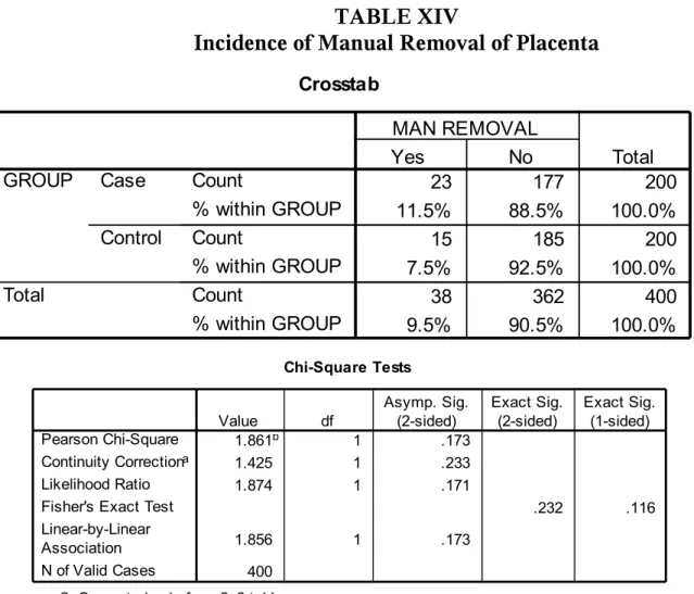
TABLE XIV
Incidence of Manual Removal of Placenta
Table XIV shows the incidence of Manual Removal of placenta in the study Group. It has been found that 23 patients (11.5%) in the study Group and 15 patients (7.5%) in the Control Group had manual removal of placenta done in the post partum
period. A Statistically significant difference(P.233) was not observed in the incidence of manual Removal of Placenta in this study.
TABLE XV
Crosstab
23 177 200
11.5% 88.5% 100.0%
15 185 200
7.5% 92.5% 100.0%
38 362 400
9.5% 90.5% 100.0%
Count
% within GROUP Count
% within GROUP Count
% within GROUP Case Control GROUP Total Yes No MAN REMOVAL Total Chi-Square Tests
1.861b 1 .173
1.425 1 .233
1.874 1 .171
.232 .116
1.856 1 .173
400 Pearson Chi-Square
Continuity Correctiona
Likelihood Ratio Fisher's Exact Test Linear-by-Linear Association N of Valid Cases
Value df Asymp. Sig. (2-sided) Exact Sig. (2-sided) Exact Sig. (1-sided)
Computed only for a 2x2 table a.
0 cells (.0%) have expected count less than 5. The minimum expected count is 19.00.
Incidence of Post partum Hemorrhage
Table XV shows the incidence of post partum Hemorrhage in the study group. It
has been observed that 11 patients (5.5%) in the study group and 7 patients (3.5%) in the control group had significant post partum hemorrhage. A Statistically significant difference(P.469) was not noted in the incidence of post partum hemorrhage in this study.
TABLE XVI
Incidence of Low Birth Weight
Crosstab
11 189 200
5.5% 94.5% 100.0%
7 193 200
3.5% 96.5% 100.0%
18 382 400
4.5% 95.5% 100.0% Count
% within GROUP Count
% within GROUP Count
% within GROUP Case Control GROUP Total Yes No PPH Total Chi-Square Tests
.931b 1 .335
.524 1 .469
.938 1 .333
.470 .235
.928 1 .335
400 Pearson Chi-Square
Continuity Correctiona
Likelihood Ratio Fisher's Exact Test Linear-by-Linear Association N of Valid Cases
Value df Asymp. Sig. (2-sided) Exact Sig. (2-sided) Exact Sig. (1-sided)
Computed only for a 2x2 table a.
0 cells (.0%) have expected count less than 5. The minimum expected count is 9.00.
b.
Crosstab
36 164 200
18.0% 82.0% 100.0%
8 192 200
4.0% 96.0% 100.0%
44 356 400
11.0% 89.0% 100.0% Count
% within GROUP Count
% within GROUP Count
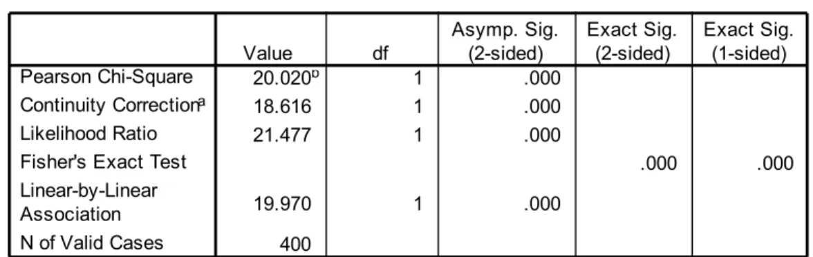
Table XVI Shows the incidence of Low Birth Weight of babies in the study group. It has been observed that36 patients ( 18% ) in the study group and 8 patients
( 4.0% ) in the control Group delivered babies with Low Birth Weight (<2500g). A Statistically significant difference (P.0001 )was noted between the study and control group in this study.
complication Case Control odds ratio 95% CI p-value
LBW 18 4 5.3 2.3-12.7 0.0001
Manual removal 11.5 7.5 1.6 0.8-3.4 0.233
PROM 6.5 1 6.9 1.5-44.8 0.008
Abr-placenta 6 0.5 12.7 1.7-264.3 0.005
PPH 5.5 3.5 1.6 0.6-4.7 0.469
Preeclampsia 5 7.5 0.7 0.3-1.6 0.409
LLP 3 1 3.1 0.6-22.2 0.284
Eclampsia 0.5 1.5 0.3 0.01-3.58 0.615
Chi-Square Tests
20.020b 1 .000
18.616 1 .000
21.477 1 .000
.000 .000
19.970 1 .000
400 Pearson Chi-Square
Continuity Correctiona
Likelihood Ratio Fisher's Exact Test Linear-by-Linear Association N of Valid Cases
Value df Asymp. Sig. (2-sided) Exact Sig. (2-sided) Exact Sig. (1-sided)
Computed only for a 2x2 table a.
0 cells (.0%) have expected count less than 5. The minimum expected count is 22.00.
DISCUSSION
It is already known that First – Trimester Vaginal bleeding affects upto 25% of all
pregnancies and reported to lead to spontaneous miscarriage in 50% of affected women. These data showed that threatened miscarriage is not only associated with miscarriage but also associated with adverse pregnancy outcome.
Results from this study confirm findings from other authors that threatened abortion is associated with an increased risk of certain pregnancy related complications namely placental abruption, preterm labour, delivery of Low Birth weight babies,
Premature Rupture of Membranes.
Generally high incidence of abortion and complications in threatened miscarriage
indicate the necessity of proper programming in care and cure and also educating high risk women.
Results of this study support other evidence to indicate that in some patients first trimester vaginal bleeding may indicate underlying placental dysfunction which may
manifest in later pregnancy by a variety of adverse outcomes that have also been related to placental dysfunction.
It has been found in this study that majority of cases belonged to the age group of 20-30 years and perhaps the reason is that majority of delivery also occur in this age
group as Mckeon (1954) concluded that the best time of women to have first baby is between the age of 20-30 years.
Table V shows that out of 200 cases with threatened miscarriage 79.5% of cases were primigravida and 20.5% of cases were multigravida. But a statistically significant difference between the case and control in this study was not noted. Basama FM (2004)
found that previous miscarriage has no influence in the outcome of Threatened miscarriage. Table VI shows that out of 200 cases with threatened miscarriage 11.0% of cases had history of previous abortion and 12% of Control group had history of previous abortion. So no significant association(P.875) is found between vaginal bleed and
history of previous abortions.
In one study by John vidaver / Robert H (2003) an increased risk of preeclampsia preterm delivery/ placental abruption and caesarean delivery was observed for patients
delivery,PPROM,Placental Abruption and caesarean delivery. In our study increased risk of preterm delivery, PROM, Abruptio placenta and LBW was observed in patients
with first trimester bleed.
In 1993, Verma et al reported that pregnancy induced hypertension was significantly more common in subjects with threatened abortion and a viable pregnancy
compared with subjects without vaginal bleeding (6% Vs. 4.7% respectively; p<0.05). Another study did not find an association between first trimester vaginal bleeding and gestational hypertension but did find that patients with light bleeding were statistically
more likely to have preeclampsia. This association carried a low OR of <2.0 Likely we don’t find any significant association between vaginal bleed and preeclampsia. Table VII Shows out of 200 cases with threatened miscarriage 5% of cases had signs and
symptoms of preeclampsia as compared with 7.5% in the control group.
Table X shows that out of 200 patients 6.5% of patients with threatened miscarriage had premature rupture of membranes as compared to 1% in the control
group. So our findings corroborate other studies that suggested an associated between threatened abortion and PROM. Although the cause is unclear it is hypothesized that disruption of the chorionic – amniotic plane by adjacent hemorrhage may make the
Table IX shows that out of 200 cases with first trimester bleed 6.0% of cases had
abruptio placenta as compared with 0.5% of Control. A statistically significant association between vaginal bleeding and Abruptio placenta was found.
Das et al. Dhaliwal LK reported an increased risk for a lowlying placenta among women with threatened miscarriage but found no difference in placental location compared with control subjects by 36 weeks of Gestation. Out data shows that in Table XI that out of 200 cases with first trimester bleed only 3% had low lying placenta. No
significant association could be demonstrated between threatened miscarriage and placenta praevia.
Preterm delivery before 37 weeks gestation occurs in 7-11% of pregnancies but is responsible for 85% of deaths of normally formed infants. Despite significant advances in perinatal medicine the incidence of preterm delivery has remained unchanged. The only potential risk factor found to be associated significantly with the risk of preterm
delivery in women with a threatened miscarriage was unexplained antepartum hemorrhage rather than other factors such as premature rupture of membranes. The association of vaginal bleed with preterm deliveries has also been noted by others.
had double the risk of preterm delivery compared with patients without bleeding. The study of Williams et al was limited to first trimester bleeding. Batzofin et al included patients with bleeding upto 20 weeks. Strobino and Pantel silverman failed to show as association between preterm delivery with light vaginal bleeding in the first trimester of pregnancy. In our study statistically significant association between study and control subjects was noted (P.0.049) Table XII shows that out of 200 cases with threatened
miscarriage 32 cases had preterm delivery and that is around 16.0% as compared with 9.0% in the control group.
Currently there is not information in the literature regarding threatened abortion and caesarean delivery. Table XIII shows that out of 200 cases with first trimester bleed 36.0% were delivered by caesarean section as compared with 44.0% in the control
group. No significant association could be demonstrated between patients with vaginal bleed in first trimester and caesarean delivery in our study. But F Davari – Tanha et al
reported statistical association between threatened abortion and the risk for caesarean delivery.
Hertz JB. Heisenberg (1985) reported that Retention of placenta was associated with threatened miscarriage and the rate of Manual Removal was 14% They postulated
threatened miscarriage. Table XIV Shows that out of 200 patients with first trimester bleed 11.5% of cases had Manual Removal of placenta done as compared to 7.5% of
control subjects. No statiscally significant association between the case and control could be demonstrated in our study.
Table XV shows out of 200 patients with first trimester bleed 5.5.% of patients
had post partum Hemorrhage as compared to 3.5% of control. No significant association could be found between case and control in postpartum hemorrhage.
Haddow et al reported an increased risk for Low birth weight (<2500g) in pregnancies that were complicated by vaginal bleeding. Table XVII shows that out of 200 patients 18% of patients with threatened miscarriage delivered babies with birth
SUMMARY AND CONCLUSION
A prospective study of outcome of pregnancy in 200 cases of patients with first
trimester bleeding was undertaken.
The patients were followed up prospectively from examination in the first
trimester until the end of pregnancy. The Pregnancy and Delivery complications like antepartum Hemorrhage (Placenta previa and Abruptio placenta) Eclampsia, Preeclampsia, Premature Rupture of Membranes, Low Lying Placenta, Pregnancy
outcome, Mode of Delivery, Manual Removal of Placenta, Post Partum Hemorrhage and Low Birth Weight were determined and the outcome data obtained.
In out study in 5% of cases had preeclampsia, 0.5% of cases had Eclampsia 6.5%
of cases had premature Rupture of Membranes, 6% of cases had Abruptio placenta, 3.0% of cases had Low Lying Placenta, 16% of cases had preterm deliveries, 36% of cases were delivered by caesarean sections and 11.5% of cases had Manual Removal of
placenta done, 5.5% of cases had significant post partum Hemorrhage and 18% of cases delivered babies of Low Birth Wt
pregnancy outcome in threatened abortion. However it is not harmful and may provide the patient with some emotional comfort.
In general most do not administer progesterone or sedatives. In most instances of threatened abortions that ultimately result in complete abortion, the embryo is already dead; thus the administration of progesterone drugs is ineffective and only prolongs the
natural course of abortion. However progesterone (Vaginal administration) may be indicated in unique circumstances including viable pregnancies achieved with advanced reproductive technology of patients with a history of an inadequate luteal phase.
Studies have shown that although progesterone administration may not necessarily change the outcome of threatened abortion, it may help reduce the severity of symptoms such as pain from cramping and uterine contractions. Empirically there is no role of
hormone therapy but undiagnosed deficiency can be corrected by 17 hydroxyl progesterone corporate or Dydrogesterone. Aspirin may be given to improve placental circulation.
The use of uterine muscle relaxants or tocolytics is not supported by adequate evidence.
Folic acid may be used to prevent neural tube defect and abruptio placenta.
tactful explanation about the pathological process and favorable prognosis when the pregnancy is viable.
It is observed that early and comprehensive prenatal care can decrease risk of threatened abortion to some extent. So Increased antenatal surveillance might identify women within this group who are at increased risk. Knowledge of this increased risk
may also facilitate decision making regarding management, mode, place and time of delivery which will inevitably improve pregnancy outcome.
Although from our study it is reassuring that the majority of women with first trimester bleeding have pregnancy outcomes comparable to those without such bleeding it is evident that they face a higher relative risk of some adverse obstetric and neonatal
BIBLIOGRAPHY
1. Batzofin JH, Fielding WL, Friendman EA Effect of Vaginal bleeding
in early pregnancy on outcome obstet Gynecol 63:515 1984.
2. Funderburk SJ, Guthrie D, Meldrum D; Outcome of pregnancies complicated by early vaginal bleeding BR J obstet Gynecol 87:10 1980
3. Nielsen S, Hahlin M; Expectant Management of first trimester threatened abortion Lancet 345:84 1995.
4. Salem HT, Ghaneimah SA, Shaaban MM, etal Prognostic Value of
biochemical tests in the outcome of threatened abortion Br J obstet
Gynecol 91:382 1984.
5. Mulik V, Bethel J, Bhal K.A. retrospective population based study of
primigravid women on the potential effect of threatened miscarriage
on obstetric outcome. J obstet Gynecol 2004 Apr; 24(3): 249-253.
6. Verma SK Premi HK, Gupta TV, Thakur S, Gupta KB, Randhawa I
Perinatal out come of pregnancies complicated by threatened
abortion J. Indian Med Assoc.1994 No; 92(110 364 -365.
abortion. Acta obstet Gynecol Scand 1985; 64(2) : 151-156
8. Bennett GL, Bromley B, Lieberman E, Benacerraf BR Subchorionic
hemorrhage in first – trimester pregnancies; prediction of pregnancy outcome with sonography. 1996 Sep; 200 (3) : 803 – 806.
9. Farrell T,Owen P. The significance of extrachorionic memberane
separation in threatened miscarriage. Br J obstet Gynaecol 1996 Sep;
103(9) : 926 – 928.
10. Williams MA, Mittendof FR, Lieberman E, Monson RR, Adverse
infant outcomes associated with first trimester uaginal bleeding
obstet Gynecol 1991 Jul; 78 (1) : 14-18.
11. Chung TK, Sahota DS, Lau TK, Mongelli JM, Spencer JA, Haines
CJ. Threatened Abortion: Prediction of viability based on signs and
symptoms. Aust NZJ obstet Gynaecol 1999 Nov; 39(4) : 443 – 447.
12. Sipila P. Hartikainen – Sorri AL, Oja H, Von Wendt L. Perinatal
outcome of pregnancies complicated by Vaginal bleeding. Br Jobstet Gynaecol. 1992 Dec; 99(12) : 959-963.
Med Assoc Thai. 2001 May 184 (5) : 661-665.
14. IY Park, CH Park, G Lee, JC Shin. Prognosis of threatened abortion
by embryonic / Fetal heart beat rate, Ultrasound in Medicine Biology 2006; 32(5) : 264 – 70.
15. Bowe P, Murphy H. Complications of pregnancy following
threatened abortion Ir. J. Med Sci. 1987 Nov; 156 (11): 328-329.
16. Nagy S. Bush M, Stone J. Lapinski RH, Gardo S. Clinical Significance of subchorionic and retro placental hematomas detected in the first trimester of pregnancy. Obstet Gynecol 2003 Jul;
102(1):94-100.
17. Tannirandom Y, Sangsawang S. Manotaya S. Uerpairojkit B. Samrit pradit P. Charoenvidhya D. Fetal loss in threatened abortion
after embryonic / fetal heart activity Int J Gynaecol obstet 2003 Jun; 81(3) : 263-266.
18. Das AG, Gopalan S, Daliwal LK. Fetal growth and perinatal
outcome pregnancies continuing after threatened abortion. Aust NZJ
obstet Gynaecol 1996 May ; 36(2): 135 – 139.
19. Weiss JL, Maone FD Vidaver J, Ball RH, Myberg DA, Comstock
Tritsch IE, D’ Alton ME; FASTER Consortium. Threatened abortion; A risk factor for poor pregnancy outcome, a population –
based screening study. AM J Obstet Gynecol 2004 Mar; 190 (3): 745 – 750.
20. Alcazar JL, Rutz – Perez ML. Uteroplacental circulation in patients
with first – trimester threatened abortion Fertil stenil 2000 Jan; 73(1):
130-135.
21. BI Patel, V. Trivedi Threatened abortion outcome in relation to
intrauterine clot site and not only volume, Inter J Gynecol & obstet
2000: 70(4): 44
22. Norwitz ER, Schust DJ, Fisher SJ. Implantation and the survival of
early pregnancy N Engl J Med. 2001 Nov 8; 345 (19) : 1400 – 1408
23. Tadmor OP, Achiron R. Rabinowiz R, Aboulafia Y, Mashiach S. Diamant YZ. Predicting first – trimester spontaneous abortion. Ratio of mean sac diameter to crown – rump length compared to embryonic heart rate. J Reprod Med. 1994 Jun; 39(6) : 459 – 462.
25. Tannirandom Y, Manotayas, Uerpairojkit B, Tanawattanacharoen S,
Wacharaprechanont T., Charoenvidhya D, Reference intervals for
first trimester embryonic / fetal heart rate in a Thai Population J obstet Gynaecol Res. 2000 Oct; 26(5): 367 – 372.
26. Makikallio K, Tekay A, Jouppila P. Uteroplacental homodynamic
during early human pregnancy: a longitudinal study Gynecology
obstet Invest. 2004; 58(1) : 49-54.
27. Strobino B, Pantel – Silvermann J. Gestational Vaginal bleeding and
pregnancy outcome. Am J epidemiol 1989 Apr; 129 (4) : 806 – 815.
28. J. Szekeres – Bartho,B. Polgar, K. Kelemen, G. Par, L. Szereday. Progesterone – mediated immunomodulation and anti – abortive effects the role of progesterone – induced blocking factor. World
Congress on the Menopause, 10-14 June 2002, Berlin.
29. Haddow JE, Knight GJ, Kloza EM, Palomati GE, Alpha – Feto protein, Vaginal bleeding and pregnancy risk. Br J obstet Gynaecol 1986 Jun; 93(6): 589 – 583.