DISSERTATION
PREDICTORS
OF
INTRACTABLE
EPILEPSY
IN
CHILDHOOD:
A
CASE
CONTROL
STUDY
Submitted in partial fulfilment of requirements for the degree of
MD paediatrics
BRANCH VIITHE TAMILNADU DR. M.G.R. MEDICAL UNIVERSITY CHENNAI
MARCH 2010
INSTITUTE OF CHILD HEALTH & HOSPITAL FOR
CHILDREN
CERTIFICATE
This is to certify that the dissertation titled “PREDICTORS OF INTRACTABLE
EPILEPSY IN CHILDHOOD: A CASE CONTROL STUDY” submitted by
Dr.SANGEETHA.P to the Faculty of pediatrics, The Tamilnadu Dr. M.G.R.
Medical university, Chennai in partial fulfillment of the requirement for the
award of M.D. Degree ( Pediatrics) is a bonafide research work carried out by
her under our direct supervision and guidance.
Dr.J.MOHANASUNDARAM,
M.D., Ph.D., DNB,
Dean,
Madras Medical College,
Chennai ‐ 3.
Dr.S.STEPHEN ABRAHAM SURESH KUMAR,
M.D.,D.M(NEURO).,
Chief, Department of Neurology
Institute of Child Health
Dr.SARADHA SURESH,
M.D., Ph.D.,F.R.C.P(Glascow)
Director & Superintendent, Institute of Child Health and Hospital for Children, Egmore, Chennai ‐ 8.
DR. P.SEKAR,
MD,DCH,
DECLARATION
I Dr.SANGEETHA.P solemnly declare that the dissertation titled “PREDICTORS
OF INTRACTABLE EPILEPSY IN CHILDHOOD: A CASE CONTROL STUDY” has
been prepared by me.
This is submitted to The Tamilnadu Dr. M.G.R. Medical University, Chennai in
partial fulfillment of the rules and regulations for the M.D. Degree Examination
in Pediatrics.
Dr.SANGEETHA.P
Place :Chennai
ACKNOWLEDGEMENT
I Express my heartfelt gratitude to Dr. S.SARADHA SURESH, MD,DCH,
MNAMS, Director and Superintendent, Institute of Child health and Hospital
for children, Madras Medical College, Chennai for her great help from the
beginning of the study, her able Guidance, Inspiration and Encouragement, in
conducting my study.
I thank my unit Chief Prof.P.Sekar, MD,DCH, and my unit Assistant
Professors for their whole hearted support, guidance and help rendered to this
work. I also thank Prof.V.T.Ramakrishnan, MD, DCH, retired professor of
paediatrics, for his guidance from the beginning.
I express my sincere thanks to my guides Prof.N. Thilothammal , MD,
DM(neuro), and Prof. Stephen Abraham Suresh Kumar, MD,DM(Neuro), for
their guidance, meticulous supervision and constant support in doing this
work. I also express my sincere thanks to assistant professors, department of
neurology for their constant support and guidance from the beginning.
My Profound thanks to Dr.Mohanasundaram MD,PhD,DNB, Dean,
Madras Medical College, Chennai for permitting me to utilise the clinical
I would fail on my part if I forget to thank all my patients and their
caretakers for their co‐operation , without which, this work would not have
been possible.
CONTENTS
S.No. Contents Page No.
1. Introduction 1
2. Review of Literature 18
3. Study Justification 24
4. Aim Of the Study 25
5. Subjects And Methods 26
6. Results 31
7. Discussion 49
8. Summary And Conclusion 53
9. Bibliography 55
10. Annexure 56
Epilepsy is a common neurological disorder affecting 3‐6/1000
children[1]. Approximately 70–80% will be controlled with single drug and
10‐15% require combination chemotherapy. However 10 – 20% of epileptic
patients are resistant to therapy ,the condition , so called ‘intractable
epilepsy’[1,3]. While a great deal is known about seizures and epilepsy, little is
known about the identification and causes of intractable epilepsy[14]. Such
intractable epilepsy, impose considerable socioeconomic and psychological
constraint on the individual patient and casts a substantial burden on health
and welfare resources. Many terms have been used for of these patients who
doesn’t respond to therapy, like ‘treatment non responder’, ‘refractory’,
‘intractable’ and ‘drug resistant’. Early identification of these children prone to
develop intractable seizures is critical for parental counselling, selecting
patients for more intensive investigations and treatment, such as early
consideration for epilepsy surgery. In addition determining risk factors for
intractability may help shed some light on the causes and mechanisms
underlying these drug resistant epilepsy.
1) There is no unifying definition of intractable epilepsy.
2) Patient do not necessarily become refractory immediately at the time of
diagnosis, nor do they inevitably remain refractory throughout the
course of illness.
3) Response to medication is assessed , without a pretreatment baseline,
as most patients are treated rapidly after diagnosis.
4) Sometimes some of these patients who are defined as intractable, will
respond readily, although not completely to therapy.
Definitions:
Seizures:
seizure or convulsion is a paroxysmal , time limited change in motor
activity and or behavior that results from abnormal electrical activity in brain(8).
Epilepsy :
Two or more unprovoked seizures occurring at an interval of more than
24 hours apart(8) .
Partial seizures
Simple partial (consciousness retained)
Motor
Sensory
Autonomic
Psychic
Complex partial (consciousness impaired)
Partial seizures with secondary generalization
Generalized seizures
Absence Typical
Atypical
Generalized tonic clonic
Tonic
Clonic
Myocolonic
Infantile spasm
Unclassified seizures
Defining intractable epilepsy
There is no uniform consensus regarding definition of intractable
epilepsy. These definition and criteria are of specific use in research purpose.
Different definitions which have been used are
1) Children who had one or more seizures per month over a period of six or
more months and who had experienced trials of atleast two different
antiepileptic drugs with adequate compliance [1]
2) Uncontrolled seizures that occurred with an average frequency of
atleast,one per month over a period of two years and during that
period, atleast three different antiepileptic drugs should have been used
daily, singly or in combination[2]
3) Seizures that continue to occur despite treatment with a maximally
tolerated dose of a first line antiepileptic drug as monotherapy or in
atleast one combination with an adjuvant medication[6]
4) Two or more seizures per month for a period of two years or more
5) Occurrence of one or more seizures per month on follow up of one year
or more with an adequate trial of two primary antiepileptic drug and or
more of the new antiepileptic drugs[7]
Intractable epilepsy as a disability
Epilepsy is sometimes called the invisible disability. But if one thinks in
terms of people with drug responsive seizures this is not entirely correct. These
people do live normal lives and reach their full potential for accomplishment in
lives. Intractable epilepsy is clearly a disability. Public seizures lead to stigma,
and to economic and social discrimination. There is possibility of head injury
due to falls. They are not allowed to drive or to swim alone. They are told to
take showers rather than baths. Their lives become limited and circumscribed.
In addition, these patients with intractable epilepsy have a number of non
physical disabilities, called the co morbidities. These are less well known and
unfortunately seldom treated, but they are often more serious than the seizure
themselves.
Aetiology of intractable epilepsy
2) Neurocutaneous syndromes
3) Developmental malformations
4) Neurodegenerative / neurometabolic problems
5) Central nervous system infection sequelae.
6) Neurocysticercosis
7) Epileptic syndromes
8) Cerebral tumors
9) Mesial temporal sclerosis
10) Porencephalic cyst
11) Head injury
12) Cerebrovascular accident
13) Idiopathic.
West syndrome
It is a triad of infantile spasms, a pathognomonic EEG pattern called
‘hypsarrythmia’ and mental retardation. Onset of seizures is between the third
and twelfth months of age. Three types of attacks occur – severe myoclonic
convulsion, nodding attacks and salaam spells. Drugs used in treatment are
ACTH and Vigabatrin(9)
Lennox – gestaut syndrome
Triad of multiple seizures, 1.5 to 2 Hz spike wave complex on EEG and mental
retardation characterize this syndrome. Age of onset is between 3‐5 years.
Multiple types of seizures occur like atypical absence, generalized tonic, atonic
and myoclonic seizures. Drugs effective are Valproate, Clonazepam, Vigabatrin,
Lamotrigene etc. Seizures are very difficult to control and most children are
mentally retarded by two years of age(9) .
Approach to intractable epilepsy
A detailed history has to be taken to know the type of seizures and frequency
of seizures. A review of the past drug records is mandatory. Detailed clinical
examination has to be done to exclude neurological deficit. A detailed
intractable epilepsy. It also helps us to rule out ‘pseudo intractability’ –
resistant seizures due to inadequate dosage, wrong drug or non‐epileptic
seizures. Patient should be requested to keep a seizure diary. Ideally patient
must be admitted and investigated.
Differential considerations in intractable epilepsy(6)
A] Errors in diagnosis
1) Failure to identify a seizure syndrome or causative condition
2) Incorrect seizure classification (partial or generalized)
3) Non epileptic seizures (syncope, pseudo seizures)
B] Errors in drug choice or management
1) Wrong drug for the seizure type / syndrome
2) Inadequate dose
C] Poor medication compliance
1) Inadequate patient instruction or education
3) Intolerable side effects
D] True intractability
Investigations
1) Detailed metabolic workup
2) EEG
3) Video EEG
4) Neuroimaging – MRI
CT
PET
SPECT
EEG
Most commonly performed neurodiagnostic study in the evaluation of
patients with seizures in EEG. Most reliable findings are primarily
generalized spike and wave discharge or focal spike or sharp wave
discharge over frontal or temporal lobes (6). A normal EEG doesn’t exclude
the diagnosis of a seizure disorder. A prolonged EEG or serial EEGs may
improve the sensitivity. Upto 2% of non seizure experiencing individuals
Video EEG recording
It’s an extremely helpful tool in the evaluation of intractable epilepsy. The
combined use of video and EEG recording improves the sensitivity and
specificity over EEG recording alone.
It has been demonstrated to accurately differentiate between epileptic and
nonepileptic seizures, to distinguish between primarily generalized and
partial onset seizures and to determine seizure onset localization and
lateralization.
Neuro‐imaging
‐Magnetic Resonance Imaging (MRI)
‐ Computed Tomography (CT)
‐ Positron Emission Tomography (PET)
‐ Single Photon Emission Computed Tomography (SPECT)
Improves our understanding about the pathological substrates of epilepsy
and also helpful in determining the long term seizure prophylaxis. In
patients considered for surgical therapy, the presence of a focal
neuroimaging abnormality can improve the prognosis for an excellent
outcome.
MRI imaging is the imaging procedure of choice in those with intractable
partial epilepsy. It has been demonstrated to be superior to CT imaging in
intractable epilepsy. Some low grade malignancies, mesial temporal
sclerosis, neuronal migration abnormalities and heterotopias might be
missed on CT imaging which are identified by MRI.
Management
Pharmacotherapy
Selection of an antiepileptic medication is the most important decision
physicians will make, after conclusion of the diagnostic evaluation.
Treatment plan should be made based on the types of seizure and epilepsy
and by the review of all antiepileptic drugs that have and have not been
used to their maximum tolerated doses, those that may have shown some
appropriate to try. It is then possible to select the AED that is most likely to
be efficacious and to have fewest side effects and to adjust the dose of this
drug to the optimum. Antiepileptic drugs that have not aided seizure
control and have produced adverse effects, should then be gradually
tapered and discontinued. If seizures remain uncontrolled, other first line
and second line drugs that have been identified as possibly helpful or
alternative treatment should be considered (7)
Elements of successful treatment (6)
Classify seizure disorder correctly
1) Balance the maximal effective dose with minimal side effects
2) Choose dosing to maximum compliance
3) Treat the patients symptoms, not the EEG findings or the serum levels
FDA approved monotherapy[12]
Partial Seizures
‐ Carbamazepine
‐ Sodium Valproate
‐ Phenytoin
‐ Lamotrigene
Primarily generalized seizures
‐ Sodium Valproate
‐ Phenytoin
‐ Phenobarbitone
‐ Ethosuximide
‐ Clonazepam
‐ Primidone
FDA appoved adjunctive therapy
‐ All agents approved as monotherapy
‐ Gabapentin
‐ Topiramate
‐ Tiagabine
‐ Levetiracetam
‐ Felbamate
Seizure type – first choice, other options
Infantile spasm : ACTH, Vigabatrin, Topiramate
Atonic seizures : Valproate, Lamotrigene, Topiramate, Phenytoin
Myoclonic seizures : Valproate, Clonazepam, Topiramate, Lamotrigene
Alternate modes of treatment of intractable epilepsy
Ketogenic diet
This is a high fat, low protein, low carbohydrate developed in 1921. It
mimics the effect of starvation[7]. The diet consists of 60% medium chain
triglycerides, 11% long chain saturated fat, 10% protein & 19% carbohydrate.
Ketogenic diet causes a prompt elevation of plasma ketone bodies that the
brain uses as an energy source. It is most effective in infants and children with
intractable seizures. The mechanism of action is not establiashed. It is effective
for the control of myoclonic seizures, infantile spasms, atonic seizures and
mixed seizures of Lennox‐Gestaut syndrome[7,9]. The side effects are abdominal
pain and diarrhea.
Vagal nerve stimulaton
Those patients who are medically refractory and are not candidates for brain
surgical procedure may be evaluated as potential candidates for vagal nerve
refractory partial epilepsies in patients who are > 12 years of age and are not
surgical candidates [6]
It utilizes a programmed stimulus from a chest implanted generator via coiled
electrode tunelled to the left vagus nerve[9]. Approximately 1/3rd of those
treated with vagal nerve stimulation have a greater than 50% reduction in their
seizure frequency. Another 1/3rd have less than 50% reduction in seizure
frequency and the remaining 1/3rd have no improvement. There are minor side
effects like neck pain, hoarseness and brief cough. Drawbacks are the need for
surgical implantation and its high cost.
Surgical treatment
In intractable epilepsy, surgical resection of the focal epileptogenic cortex is
the most effective method currently available to render children and adults
seizure free. Potential candidates include patients with intractable seizures,
who have seizures emanating from a localized region of brain that can be
resected without neurological morbidity. Indications include mesial temporal
sclerosis, heterotopias, and surgically remediable syndromes. A comprehensive
pre surgical evaluation is necessary to pinpoint the seizure onset site and to
identify risk of neurological morbidity. These include ictal video EEG
The surgical procedures available are
1) Hemispherectomy
2) Interhemispheric commissurotomy
3) Temporal lobectomy
4) Lesionectomy
5) Multilobar resection
1] Hemispherectomy
Exclusively for children with intractable epilepsy with hemiplegia. Best
results are for children with disease affecting only one hemisphere, like Sturge
Weber syndrome and Rasmussen encephalitis. The original procedure
consisted of removing the cortex of one hemisphere along with variable
portion of the underlying basal ganglia. Modified procedure includes removal
of most of the damaged hemisphere, but with portions of frontal and occipital
lobe kept in place, but disconnected from other hemisphere and brainstem [9].
Complications are hemorrhage and hydrocephalus.
It includes disconnecting the hemisphere from each other and from the
brainstem. It is an alternative to hemispherectomy in children with intractable
epilepsy and hemiplegia. Another use is to decrease occurrence of secondary
generalized tonic clonic seizures from partial seizures. Two types are there –
complete and partial commisurotomy. Side effects are hemiparesis, mutism,
apraxia and urinary incontinence.
3] Temporal lobectomy
It is the most common surgical procedure performed for medically
intractable epilepsy [6]. Complete seizure relief should be attainable in more
than half of the children with intractable partial complex seizures of temporal
lobe origin[9]. Only children who have a unilateral temporal lobe focus (mesial
temporal sclerosis) are candidates for surgery. Surgical procedures include
resection of superior temporal gyrus, hippocampus and amygdala. The most
common complications are superior quadrantanopia, aphasia, but they are
transitory.
Etiology & clinical predictors of intractable epilepsy:
Chawla et al conducted this case control study comprising 50 cases and
50 control subjects in Kalawati Saran Children’s hospital, New Delhi. Patients
included children who had more than one seizure per month over at least 6
months. Control subjects included children with epilepsy who had been
seizure-free for more than 6 months. Patients were evaluated with special reference to
birth history and development. Clinical examination and neurodevelopmental
assessment were performed in all the patients. Drug monitoring was performed
to exclude pseudointractability. Epilepsy in the study group was caused by
perinatal problems (48%) and sequelae of central nervous system infection
(24%) and was idiopathic in 20%. In the control group, epilepsy was idiopathic
in 72%, a result of calcified granuloma in 22%, and perinatal problems
comprised 6% of the subjects. On univariate analysis, strong association was
evident between intractable epilepsy and several factors, including age at onset
of seizure, remote symptomatic epilepsy, initial seizure type, history of neonatal
seizure, high initial seizure frequency, microcephaly, and neurologic
impairment. On multivariate analysis, neurologic impairment (odds ratio [OR]
12.25; 95% confidence interval [CI] 3.58-41.89), age at onset of seizure less
(OR 10.36; 95% CI 2.39-44.93), and remote symptomatic epilepsy (OR 2.9;
95% CI 1.13-7.43), were independent predictors of intractability.
Predictors of intractable epilepsy in childhood; a case control study:
This case control study was done by Berg et al in the department of
paediatrics and neurology, Yale university, New Haven, Connecticut.Cases
were children who had an average of one seizure or more a month over a 2-year
period and who, during that time, had failed trials of at least three different
antiepileptic drugs. Controls were children who had epilepsy, who had been
seizure-free for >2 years, and who had never, before becoming seizure-free, met
the definition for intractable epilepsy. Strong univariate associations were noted
between intractability and several factors: infantile spasms (IS), remote
symptomatic epilepsy, a history of status epilepticus (SE) before the diagnosis
of epilepsy, neonatal seizures, and microcephaly. Cases were significantly
younger than controls at onset (1.8 vs. 5.8 years); this was not due solely to
cases with onset during the first year of life but was an association apparent
throughout the age range studied. With multiple logistic regression,
independent predictors of intractability were infantile spasms , odds ratio (OR)
= 10.42, p = 0.03; age at onset with a decreasing risk with increasing age, OR =
0.77 per year, p < 0.0001; remote symptomatic epilepsy, OR = 2.24, p = 0.04;
Risk factors predicting refractoriness in epileptic children with partial seizure:
This retrospective study was done by Sakir et al in the department of
paediatric neurology, Cukurova university, Adana, Turkey. Fifty children with
intractable epilepsy as evidenced by at least 1 epileptic fit per month were
included in the study group, whereas the control group consisted of children
who did not experience any recurrent seizure for at least 1 year at the time of the
study. Chi square test test was used to evaluate the relationship between the
test variables for the 2 groups, and the estimated relative risk (odds ratio) for
each variable was calculated. The risk factors were subsequently determined by
logistic multiple regression analysis. Univariate analysis showed that mental
retardation, neurological abnormality, neuroradiological abnormality, perinatal
anoxia, neonatal convulsion, presence of status epilepticus, and symptomatic
etiology were significant risk factors for the development of refractory epilepsy
(P < .05). For multivariate logistic regression analysis, age at seizure onset,
status epilepticus, mixed type of seizures, and history of frequent seizures (more
than once a month) were all found to be significant and independent risk factors
for refractory epilepsy, and the number of drugs used in the study group was
significantly higher than that in the control group (P < .05). In line with these
findings, it was concluded that children who present with epilepsy and have
these risk factors should be referred to a center where epileptic surgery is
Profile of intractable epilepsy in a tertiary referral centre
Singhvi et al did this study in the department of neurology, institute of
medical education and research, Chandigarh in 100 patients over 14 months.
Detailed history, examination, investigatons like EEG, CT scan and details
regarding pharmacotherapy were analysed. Factors which predicted poor
prognosis were found to be organic brain lesions, partial seizures, multiple
seizure types, high seizure frequency, seizure onset in infancy and abnormal
EEG.
Predictive factors in pediatric intractable seizures
Javad akhondian et al conducted this case control study in Children’s
neurology clinic of Imam Reza hospital in north eastern Iran with 51 cases and
80 controls. ‘Intractable seizure‘ was defined as seizure frequency of atleast
one attack per month during six months, despite receiving two antiepileptic
drugs. ‘Well controlled seizures’ was defined as no seizure episode during six
months after the start of treatment. Factors affecting the occurrence of
refractory seizures with a p value < 0.05 were found to be age < 1 year,
multiple seizures, male gender, myoclonic seizures, neurological deficit,
Epidemiology and etiology of intractable epilepsy in Qatar
Al Hail et al conducted this study in Hamad medical corporation, Doha,
Qatar. The medical records of 219 patients seen over a period of eight years
were reviewed to determine incidence and causes of intractable epilepsy. Out
of these only 39 patients fulfilled the criteria of intractable epilepsy. Intractable
epilepsy was defined as occurrence of ≥ 2 seizure per month despite receiving
adequate mono or polytherapy of AED medication for at least two years. The
most common type of intractable epilepsy was idiopathic generalized
epilepsy(75%), followed by symptomatic epilepsy(19%), and temporal lobe
epilepsy(6%).
Early development of intractable epilepsy in children: a prospective study
Berg et al conducted this prospective study in Northern Illinos
University. Children with newly diagnosed epilepsy were prospectively
followed up for occurrence of intractable epilepsy. Out of 599 children
followed up, 60 children(10%) have met the criteria for intractable epilepsy,
including 34.6% with cryptogenic/symptomatic generalized , 2.7% with
idiopathic, 10.7% with other localization related and 8.2% with unclassified
slowing(p=0.02), and neonatal status epilepticus(p=0.001) were associated
with an increased risk of intractable epilepsy
If a first antiepileptic drug fails to control a childs epilepsy, what are the
chance of success with next drug?
Camfield et al conducted this study to determine how often a child’s
epilepsy is controlled and remits if a first AED fails to control seizures. 17% of
patients had inadequate seizure control with their first AED and received a
second AED, with only 42% of them having complete remission of their
epilepsy. In these children in whom seizures were not controlled with the first
AED were more likely to have neurological deficits(p=0.01) and complex partial
seizures(p=0.01) and 29% had intractable epilepsy(p<0.0001)
STUDY JUSTIFICATION
Of all the epileptic children, 10 – 20 % have intractable epilepsy. Though
intractable epilepsy has been extensively studied in developed countries, there
are not many studies done in developing countries. In India, there are some
studies done in north India, but not in South India. This study will be useful in
identifying children prone for intractability at the earliest, so that we can start
treatment accurately.
AIM OF THE STUDY
To find out the predictors of intractable epilepsy in childhood & to
identify these children prone for intractability at the earliest
SUBJECTS AND METHODS
Study design:
Case Control Study
Study place:
Dept. of paediatric Neurology, Institute of Child Health & Hospital for
Children, Egmore.
Study Period:
2 Years(october 2007 to august 2009)
Study Population:
All epilepsy cases attending neurology OP in the age group 6months to
12 yrs
Sampling technique:
Cases:
Epileptic patients who have seizure frequency of atleast 1 attack per
month during 6 months despite receiving 2 antiepileptic drugs with adequate
dosage & compliance
Controls:
Children with epilepsy who have complete control of seizure for >
6months with one or two drugs.
Inclusion criteria:
Epileptic children in age group 6 months to 12 years who meets the
definition of cases & controls
Exclusion criteria:
People from outstation
Poor compliance
Pseudo intractability
• Wrong drug for seizure type
• Non epileptic seizure
Study maneuvre
This case control study was conducted in Neurology Department of
institute of child health and Hospital for Children, Chennai. Epileptic children in
the age group 6 months to 12 years were included in the study. A total of 50
cases and 50 controls were enrolled.
Detailed history and a detailed neurological examination was done in all
patients and entered in a prestructured proforma. Seizures were classified
using the international classification of epileptic seizures.
Electroencephalogram and neuroimaging was performed in all patients to
determine etiology and classification of epilepsy. Cranial computed
tomography was performed in all patients. MRI scan was not done in some
patients in whom CT scan was diagnostic and in a small number of patients
who couldn’t afford MRI.
Factors analyzed included the following
1) Details of pharmacotherapy
Number of drugs, duration after starting two drugs, whether drug
dosage is adequate or not, compliance
2) Age at onset of seizures : <1 year or >1 year
3) Type of seizures
4) Frequency of seizures before starting treatment
5) Frequency of seizures after starting two drugs
6) History of birth asphyxia
7) History of neonatal convulsions
8) History of developmental delay
9) History of status epilepticus
10) Presence of microcephaly
11) Presence of focal neurological deficit
12) CT/MRI findings
13) EEG findings
Statistical analysis
Association between factors analyzed and outcome was determined
using chi‐square test. Odds ratio and 95% confidence interval was calculated
using univariate analysis. Multivariate analysis by logistic regression method
was done to rule out confounding factors and to find out independent
predictors of outcome.
A tot
contr
Age
6 mo >2 yr >5 yr Total 0 5 10 15 20 25 30
tal of 50
rolled seizu
on – 2 yr
r – 5 yr
r – 12 yr
6 MON
patients w
ures were
Age case number 4 16 30 50
N ‐2 YR
RE
with intra
studied. distributio % 8% 32 60 10
>2 YR ‐5 Y
ESULTS
ctable epi
Table‐1
on of stud
% % % 0% YR >
S
ilepsy and
y populati
contro numbe 3 21 26 50
>5 YR ‐12 YR
d 50 patie
ion ol er % 6 4 5
ents with
[image:37.612.108.521.411.665.2]Sex fema Male Total 0 5 10 15 20 25 30 35 Majority
years – 1
age grou
group.
cases
n
ale 27
e 23
50
0 5 0 5 0 5 0 5
of patient
12 years. T
up and 16
Sex
s
%
54 %
46 %
100 %
FEMALE
ts in case g
There were
patients (3
distributio contr n 16 34 % 50
group, 30 (
e only 4 p
32%) belo
Table‐
on of study
rol
% 32 % 68 % 100
MALE
60%) belo
atients (8%
onged to t
‐2
y populati
p val
0.02 %
% %
E
nged to th
%) in 6 mo
he 2 year
ion
lue odds
ratio
6 2.49
he age grou
onths – 2 y
to 5 year
s o 95% 95 1.10 ‐ 5.62 CASE CONTROL
up 5
year
age
% ci
05
29
[image:38.612.101.533.346.712.2]A fem
54%
was o
age a
<1 ye
≥1 yr
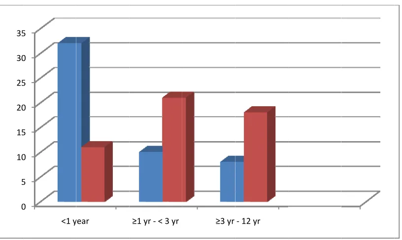
≥3 yr total 0 5 10 15 20 25 30 35
male prep
and males
of 0.026 an
at onset
ear
r ‐ <3 yr
r – 12 yr
0 5 0 5 0 5 0 5
<1 y
onderance
s 46 %. Th
nd odds ra
case n 32 10 8 50 year
e was see
he differen
atio of 2.49
Age at o
% 64
20
16
10
≥1 yr ‐< 3 yr
en among
nce was st
95.
Table‐3
onset of se
% % % 0 %
≥3 yr
case grou
tatistically eizures Contro n 11 21 18 50
‐12 yr
up, female
significant ol % 2 4 3 1
es constitu
t with p va
%
22 %
42 %
36 %
100 %
uting
alue
[image:39.612.110.518.468.712.2]Majo
as ag
signif
interv
no of
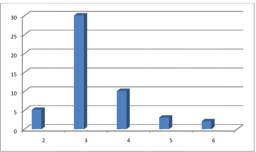
2 3 4 5 6 total 0 5 10 15 20 25 30
ority of pat
gainst pati
ficant with
val of 2.60
f drugs
0 5 0 5 0 5 0 2
tients in ca
ents (22%
h a p valu
04 to 15.25
Numbe
ase group,
%) in contro
ue of 0.00
5
r of drugs
Cases N 5 30 10 3 2 50 3
32 (64%)
ol group.
00, Odds r
Table‐4
being take
4
had seizur
This was f
ratio of 6
en by case
% 1 6 2 6 4 1 5
re onset <
found to b
.3 and 95
e group
10 % 60 % 20 % 6 % 4 % 100 %
6
1 year of
be statistic
5% confide age
cally
ence
[image:40.612.108.518.464.707.2]
Maxi
patie
30 (6
seizu gtcs tonic absen myoc simp comp mixe 0 5 10 15 20 25 30 35
mum num
ents. In this
60 %) were
re type
c seizures
nce seizure clonic seizu le partial s plex partia
d 0 5 0 5 0 5 0 5
mber of ant
s study, m
taking thr
es
ures
seizures
l seizures
tiepileptic d
aximum n
ree drugs.
Typ
drugs take
umber of
Table‐5
pe of seizur
case n 32 6 2 7 7 3 13
en was 6 dr
patients w
res
% 64 % 12 % 4 % 14 % 14 % 6 % 26 %
rugs, being
with intract
Contr n 28 7 2 0 9 2 1
g taken by
table epile
rol
%
56 % 14 % 4 % 0 % 18 % 4 % 2 %
case
control
two
psy,
[image:41.612.106.529.293.720.2]
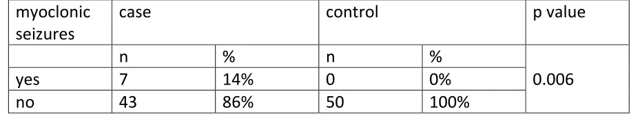
Most common seizure type among cases as well as controls was generalized
tonic clonic convulsions (64 % in case group, 56 % in control group). Seven
patients (14 %) in case group had myoclonic seizures, where as none among
control group had myoclonic seizures. This difference was found to be
statistically significant with a p value of 0.006. Mixed seizure type was also
found to be more common in case group.
Most of the patients, 34 (68%) in case group had daily seizures, before
starting treatment when compared to 8 (16%) in control group. This was found
to be statistically significant with a p value of 0.000, odds ratio of 11.15 and
95% confidence interval of 4.26 – 29.18.
Table‐6
Myoclonic seizures
myoclonic
seizures
case control p value
n % n %
0.006
yes 7 14% 0 0%
no 43 86% 50 100%
[image:42.612.96.554.566.649.2]param deve delay h/o epile birth neon
34 pa
(24 %
epile contr 0 5 10 15 20 25 30 35 meter lopmental y stat pticus asphyxia
natal seizur
atients (68
%) in cont
psy had h
rol group h
0 0 0 developm delay case n 34
tus 30
18
res 15
8 %) in case
rol group.
history of
had status
mental
y
status
% 68 %
60 %
36 %
30 %
e group ha
(p value
status ep
epilepticu
s epilepticus
Table‐7
control n 12 3 4 4
ad develop
– 0.000).
pilepticus w
s (p value
birth asphyxia
p %
0 24 %
6 % 0
8 % 0
8 % 0
pmental de
60% of p
whereas o
= 0.000)
a neonatal s
p value or
0.000 6. 0.000 23 0.001 6. 0.005 4.
elay, again
atients wi
only 6 % o
seizures
r 95%
.72 2.7
16.
3.5 6.4
85.
.46 2.0
20.
.92 1.5
16.
nst 12 patie
ith intracta
of patient
case
control
% ci
9 –
22
2 – 9
0 – 91 0 – 15 ents able
[image:43.612.104.528.123.560.2]
18 patients (36%) with intractable epilepsy had history of birth asphyxia
whereas only 4 patients (8%) with well controlled epilepsy had birth asphyxia
(p value = 0.001). history of neonatal seizures was found in 15 (30%) cases, as
against 4 (8%) controls (p value = 0.005)
Only two patients with intractable epilepsy and four patients with well
controlled epilepsy reported some reaction to antiepileptic drug history of
febrile seizures was found in 12 % of cases and 18 % of controls
Table‐8
Neurological examination
no examination case Control
n % n %
1 hemiplegia 7 14 % 2 4 %
2 quadriplegia 13 26 % 3 6 %
3 microcephaly 17 34 % 4 8 %
4 visual
disturbances
2 4 % 0 0 %
5 hearing deficit 1 2 % 0 0 %
6 aphasia 0 0 % 0 0 %
7 movement
disorder
0 0 % 0 0 %
8 no deficit 23 46 % 43 86 %
[image:44.612.101.539.442.640.2]
Amon
patie
of mi
Only
the c
0 5 10 15 20 25 30 35 40 45
ng cases,
ents had m
icrocephaly
4 cases (8
ase and co
0 5 0 5 0 5 0 5 0 5
7 patient
microcepha
y was foun
8%) and 3
ontrol grou
s had hem
aly and in 2
nd to be st
controls (
up, the mo
miplegia, 1
23 patient
atistically s
(6%) had f
st commo
13 patient
ts there w
significant
family hist
n precipita
ts had qu
ere no def
.
ory of epi
ating facto
adriplegia,
ficit. Prese
lepsy. In b
r was feve
CASE
CONTROL
, 17
ence
both
Table‐9
Neurological examination
examination case Control
n % n %
abnormal 27 54 % 7 14 %
normal 23 46 % 43 86 %
Any abnormality detected in neurological examination was to be statistically
significant significant with a p value of 0.000
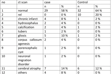
Table‐10
CT scan
no ct scan case Control
n % n %
1 normal 29 58 % 32 64 %
2 rel 0 0 % 10 20 %
3 chronic infarct 4 8 % 1 2 %
4 hydrocephalus 2 4 % 0 0 %
5 calcification 2 4 % 1 2 %
6 tubers 1 2 % 0 0 %
7 gliosis 5 10 % 1 2 %
8 corpus callosum
agenesis
2 4 % 0 0 %
9 porencephalic
cyst
1 2 % 0 0 %
10 neuronal
migration
disorder
1 2 % 0 0 %
11 cerebral atrophy 7 14 % 6 12 %
[image:46.612.105.540.154.225.2] [image:46.612.101.543.421.715.2]
CT Sc
abno
0 1 1 2 2 3 3
can was n
ormality wa
0 5 0 5 0 5 0 5
normal in
as cerebral
most of t
l atrophy 7
the cases
7 (14%), fo
29 (58%).
llowed by
The most
gliosis 5 (1
t common
10%)
CASE
CONTROL
Table‐11
MRI
no mri case Control
n % n %
1 normal 12 24 % 17 34
2 rel 0 0 % 8 16
3 c/c infarct 2 4 % 1 2
4 hydrocephalus 1 2 % 0 0
5 tubers 2 4 % 0 0
6 gliosis 5 10 % 1 2
7 corpus callosum
agenesis
4 8 % 0 0
8 porencephalic
cyst
1 2 % 0 0
9 neuronal
migration
disorder
6 12 % 0 0
10 cerebral atrophy 9 18 % 6 12
11 others 7 14 % 0 0
12 not taken 9 18 % 16 32
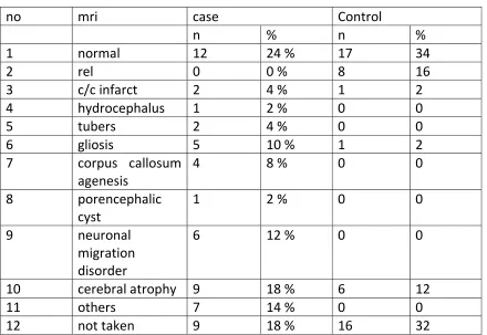
[image:48.612.102.542.136.439.2]Most enha case 4(8%
In th
signif 0 4 1 1 14 1 1
t common
ncing lesio
group wer
) and glios
his study n
ficant with
0 2 4 6 8 0 2 4 6 8
MRI findi
on 8(16%)
re neurona
sis 5(10%).
neither CT
h intractabl
ngs were
in contro
al migratio
MRI was n
T nor MRI
le epilepsy
cerebral a
ls. The oth
on disorder
normal in 1
abnorma
y in the stu
atrophy 9
her comm
rs 6(12%),
12 cases an
lity was fo
udy popula
(18%) in c
on MRI fin
corpus cal
nd 17 cont
ound to b
tion.
cases and
ndings am
llosal agen
trols
be statistic
no 1 2 3 4 5 0 5 10 15 20 25 30 35 ee su se ac fo se ac ba ab ot no 0 5 0 5 0 5 0 5 subcortica seizure activity eg ubcortical eizure ctivity ocal eizure ctivity ackground bnormality thers ormal
al focal seiz
activity EEG case n 15 2 y 12 4 17 zure y backgro abnorm
Table‐12
abnormal
% 30 %
4 %
24 %
8 % 34 %
ound mality othe lity c n % 3 5 % 7 2 % 3 ers nor control n 3 5 7 2 33 rmal %
6 %
10 %
14 %
4 %
66 %
case
control
[image:50.612.109.518.416.660.2]
Table‐13
EEG abnormality
eeg case control p value or 95 % ci
n % n %
0.001
3.76 1.64 –
8.62
abnormal 33 66 % 17 34 %
normal 17 34 % 33 66 %
The most common EEG abnormality was subcortical seizure activity 15 (30 %)
followed by background abnormality 12 (24 %). EEG was normal in 17 (34 %)
cases and 33 (66 %) control. EEG abnormality was found to be statistically
significant with a p value of 0.001.
[image:51.612.101.541.185.252.2]
Table‐14
Distribution of etiology among cases
no etiology number %
1 post meningitic/
post encephalitic
sequelae
2 4 %
2 cerebral palsy 18 36 %
3 neurodegeneration/
neurometabolic
syndrome
5 10 %
4 neurocutaneous
syndrome
3 6 %
5 cerebrovascular
accident
1 2 %
6 epileptic syndrome 2 4 %
7 cns malformaton 4 8 %
8 others 15 3 %
[image:52.612.102.540.169.418.2]
Multivariate analysis:
Multivariate analysis was done by multiple logistic regression method to
examine association between intractable epilepsy and potential predictive
factors. Female sex, multiple seizures before starting treatment, neurological
impairment, microcephaly and EEG abnormalities were found to be
independent predictors of mortality.
DISTRIBUTION
OF
ETIOLOGY
POST MENINGETIC / POST
ENCEPHALITIC SEQULAE
CEREBRAL PALSY
NEURODEGENERATION /
NEUROMETABOLIC SYNDROME
NEUROCUTANEOUS SYNDROME
CEREBROVASCULAR ACCIDENT
EPILEPTIC SYNDROME
Table‐15
Independent predictors of intractable epilepsy after multivariate analysis
Serial
number
factor Standard
error
significance Odds ratio 95%
confidence
interval
1 sex 0.416 0.017 2.68 1.18‐6.07
2 Multiple
seizures
before
treatment
0.502 0.000 9.6 3.61‐25.87
3 Neurological
impairment
0.698 0.0015 9.2 2.33‐36.19
4 microcephaly 0.793 0.0007 14.87 3.13‐70.50
5 EEG
abnormality
0.439 0.000 5.52 2.33‐13.07
[image:54.612.104.549.168.425.2]
DISCUSSION
Intractable epilepsy does not have a common agreed‐upon definition. It
makes the subject very complex and comparison of results becomes difficult.
In our study intractable epilepsy was defined as seizure frequency of atleast
one attack per month during six months despite receiving 2 antiepileptic
drugs,with adequate dosage and compliance. Definition by Chawla et al(1) is
similar to our definition. The aim of our study was to find out the predictors of
intractable epilepsy in childhood so that early identification of these children
prone for intractability is possible. This is essential for selecting patients for
more intensive investigations and treatment such as early consideration of
epilepsy surgery and for parental counselling.
In this study, majority of cases 60%, patients belonged to the age
group 5 years to 12 years. Majority of controls 52%, also belonged to the
same age group. Female gender is found to be a risk factor for developing
intractable epilepsy in this study, but this is in contrast with the results of
Javad akhondian et al (4) , where male gender was found to be a risk factor.
at onset of seizures less than one year and intractability. This is similar to the results of Chawla et al (1) and Javad Akhondian et al(4).
In our study , generalised seizures were more common than
partial seizures among cases as well as controls. Presence of myoclonic epilepsy was associated with intractability with a p value of 0.006. This is in concordance with the results of Javad Akhondian et al(4) and Chawla et al (1). However Camfield et al (12) didn’t find seizure type as a predictor of
intractable epilepsy even in univariate analysis. Multiple seizure type before starting treatment was found to be a predictor of intractable epilepsy in our study which is similar to the results of Chawla et al(1) , singhvi et al(5) and Javad Akhondian et al(4) .
In our study 68% of patient of intractable epilepsy had
developmental delay which was found to be statistically significant. 36% of patients had history of birth asphyxia in newborn period which was also statistically significant with a p value of 0.001.
We noted , as did Sillanpaa et al(11) that status epilepticus and
intractable seizures were strongly associated, partly because children who had symptomatic epilepsy were more likely to have an episode of status
epilepticus. This association was also found in the results of berg et al (2). The
odds ratio of 23.5 , suggests that occurrence of status epilepticus may be of
some prognostic significance, it may be a marker for an underlying etiology
even if none is detected at the time of initial presentation and evaluation.
epilepsy, with a p value of 0.005 with odds ratio of 4.92 and 95%
confidence interval of 1.50‐ 16.15 . This is comparable to the results of Chawla
et al (1) and Berg et al(2) . A history of febrile seizures was not found to be a predictor of intractable epilepsy. Therefore although febrile seizures are a risk factor for epilepsy, our data indicates that they are not associated with poor prognosis if epilepsy develops.
The most common examination finding in our study population
was microcephaly (34%) , followed by quadriparesis (26%) and
hemiplegia(14%). Abnormal neurological examination was found to be a predictor of intractable epilepsy, similar to the results of Chawla et al (1) and Berg et al (2) .
CT scan or MRI abnormalities, unlike other studies was not found
to be a predictor of intractable epilepsy. The most common CT scan
abnormalities detected were cerebral atrophy followed by gliosis. Common MRI findings were cerebral atrophy, neuronal migration disorders& corpus callosal agenesis. EEG abnormality was found to be a predictor of intractable epilepsy, with a p value of 0.001. This is comparable to the results of singhvi et al(5) .
Most common cause of intractable epilepsy detected was cerebral palsy found in 18 of patients.3 patients had neurocutaneous syndromes out
patients had neurodegenerative syndrome, 2 patients had neuro metabolic
syndrome and 4 patients had neuronal migration disorders. 2 patient had
lennox gestaut syndrome and 2 patient had post meningitic sequelae/post
encephalitic sequelae. Etiology couldn’t be found in 15 patients.
Independent predictors of intractable epilepsy after multiple logistic
regression analysis were female sex, multiple seizures before the onset of
treatment, neurological impairment, microcephaly and EEG abnormality.
Chawla et al study revealed the independent predictors to be neurological
impairment, age at seizure onset less than one year, myoclonic seizures and
remote symptomatic epilepsy(1) . Sillanpaa et al (5) found that occurrence of
status epilepticus, high initial seizure frequency and remote symptomatic
etiology were the only independent predictors of seizure intractability
SUMMARY
• Female gender was found to be a risk factor for developing intractable
epilepsy.
• Significant association with p value <0.001 was found between age at
onset of seizures less than one year and intractable epilepsy.
• Generalized seizures were the most common seizure type in case and
control group.
• Presence of myoclonic epilepsy was associated with intractability with a
p value of 0.006.
• History of birth asphyxia , neonatal seizures, developmental delay and
status epilepticus were also significant risk factors for development of
intractable epilepsy.
• The most common examination finding among patients with intractable
epilepsy was microcephaly followed by quadriparesis. Abnormal
neurological examination was found to be a independent predictor of
intractable epilepsy with p value of 0.0015.
• The most common CT abnormality detected were cerebral atrophy
followed by gliosis
• Common MRI findings were cerebral atrophy, neuronal migration
• EEG abnormality was found to be a independent predictor of
intractability with p value of 0.001
• Most common etiology of intractabile seizures was cerebral palsy seen
in 36% of cases, followed by neuronal migration disorders and
CONCLUSION
Factors associated significantly with intractable epilepsy both by
univariate and multivariate analysis were female sex, multiple seizures before
starting treatment, neurological impairment, microcephaly and EEG
abnormality. Early identification of these children prone to develop intractable
seizures is critical for parental counselling, selecting patients for more intensive
investigations and treatment, such as early consideration for epilepsy surgery.
BIBLIOGRAPHY
1.Chawla S, Aneja S, Kashyap R, Mallika V. Etiology and clinical predictors of
intractable epilepsy. Paediatr neurol 2002;27:186‐191.
2. Berg AT, Levy SR, Novotny EJ, Shinnar S. Predictors of
intractable epilepsy in childhood: A case control study. Epilepsia
1996;37:24-30
3. Altunbasak S, Herguner O, Burgut HR. Risk factors predicting refractoriness
in epileptic children with partial seizures. J Child Neurol.
2007;22:195-199
4. Javad akhondian, Farhad H, Seyed AJ. Predictive factors of paediatric
intractable seizures. Archives of Iranian medicine, vol 9,number
3,2006:236-239.
5.SinghviJP, Sawhney IMS, Lal V, Pathak A, Prabhakar S.
Profile of intractable epilepsy in a tertiary referral center. Neurol India
2000;48:351-6.
6. David R Chabolla. Medically refractory seizures. Jacksonville Medicine,
august 2000;12:45-56
7. AS Girija. Medical management of intractable epilepsy. Calicut Medical
8. Kleigman , Behrman , Jenson , Stanton : Nelson Textbook of Pediatrics; 18:
2457‐2473
9. Gerald M Fenichel:clinical paediatric neurology;5:19-43
10. Al Hail h, Sokrab T, Hamad A. epidemiology and etiology of intractable
epilepsy in Qatar. Qatar medical journal,june 2004;vol 13:no 1
11. SillanpaaM. Remission of seizures and prediction of intractability
in long term follow up. Epilepsia 1993;34:930-6.
12. Camfield C, Camfield P, Gordon K, Smith B, Dooley J.
Outcome of childhood epilepsy: A population-based study with a simple
scoring system for those treated with medication. J Pediatr 1993;122:
861-8.
13. Eriksson KJ, Koivicco MJ. Prevalence, classification and
severity of epilepsy and epileptic syndrome in children. Epilepsia
1997;38:1275-82.
14. Jacqueline A French. Refractory epilepsy- one size doesn’t fit all. Epilepsy
curr 2006 nov:6(6);177-180
15. Aggarwal A, Aneja S, Taluja V, Kumar R, Bhardwaj K.
Etiology of partial epilepsy. Indian Paediatr 1998;35:49-52
16. Udani VP, Dharindharka V, Nair A. Difficult to control
epilepsy in childhood—A long term study of 123 cases. Indian Pediatr
17.Houser WA, Nelson KB. Epidemiology of epilepsy in children.
Cleve Clin J Med 1989;56(Suppl 2):S185-94.
18. Sander JWAS, Shorvan SD. Incidence and prevalence studies in
epilepsy and their methodological problems: A review. J Neurol Neurosurg
Psychiatry 1987;50:829-39.
19. Blume WT. Clinical profile of partial seizures beginning at less
than four years of age. Epilepsia 1989;30:813-9.
20. Commission on Classification and Terminology of International
League Against Epilepsy. Proposal for revised classification of
epilepsies and epileptic syndromes. Epilepsia 1989;30:389-399.
21. Commission on Epidemiology and Prognosis of the International
League Against Epilepsy. Guidelines for epidemiological studies
on epilepsy. Epilepsia 1993;34:592-6
22.Chevrie JJ, Aicardi J. Convulsive disorders in the first year of
life: neurological and mental outcome and mortality. Epilepsia 1978;19:
67-74.
23. Shinnar S, Maytal J, Krasnoff L, Moshe SL. Recurrent status
epilepticus in children. Ann Neurol 1992;31:598-604
24. Matsumoto A, Watanobe K, Sugiura M, Negoro T, Takaiso E,
Iwase K. Long term prognosis of convulsion disorder in first year of life.
Mental and physical development and seizure persistence. Epilepsia
25. Datta AN, Wirrell EC. Prognosis of seizures occurring in the
first year. Pediatr Neurol 2000;22:386-91
26. Huttenlocher PR, Hapke RJ. A follow-up study of intractable
seizures in childhood. Ann Neurol 1990;28:699-705.
27. Maytal J, Shinnar S, Moshe SL, Alvarez LA. Low morbidity
and mortality of status epilepticus in children. Pediatrics 1989;83:
323-31.
28. Gross-Tsur V, Shinnar S. Convulsive status epilepticus in
children. Epilepsia 1993;34(Suppl 1):S12-20.
29. Kwan P, Brodie MJ. Early identification of refractory epilepsy.N Engl J
Med2000;324:314-319
30. Scarpa P, Carassini P. Partial epilepsy in childhood. Clinical and
EEG study of 261 cases. Epilepsia 1982;23:333-340
PROFORMA
REGISTER NO.
Name :
Age in
years
:
Sex :
1. Male
2. Female
Address :
Area :1. Rural
Weight in
Kg
:
Age of
onset of
seizures
:1. < 1 year
2. >= 1 – < 3 years
3. >= 3 – 12 years
Drugs
started
with
:
1.
2.
3.
Drugs Now
on
No of
drugs:
:
:
:
:
1.
2.
3.
4.
Duration
after
starting 2
drugs:
Response
to
treatment
:1. Intractable epilepsy
2. Responding to treatment
History of
any
reaction to
AED
:
1. Yes
2. No
Developme
nt
:1. Normal
2. Delayed
Type of
Seizures
:1. Generalised Tonic Clonic Seizures
2. Generalised Tonic seizures
3. Absence
4. Atonic
6. Simple Partial Seizures
7. Complex Partial Seizures
8. Mixed
9. Others
Frequency
of seizures
before
starting
treatment
:1. Daily
2. > 1 / week
3. 1 – 4 / months
4. < 1 / month
Frequency
of seizures
after
starting 2
drug on
average in
6 months
:1. 1
2. 2‐3
3. 4‐5
4. 6‐7
5. 8‐9
History of
status
epilepticus
:1. Yes
2. No
History
Birth
Asphyxia
:1. Yes
2. No
History of
Neonatal
Convulsion
:1. Yes
2. No
History of
Febrile
Seizures
:1. Yes
2. No
Family
History of
Epilepsy
:1. Yes
2. No
g Factors 1. Fever / Intercurrent infection
2. Lack of Sleep
3. Physical stress
4. Mental Stress
5. Watching TV
6. Others
Etiology of
Intractable
Epilepsy
:
1. Post Meningitic / Encephalitic Sequelae
2. Neurocysticercosis/REL
3. CP/MR
4. Neurodegenerative syndrome
5. Neurocutaneous syndrome
6. Head injury
7. CVA
8. Epileptic Syndrome
9. CNS Malformation
10.Mesial temporal sclerosis
11.Others
Neurologic
al
Examinatio
n
:1. Hemiplegia
2. Quadriplegia
3. Visual disturbance
4. Hearing deficit
5. Aphasia
6. Movement disorders
7. Microcephaly
8. No deficit
CT Scan :1. Normal
2. REL