PULMONARY FUNCTION TEST ANALYSIS IN
SMOKERS
Dissertation submitted to
THE TAMIL NADU DR. M.G.R. MEDICAL UNIVERSITY
CHENNAI – 600 032.
In partial fulfillment of the Regulations for the Award of the Degree of
M.D. BRANCH - I, PART - II
GENERAL MEDICINE
DEPARTMENT OF GENERAL MEDICINE
KILPAUK MEDICAL COLLEGE
CERTIFICATE
This is to certify that Dr. T. MUTHU , Post -Graduate Student (July 2003 to September 2006) in the Department of Internal Medicine, Kilpauk Medical College, Chennai- 600 010, has done this dissertation on “PULMONARY
FUNCTION TEST ANALYSIS IN SMOKERS” under my
guidance and supervision in partial fulfillment of the regulations laid down by the Tamilnadu Dr.M.G.R. Medical University, Chennai, for M.D. (General Medicine), Degree Examination to be held in September 2006.
Prof. K.S. SAIKUMAR,
M.D.,Professor & HOD
Department of Internal Medicine, Kilpauk Medical College
Chennai.
Dr. THIYAGAVALLI KIRUBAKARAN M.D.,
The Dean
Kilpauk Medical College Chennai 600 010.
ACKNOWLEDGEMENT
I am very much thankful to the Dean, Government Kilpauk Medical College and Hospital, Chennai, for granting me permission to utilize the facilities of the hospital for the study.
I express my profound thanks to my esteemed Professor and Teacher Prof. K.S. SAIKUMAR, M.D., Professor of Medicine, Government Kilpauk Medical College and hospital, encouraging and extending invaluable guidance to perform and complete this dissertation.
Iam also thankful to Assistant
Professors of medicine Dr. S. RAGUNANDAN M.D.,
Dr. K.T. JAYAKUMAR M.D., for their valuable suggestions during this study.
I wish to thank Dr. M. GUNASEKARAN, MD., DTCD,
Dr. S. KULOTHUNGAN, MD., and Dr. P. RAJASEKAR MD,
Assistant Professors, Department of Medicine, Government, Kilpauk Medical College Hospital for their valuable suggestions.
I sincerely thank Mr. S. PADMANABAN, Research Officer (Statistics), ICMR, KMCH for his guidance in analyzing this study.
I thank all our Postgraduates, House Surgeons, Staff of our College for their contribution in this study.
Last, but not the least, I express my gratitude to all the patients, but for whose cooperation this study would not have been successful.
CONTENTS
Sl.No. Chapters Page No.
Abbreviations
1. Introduction 1
2. Aim of Study 5
3. Review of Literature 6
4. Patients and Methodology 29
5. Observations 36
6. Discussion 57
7. Conclusion 62
ABBREVIATIONS
FVC - Forced vital capacity VC - Vital capacity
FEV1 - Forced expiratory volume in 1s FEV1/FVC - Ratio of FEV1 to FVC
FEF25-75% - Forced expiratory flow rate between 25 and 75% of the VC
TLC - Total lung capacity
RV - Residual volume
MMFR - Maximal midexpiratory flow rate
MIP - Maximal inspiratory pressure
INTRODUCTION
Pulmonary Function Tests involves the measure of airflow rates, lung volumes, and the ability of the lung to transfer gas across the alveolar capillary membrane. Pulmonary function parameters are very useful to diagnose obstructive lung disease. Smoking is the most important risk factor for chronic obstructive pulmonary disease.
SMOKING
Smoking is major public health problem. Cigarette smoke contains more than 40001 compounds of which a few toxins have been studied in detail. Among the most prevalent diseases with which cigarette smoking has been implicated are cancers, chronic obstructive lung disease and atherosclerotic cardiovascular disease.
In the respiratory tract smoking predisposes to malignant conditions like lung cancer, laryngeal cancer and non-malignant conditions like COPD, sleep apnea, eosinophilic granuloma of lung, respiratory bronchiolitis, Goodpature syndrome and pneumothorax.
Smoking is the major risk factor for the development of chronic bronchitis.
SPIROMETRY
Spirometry is a simple method for studying the pulmonary function by recording the volume of air movement into and out of the lungs. In this test, the subject inhales maximally to total lung capacity (TCL) and then exhales as rapidly and forcefully as possible into the turbine of spirometer, which calculates the flow rates and volume measurements. The flow rates can be calculated from the ‘spirogram’ which is a plot of volume versus time, and the volume can be calculated from the ‘ Flow – volume tracings‘ which is a plot of airflow versus the expired or inspired lung volume.
FLOW RATES
the airway obstruction that occurs in varied pathological conditions of lung.
A more sensitive means of evaluating airway obstruction is, to express the forced expired volume as percent of vital capacity, abbreviated as FEV1/FVC. This ratio is relatively independent of patients restriction of lung volumes. Normally it is 70% or greater.
Another way of assessing airflow obstruction is to measure specific flow rates. Peak expiratory flow rate (PEFR) is defined as the maximum flow achievable from a forced expiration starting at full inspiration with an open glottis. It measures the maximum expiratory flow rate over the first ten milliseconds. PEFR is reduced in large airway narrowing due to asthma, COPD, vocal cord palsy and expiratory muscle weakness etc.
FLOW VOLUMES
Flow volume curve, a recording during spirometry, of the expiratory flow plotted against expired volume, instead of time, resembles a triangular shaped envelope. At the point where 25% of vital capacity has been exhaled, this flow rate is termed Vmax25 or FEF25%, when 50% of the vital capacity has been exhaled is is termed Vmax50 or FEF50% and at 75% of vital capacity it is Vmax75 or FEF75%
The inspiratory portion of the curve is helpful in distinguishing large airway obstruction which occurs above the level of the thoracic inlet from obstruction that occur below this level. Large airway obstruction above the thoracic inlet results in a plateau of the flow rate on the inspiratory portion of the curve, while the expiratory portion in affected when
the flow limiting portion is within the thoracic cavity.
FEF75% is thought to be very sensitive to detect early small airway obstruction. In early small airway disease, the only abnormality detected may be reduced FEF75% and FEF50% with normal PEFR and FEV1.
PATTERNS OF ABNORMAL VENTILATORY CAPACITY
Obstructive ↓↓ ↓/ N ↓
Restrictive ↓ ↓↓ ↑/ N
Smoking being the most important risk factor for COPD, this study was undertaken to assess the pulmonary dysfunction in asymptomatic smokers, so that early intervention by smoking cessation would reduce the disease burden of COPD.
AIM OF STUDY
1. To evaluate the pulmonary function test parameters in asymptomatic smokers.
2. To compare the spirometric findings in asymptomatic smokers to their expected values.
3. To identify the asymptomatic COPD among smokers, so that cessation of smoking would halt their progression.
4. To identify the degrees of deterioration in PFT.
5. To correlate the dysfunction with the quantum of smoking.
REVIEW OF LITERATURE
HISTORICAL PERSPECTIVE
Chronic obstructive pulmonary disease (COPD) has been defined by the Global Initiative for Chronic Obstructive Lung Disease (GOLD) as a disease state characterized by airflow limitation that is not fully reversible.
By 1964, the Advisory committee to the surgeon general of the unitedstates concluded that cigarette smoking was a major risk factor for mortality from COPD. Subsequent longitudinal studies have shown accelerated decline in the volume of air exhaled within first second of the forced expiratory maneuver (FEV1) in a dose dependent manner to the intensity of cigarette smoking, which is typically expressed in pack years (average number of packs of cigarette smoked per day multiplied by total number of years of smoking).
increasing as the gender gap in smoking rates is diminishing globally in the past 50 years.
COPD includes all of the following clinical labels namely,13
1. Chronic bronchitis 2. Emphysema
3. Chronic airway obstruction
4. Chronic non-specific lung disease
5. Non-reversible obstructive airways disease 6. Small airway disease( obliterative
bronchiolitis)
7. Cases of chronic asthma with fixed airway obstruction
INDIAN SCENARIO
beedies. In urban area, smoking with superimposed exposure
to dust, fumes and industrial particles predispose to COPD.
DEFINITION
The American Thoracic society defines chronic obstructive pulmonary disease as a disease state characterized by the presence of airflow obstruction due to chronic bronchitis or emphysema; Airway obstruction is generally progressive, may be accompanied by airflow hyperreactivity, and may be viewed as partially reversible.
COPD is usually diagnosed by demonstration of irreversible airflow limitation. Most groups such as the American Thoracic Society (1987), and the European Respiratory Society (1995), specifically excluded asthma. However American Thoracic Society (1995) guidelines included asthma with an irreversible component within COPD. Some studies mean COPD as smoking related chronic airway disorder2,3. While others include all disorders causing chronic airway obstruction (eg. Bronchiectasis and obliterative bronchiolitis).4,5
pathological processes leading to recognition of subgroups that may have their own characterisations and natural history.6
The Dutch hypothesis originally put forward by Orie et al proposed a unitary hypothesis for all chronic airway disorders which included chronic asthma and COPD were due to constitutional disturbance, airway hyper-responsiveness (AHR) and atopy.7 However, as most workers considered asthma and COPD are different entities and COPD was considered to be due to an exogenous factor i.e. smoking, Dutch hypothesis was not accepted.
Although the attributable risk of tobacco smoking to COPD is about 85-90%8, only 15% of smokers develop COPD. Polymorphism for the genes controlling xenobiotic metabolism, hence oxidant-antioxidant balance may explain this susceptibility.9,10 About 10-15% of COPD have been thought due to other risk factors like environmental tobacco smoke (ETS), occupational exposures and genetic factors. Causal association between COPD and passive smoking requires additional evidence, as the magnitude of association is small.8
Recently COPD is defined more clearly using the following clinical diagnostic criteria.11
2. Objective evidence of airway obstruction i.e. FEV1 <80% predicted and FEV1/FVC <70% which is irreversible.
3. Usually a smoking history of more than 20 pack years.
Milder forms of chronic bronchitis may be relatively asymptomatic with only cough due to mucus production and normal airflow called “chronic simple bronchitis”.12 Most patients with chronic bronchitis do not have airflow obstruction and hence do not have COPD. However 10-15% of smokers are susceptible to more rapid decline in airflow than normal.
Emphysema is defined in anatomic terms – abnormal permanent enlargement of gas exchanging units of the lung in association with destruction of alveolar walls and without obvious fibrosis. The predominant physiological consequences of these anatomical abnormality is a decrease in elastic recoil of the lung – which in turn causes outward displacement of the chest wall, flattening of the diaphragm, hyper inflation of the lungs and increased resistance to airflow due to circumferential traction on the small airways by the over distended lung.
age. The characteristic features are reversibility of airflow obstruction and hyperresponsiveness of the airways to diverse inciting influences. In asthma the larger bronchi are predominantly affected, goblet cells undergo hyperplasia, bronchial glands hypertrophy, the basement membrane thickens, airway smooth muscle hypertrophies and the walls and lumina are invaded by eosinophils.
Localized bronchiectasis is often present in patients with COPD. As a rule, however, bronchiectasis is characterized by dilatation of airways rather than narrowing. Bronchiectasis does not contribute to obstruction of airflow. Characteristically, proximal airways are dilated and distal airways are obstructed by mucopurulent exudate and by narrowed obliterated terminal airways.
In a recent study O’Brien and colleagues evaluated cases admitted as acute exacerbation of COPD with full lung function and high resolution CT after they were clinically stable. An interesting finding of the study was that evidence of bronchiectasis was present in 29% of the cases suggesting that CT scanning may play an important role in defining the exact nature and subgroup of COPD.
disease was coined by Hogg et al 14 and consists of involvement of the peripheral airways. Small airways are those less than 3 mms in diameter most of which are respiratory bronchioles. They have a large cross sectional area and hence contribute very little to airways resistance, also called the silent zone of the lung.
A recent epidemiological study13 on prevalence of COPD in spain used spirometric criteria for diagnosis. Irreversible airway obstruction was seen in majority of the cases ( 74 .1%), though 21.7% of cases had good reversibility of airway obstruction after bronchodilator test. The prevalence is reported as 15% in smokers, 12.8% in ex-smokers and 4 .1% in non-smokers.
RISK FACTORS
risk, it is not possible to predict which person will actually develop COPD. Third, even though smoking and alpha1 antitrypsin deficiency dominate as risk factors, there are definitely other risk factors for developing COPD. Low socioeconomic status is one such risk factor22.
EPIDEMIOLOGY
COPD occurs worldwide, but is a major health problem principally in societies where cigarette smoking is common and average lifespan extend into the fifth decade and beyond. In western population it is the fourth most common cause of death and the death percent accounted for about 4.5% of all deaths.
In 1995, in the developed countries 2 million death were due to tobacco and in developing countries it could be 1 million. By 2020 it is likely to increase to 3 million and 7 million in developed and developing countries respectively 24.
In a meta analysis by Halbert RJ et al30, the prevalence of COPD was 7.5% , the prevalence of chronic bronchitis was 6.4% and that of emphysema was 1.8%. prevalence of COPD by spirometric estimates was 8.9%.
Smoking usually begins for psychological reasons, such as curiosity, parental smoking, peer pressure, rebelliousness, and assertion of independence.
Pattern of smoking is different in developing and developed countries.
In developing countries over 50% men smoke while only 10% of women smoke, compared to developed countries where 25-30% of both men and women smoke.
SMOKING AND DISEASE ASSOCIATIONS
Smoking increases the absolute number of deaths from lung cancer, cancer of other respiratory sites, chronic bronchitis/ emphysema and cor pulmonale. Death from lung cancer is 8-25 times more common among smokers with a clear dose-response relationship
. Smoking is also associated with gastro intestinal diseases like peptic ulcer disease, gastro esophageal reflux, chronic pancreatitis, crohns disease, colonic adenomas.
It can cause reproductive disease like ovarian failure, decreased sperm quality, pregnancy related events like spontaneous abortion, prematurity, fetal effects like low birth weight, SIDS, impaired lung growth, atopy and asthma.
It can also be related to renal diseases like glomerulonephritis, and others such as benign prostatic hypertrophy, cataract and peripheral vascular disease.
SMOKING AND COPD
Nearly all patients with clinically significant emphysema are smokers. Smoking is also a major risk factor for chronic bronchitis. Heavy smokers are at greater risk of developing COPD, than moderate smokers. Low exposures to cigarette smoke as encountered during passive smoking also seem to be harmful.
Fletcher and Peto16 showed that smoking was associated with irreversible obstructive changes in airways in some subjects while not in others.
study in 1393 middle aged Norwegian males and followed up the patients for about 7 years. The study stated that smoking is definitely associated with increased risk of COPD. But other factors also determine its predisposition.
Marcus et al18 described that smoking causes decline in pulmonary function in dose dependent manner and cessation of smoking improves the obstructive pattern.
On an average, the rate of expiratory airflow in smokers decrease twice as fast in smokers (40 ml per year) as in non- smokers( 20ml per year). 19
Nonetheless, most smokers do not develop symptomatic COPD, probably because of the large physiological reserve of the lung. However some smokers (15%) do experience accelerated loss of lung function and develop symptomatic disease. The basis of the heightened susceptibility in this group is not known.
The degree of cough, phlegm and wheeze are directly related to number of cigarette smoked daily. The tar yield in the cigarette is related to the volume of sputum production.20
exercise tolerance is aggravated by high levels of carboxyhemoglobin 21
PATHOPHYSIOLOGY
Many pulmonary function abnormalities occur in COPD. Increases in residual volume, and the residual volume to lung capacity ratio, non uniform distribution of ventilation and ventilation perfusion mismatch are also typical findings but, persistent reduction in forced expiratory flow rates is the most typical finding. Reduction in FEV1 and FEV1/FVC are the characteristic physiological abnormalities of COPD.
Airflow during forced exhalation is the result of balance between the elastic recoil of the lungs promoting the flow and the resistance of the airways limiting the flow. The relative contribution of diminished elastic recoil and increased resistance in reducing maximal expiratory airflow can be quantified from the flow – pressure curves. With decreased elastic recoil, the curve has a normal slope, but it terminates prematurely. In contrast, with increased airway resistance the slope becomes less steep, reflecting the necessity for increased driving pressure for any level of airflow.
abnormalities in COPD, certain generalisations can be made. The arterial Po2 (Pao2), usually remains near normal until the FEV1 is decreased to about half of the predicted value. Even a much lower FEV1 may be associated with normal Pao2, atleast at rest. An elevation of Paco2 is not expected in COPD until the FEV1 is less than one-fourth of the predicted value.
PATHOGENESIS
COPD is characterized by mild chronic inflammation throughout the airways, lung parenchyma and pulmonary vasculature.22
Various proposed hypothesis for COPD are
1. Inflammatory repair hypothesis
2. Proteinase anti-proteinase hypothesis
INFLAMMATORY REPAIR HYPOTHESIS
Neutrophils, macrophages and CD8+ lymphocytes are the major cells involved in the inflammatory response in COPD patients. Smoking plays a major role in producing such inflammatory reaction in the airways and lung parenchyma.
Smoking recruits macrophages, neutrophils and lymphocytes and these cells are increased in various parts of the lung. The inflammatory cells get activated and initiate the inflammatory process through various mediators.
Activated inflammatory cells release a variety of mediators including Leukotriene B4, IL-8, TNF and others capable of damaging lung structure or sustaining neutrophil inflammation.
Any stimulus that increases either the number of leukocytes ( neutrophils, macrophages ) in lung or release their elastase containing granules, increase the elastolytic activity thereby triggering a chronic inflammatory response in the lung.
In smokers, neutrophils and macrophages accumulate in the alveoli and pathogenesis may involves the following processes.
● Direct chemoattractant effects of nicotine and also due to the effects reactive oxygen species in smoke
● Accumalated neutrophils are activated and release their granules rich in variety of their cellular proteases ( Neutrophil elastase, Proteinase 3,
Cathepsin G).
● Various cellular proteinases released cause tissue destruction.
● Smoking enhance the elastase activity of the macrophages.
● Macrophage elastases are not inhibited by alpha 1 AT and hence the
elastase activity is unchecked causing tissue destruction.
● Matrix metalloproteinases from neutrophils and macrophages also
play an important role in tissue destruction.
● Oxidant and anti-oxidant balance is essential and smoking disturbs it
causing an imbalance, once again predisposing to tissue destruction.
● Normal lung contain a lot of anti-oxidants like SOD,
● Tobacco smoke contain an abundant reactive oxygen species ( Free
radicals) that deplete these anti-oxidant mechanism thereby inciting tissue
damage.
● Secondary consequence of oxidative injury is inactivation of
native anti - proteases resulting in functional alpha1 anti- trypsin deficiency
even in patients without enzyme deficiency.
PROTEINASE ANTI-PROTEINASE HYPOTHESIS
According to this theory there is steady and episodic release of proteolytic enzymes into the lung parenchyma. Normally, plasma proteinase inhibitors, especially alpha1 anti-trypsin, permeate lung tissue and prevent the proteolytic enzymes from digesting the structural proteins of the lung. The uninhibited activity of proteinases cause destruction of the Elastin in the lung parenchyma which is the key event in the development of emphysema. Elastin is the principal component of elastic fibers, which are prominent part of lung’s extra cellular matrix.
protein. Elastin synthesis in the lung begins in the late fetal life and stops in adult life. Destruction of the lung elastin plays an important role in the pathogenesis of emphysema.
COMPLICATIONS
• Secondary erythrocytosis
• Recurrent acute exacerbations
• Pneumothorax
• Chronic and acute on chronic respiratory failure
• Pulmonary hypertension
• Chronic Cor Pulmonale
• Right ventricular failure
• Weight loss
TREATMENT FOR ACUTE EXACERBATION
1. Oxygen therapy
2. Bronchodilator- nebulised salbutamol (5mg) or terbutaline (10mg) + ipratropium 500µg
3. Corticosteroids – inhaled, in severe cases parenteral steroids.
5. Antibiotics
6. Respiratory stimulants like Doxapram
7. Chest physiotherapy
8. Mechanical ventilator – Non invasive positive pressure ventilation is the procedure of choice
TREATMENT FOR CHRONIC COPD
1. Smoking cessation
2. Avoid dusty environment
3. Bronchodilator
4. Steroids
5. Theophylline or aminophylline
6. Cilomilast31 is an orally active phospho diesterase inhibitor-4 and is effective in reducing the exacerbations and maintaining lung function.
SMOKING CESSATION
euphoria inducing drugs such as cocaine, amphetamine and morphine.
Nicotine has number of other effects on CNS. It may improve task performance and attention time, measurably in non habituated subjects. It may also alleviate anxiety and depression and induce a sense of well being, while causing a state of arousal. Unfortunately nicotine is also an addicting substance.
Smoking is a major public health problem. Health hazards attributable to smoking parallel smoking prevalence. Hence smoking cessation is very important for reducing the disease burden of COPD which is the fourth most common cause of death worldwide. Smoking cessation can be attained by
1. Non pharmacological methods
2. Pharmacological
Non pharmacological methods involve various behavioral techniques.
HYPNOSIS is to enable the smokers to achieve an altered state of consciousness that enhances the ability to quit. However, it is of low reliability with published qui rates between 0 and 88%.
ADVERSIVE CONDITIONING is extinguishing smoking by associating it with negative sensation like electric shock, nausea inducing drugs, hot, smoky air treatment. It is of limited value.
GROUP COUNSELING offered by several commercial and voluntary health organizations. It typically include lectures, group interaction, some form of tapering method like quit a day development of coping skills. This method is largely limited to cities and one year success rate ranges between 15-35%.
PHARMACOLOGICAL METHODS
It includes tranquilizers, antidepressants, topical anaesthetics, anti anxiety agents, anti cholenergics, clonidine and nicotine replacement therapy.
Lobeline sulphate is pharmacologically similar to nicotine and was selected as stop-smoking drug. Interest in it has declined but recently sublingual formulation are being tested.
Bupropion, originally developed as anti depressent is very useful in nicotine deaddiction.
Nicotine replacement therapies
Nicotine replacement therapy dates back to 1950s in the form of lozenges. Today there are several forms of nicotine replacement therapy available, including nicotine polacrilex, Transdermal nicotine, nicotine nasal spray, nicotine vapours and aerosols, and nicotine toothpicks. Two widely adopted therapies are
1. Nicotine polacrilex - Nicotine is bound to a resin and is available in two doses of 2mg and 4mg. Blood nicotine levels are less than 40% associated with customary smoking. It is proved to be effective in reducing the nicotine withdrawal symptoms.
2. Transdermal nicotine – primary advantage of transdermal delivery system is ease of use and controlled drug delivery. They reduce withdrawal symptoms and studies have proved that abstinence associated with transdermal patch is double that of placebo.26
1. Persistent hypoxemia with failure to maintain Pao2 55-60mm of Hg or oxygen saturation <88-90% despite maximum medical treatment
2. Worsening respiratory acidosis
3. Signs of progressive respiratory muscle fatigue 4. Deterioration of mental status
5. Inability to protect airway
6. Inability to clear copious secretions.
PATIENTS AND METHODS
PLACE OF STUDY
This study was conducted in the general medical ward of Government Kilpauk medical college Hospital, Chennai.
PERIOD OF STUDY
From April 2004 to February 2005.
DESIGN
Observational prospective cohort study of patients who are smokers admitted in the hospital for complaints other than respiratory symptoms. A total of 100 patients were included in the study.
METHODOLOGY
A. Subject selection
1. Inclusion criteria
a. Subjects randomly selected from medical ward in age group 20 -60 years.
2. Exclusion criteria
a. Patients who had symptoms as per American Thoracic Society scale for dyspnoea
b. Known case of cardiac disease. c. Known diabetic patients.
d. Patients with spinal deformity. e. Age > 60 years
All patients included in the study were subject to thorough clinical examination. All were subject to laboratory investigation as per the proforma.
After the investigations, patients were subjected to spirometry in medical department, kilpauk medical college, Chennai by using FUKUDA SANGYO spirometer and results were recorded and analyzed.
FUKUDA SANGYO
The spirometer used was FUKUDA SANGYO. This spirometer can measure and analyze the pulmonary function by means of Fleish type flow sensor and variable reluctance type transducer.
UDWADIA’S FORMULA
VARIABLES MALE FEMALE
FEV 1(L) Adults <30years Adults <30years 0.039×H-0.010 × A-3.266 0.025× H-0.011 × A-1.424
Adults >30years Adults >30years 0.037×H-0.022× A-2.650 0.032×H-0.012×A-2.580 FVC(L) Adults <30years Adults <30years
0.055×H+0.019×A-6.058 0.030×H+0.006×A-2.284 Adults >30years Adults >30years 0.054×H-0.018×A-4.832 0.043×H-0.010×A-3.755 PEF 0.085×H-0.0187×A-6.2083 0.0497×H-0.0336×A-0.1399
FEF25-75% 0.0173×H-0.0407×A+ 0.0245×H-0.0336×A0.1399
1.6108
FEF50 0.0195×H-0.0365× 0.0272×H-0.279×A-0.2704
A+1.7383
FEF75 0.0088×H-0.0301×A+1.0402 0.0113×H-0.0288×A+0.5012
A - Age in years H - Height in cms
Interpretation
Various abnormalities are
1. Obstructive pattern
FEV1 < 70% FVC
2.
Restrictive
patternFEV1 > 80% FVC
In normal subjects a full VC may be expired in 3 seconds and a slow VC equals FVC. So %FVC is taken for grading restriction.
Normal > 80%
Mild restriction ≥66 - 80%
Moderate restriction ≥50 - 65%
Severe restriction <50%
3. Mixed pattern
All datas obtained were statistically analyzed and their significance studied.
However, patients cooperation, standardization of the equipment and the procedure are crucial for any meaningful interpretation. It is also important to calculate the percent predicted values based on regression equations drawn from the population to which the patient belongs. From tropical countries like India there are large number of reports on normal spirometric values which can be used for prediction.
A value less than the lower limit of normal can be considered normal provided the technique is standardized and the measurement well judged.
The patient is then connected to the spirometric tubing using a mouthpiece and all leaks around the mouthpiece are seen to be occluded.
Nasal leaks are prevented by occluding the nose with nasal clips.
The patient is then asked to breathe normally in the spirometer circuit for a couple of breathes and then patient is asked to take maximum inspiration and stop. Then patient is instructed to expire forcefully through the mouthpiece.
The computer analyzes these flows and volumes in flow-volume and volume-timed formats to give results comparing them with the normal predicted values. Spirometry can find VC, FVC, FEV1, FEV1/FVC, FEF 25-75%.
Clinical spirometry detects obstructive lung disease by measuring dynamic flow rates. It identifies whether flow obstruction is present or not, and characterizes whether the obstruction is inspiratory, expiratory or both.
grantifies the stage of the disease with broad limits of mild, moderate and severe.
General rule, expiratory flow rate < 70% of predicted normal value of FEV1/FVC ratio is suggestive obstructive lung disease. All the values obtained were statistically analyzed.
GOLD classification of COPD
STAGE CHARACTERISTICS
0 : at risk Normal spirometry
I : mild COPD FEV1/FVC <70%
FEV1≥ 80% predicted
II : moderate COPD FEV1/FVC <70%
30% ≤ FEV1 < 80% predicted
III: severe COPD FEV1/FVC <70%
FEV1 < 30% predicted OR presence of
RESULTS AND ANALYSIS
In this study, 100 patients admitted for other complaints were selected according to inclusion criteria and studied.
DETAILS OF STUDY POPULATION
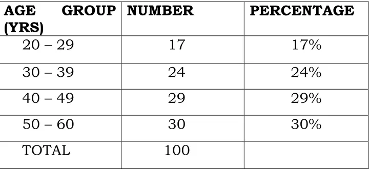
[image:42.595.97.457.603.769.2]Total Number of Patients - 100
TABLE –1
Age Distribution
AGE
GROUP
(YRS)
NUMBER
PERCENTAGE
20 – 29
17
17%
30 – 39
24
24%
40 – 49
29
29%
50 – 60
30
30%
Age Distribution
17%
24%
29%
30%
TABLE – 2
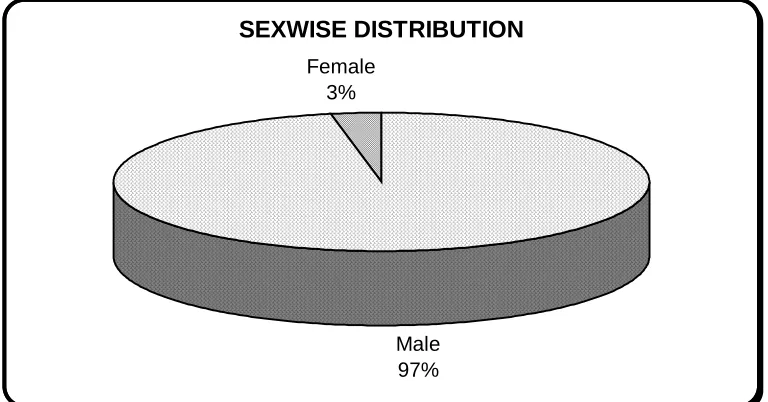
SEXWISE DISTRIBUTION
S.No. Sex Number Percentage
1. Male 97 97%
2. Female 3 3%
Among the patients 97 were male and remaining were female
SEXWISE DISTRIBUTION
Male 97% Female
3%
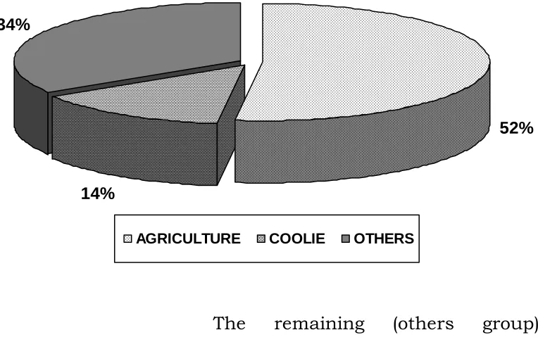
OCCUPATION
[image:46.595.94.517.275.437.2]Patients of varied occupation were studied . Among them most of them belonged to the group of agricultural workers
TABLE -3
CASES PERCENT
FARM WORKERS 52 52 MANUAL
LABOURERS
14 14
52%
14% 34%
AGRICULTURE COOLIE OTHERS
[image:47.595.126.508.112.355.2]The remaining (others group) patients comprised of factory workers, mansons, butchers etc.,
TABLE – 4
DURATION OF SMOKING
DURATION IN
PACK YRS
CASES
%
0 -5
6
6
5 – 10
38
38
10 – 15
20
20
Most of the patients (38%) belonged to 5-10 pack years of smoking. Another 36% belonged to > 15 pack years of
0%
5%
10%
15%
20%
25%
30%
35%
40%
0-5
5-10yrs
10-15yrs
>15
TABLE – 5
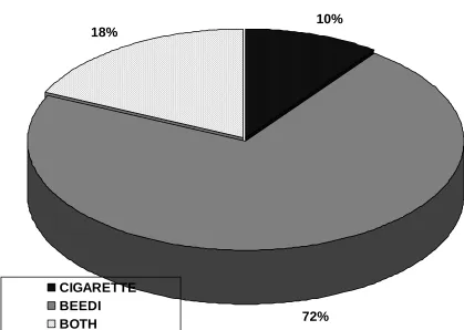
DISTRIBUTION OF SMOKERS
TYPE CASES %
CIGARETTE 10 10
BEEDI 72 72
BOTH 18 18
10%
72% 18%
TABLE – 6
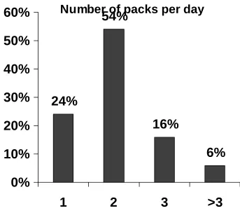
BEEDIES CONSUMPTION
NUMBER OF PACKS PER DAY
CASES %
1 24 24
2 54 54
3 16 16
>3 6 6
Number of packs per day
24%
54%
16%
6%
0%
10%
20%
30%
40%
50%
60%
Other habits
74% were alcoholic, usually combined with smoking beedies
GENERAL EXAMINATION
The general examination of the patients showed the following abnormalities. Of the 100 cases studied, 38 patients had pallor. Ten patients had clubbing.
General examination
0
5
10
15
20
25
30
35
40
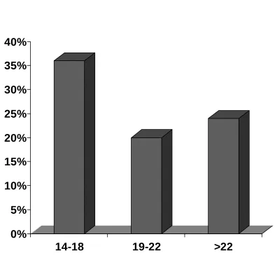
TABLE - 7
RESPIRATORY RATE RANGE
RESPIRATORY
RATE
CASES PERCENT
14 – 18 36 36 19 -22 20 20 >22 24 24
0%
5%
10%
15%
20%
25%
30%
35%
40%
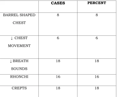
TABLE - 8
RESPIRATORY SYSTEM EXAMINATION
CASES
PERCENTBARREL SHAPED CHEST
8 8
↓ CHEST
MOVEMENT
6 6
↓ BREATH
SOUNDS
18 18
RHONCHI 16 16
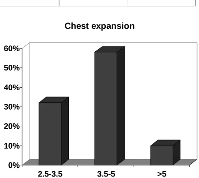
TABLE – 9
CHEST CIRCUMFERENCE
CHEST
EXPANSION
CASES PERCENT
2.5 -3.5 32 32 3.5 – 5 58 58 >5 10 10
0%
10%
20%
30%
40%
50%
60%
2.5-3.5
3.5-5
>5
TABLE
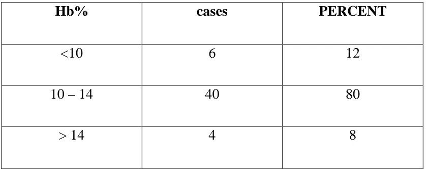
- 10
HAEMOGLOBIN
Hb%
cases
PERCENT
<10
6
12
10 – 14
40
80
> 14
4
8
TABLE
-11
LEUKOCYTE COUNT
TC CASES PERCENT
< 6000 1 5
6000 – 10000 60 60
10001 – 14000 36 36
[image:56.595.94.513.155.322.2]ESR VALUES
Out of the 100 cases studied , 42 had ESR 6-10/ hr
58 had 11- 30/ hr
Random blood sugar, blood cholesterol, and serum creatinine were in normal range.
PCV values – none had polycythemia.
CHEST X-RAY
It was normal in most of the patients ( 94%).
6 patients had early emphysematous changes.
NORMAL EXPECTED SPIROMETRIC VALUES IN
INDIA (MEAN + SD)
Male
15-19
20-24
25-34
35-44
45-54
>55
FVC 3.49±0.57 3.65±0.53 3.59±0.54 3.31±0.62 3.14±0.65 2.98±0.67 FEV1 3.04±0.52 3.09±0.51 2.9±0.47 2.6±0.53 2.42±0.42 2.35±0.51 FEV1/FEC% 87.1±11 84.6±7 80.6±9 78.4±10 72.4±10 68.9±9 PEFR 530±72 553±72 559±87 512±98 471±91 451±102SPIROMETRY
32 Patients showed combined defect ( both restrictive and
obstructive)
46 Cases showed obstructive pattern
8 cases showed restrictive pattern
14 showed normal study.
Spirometric patterns
32%
46%
8% 14%
FEV
1EXPECTED VS ACTUAL
AGE
GROUP
(YRS)
EXPECTED ACTUAL
PERCENTAGE
20
–
29
2.99
±
0.49
2.38
79%
30
–
39
2.7
±
0.47
1.99
73%
40
–
49
2.51
±
0.51
1.32
53%
50
–
60
2.34
±
0.49
1.01
43%
MEAN 2.56
1.67
The expected FEV
1among subjects studied was 2.51± 0.42 L,
however, the actual mean FEV
1was 1.67± 0.5 L, 65.2% of the
FEV 1 Expected vs Actual
0
0.5
1
1.5
2
2.5
3
3.5
20-29
30-39
40-49
50-60
FEV
1%
STAGES OF FEV1 CASES PERCENT
> 80% 18 18
60-80% 22 22 <60% 60 60
18%
22% 60%
>80% 60-80% <60%
FEV
1%
AND PACK YEARS
PACK YEARS
FEV1%
0-5
81%
5-10
71%
>15
41%
0% 10% 20% 30% 40% 50% 60% 70% 80% 90%
0-5 5--10 10--15 >15
DISTRIBUTION OF CASES ACCORDING TO FEV
1/FVC%
FEV1/FVC CASES PERCENT
40 – 60 32 32 < 40 0 0
Actual mean FEV1/FVC was 70.49% of the expected value.
FEV1/ FVC
34%
34% 32%
>80% 60-80% 40-60
DISTRIBUTION OF CASES ACCORDING TO FEF
25-75%60 -80
18
18
<60
74
74
FEF25-75%
8%
18%
74%
>80
60-80
<60
FEF
25-75%is a good indicator of small airway disease.
The mean value is 49.04%
GENDER DIFFERENCE
MALE
FEMALE
FEV
11.01(39%)
0.92(43%)
DISCUSSION
In a similar study by Zielensky.J et al27., spirometric analysis, airway limitation was observed in 20.3% patients and restrictive pattern was observed in 8.3% patients comparable to this study.
In this study, though patients did not complain subjective respiratory symptoms, 10 patients had clubbing. In the present study, decreased chest movement was seen in 12% patients, decreased breath sounds in 18% and barrel shaped chest was seen in 8% subjects. In a similar study by K.V.Thiruvengadam et al., decreased chest movement, decreased breath sound, and barrel shaped chest were seen in 50%, 43.3% and 16.7% patients.
An important finding in this study is that 88% patients had reduced FEF25-75% and 74% showed significant fall in FEF25-75% < 60%. FEF25-75% is a very good indicator of small airway disease. Statistically the percentage of reduction occurred in 88% of patients which is significant.
but that these airways become the principal sites of increased resistance in COPD. This study gave rise to the concept that COPD is a small airway disease.
In male, the reduction in forced expiratory volume reduction in 1s (FEV1) per year above the normal decline in adults for each pack-year of smoking is 9 ml; in female the excess rate of decline is about 6 ml. Based on these rates of decline, a man who has smoked one pack per year for the past 20 years will have FEV1 that is 180 ml less than it would have been if the person had not smoked. In this study it is observed that patients with longer duration of smoking had much reduced FEV1 than those with shorter duration of smoking.
In the present study, there was significant reduction in FEV1. The mean FEV1 was 1.67±0.05 L against the expected 2.56±0.49L which was 65% of the expected for the age. Reduction in FEV1 was maximum in the age group of 50-60 years - 43% of the expected and minimum in 20-29 yrs -79%.
the rate of decline of FEV1 that normally begins by 25 years of age, was markedly increased in 10-15 % of susceptible smokers. Clinical disability did not occur until late in the disease when FEV1 was reduced by about 70% of the expected value. This study results may help to explain why many patients even when FEV1 was decreased to 60% did not have respiratory symptoms like breathlessness. The findings of Fletcher’s study have been confirmed by the Lung Health Study.23
The Lung Health Study revealed that among middle- aged smokers with FEV1 between 55 and 90% of predicted, differences of several hundred ml in FEV1 developed within five years between those who quit and those who did not quit smoking. Such deleterious effect of smoking was seen in 10-15% of the susceptible smokers.
The mean FEV1 in this study was 1.675 L and the expected mean FEV1 was 2.56± 0.42 L, which is 65.5% of the expected value. In a similar study by Vaidya PR et al the observed FEV1 was 1.28 ±0.66L.
The mean expected FVC was 3.25 ±0.48L , the actual mean was 1.94 ± 0.64 L which was about 60% of the expected
On an average, the rate of expiratory airflow in
smokers decrease twice as fast in smokers ( 40ml per year) as in non smokers( 20ml per year). 19
The small sample of 3% female patients were matched for age
and smoking habits and it was found that FEV1 was 0.92L (39%) compared to 1.01 (43%) in male subjects.
Though definitive conclusion could not be drawn due to small
sample size, the accelerated decline in fev1 was noted in women than men.
In a study by Gan WQ et al28., it was concluded that women as
they age, are at increased risk of accelerated decline in FEV1. In another
study by Johannessen et al29., sex and residential area did not influence the
incidence of COPD.
CONCLUSION
In this study, 46% patients showed obstructive pattern, 32% showed mixed pattern, 8% showed restrictive pattern and 14% subjects had normal lung function.
Progressive decline of FEV1 ( more than that of age
related decline) was observed in asymptomatic smokers as the f was 79%, 73%, 53%,and 43% in the age groups of 20-29, 30-39, 40-49 and 50-60 respectively.
Age, increased quantum of smoking, female gender all are compounding factor in obstructive lung disease as evidenced by more FEV1 reduction with increasing age, increased pack years of smoking. Mean FEV1 was
65.5% of the expected normal value.
Though definitive conclusion could not be drawn, due to small sample size, the accelerated decline of FEV1
was noted in women than men, as age matched FEV1 was 39% in women as compared to 43% in men of same age.
Routine Pulmonary function test in asymptomatic smokers is an important investigation to identify early obstructive/mixed/restrictive lung disease, lest such identified persons could be enrolled for nicotine
deaddiction. Early nicotine deaddiction if
BIBLIOGRAPHY
TEXT BOOKS
1. Steven E. Weinberger, Jeffrey M. Drazen : disturbance of respiratory function, Harrison’s principles of internal medicine – 16 th edition
3. Stofalar NM, Vermiere P et al., optimal assessment and management of copd. Eu. Resp . J. 1995 ; 8: 1398-1420
4. Bergund DJ , Abbey DE et al., respiratory symptoms and pulmonary function in elderly non- smoker population. Chest 1999; 115: 49-59.
5. Avitol A , Springer C , et al ., Adenosine, methacholine and exercise challenges in children with asthma or pediatric COPD. Thorax1995 ; 50 : 511 -516.
6. Wedzicha J et al ., The heterogenecity of COPD. Thorax 2000; 5: 631 – 632.
7. Orie NGM, de Vrest K et al., the Host factors in Bronchitis: an international symposium, Assen, Netherlands. 1961
8. Thun MJ , Myers DC, Day Lally C et al., Age and exposure – response relationship between cigarette smoking and premature death in cancer prevention study.
9. Koyane H , Geddes DM ; Oxidative stress and the risk of COPD: Thorax 1998 ; 53 ( suppl 2)
11. Calverly P , Bellamy D. The challenge of providing better care for patients with COPD Thorax 2000, 55: 78 – 82.
12. Peto R , Spalzer FE, Cochrane AL et al ., The relevance in adults of airflow obstruction but not mucopus hypersecretion from chronic lung diseasse : Am Resp Disease 1983 ; 128: 491 – 500.
13. Joshi J.M et al ., COPD : The status today ; API Medicine update.
14. Hogg JC, Macklem PI et al ., Site and nature of airway obstruction in COPD. NEJM 1968 : 278: 1355 – 1360.
15. Evan DJ , Green M. Small airways – A Time to revisit ? Thorax 1008, 53: 629 – 630.
16. Fletcher C, Peto R. The natural history of chronic airflow obstruction. BMJ 1977.
17. Sandrick L et al ., Long term effects of smoking on lung function test. BMJ 1995 ( 311 : 715 – 720)
19. Fletcher C, Peto R, The natural history of chronic bronchits and emphysema, New York Oxford clinical press, 1976 , p 272.
20. Higenbottom T, Clark TJH et al ., lung function and symptoms of cigarette smoking related to Tar yield and number of cigarette smoked. Lancet 1980; 1:409.
21. Calverly PMA, Leggett RJE et al., Smoking and exercise tolerance in chronic bronchitis and emphysema BMJ 1981 ; 2: 878.
22. Bake PS, Hanoa R et al.,: Educational level and obstructive lung disease given smoking habits and
occupational airborne exposure: a Norwegian
community study . Am J. EPI 141:1080-1088-1995
23. Anthonben NR, Connelt JE et al: Effects of smoking intervention and the use of an inhaled anti cholinergic broncho dilator on the rate of decline of FEV1. The Lung Study. JAMA272: 1497-1505; 1994
24. Crofton textbook of respiratory medicine.
25. Fishman’s pulmonary disease and disorders
27. Zielenzki J, Bednerak M et al., Increasing COPD Awareness. Eur resp J 2006; APR27(4) 833-852.
28. Gan WQ, Man SF et al Female smokers beyond perimenopausal period are at increased risk of COPD: a systemic review and meta analysis
29. Johannessen A, Omenaas e et al., incidence of GOLD defined COPD in general adult population. Int. J TB lung disease 2005 aug; 9(8) 926-32.
30. Halbert RJ, Natoli JL et al., Global burden of COPD; Eur Resp J : 2006 ; apr12
31. Jindal SK, Aggarwal AN et al., A multicentric study on epidemiology of COPD and its relationship with tobacco smoking and environmental smoke. Indian J of chest dis all sci; 2006 JAN-MAR; 48(1) 23-9.
32. Sommerhoff CP, Nadel JA ,et al : Neutrophil elastase and cathepsin G Stimulate secretion from cultured airway gland serous cells.J clin invest 85 : 682 – 689, 1990
33. Thurlbeck W: Lung function and structure in Cigarette smokers. Thorax 49:1276, 1994
35. Jorgen V, Prescott E,Lange P: Assosiation of chronic mucus hypersecretion with FEV1 decline and COPD morbidity. Am J Respir Crit Care Med 153: 1530 – 1535, 1996
PROFORMA
Case No.
Name : Age : Sex :
IP No. : DOA : DOD:
FINAL DIAGNOSIS: H/o Hypertension H/o diabetes mellitus
Past History
IHD TB RHD BA
Personal History
TOBOCCO use: Smoking Beedi/ Cigarette/ pipe / cigar
Quantity
Period Other forms Alcohol
GENERAL EXAMINATION
Height
Weight
BMI
PR : RHYTHM : CHARECTOR :
Temperature:
BP:Respiratory rate: Type
:Pallor:
Cyanosis:
Lymph node: Clubbing:
JVP:
RESPIRATORY SYSTEM
UPPER RESP.TRACT NONE PNS THROAT
INSPECTION
Shape of chest: B/L Symmetrical / Not Trachea position: R / Central / L
Any spinal deformity: Scoliosis / kyphosis / Normal Any chest deformity: Hollowing / Bulging / Normal Movement of chest: Equal / Not
Any dilated veins/pulsations: Present / Not Any scar / sinuses : Present / Not
PALPATION
Tracheal position: R / Central / L Movement of chest: Equal / Not Apical impulse: Normal / Not
MEASUREMENTS
AP Diameter:
Transverse diameter: Chest circumference: Expansion:
Palpable rales/ ronchi: Yes / No Site Percussion Anteriorly Kronig isthmus Infraclavicular Mammory
Liver dullness 5th ICS Yes / No Traubes area Tympanic / Not Tidal percussion: Posteriorly Suprascapular: Infrascapular: Interscapular: Axillary: Infra axillary: AUSCULTATION
Breath sounds - normal / diminished
Type - Vesicular / Bronchovesicular / Bronchial Added sounds - rales / rhonchi
Site :
VR Equal / Not
CVS
ABDOMEN CNS
Investigations :
Blood Sugar : URINE :
Serum Creatinine : CXR PA :
ECG :
Spirometry : Others
Bronchoscopy HRCT
MASTER CHART
S.
No. IP No. Age Sex occupation
Duration of
smoking FEV1% FEC1/FVC FEF25-75%
1 107402 23 1 2 1 1 1 1
2 107569 30 1 1 2 1 1 2
3 107600 26 1 1 2 1 1 2
4 107771 38 1 3 2 1 1 1
5 107772 57 1 1 4 3 3 3
6 107974 24 1 1 1 1 1 1
7 108064 58 1 2 4 3 3 3
8 107984 46 1 1 3 3 2 3
9 108411 49 1 3 3 3 3 3
10 108481 39 1 1 2 2 1 3
11 108492 60 1 1 4 3 3 3
12 108596 31 1 2 2 1 1 1
13 108730 40 1 3 2 2 1 3
14 109025 59 1 1 4 3 2 3
15 109026 54 1 1 3 3 2 3
16 109137 59 1 2 4 3 3 3
17 109146 41 1 1 3 3 2 3
18 109289 42 1 1 3 3 3 3
19 109349 44 1 3 3 3 2 3
20 109360 53 1 2 4 3 3 3
21 109681 51 1 1 4 3 2 3
22 109779 59 1 1 4 3 3 3
23 109980 42 1 2 2 2 1 3
24 109984 25 1 1 1 1 1 1
25 110026 33 1 3 2 2 1 2
26 110139 57 1 1 4 3 2 3
27 110163 35 1 3 2 2 1 2
29 110260 41 1 1 2 3 1 3
30 110288 58 1 3 4 3 2 3
31 110575 36 1 1 2 2 1 2
32 110585 55 1 3 4 3 3 3
33 110684 48 1 1 3 3 2 3
34 110857 53 1 2 4 3 3 3
35 110870 44 1 1 3 3 2 3
36 111042 59 2 3 4 3 3 3
37 111138 35 1 1 2 2 2 3
38 111542 40 1 3 2 2 2 3
39 111630 51 1 3 4 3 3 3
40 111748 28 1 1 2 1 1 3
41 112044 60 2 3 4 3 3 3
42 112104 59 1 1 4 3 3 3
43 112402 49 1 3 3 3 2 3
44 112455 35 1 3 2 2 1 2
45 112459 39 1 1 2 2 2 2
46 112582 29 1 3 2 1 1 1
47 112586 36 1 1 2 3 3 3
48 112661 51 1 3 3 3 2 3
49 112786 56 2 1 4 3 3 3
50 112911 42 1 1 2 2 2 3
51 112961 58 1 1 4 3 2 3
52 113053 31 1 1 2 1 1 1
53 113211 52 1 1 4 3 2 3
54 113146 44 1 1 3 3 2 3
55 113204 58 1 1 4 3 3 3
56 113284 40 1 1 2 2 1 2
57 113361 42 1 2 2 2 1 2
58 113529 37 1 1 2 2 1 2
59 113619 56 1 3 4 3 3 3
61 113800 52 1 2 4 3 3 3
62 113820 59 1 1 4 3 3 3
63 113815 43 1 1 3 3 3 3
64 113828 60 1 2 4 3 3 3
65 114140 36 1 1 2 2 2 2
66 114165 27 1 1 2 1 1 1
67 114263 51 1 3 4 3 2 3
68 114301 59 1 1 4 3 3 3
69 114364 40 1 1 2 2 2 3
70 114424 49 1 3 3 3 3 3
71 114624 24 1 2 1 1 1 1
72 114648 51 1 3 4 3 3 3
73 114653 48 1 3 3 3 2 3
74 114686 29 1 3 2 1 1 2
75 115074 60 1 1 4 3 3 3
76 115092 25 1 1 1 1 1 1
77 115205 60 1 1 4 3 3 3
78 115094 45 1 3 3 3 2 3
79 115291 35 1 1 2 3 3 3
80 115319 59 1 2 4 3 3 3
81 115332 26 1 1 1 1 1 1
82 115555 34 1 3 2 2 1 2
83 115706 58 1 3 4 3 3 3
84 115782 41 1 1 2 2 2 3
85 115783 53 1 3 3 3 2 3
86 115813 34 1 3 2 2 1 3
87 115936 53 1 1 3 3 2 3
88 116013 59 1 3 4 3 3 3
89 116031 40 1 1 2 3 1 3
90 116242 30 1 2 2 1 1 3
91 116463 57 1 3 4 3 2 3
KEY
SEX: MALE-1; FEMALE-2
OCCUPATION: FARM WORKER-1; MANUAL LABOURER-2; OTHERS-3 DURATION: 0-5YRS-1; 5-10YRS-2; 10-15YRS-3; >15YRS-4.
FEV1 : >80%-1 ; 60-80%-2 ; <60%-3
FEV1/FVC : >80%-1 ; 60-80%-2 ; 40-60%-3 ; <40%- 4 FEF25-75 : >80-1 ; 60-80-2 ; <60-3.
93 116977 27 1 1 2 1 1 1
94 117082 56 1 1 4 3 2 3
95 117131 39 1 3 2 1 1 2
96 117135 34 1 3 2 2 1 3
97 117194 51 1 2 4 3 3 3
98 117200 41 1 3 2 2 1 3
99 117218 47 1 1 3 3 2 3