AN ANALYSIS OF
CARCINOMA TONGUE
Dissertation submitted to
THE TAMIL NADU
DR. M.G.R. MEDICAL UNIVERSITY
CHENNAI – 600 032
with fulfillment of the Regulations for the Award of the Degree of
M.S. GENERAL SURGERY (BRANCH – I)
DEPARTMENT OF SURGERY
GOVERNMENT KILPAUK MEDICAL COLLEGE CHENNAI – 600 010.
ACKNOWLEDGEMENT
I thank the DEAN of Kilpauk Medical College and Hospital,
Prof. Dr. M. DHANAPAL, M.D., D.M., (Cardiology) for permitting me to
conduct this study in the Department of General Surgery of the Government
Kilpauk Medical College and Hospital, Chennai.
I thank Prof. Dr. R.N.M. FRANCIS, M.S., Head of Department of
General Surgery, for helping and guiding me during the study.
My heartful gratitude to Prof. S. UDAYAKUMAR, M.S., for his
esteemed guidance and valuable suggestions. It is my privileged duty to
profusely thank my teacher, guide and mentor
under whom I have the great honour to work as a postgraduate student.
I am greatly indebted to my Unit Assistant Professors
Dr. T.S JAYASHREE, D.G.O., M.S., Dr.S.THIRUNAVUKKARASU,
M.S., who have put in countless hours in guiding me in many aspects of this
study and also in honing my surgical skills.
I thank the Department of Surgical Oncology and Medical Oncology,
Radiation Oncology in Government Royapettah Hospital, Chennai.
Last but not the least, my heartfelt gratitude to my patients without
whom this study could not have been possible and also to Medical Records
CERTIFICATE
This is to certify that this dissertation in “AN ANALYSIS OF
CARCINOMA TONGUE” is a work done by DR. SANTHI, under my
guidance during the period 2005-2007. This has been submitted in partial
fulfillment of the award of M.S. Degree in General Surgery (Branch – I)
by the Tamilnadu Dr. M.G.R. Medical University, Chennai – 32.
Prof. Dr. R.N.M. FRANCIS, M.S.,
Professor and Head of the Department, Department of Surgery,
Government Kilpauk Medical College and Hospital, Chennai.
Prof. Dr. S. UDAYAKUMAR, M.S.,
Professor and Unit Chief, Department of Surgery,
Government Kilpauk Medical College and Hospital, Chennai.
THE DEAN
Prof. Dr. M. DHANAPAL, M.D., D.M.,
CONTENTS
CHAPTER TITLE PAGE NO.
1 INTRODUCTION 1
2 AIM OF STUDY 3
3 MATERIALS AND METHODS 4
4 REVIEW OF LITERATURE 9
Anatomy 9
Etiology and Premalignant Conditions 15
Clinical features and investigations 20
Staging 24
Principles of Radiotherapy 29
Principles of Surgery 34
Principles of chemotherapy 43
5 OBSERVATIONS AND RESULTS 46
6 DISCUSSION 52
7 CONCLUSION 58
8 PROFORMA
9 BIBLIOGRAPHY
INTRODUCTION
Carcinoma of tongue, one of the commonest site for malignancies of
oral cavity has gained importance by virtue of the organ’s anatomical and
functional importance. Along with other cancers of oral cavity like cheek,
floor of mouth, lip it has a deleterious effect on functions like swallowing,
speech, taste and respiration.
The presence of premalignant conditions has provided the way for
early detection of susceptible persons. With changing life style and
increased incidence of tobacco abuse the tongue cancer is on the rise. It is of
interest that an increase in tongue cancer has been reported among young
adult men. The median age for individuals with tongue cancer is
approximately 40 years. Knowledge of tongue’s anatomy along with neck
nodes, about 200 in number, is of utmost importance for managing these
lesions and to get a better loco-regional control, as most of the failures occur
in this level.
As with other oral cavity cancers, carcinoma tongue patients, also
have the likelihood of developing a second primary tumour elsewhere in the
effect and this should be kept in mind during pre-treatment investigations
and post-treatment follow up. Management of these patients should provide
a good control of the disease and a better functional outcome in terms of
speech and most importantly swallowing. Post-treatment rehabilitation is an
AIM OF THE STUDY
¾ To evaluate association of risk factors with tongue cancers
¾ To find out the age specific incidence and male:female ratio
¾ To find the stage at presentation
¾ To find out the site of commonest presentation anterior or posterior tongue
¾ To assess the pattern of cervical metastasis from tongue cancer in
relation to T status and location of cancer
¾ To find out the number of patients taken for primary radiotherapy and
its response
¾ To study the role of surgery in management of primary and neck nodes
¾ Compare the outcome of surgery following chemotherapy and
radiotherapy
¾ To study the role of chemotherapy in management of tongue cancer
MATERIALS AND METHODS
All patients who reported at oncology department diagnosed as a
carcinoma tongue at Government Royapettah Hospital were included in the
study.
The patients either came to department directly or were referred from
other departments and other hospitals after proving the malignancy by
Histopathological examination.
The study period was 25 months from September 2005 to September
2007.
Diagnosis was confirmed by HPE of specimen obtained by wedge
biopsy of ulcer / growth.
Detailed history regarding duration of symptoms, habits like smoking,
alcoholism, tobacco chewing were obtained. Baseline investigation which
included a complete hemogram, blood biochemistry, X-Ray chest and X-ray
A thorough physical examination was done to assess the size and
extent of tumour presence or absence of Ankyloglossia, involvement of
adjacent structures. Nodal status was assessed clinically. Finally staged
according to TNM staging. Early cases categorized to T1-T2 advanced
cases to T3-T4.
The protocol followed at oncology department GRH is to subject the
patient with advanced disease to combined modality compiling
Radiotherapy and surgery.
Radiotherapy was given as external beam Radiotherapy using
radioactive cobalt 60.
Dosage used in a range of radiation schedule was 4000-6000 CGY.
200 CGY / day for 5 days a week for 5-7 weeks.
Criteria for Response
Complete regression of tumour and node was taken as complete
Surgery was done in the form of Hemi glossectomy, Partial
glossectomy, sub total / Total glossectomy and composite resection.
Management of neck nodes
Supra omo hyoid neck dissection (SOHND) in selective cases,
Modified radical neck dissection (MRND) type I and Radical neck
dissection (RND).
Few cases towards end of my study protocol underwent palliative
chemotherapy.
FOLLOW UP
Complete regression of the lesion were followed up with observation.
Residual lesions were followed with Radiotherapy chemotherapy or
surgery.
After complete surgical resection adjuvant radiotherapy was given for
Case Criteria
US intergroup R9501 as follows
Major criteria Minor criteria
1. Margins positive 1. High grade tumour
2. Extra capsular nodalspread 2. Lymphovascular invasion
3. Perineural invasion
4. Multiple positive nodes without
extracapsular nodalspread
Presence of one major or two minor criteria in post operative patients
should receive standard post operative Radiotherapy or same radiotherapy
LIMITATIONS
1. There was a very high drop out rate even at the initial stage of study.
Some patients were not willing for salvage surgery after RT.
2. Because of short period available, patients could be followed for a
minimum period only. Hence adequate data regarding tumour free
interval, survival period, exact recurrence rate were not available.
3. Since most of the patients present at late stage of disease, proper
evaluation of primary RT and surgery in early stage of disease could
REVIEW OF LITERATURE
ANATOMY
The tongue, organ of taste, speech, mastication and deglutition, is a
mass of skeletal muscle covered by mucous membrane and with amidline
fibrous septum separating the two muscular halves. It is divided into
anterior and posterior part by a V-shaped sulcus called sulcus terminals. The
stratified squamous epithelium is keratinized on the oral part and
non-keratinised on the pharyngeal part.
The mucous membrane of the anterior two thirds of the dorsum is
roughened by the presence of three types of papillae:
Filliform papillae - No taste buds
Fungiform papillae - Few taste buds
Vallate papillae - In front of sulcus terminalis
Numerous taste buds and glands in the sulcus
The posterior third of the tongue is behind the sulcus terminalis and at
the apex of the sulcus is a small depression, the foramen caecum. There are
no papillae behind the sulcus. The smooth mucous membrane has a nodular
appearance from the presence of underlying masses of mucous and serous
glands and aggregation of lymphoid follicles. Foliate papillae are a series of
parallel folds of mucous membrane on the side of posterior part, site of
numerous taste buds.
MUSCLES
Muscles of tongue are divided into intrinsic and extrinsic groups
The muscles of the intrinsic group are the superior longitudinal,
inferior longitudinal, transverse and vertical.
The extrinsic group comprises genioglossus, Hyoglossus, styloglossus
and palatoglossus.
DEVELOPMENT
Epithelium
a) Anterior 2/3rd – tuberculum impar and two lingual swellings i.e.
from first branchial arch.
b) Posterior 1/3rd – Midline Hypobranchial eminence (third arch) and
posterior most from the fourth arch.
BLOOD SUPPLY
Artery
¾ Lingual artery, a branch of external carotid artery
¾ Small contribution to root from tonsillar branch of facial artery and
ascending pharyngeal artery
¾ The fibrous septum dividing the two halves of tongue prevents any
significant anastomosis of blood vessels across the midline
Venous Drainage
¾ Lingual vein
¾ DLV joined by sublingual vein to form vena comitans of the
hypoglossal nerve
¾ DLV joins the lingual vein or facial or anterior jugular vein
¾ Lingual veins usually joins the IJV
Nerve Supply
Motor
Hypoglossal nerve (except palatoglossus)
Sensation – Anterior 2/3rd
Taste – Chorda tympani of lingual nerve
Common sensation – Trigeminal Component of lingual nerve
Posterior 1/3rd
Lymph drainage
A significant feature of tongue’s lymph drainage is that lymph from
one side, especially of the posterior part, may reach nodes on both sides of
neck.
Tip - Submental nodes or directly to deep cervical nodes
Lateral - Ipsilateral submandibular or directly to deep
cervical nodes
Central - Deep cervical nodes of either side
Posterior part- Directly and frequently bilateral to deep cervical
nodes
Deep nodes usually involved are the jugulodigastric and
jugulo-omohyoid nodes.
LEVEL OF NECK NODES
Level I - Submental group
from the skull base down to level of carotid bifurcation
(Hyoid bone on CT)
Level III - Around the middle third of IJV from Carotid bifurcation
to upper part of cricoid cartilage (where omohyoid
crosses the IJV)
Level IV - Lower third of IJV from cricoid cartilage to clavicle
Level V - Along the lower half of spinal accessory nerve and
transverse cervical artery. (Posterior triangle group
including the supraclavicular nodes)
Level VI - Anterior compartment group from the Hyoid bone to
suprasternal notch, laterally between the medial border of
the two sternomastoid muscle.
ETIOLOGY AND PREMALIGNANT CONDITIONS
Life-style habits and social factors have a major impact on
development of tongue cancer. Epidemiological studies have shown that use
of alcohol, tobacco (smoked or smokeless) and betelnut and panproducts is
associated with a high risk.
TOBACCO
The relationship between tobacco exposure to oral mucosa and
disease development has been demonstrated strongly. A clear dose-response
relationship has been identified, with a greater risk being directly
proportional to intensity and duration of exposure. Risk decreased only
gradually after cessation of tobacco.
Tobacco exposure can be by means of smoking or by smokeless
tobacco. The risk of malignancy is six-times that of non-smokers.
The risk of cancer was found to increase four-times in use of
smokeless tobacco. Smokeless tobacco is available in many forms like it is
chewed alone or with pan (betel leaf, catechu, areca nut and slaked lime) or
as a commercially available pan masala. Smoking and chewing tobacco was
ALCOHOL
Alcohol is another strong independent risk factor for cancer with a
multiplicative effect from combined exposure with tobacco. Alcohol may act
as a promoter, an irritant or solvent to increase the solubility of carcinogens
from tobacco leading to development of cancer. Experimental evidence
suggests that alcohol suppresses the efficiency of DNA repair after exposure
to nitrosamine compounds. Indirectly vitamin deficiency and poor
detoxifying capability due to alcohol – induced liver function may promote
carcinogenesis.
DIET AND NUTRITION
Dietary factors may also have a role in the development of cancer.
Diets having fresh fruit and vegetables, particularly those rich in vitamin A,
have a protective effect against cancer and precancer.
OTHER RISK FACTORS
Poor dental hygiene and chronic irritation from ill-fitting denture or a
proven risk factor. A direct link between human papilloma virus and oral
cancer remains to be established.
Smoke, spirit, spicy food, syphilis, sepsis sharp tooth and
susceptibility the S’s to be remembered in the etiology of oral cancer.
PREMALIGNANT CONDITIONS
Leukoplakia
Any white patch or plaque that cannot be characterized clinically or
pathologically as any other disease. Tobacco smoking and chewing are
undoubtedly important etiological factors. Leukoplakia may persist, regress
spontaneously, recur or progress to cancer. The incidence of ultimate
malignant change in oral leukoplakia increases with increasing age of the
lesion. Studies have shown that leukoplakia of floor of mouth and ventral
surface of tongue has a particularly high incidence of malignant change.
This was due to pooling of soluble carcinogens in the sump of the floor of
the mouth. Oral cancer develops in 3-6% of leukoplakias. Nodular or
ERYTHROPLAKIA
Any lesion of the oral mucosa that presents as bright red velvety
plaques which cannot be characterized clinically or pathologically as any
other recognizable condition. The incidence of malignant change in
erythroplakia is 17-fold higher than in leukoplakia.
SYPHILITIC GLOSSITIS
Prior to the antibiotic era syphilis was an important predisposing
factor in the development of oral leukoplakia and cancer. The syphilitic
infection produces an interstitial glossitis with an endarteritis which results
in atrophy of the overlying epithelium. This atrophic epithelium appears to
be more vulnerable to other irritant which cause cancer. It should be noted
that squamous cell carcinoma may arise in syphilitic glossitis even in the
absence of leukoplakia.
SIDEROPENIC DYSPHAGIA (Plummer – Vinson Syndrome)
The sideropenic dysphagia leads to epithelial atrophy, which in itself
SUBMUCUS FIBROSIS
Most important precancerous condition in India. It is characterized by
Juxta-epithelial fibrosis with atrophy or hyperplasia of overlying epithelium
which also shows areas of dysplasia.
ORAL LICHEN PLANUS
The relation of lichen planus with malignancy exists with erosive or
CLINICAL FEATURES AND INVESTIGATIONS
The goal of evaluating a patient with oral cancer is to assess the extent
of disease and to define the tumour type histologically.
The majority of tongue cancer present with persistent ulcer or sore. It
can also present with growth which is exophytic with areas of ulceration.
The ulcer is hard in consistency with heaped-up and everted edges. The
floor is granular, indurated and bleeds readily. Infiltration into deeper
tissues cause pain. Pain can also radiate to ears due to irritation of the
lingual nerve which is referred to auriculotemporal nerve.
Patient can also present with profuse salivation, this is partly due to
irritation of nerves of taste and partly due to difficulty in swallowing.
Ankyloglossia, that means inability to protrude the tongue is due to
infiltration of lingual musculature by cancer. There may be fetor oris
(offensive smell), difficulty in speech and swallowing more often in
Growths at the posterior third of tongue often escapes notice and in
these cases alteration of the voice, dysphagia and neck swelling will be the
primary symptoms.
INVESTIGATIONS
The clinician should evaluate any medical and nutritional problems.
Common problems in patients with cancer are hepatic disease, pulmonary
disease and malnutrition. Dental consultation should be obtained for any
sharp tooth or other precipitating factors and if radiation is being planned.
1. Biopsy of the lesion is mandatory before treatment. This can be done
under local anaesthesia. The biopsy should be deep and encompass a
portion of tumour as well as adjacent normal appearing mucosa
(Wedge biopsy). Areas of necrosis or gross infection should be
avoided as they may confuse the diagnosis. Toludine blue staining
can be used to target biopsies but has a high false positivity as
inflammatory and non-cancerous lesions also stain. It is not routinely
2. Fine needle aspiration cytology is applicable mainly to lumps in the
neck, especially suspicious lymph nodes in a patient with known
primary carcinoma.
3. Chest X-Ray
4. Plain radiograph is of limited value in assessing mandible
involvement become atleast 50 percent of the calcified component of
bone must be lost before any radiographic change is apparent.
5. In this situation orthopantomogram will be effective but is limited in
the ability to evaluate the symphysis and lingual cortex.
6. CT of the mandible can be used in selected cases where the above
imaging is inadequate. CT gives information regarding the extent of
mandibular involvement, malignant infiltration and cervical nodal
disease.
7. MRI can be used to determine soft tissue and perineural involvement.
MRI gives excellent definition of the extent of cancer involving the
Indication for CT are
a. in lesions abutting the mandible
b. where marginal mandibulectomy is being planned
c. to evaluate the clinically negative (No) neck
d. in patients with large nodes to look for carotid artery
involvement
8. Laboratory investigations – These are non-specific and done to
evaluate the patient’s fitness for surgery and to exclude concurrent
medical illness. These usually include a full blood count and renal
and liver function tests.
9. Dental consultation – dental scaling, removal of caries tooth.
10. Laryngoscopy, bronchoscopy and oesophagoscopy for finding out any
STAGING
TNM staging is a clinical staging system that allows to design
treatment modality compare results and assess prognosis.
PRIMARY TUMOUR (T)
Tx - Primary tumour cannot be assessed
T0 - No evidence of primary tumour
Tis - Carcinoma insitu
T1 - Tumour not more than 2 cm in greatest dimension
T2 - Tumour more than 2 cm and not more than 4 cm in greatest
dimension
T3 - Tumour more than 4 cm in greatest dimension
T4 - Tumour invades adjacent structures
NODAL INVOLVEMENT
Nx - Nodal status cannot be assessed
N0 - No regional lymph node
N1 - Presence of single ipsilateral lymphnode not more than 3 cms in
greatest dimension
N2:
N2a - Single ipsilateral node more than 3 cm but not more than 6 cms
in greatest dimension
N2b - Multiple ipsilateral lymphnode none more than 6 cm in greatest
dimension
N2c - Bilateral or contralateral lymphnode none more than 6 cms in
greatest dimension
DISTANT METASTASIS
Mx - Distant metastasis cannot be assessed
M0 - No distant metastasis
M1 - Presence of distant metastasis
STAGE GROUPING
Stage 0 Tis N0 M0
State I T1 N0 M0
Stage II T2 N0 M0
Stage III T3 N0 M0
T1 N1 M0
T2 N1 M0
Any T N1 M0
Any T N2 M0
Stage IVB Any T Any N M1
Stage IVC Any T Any N M1
LIMITATION OF TNM STAGING
Important prognostic factor such as fixation of nodes and level of
nodal status is not included. False negative physical examination varies
from 16-60%.
NODAL STATUS
The risk of nodal metastasis from oral cancer is related to several
factors site of the primary, T stage, depth of invasion, histological grade,
vascular and perineural invasion.
Tumours of tongue have the highest incidence of neck nodes in oral
Tumour thickness > 2cm carry a higher risk of metastasis to regional
lymphnodes.
Endophytic tumours, poor differentiation, increasing T stage and
neurovascular invasion increase the risk of lymph nodal spread.
Positive neck nodes, multiple nodal metastasis carries worse
prognosis. Extranodal spread is associated with 50% reduction in survival
when compared with nodes without extranodal spread. Nodes with
lymphocyte predominant pattern is associated with a good prognosis.
There is early nodal involvement in posterior third of tongue lesions
and incidence of bilateral nodal involvement was more in the same.
The level of node in anterior lesion was level II (jugologastric nodes).
Nodes within level I, III and IV are more frequently involved than many
other cancers of the head and neck such as oral cavity lesions.
DISTANT METASTASIS
disease and failure at primary site or neck after treatment are often
associated with a high risk of distant metastasis.
The most common site for distant metastasis are lungs and bones. T4
lesions and four or more positive nodes are associated with increased
incidence of metastasis.
PRINCIPLES OF RADIOTHERAPY
Cancer of tongue are of particular interest to radiation oncologists.
They have an active role in early and advanced disease. The twin advantage
is easy accessibility and relative tolerance of normal tissue.
TECHNIQUES
1. Total dose of definitive radiation therapy is usually 60-65gy to
primary and neck for clinically evident disease and atleast 60 Gy for
low-risk cervical disease.
2. Post operative adjuvant RT – 60 Gy or more to primary and neck.
INDICATIONS
Tumour Control
Particularly for small tumour (T1-T2) in both anterior and posterior
tongue.
Clinically N0 neck
If primary tumour is being treated with definitive RT, then nodes are
treated electively with external beam radiotherapy.
Pre-operative
For advanced (stage III/IV) or inoperable stage-help in down- staging
to more operable stage.
Disadvantages
- Wound healing delayed due to fibrosis,
Post-operative
To Primary
i) In patients with T3/T4 primary.
ii) Residual microscopic tumour or positive margins.
To neck
i) Extracapsular spread.
ii) Pathologically positive lymph nodes after selective neck
dissection.
iii) Multiple positive lymphnodes after radical neck dissection.
Advantages
i) Sterilization of residual microscopic cells.
Disadvantages
i) Surgical complication may delay the initiation of radiotherapy.
Palliative
When tumour is considered surgically or radio therapeutically
incurable, a few high dose fraction are given in order to decrease symptoms
such as pain & bleeding.
Tumour Recurrence After Surgery
EBRT is used for local control. Reexcision surgery can also be
selected if feasible.
ADVANTAGES OF RT
- Centripetal Shrinkage of Tumour.
- Sterilises lymphatics
- Facilitates adequate clearance.
SIDE EFFECTS OF RT
Acute
- Mucositis
- Xerostomia (Dry mouth)
- Dysguesis (Loss of taste)
- Dysphagia
- Erythema
- Epidermolysis
Late
- Soft tissue necrosis
- Osteroradionecrosis of mandible
PRINCIPLES OF SURGERY
Early stage disease can be managed either by radiotherapy or surgery.
Both are proved to give equally good results.
Advanced lesions need a combination of both radiotherapy and
surgery. Nodes larger than 3 cms, nodes with capsular infiltration and
multimodality treatment.
When combination of surgery or radiotherapy is used, either could be
used first depending on the philosophy of treating institution.
Resected margins should be free from microscopic involvement of
cancer. Positive surgical margin carries poor prognosis. Frozen section
histopathology can be utilized to verify microscopically negative margins.
Whenever composite resection is carried out for advanced lesions, the
resulting excisional defects should be primarily reconstructed. This allows
prompt healing, early resumption of activities effective rehabilitation and
ADVANTAGES
Many cancers of oral cavity are amenable to surgical excision by oral
approach.
Surgical treatment is required in almost all patients with advanced
tumours.
Requires less time and provides fewer long – term sequelae.
Delineation of anatomy will be easier with primary surgery compared
to surgery after radiotherapy.
Histopathological examination of post – operative specimen will be
accurate after primary surgery.
DISADVANTAGES
Potential risk anaesthesia
Post- operative complications
- Local
Functional Disabilities
- Speech
- Deglutition
- Cost
Available Surgical procedures for primary
- Wide local excision
- Hemiglossectomy
- Total glossectomy
- Total resection of tongue base with total largectomy
Primarily for T4 posterior lesions.
Composite resection – refers to resection of a portion of tongue, floor
Surgical approaches for tongue resection
- Transoral
- Mandibulotomy
- Pull through procedure
- Lateral pharyngotomy
Cervical node metastasis
The surgical options available managing neck node are.
1) Selective neck dissection
2) Radial neck dissection
3) Modified radical neck dissection
Selective neck dissection
For T1-T4 lesions and clinically No, neck, provided the neck is not
The selective neck dissection in case of tongue cancer is
supraomohyoid neck dissection (SOHND). In SOHND, the nodal levels
dissected are I-III. The three non-lymphatic structures removed in radical
neck dissection are preserved in SOHND. There is controversy regarding
managing N0 neck, whether to observe or to carry out SOHND.
Arguments for neck surgery
- High incidence of occult metastatic disease
- If limited, neck dissection has a low morbidity and mortality.
- It is impossible to provide the clinical follow-up necessary to detect
the earliest from N0 to N1.
- Allowing neck metastasis to develop increases the incidence of distant
metastasis
- Cure rate for neck dissection is decreased if gland enlargement occurs
Arguments against elective neck surgery
Radiation is as effective as neck dissection for non-palpable disease.
Results in a number of unnecessary surgical procedures associated
with inevitable morbidity.
Elective neck dissection removes the barrier to the spread of disease
and may have a detrimental immunological defect.
Elective neck dissection gives no guarantee against recurrence of
tumour in the neck.
There is no prospectively controlled trial to support the argument that
elective neck dissection does improve the prognosis.
Radical neck dissection
The operation removes the lymph-node in the levels I – V and all
three non- lymphatic structures (spinal accessory nerve, sternomastoid and
Indicators
Significant operable neck disease (N1, N2a, N2b)
Some cases of N2c / N3
Access prior to pedicled flap reconstruction.
Contra- indications
Untreatable primary tumour
Patients unfit for major surgery
Distant metastasis significant bilateral neck disease
Inoperable neck disease
Modified Radical Neck Dissection
The operation consists of the removal of lymph node levels I- V with
MRND Type Structures preserved
I Spinal accessory nerve
II SAN and IJV
III SAN, IJV and sternomastoid
RECONSTRUCTION
When performing surgical excision of less than one third of the
tongue, formal reconstruction is not necessary. Indeed the best results are
obtained by attempting to close the defect or to apply a quilted split- skin
graft. The base of the residual defect should be fulgrated and then allowed to
granulate and epitheliase spontaneously.
If the volume of the tongue defect does not exceed thirds of the
original tongue, a radial forearm free flap with microvascular anastomosis
For very large volume defects, for total glossectomy or for deeply
infiltrating tumours, when the resection extends to the hyoid bone, more
bulky flaps are required to fill in the deadspace and prevent food pooling. A
PRINCIPLES OF CHEMOTHERAPY
The role of chemotherapy in tongue cancer is very limited.
Chemotherapy can not be used as definitive single modality treatment.
Chemotherapy has been in the palliative, neo-adjuvant, adjuvant settings and
concurrently with radiation.
1. Palliative - In recurrent cases after surgery of RT
- Very advanced cases.
2. Previously Untreated cases
- Neo adjuvant (On going trials – Result awaited)
- Adjuvant
- Concurrent with radiation
Drugs commonly used are
- Methotrexate
- Cisplatin
- Bleomycin
- Ifosfamide
Cis-platin based combination chemotherapy is more effective than
single-agent chemotherapy.
Patients with poor performance status should not be given palliative
OVERVIEW OF TREATMENT
ANTERIOR LESIONS
Stage I and II - Surgery or Radiotherapy
↓
Equally effective
Stage III and IV - Surgery followed by Post-operative
RT or RT followed by surgery
Posterior lesion
Early stage - Surgery or Radiotherapy
Advanced Stage - Surgery with post-operative RT or
Chemotherapy and RT, reserving surgery for
OBSERVATIONS AND RESULTS
A total 63 patients were treated at the Department on Oncology,
Government Royapettah Hospital, Kilpauk Medical College from September
2005 to September 2007.
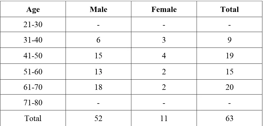
The following observations were made. Among the total 63 cases of
tongue cancer, the maximum amount of cases occurred in 4th to 6th decades
of life accounting for 62% of cases. Of these, males predominate and
constituted 73% of cases. The male:female ratio was 3:1.
[image:50.612.95.544.487.703.2]Table I
AGE AND SEX INCIDENCE
Age Male Female Total
21-30 - - -
31-40 6 3 9
41-50 15 4 19
51-60 13 2 15
61-70 18 2 20
Regarding the site of cancer, among the 63 cases, 42 cases had cancer
in oral (anterior) tongue and 21 cases were found to have lesion in the
posterior tongue.
77% of cases were T3 and T4 lesions. 64.5% of cases were N1 and
N2 nodal involvement.
Table II
STAGE AT PRESENTATION
Primary Lesion Node Metastasis
T1 12 N0 16 M0 63
T2 16 N1 18 M1 0
T3 20 N2 24
Table III
RISK FACTORS
Risk No. of Cases
Smoking and alcohol 40
Tobacco chewing 6
Pan masala 5
No risk factor 12
Nodal involvement in T1 & T2 lesions were present only in 27.3% of
cases. But 83.8% of T3 and T4 lesions presented with palpable neck nodes.
Nodal involvement was more in posterior tongue lesions (85%) compared to
Table IV
PATTERN OF CERVICAL METASTASIS
Total Cases Palpable nodes No neck
T1 & T2 28 13 14
T3 & T4 33 29 3
Anterior tongue 40 23 16
Posterior tongue 23 17 2
External beam radiotherapy played an important role in management
of tongue cancers. Thirty cases were taken for primary radiotherapy. Of
these five cases defaulted their treatment. There was residual lesion in 3
cases. Among these 3 cases, 2 were taken for surgery like composite
resection and hemiglossectomy. Even though surgery was feasible in some
30 cases were taken for primary radiotherapy. 3 cases had residual
lesions, thus can be improved if external beam radiotherapy is combined
with brachytherapy.
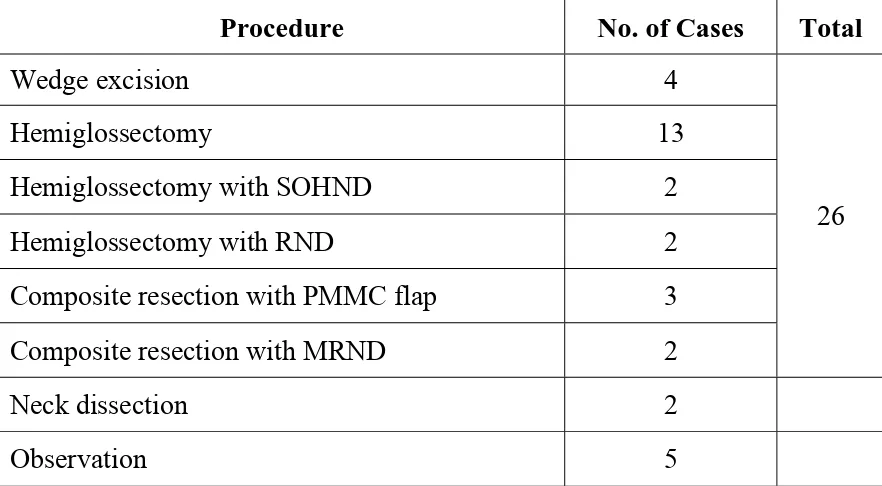
Total 26 patients were taken up for primary surgery
(Hemiglossectomy, Hemiglossectomy with neck dissection or composite
resection).
[image:54.612.96.537.402.645.2]Table V
PRIMARY SURGERY
Procedure No. of Cases Total
Wedge excision 4
Hemiglossectomy 13
Hemiglossectomy with SOHND 2
Hemiglossectomy with RND 2
Composite resection with PMMC flap 3
Composite resection with MRND 2
26
Neck dissection 2
RESULT OF THE STUDY
Total number of patients 63
4th and 6th decade 61%
Male:Female 3:1
Anterior tongue lesion 42
Posterior tongue lesion 23
Stage T3 and T4 N1 and N2
80% 63%
Smoking and alcoholism in 70% of cases
Nodal involvement T1 & T2 T3 & T4
Anterior tongue Posterior tongue 27.3% 83.8% 60.7% 85%
Radiotherapy Palliative Primary RT No residue Residue Defaulted 20 30 24 2 5 Primary Surgery 24
Post – RT Surgery 2
DISCUSSION
In a total of 63 patients tested in present study the maximum age
incidence was in 4th to 6th decades of life. Nil cases found in the age group
of 2nd and 3rd decades, 7th and 8th decades of life.
Majority of cases were male predominance because of uses of
smoking, alcoholism, panproducts. This was in comparison to study by
MEHROTRA published in Indian Journal Medical Science in which the
maximum incidence was in age group 51-60 years and male:female ratio
was 3.27:1.
COMPARISON STUDY
AGE AND SEX INCIDENCE
Contents Present Study Percent
KMC Study Percent
Literature Percent
Male 73 70 75 Sex
Female 27 30 25
40-50 yrs 30 37
50-60 yrs 24 23
There is a strong association of cancer tongue with addiction habits
like smoking, alcoholism, tobacco chewing and use of panproducts. Among
the cases, the main risk factor smoking and alcoholism was found in 70% of
cases. According to study by BARASCH, the percentage of smokers among
cases of tongue cancer was 64%.
RISK FACTORS
Contents Present Study Percent KMC Study Percent Literature Percent Smoking Alcoholism Panproducts
70 63 64
SITE
Anterior site lesions in the tongue are about 70%, Posterior lesions are
30%. According to study by MITCHELL anterior lesions are about 72%,
posterior lesions are 28%.
Present Study Percent KMC Study Percent Literature Percent
Anterior 70 71 72
Regarding the stage of presentation at the time of diagnosis, most of
them were advanced lesions. Public awareness is to be created so that
patient will present at the department in early stages which would offer a
better cure.
STAGE
Present Study Percent
KMC Study Percent
Literature Percent
T1-T2 33 35 33
t3-t4 77 75 72
The neck node involvement was influenced by the T status and
location of tumour. According to a study in Trivandrum (world Journal of
Surgery Oncology) cervical metastasis was present in 35.6% of T1, T2
NODES
Present Study Percent
KMC Study Percent
Literature Percent
N0 25 27 24
N1 28 25 27
N2 38 37 36
N3 7 6 8
According to study by LINGC and GRAS JR, Clinically negative
neck patients in stage I can be observed, but for T2 lesions – SOHND and
T3, T4 lesions – functional neck dissection should be done. So prophylactic
treatment of No neck would be a better option, for better regional control in
lesions above T2.
According to study by REGUEIRO, the response rate to EBRT was
TREATMENT
Present Study Percent
KMC Study Percent
Literature Percent
Surgery 41 20 35
RT 47 66 52
CT 4 4 5
The role of chemotherapy was limited in tongue cancer.
Primary RT - 30 cases
Palliative CT - 3 cases with advanced lesions
Post RT chemotherapy for residue - 2 cases
Palliative RT - 20 cases
Most of the cases presented to the department at advanced stage. So
measures should be taken to teach the public about early reporting to the
hospital so that better cure can be obtained.
Smoking and alcoholism was present in more than 62% of the cases
necessitating the need to bring public awareness about ill-effects of tobacco.
Nodal involvement was more in T3 and T4 posteriorly placed lesions.
CONCLUSION
¾ Smoking, alcoholism, panproducts are most commonly associated
risk factors for carcinoma tongue
¾ Most common in 4th to 6th decade of life Males are more commonly
affected than females in the ratio of 3:1
¾ Stage III, IV are common presentation in Carcinoma tongue
¾ Anterior 2/3rd is the commonest site of involvement
¾ Nodal involvement was common in T3 and T4 lesions, posteriorly
placed lesions
¾ Majority of early lesions (T1 and T2) are best treated surgicaly
¾ Few cases were down staged by using pre operative radiotherapy and
chemotherapy followed by surgery.
¾ Chemotherapy is mainly palliative measure
BIBLIOGRAPHY
1. Last’s Anatomy – Regional and applied – Tenth edition – Chummy S.
Sinnatamby FRCS. 372-375.
2. Mastery of Surgery – Third Edition – Lloyd M. Nyhus, M.D., Robert
J. Baker, M.D., Josef E. Fischer, M.S.
3. Essential surgical Practice – Higher Surgical training in General
Surgery – 4th Edition – Sir Alfred Cuschieri, 1093-1099.
4. Bailey and Love’s short practice of surgery – 23rd edition. 635-650.
5. Sabiston Textbook of surgery – The Biological Basis of Modern
Surgical Practice – 16th Edition. 533-553.
6. Recent Advances in surgery No.25-I. Taylor MD ChM FRCS, C.D.
Johnson Mchir FRCS, 71-76.
7. Stell & Maran’s Head & Neck Surgery – 4th edition – J.C. Watkinson,
M.N. Gaze, J.A. Wilson, 197-232, 275-316.
9. Treatment for early stage (T1-T2 No) SCC of mobile tongue – GRAS
JR – Acta Otorrhinolarynogol 2003 Jun – Jul 54(6): 443-8.
10.Management of patients with cancer tongue – MITCHELL R-British
Journal of oral maxillofacial surgery 1993 Oct 31(5): 304-8.
11.Age specific incidence rate and pathological spectrum of tongue
cancer – MEHROTRA R – Indian Journal of medical science 2003
Sep. 57 (9): 400-4.
12.Smoking, gender and age risk factor for intra – oral
SCC-BARASCHA – Cancer 1994 Feb. 1:73(3): 509-13.
13.Risk factor for cancer of tongue – OREGGIE F – Cancer 1991 Jan. 1:
67 (1) 180-3.
14.Oral Health consequence of chewing arecanut – Trivedy CR – Addict
Biol. 2002 Jan (7): 115-257.
15.Influence of EBRT and Brachytherapy on outcome of patients with
16. Pattern of cervical metastasis from cancer tongue C NITHYA –
World Journal of Surg. Oncology 2003 Jan: 38 (1) 5-8.
17.Relevant factor in management of patients with SCC in tongue
without clinical cervical lymph nodes – LINGC – Zhanghua Kou
Qians Yixue Zazhi 2003 Jan: 38 (1) 5-8.
18.Brachytherapy in management of initial stage of cancer tongue –
BALNER A – Acta otorrhinolaryngol – 1999 April 19 (2): 80-6.
19.Regional lymph node involvement affecting increased incidence of
distant metastasis in tongue SCC – SHINTANI – Anticancer Resi –
PROFORMA
Name :
Age :
Sex
Registration No. :
Date of Admission :
Date of Discharge :
Risk factors & Duration
Symptoms
- Ulcer
- Growth
- Pain, Bleeding
- Functional Disability
Examination
- Site
- Extension
- Size
Staging - TNM
Investigation - Biopsy
- Hemogram
- CXR
- CT, MRI
HPE report
Primary Treatment
Outcome
Adjuvant Treatment
MASTER CHART
S.No. Name Age/Sex IP No. Risk factors Site TNM HPE Primary Treatment Outcome Adjuvant
RT/CT
Post RT Surgery
1 Dhanam 75/f 1511/06 - Postetrior1/3 T3N0M0 adenosquamous Pallattive RT 2 Nagaraj 60/m 1300/06 S,A Postetrior1/3 T2N 2MO SCC L Hemiglossectomy
withRND
CT RT
3 Abdul 63/M 141106 C Postetrior1/3 T2N2M0 SCC wd
RT 4 Swaminathan 43/M 1810/06 S,C Postetrior1/3 T4N2M0 SCC
PD
RT POST RT
resection
Totalglossectomy Compposite resection 5 Muthu 72/M 1076/06 S,c Postetrior1/3 T4N1MO SCC
md
RT 6 Mari 54/m 1236/06 S Postetrior1/3 T4N3M0 SCC Palliative RT
7 Anandha raj 42/m 1526/06 S,A Anterior 2/3
T4N 2MO SCC wd
RT 8 Hussain 65/m 1450/06 S,C Anterior
2/3
T2N0M0 adenosquamous Wide excision 9 Sundaram 40/m 1417/06 s Anterior
2/3
T4N 2MO SCC wd RT 10 Ramani 64/f 1383/06 c Anterior
2/3
T4N 0MO SCC R. Hemiglossectomy with SOHND
CT 11 Nataraj 64/m 710/06 S,C Anterior
2/3
T2N 1O SCC Compposite resection with PMMCflap
12 lalitha 60/f 1546/06 Anterior 2/3
T1N2M0 SCC R. Hemiglossectomy
13 Saravanan 60/M 230/07 SC Posterior1/3 T4N1M0 SCC RT CT
14 Danasekar 72/M 1206/06 S,C Anterior 2/3 CROSSDING MIDLINE
T2N2M0 SCC wd RT NWS
15 Lakshmi 46/f 1129/06 C Anterior 2/3
T4N2M0 SCC PD
RT 16 Jeyaraman 32/M 1338/06 S,C,P Anterior
2/3
T4n2m0 SCC RT
17 selva 50/m 1631/06 S/C Anterior 2/3
T1N0M0 SCC Wd R
Hemiglossectomy withSOHND 18 babu 39/m 11631/06 S,P Anterior
2/3
T1N0M0 SCC Wd R
Hemiglossectomy
Nodes after 8 mnths
RND 19 Dhayalan 50/M 864/06 S,C Anterior
2/3
T4N1M0 SCC PD RT
20 Syed 42/m 488/06 S,C,P Anterior 2/3
T2N1M0 SCC Lt Hemiglossectomy with PMMCflap
RT
21 Mohan 60/m 981/06 Anterior
2/3
T3N0M0 SCC RT
22 Palani 63/m 1031/06 S,C Anterior 2/3
T3N1M0 SCC RT
23 RANI 45/F 1061/06 C Anterior 2/3
T2N2M0 SCC RT NWD
32 Muthu 43/m 403/07 S Central T2N2M0 SCC RT,CT 33 Basha 65/m 380/07 S,C Anterior
2/3
T2N0M0 SCC Compposite resection with Type 1 MRND
34 Kumar 60/m 1081/07 S,C Hemiglossectomy T34N3M0 SCC RT CT
35 Raja 34/m 1308/07 S,P Anterior 2/3
T4N2M0 SCC RT,CT CT
36 Sakunthala 35/f 434/07 Anterior 2/3
T1N0M0 SCC Wide Excision 37 Mallika 53/f 238/07 C Anterior
2/3
T2N0M0 SCC RT
38 Annamalai 45/f 217/0-7 C Anterior 2/3
T1N0M0 SCC Hemiglossectomy 39 Sandhanam 30/m 155/07 S,P Posterior1/3 T3N3M0 SCC RT
40 Lalitha 60/f 153/07 C Anterior 2/3
T1N1M0 SCC Hemiglossectomy with RND
41 Madhavan 40/m 541/07 S,C Posterior1/3 T4N3M0 SCC RT CT
42 Kumari 65/f 66/07 Anterior
2/3
T1N0M0 SCC Hemiglossectomy 43 Shanmugam 45/m 25/07 S,C Anterior
2/3
T4N2M0 SCC RT
44 Prama 64/m 24/07 S,C Posterior1/3 T4N3M0 SCC RT,CT
45 Kasim 53/m 93/07 S,C Posterior1/3 T3N0M0 SCC RT
46 Veeran 45/m 44/07 S Posterior1/3 T4N1M0 SCC RT
47 Rethinam 53/m 400/07 C Posterior1/3 T4N1M0 SCC RT 48 Selvi 64/f 1383/07 Anterior
2/3
T1N2M0 SCC Hemiglossectomy with SOHND
49 Sundar 44/m 1250/07 S Anterior 2/3 T2N1M0 SCC Compposite resection
50 Mani 65/m 1349/07 S,C Anterior 2/3 T3N1M0 SCC Hemiglossectomy with RND CT,RT Hussian 44/m 1450/07 S Anterior 2/3 T2N0M0 SCC Wide Excision
51 Murugan 54/m 1542/07 S,C Anterior 2/3 T2N0M0 SCC Compposite resection with PMMCflap
52 Thangam 56/f 1546/07 Posterior1/3 T4N2M0 SCC RT
With Hemiglossectomy with RND
53 Sethu 60/m 1310/07 S,C Posterior1/3 T4N1M0 SCC RT
54 Syed 40/m 485/07 S,C Anterior 2/3 T2N1M0 SCC Hemiglossectomy with PMMC flap 55 Raman 50/m 864/078 S,C Anterior 2/3 T4N1M0 SCC RT
56 Raja 48/m 865/07 S Anterior 2/3 T4N2M0 SCC RT
57 Ramaraj 37/m 1089/06 P Anterior 2/3 T3N1M0 SCC RT Residue Compposite resection 58 Arun 32/m 1295/07 P,S Anterior 2/3 T2N1M0 SCC Hemiglossectomy with
MRND
59 Manoj 39/m 1107/07 S,A Anterior 2/3 T1N0M0 SCC Hemiglossectomy Nodes after 7 mnths
MRND done
60 Manju 46/f 1267/07 C Anterior 2/3 with posterior extension
T4N2M0 SCC RT, CT CT
61 Jeyaraman 45/m 138/07 S,C Posterior1/3 T4N2M0 SCC RT CT
62 Palayam 50/m 1031/07 S,C Anterior 2/3 T3N3M0 SCC RT
63 Seetharaqman 65/m 381/07 S,C Anterior 2/3 T2N1M0 SCC Compposite resection with PMMC flap
S - Smoking SOHND - Supra omohyoid node dissection
A - Alcoholism RND - Radical neck dissection
P - Panproducts MRND - Modified radical neck dissection
C - Tobacco chewing NWS - Not willing for sugery