D
D
R
R
U
U
G
G
-
-
I
I
N
N
D
D
U
U
C
C
E
E
D
D
U
U
P
P
P
P
E
E
R
R
G
G
A
A
S
S
T
T
R
R
O
O
I
I
N
N
T
T
E
E
S
S
T
T
I
I
N
N
A
A
L
L
B
B
L
L
E
E
E
E
D
D
I
I
N
N
G
G
–
–
A
A
R
R
E
E
G
G
I
I
O
O
N
N
A
A
L
L
S
S
T
T
U
U
D
D
Y
Y
D
DIISSSSEERRTTAATTIIOONN
Submitted for
M
M..DDBBRRAANNCCHH11 (
(GGEENNEERRAALLMMEEDDIICCIINNEE))
D
D
E
E
P
P
A
A
R
R
T
T
M
M
E
E
N
N
T
T
O
O
F
F
I
I
N
N
T
T
E
E
R
R
N
N
A
A
L
L
M
M
E
E
D
D
I
I
C
C
I
I
N
N
E
E
T
T
H
H
A
A
N
N
J
J
A
A
V
V
U
U
R
R
M
M
E
E
D
D
I
I
C
C
I
I
A
A
L
L
C
C
O
O
L
L
L
L
E
E
G
G
E
E
,
,
T
T
H
H
A
A
N
N
J
J
A
A
V
V
U
U
R
R
T
T
H
H
E
E
T
T
A
A
M
M
I
I
L
L
N
N
A
A
D
D
U
U
D
D
r
r
.
.
M
M
G
G
R
R
M
M
E
E
D
D
I
I
C
C
A
A
L
L
U
U
N
N
I
I
V
V
E
E
R
R
S
S
I
I
T
T
Y
Y
,
,
C
C
H
H
E
E
N
N
N
N
A
A
I
I
M
C
C
e
e
r
r
t
t
i
i
f
f
i
i
c
c
a
a
t
t
e
e
This is to certify that this dissertation entitled ““DDrruugg--IInndduucceedd U
Uppppeerr GaGassttrrooiinntteessttiinnaall BBlleeeeddiinngg - - A A RReeggiioonnaall ssttuuddyy””, is the bonafide record work done by Drr..PP..SSuubbrraammaanniiaann submitted as partial fulfillment for the requirements of M.D Degree Examination, General Medicine (Branch I) to be held in March 2008.
P
Prrooff.. DDrr.. SS..MMuutthhuu kkumumaarraann,, MM..DD.. PPrrooff.. DDrr.. ((MMrrss))..NN..JJeeeevvaa,, MM..DD..
Professor and HOD, Professor of Therapeutic Medicine, Department of Internal Medicine, Department of Internal Medicine, Thanjavur Medical College, Thanjavur Medical College, Thanjavur - 613004. Thanjavur - 613004.
D Deeaann T
Thhaannjjaavvuurr MMeeddiiccaall CCoolllleeggee T
A
A
c
c
k
k
n
n
o
o
w
w
l
l
e
e
d
d
g
g
e
e
m
m
e
e
n
n
t
t
s
s
I am greatly indebted to my guide DDrr.. ((MMrrss))..NN..JJeeeevvaa,,MM..DD.., Professor of Therapeutic Medicine, Department of Internal Medicine, Thanjavur Medical College, Thanjavur for having accepted me as her student and for giving a chance to undertake this dissertation work under her guidance. Also I express my deep sense of gratitude for her encouragement, directions, periodical discussions, rigorous reviews and precious suggestions for shaping my dissertation.
I express my gratitude to DDrr.. SS.. MMuutthhuukkuummaarraann,, MM..DD.., Professor and Head, Department of Internal Medicine, Thanjavur Medical College, for his kind encouragement and review of my work, besides providing me with all the required facilities.
I am extremely grateful to DDrr.. RR..MM.. NNaattaarraajjaann,, MM..SS..,, Dean, Thanjavur Medical College for granting me the permission to do this dissertation in Thanjavur Medical College Hospital, Thanjavur.
I recall with gratitude all the Unit Chiefs of Department of Internal Medicine, for their thoughtful guidance in conducting the study.
I extend my thanks to DDrr.. CC.. KKrriisshhnnaann,,MM..DD..,,DD..MM..,, Assistant Professor in Department of Medical Gastro Enterology, who has done UGI endoscopy for the patients I studied and helped me a lot.
I am thankful to my unit Assistant Professors DDrr.. DD.. SSeekkaarr,, MM..DD.. and
D
Drr..GG..GGoowwtthhaammaann,,MM..DD.. who have helped me a lot in this dissertation.
I thank our college Librarian who gave me full cooperation in collecting literature, references etc.
I thank the paramedical staffs of Thanjavur Medical College hospital for helping me in doing this dissertation work.
B
B
r
r
i
i
e
e
f
f
C
C
o
o
n
n
t
t
e
e
n
n
t
t
s
s
Chapter 1: Introduction……… 1
Chapter 2: Aim of the study……….……….. 4
Chapter 3: Literature review……….. 5
Chapter 4: Materials and methods……… 31
Chapter 5: Results and observations……….………. 38
Chapter 6: Discussion……… 50
Chapter 7: Conclusion ……….………. 61
References………. i
Appendix A: Proforma –– Study on Drug-induced UGI Bleeding……… xxv
T
T
a
a
b
b
l
l
e
e
o
o
f
f
c
c
o
o
n
n
t
t
e
e
n
n
t
t
s
s
List of abbreviations List of Figures and Boxes List of Tables
ABSTRACT
CHAPTER 1: INTRODUCTION
1.1 International scenario……….. 1
1.2 National scenario……….. 3
CHAPTER 2: AIM OF THE STUDY 2.1 Aim of the study………. 4
CHAPTER 3: LITERATURE REVIEW 3.1 Gastric mucosal barrier……… 5
3.2 Drugs causing UGI bleeding……….………. 7
3.3 Mechanisms of drug-induced UGI bleeding….………. 9
3.4 Clinical and endoscopic features of drug-induced gastro intestinal damage ……….……….. 11
3.4.1 Oesophagus………..12
3.4.2 Stomach and duodenum……….12
3.4.3 Small intestine………..13
Table of contents continued…
3.5 Sustained release and enteric coated NSAIDs….……….15
3.6 COX-2 selective inhibitors……….16
3.7 Risk factors of drug-induced UGI bleeding……..……….16
3.7.1 Old age………...16
3.7.2 High doses/Chronic drug intake………..17
3.7.3
Self medication / OTC drugs………..17
3.7.4 Use of Gastro protective agents………..18
3.7.5 Known PUD……….19
3.7.6
Alcoholism………..20
3.7.7 Smoking………20
3.7.8 Stress and Serious systemic illnesses………21
3.7.9 Concomitant use of other gastro toxic drugs 22 3.7.10 ‘O’ Blood group………. 23
3.7.11 Helicobacter pylori……….24
3.7.12 Genetics and other causes……… 26
3.8 Management of drug-induced UGI bleeding……… 27
3.8.1 Medical management of unstable patient with bleeding ulcer……… 27
3.8.2 Medical management of stable patient with NSAID induced ulcer………. 28
3.8.3 Surgical care……….. 29
CHAPTER 4: MATERIALS ANDMETHODS 4.1 Materials and methods……… 31
Table of contents continued…
4.2.1 Inclusion criteria………..……….32
4.2.2 Exclusion criteria ………. 32
4.3 Study method……….33
4.3.1 History……….33
4.3.2 Clinical examination……….34
4.3.3 Laboratory investigations……….35
4.3.4 Upper gastrointestinal endoscopy……….35
4.4 Study approach……….37
CHAPTER 5: RESULTS AND OBSERVATIONS 5.1 Study results and observations…………..………. 38
CHAPTER 6: DISCUSSION 6.1 Discussion 6.1.1 Sex………..………. 50
6.1.2 Age………. 50
6.1.3 Severity of hematemesis…………..………... 51
6.1.4 Causative drugs………... 52
6.1.5 High doses / Chronic drug intake………. 53
6.1.6 Self medication / OTC ………. 53
6.1.7 Drug intake without Gastro protective agents………. 54
6.1.8 Known PUD……….… 54
Table of contents continued…
6.1.10 Smoking ………...56 6.1.11 Stress and Serious systemic illnesses ……. 56 6.1.12 Concomitant use of Steroids………….………… 57 6.1.13 Concomitant use of Anticoagulants…….…… 57 6.1.14 ‘O’ Blood group ……….………... 58 6.1.15 H.pylori……….………….58 6.1.16 Relationship between OTC and GPA………..……. 59
6.1.17 Endoscopic findings………....59 6.1.18 Prevalence of number of risk factors………. 60
CHAPTER 7: CONCLUSION
7.1 Conclusion……….………61
References ……….i
Appendix A: Proforma –– Study on Drug-induced UGI Bleeding………. xxv
L
L
i
i
s
s
t
t
o
o
f
f
a
a
b
b
b
b
r
r
e
e
v
v
i
i
a
a
t
t
i
i
o
o
n
n
s
s
1) ARAMIS - Arthritis, Rheumatism and Aging Medical Information System
2) CAHD - Coronary Artery Heart Disease
3) COPD - Chronic Obstructive Pulmonary Disease 4) COX - Cyclo oxygenase
5) EC - Enteric Coated
6) EGF - Epidermal Growth Factor 7) FDA - Food and Drug Administration 8) GI - Gastro Intestinal
9) GIT - Gastro Intestinal Tract 10) GPA - Gastro-Protective Agents 11) H/O - History Of
12) H. pylori - Helicobacter pylori
13) hr. - hour
14) H2RA - Histamine-2Receptor Antagonists
15) NSAIDs - Non-Steroidal Anti-Inflammatory Drugs 16) IgG - Immunoglobulin G
17) IgA - Immunoglobulin A 18) LP - Lipid Peroxidation
19) mg - milli gram
20) ml - milli liter 21) NO - Nitric Oxide 22) OTC - Over-The-Counter 23) PGE2 - Prostaglandin E2
24) PPI - Proton Pump Inhibitor 25) PUD - Peptic Ulcer Disease 26) SR - Sustained Release
27) SSI - Serious Systemic Illnesses
28) SSRIs - Selective Serotonin Reuptake Inhibitors 29) TGF - Transforming Growth Factor
L
L
i
i
s
s
t
t
o
o
f
f
F
F
i
i
g
g
u
u
r
r
e
e
s
s
a
a
n
n
d
d
B
B
o
o
x
x
e
e
s
s
Figure 3.1
Naproxen induced duodenal ulcer………. 13 Figure 3.2
Sustained release tablet diclofenac induced colonic ulcer……..….. 15
Figure 3.3
Electron microscopic picture of H.pylori ……….24
Figure 4.1
PENTAX video endoscopic system at Department of Medical Gastro Enterology, Thanjavur Medical College………. 36
Figure 4.2
Endoscopy showing antral ulcer (done on 17-7-2007)………. 36
Figure 5.1
Sex distribution ……….………. 45
Figure 5.2
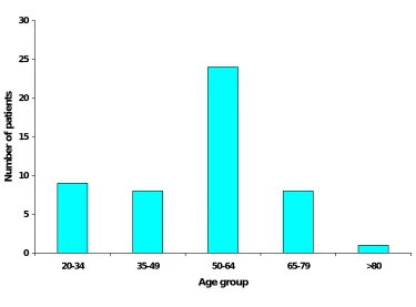
Age group distribution.………..……… 45
Figure 5.3
Frequency of hematemesis……….………. 46
Figure 5.4
List of Figures and boxes continued…
Figure 5.5
Prescription vs. Self medication / OTC ………. 47
Figure 5.6
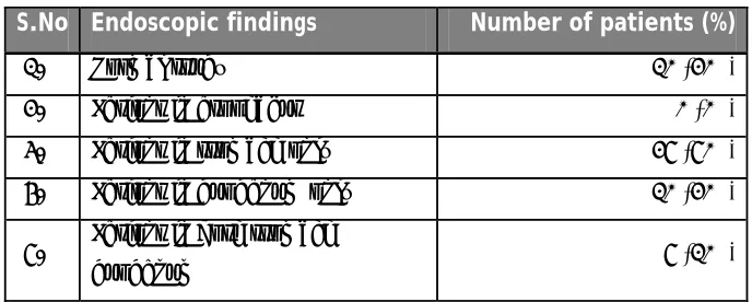
Prevalence of site of lesions on endoscopic study………. 47
Figure 5.7
Prevalence of nature of lesions on endoscopic study………... 48
Figure 5.8
Prevalence of individual risk factors among the patients……… 48
Figure 5.9
Prevalence of patients with drug-induced UGI bleeding with respect to number of risk factors………….………..……… 49 Box 3.1
L
L
i
i
s
s
t
t
o
o
f
f
T
T
a
a
b
b
l
l
e
e
s
s
Table 3.1
Classification of NSAIDs………. 8 Table 3.2
NSAID ingestion and GI injury………. 13
Table 5.1
Prevalence of drug-induced UGI bleeding related to age group and sex………...………. 39
Table 5.2
Frequency of hematemesis……… 40 Table 5.3
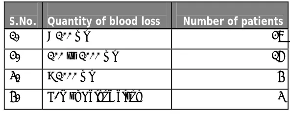
Approximate quantity of blood loss ……….… 40
Table 5.4
Causative drugs and number of patients………..……… 40
Table 5.5
Prevalence of site of lesions on endoscopic study………. 41
Table 5.6
List of tables continued….
Table 5.7
Prevalence of risk factors of drug-induced UGI bleeding…...………… 41
Table 5.8
A
A
b
b
s
s
t
t
r
r
a
a
c
c
t
t
Study Objective: To study the clinical profile and risk factors in fifty cases of drug-induced UGI bleeding.
Design and Setting: Prospective study, Thanjavur Medical College hospital.
Patients: Fifty patients admitted with drug-induced hematemesis and/or malena.
Study period: Between March 2006 and August 2007.
Results: Prevalence of risk factors among the patients suffering from drug-induced UGI bleeding are as follows: [1] Old age > 50 years of age - 66% [2] ‘O’ Blood group – 50% [3] Alcoholism – 42% [4] Not using Gastro protective agents – 40% [5] Self medication / OTC drugs – 36% [6] Smoking – 30% [7] Stress and Serious systemic illnesses – 12% [8] Helicobacter pylori – 12% [9] Known Peptic ulcer disease – 10% [10] High doses / Chronic drug intake – 10% [11] Concomitant use of Steroids – 8% and [12] Concomitant use of anticoagulants – 4%.
Conclusion: NSAIDs were the causative drugs for UGI bleeding in all the fifty cases studied. All those fifty cases had at least one known risk factor and majority (80%) had more than one risk factors of drug-induced UGI bleeding.
C
C
H
H
A
A
P
P
T
T
E
E
R
R
-
-
1
1
I
I
N
N
T
T
R
R
O
O
D
D
U
U
C
C
T
T
I
I
O
O
N
N
I
I
n
n
t
t
e
e
r
r
n
n
a
a
t
t
i
i
o
o
n
n
a
a
l
l
s
s
c
c
e
e
n
n
a
a
r
r
i
i
o
o
N
It is highly unfortunate that many patients are admitted daily with hematemesis and/or malena due to the adverse effects of drugs either prescribed or self medicated. Incidence of such cases can be greatly reduced if medical practitioners are not only aware of the adverse effects of drugs on gastrointestinal tract but also assess the patients for the risk factors of drug-induced UGI bleeding before prescribing these drugs and also by properly educating the patients. Non-steroidal anti-inflammatory drugs (NSAIDs) including aspirin are among the most frequently prescribed drugs worldwide and are available ‘Over-The-Counter’ (OTC) also. Though reasonably safe in most cases in prescribed dosages and for short durations, these drugs cause serious gastrointestinal toxicity in a large number of cases, particularly in older age, history of peptic ulcer disease, alcoholism, smoking, stress, poor general health and concurrent use of steroids and anticoagulants. They can affect all segments of the gastrointestinal tract causing ulcers, severe bleeding, perforation, and obstruction. It is therefore imperative for physicians to be aware not only about their serious adverse effects but also about the risk factors of drug-induced gastrointestinal (GI) bleeding and use these drugs with caution and only when genuinely indicated.
1.1 International
scenario
for NSAID-related gastrointestinal complications, and at least 16,500 NSAID- related deaths occur each year among arthritis patients alone. Surprisingly, the management of this problem has undergone little change in the past 50 years, and is not only frequently under-diagnosed but also under-treated (Vikas et al., 2003).
There were approximately 11.14 crore NSAID prescriptions in the United States for the year ending in August 2000 (Retail and Provider Perspective, 2000). In addition, annual United States sales of over-the-counter oral analgesics approach three billion dollars; The prevalence of at least once-weekly NSAID use among people 65 years old or older has been reported to be as high as 70%; half of this group takes at least seven doses a week (Talley et al., 1995; Loren, 2001).
Based on an analysis of acute hospital admissions in one health district in United Kingdom, it has been calculated that NSAID-related upper GI events account for around 12,000 admissions annually in England and result in approximately 2500 potentially avoidable deaths each year. Most of the dead were taking higher doses of NSAIDs for long duration or elderly or chronic alcoholic or known peptic ulcer disease patients (Belsey, 2003).
serious gastrointestinal side effects. Almost all deaths from NSAID related gastrointestinal side effects occur in elderlypersons; elderly women seem particularly susceptible (Robyn et al., 1997).
1.2 National
scenario
Indian studies have shown that NSAIDs including aspirin are among the most common drugs responsible for adverse drug reactions seen in clinical practice. In general, at least 10 to 20 percent of patients have dyspepsia while taking an NSAID, although the prevalence may range from 5 to 50 percent.
Incidence of new ulcer cases following NSAID intake, ranges from 10% to 40% for gastric ulcers and 5% - 15% for duodenal ulcers. Most patients are, however, asymptomatic (Simon et al., 1999). Seventy percent of the patients admitted with drug induced UGI bleeding were > 50 years of age (Vikas et al., 2003).
According to prospective data from the Arthritis, Rheumatism, and Aging Medical Information System (ARAMIS), 13 of every 1,000 patients with rheumatoid arthritis who take NSAIDs for one year have a serious gastrointestinal complication (Singh, 1998; Simon et al., 1999). In India, quacks also practice these drugs commonly and elderly are the most affected (Vikas et al., 2003).
C
C
H
H
A
A
P
P
T
T
E
E
R
R
-
-
2
2
A
A
I
I
M
M
O
O
F
F
T
T
H
H
E
E
S
S
T
T
U
U
D
D
Y
Y
A
2.1 Aim of the study
• To study the clinical profile of patients admitted with drug-induced upper gastrointestinal bleeding.
• To find out the risk factors for the drug-induced UGI bleeding in those patients.
• To find out the prevalence of the risk factors among them.
C
C
H
H
A
A
P
P
T
T
E
E
R
R
-
-
3
3
L
L
I
I
T
T
E
E
R
R
A
A
T
T
U
U
R
R
E
E
R
R
E
E
V
V
I
I
E
E
W
W
G
G
a
a
s
s
t
t
r
r
i
i
c
c
m
m
u
u
c
c
o
o
s
s
a
a
l
l
b
b
a
a
r
r
r
r
i
i
e
e
r
r
D
D
r
r
u
u
g
g
s
s
c
c
a
a
u
u
s
s
i
i
n
n
g
g
U
U
G
G
I
I
B
B
l
l
e
e
e
e
d
d
i
i
n
n
g
g
M
M
e
e
c
c
h
h
a
a
n
n
i
i
s
s
m
m
s
s
o
o
f
f
d
d
r
r
u
u
g
g
-
-
I
I
n
n
d
d
u
u
c
c
e
e
d
d
U
U
G
G
I
I
B
B
l
l
e
e
e
e
d
d
i
i
n
n
g
g
C
C
l
l
i
i
n
n
i
i
c
c
a
a
l
l
a
a
n
n
d
d
e
e
n
n
d
d
o
o
s
s
c
c
o
o
p
p
i
i
c
c
f
f
e
e
a
a
t
t
u
u
r
r
e
e
s
s
o
o
f
f
d
d
r
r
u
u
g
g
-
-i
i
n
n
d
d
u
u
c
c
e
e
d
d
g
g
a
a
s
s
t
t
r
r
o
o
i
i
n
n
t
t
e
e
s
s
t
t
i
i
n
n
a
a
l
l
d
d
a
a
m
m
a
a
g
g
e
e
S
S
u
u
s
s
t
t
a
a
i
i
n
n
e
e
d
d
r
r
e
e
l
l
e
e
a
a
s
s
e
e
a
a
n
n
d
d
e
e
n
n
t
t
e
e
r
r
i
i
c
c
c
c
o
o
a
a
t
t
e
e
d
d
N
N
S
S
A
A
I
I
D
D
s
s
C
C
O
O
X
X
-
-
2
2
s
s
e
e
l
l
e
e
c
c
t
t
i
i
v
v
e
e
i
i
n
n
h
h
i
i
b
b
i
i
t
t
o
o
r
r
s
s
R
R
i
i
s
s
k
k
f
f
a
a
c
c
t
t
o
o
r
r
s
s
o
o
f
f
d
d
r
r
u
u
g
g
-
-
i
i
n
n
d
d
u
u
c
c
e
e
d
d
U
U
G
G
I
I
b
b
l
l
e
e
e
e
d
d
i
i
n
n
g
g
M
Peptic Ulcer Disease (PUD) results from the imbalance between the defensive factors that protect the mucosa and offensive factors that disrupt the important gastric mucosal barrier. Many of the primary ulcers seen in teenagers are now thought to be associated with Helicobacter pylori infection while many of the secondary ulcers are due to use of NSAIDs including aspirin.
3.1 Gastric
mucosal
barrier
The gastric mucosal barrier is composed of several hierarchically organized anatomical and functional components, which mutually contribute to the mucosal integrity endangered by the offending luminal acid. The barrier consists of three protective components (Source: http://en.wikipedia.org/wiki; Last accessed 9 September 2007). The three components include:
(1) A compact epithelial cell lining
(2) A special mucus covering
Derived from mucus secreted by surface epithelial cells and mucosal neck cells this gel-like coating protects the entire surface of the gastric mucosa from auto digestion by e.g. pepsin and from erosion by acids and other caustic materials that are ingested. The mucus gel contained numerous phospholipids, and its luminal surface was coated with a film of phospholipid - a surfactant layer which accounts for the remarkably strong hydrophobic nature of the gastric luminal surface, which, in turn, provides protection against damaging agents. Physico-chemical effect of the very high viscosity of the mucus layer and its ability to retain bicarbonate secreted by the epithelium with maintenance of a pH gradient is also an important factor in protection.
The protective properties of prostaglandins are not based only on their
antisecretory but also on their cytoprotective action. They stimulate glycoprotein and bicarbonate secretion and enhance the microcirculation,
cellular oxygen supply, restitution and repair (Robert et al., 1979).
(3) Bicarbonate ions
The bicarbonate ions are secreted by the surface epithelial cells of the stomach and duodenum. The pre-epithelial mucus-HCO3 layer buffers back diffusing luminal H+ preventing its entry inside the epithelial cells.
production. Cytokines such as fibroblast growth factor and hepatocyte growth factor are found to enhance healing of gastrointestinal ulcers in experimental models (Murphy, 1998).
Trefoil proteins are a family of small peptides that are secreted abundantly by goblet cells in the gastric and intestinal mucosa, and coat the apical face of the epithelial cells and render them resistant to proteolytic destruction. Trefoil peptides play an important role in mucosal integrity, repair of lesions, limitation of epithelial cell proliferation and protection of the epithelium from a broad range of toxic chemicals and drugs.
Nitric oxide (NO) plays a crucial role in mucosal integrity and barrier function with stimulation of fluid and mucus secretion, enhancement of microcirculation and maintenance of epithelial barrier function. It is synthesized from arginine through the action of nitric oxide synthease (NOS). In several models, co-administration of NO donors such as glyceryltrinitrate and NSAIDs results in anti-inflammatory properties comparable to NSAIDs alone, but with less damage to the gastrointestinal mucosa (Muscara, 1999). Several studies have emphasized the roles of NOsynthase (NOS) and COX in maintaining gastric mucosal integrity and
epithelial restitution (Wallace and Granger, 1992).
3.2 Drugs causing UGI bleeding
stomach or duodenum. Although the bleeding risk increases in proportion to NSAID dose, any amount (including low-dose aspirin taken for cardiovascular prophylaxis) may cause bleeding particularly when taken on empty stomach. Use of selective serotonin reuptake inhibitors (SSRIs) has recently been found to be associated with a higher risk of upper GI bleeding, especially in patients who are also taking NSAIDs or low-dose aspirin (Dalton et al., 2003). Alendronate and potassium chloride also cause upper GI bleeding in very few cases. Role of steroids in ulcerogenesis is relatively small, although hemorrhage seems to be the most common complication from peptic ulcers in steroid-treated patients (Messer et al.,
[image:26.612.119.519.439.719.2]1983; Fadul et al., 1988). Anticoagulants do not cause GI bleeding per se, but they can unmask or aggravate hemorrhage from preexisting lesions (Dalton et al., 2003).
Table 3.1 Classification of NSAIDs
Non-steroidal anti-inflammatory drugs
Salicylates Aspirin (Acetylsalicylic Acid), Diflunisal, Ethenzamide, Salicin
Arylalkanoic acids Diclofenac, Etodolac, Indometacin, Nabumetone, Sulindac
2-Arylpropionic acids (profens)
Carprofen, Flurbiprofen, Ibuprofen, Ketoprofen, Ketorolac, Loxoprofen, Naproxen, Oxaprozin, Suprofen, Tiaprofenic acid
N-Arylanthranilic acids (fenamic acids) Mefenamic acid
Pyrazolidine derivatives Phenylbutazone
Oxicams Meloxicam, Piroxicam
Coxibs Celecoxib, Etoricoxib, Parecoxib,
Rofecoxib, Valdecoxib, Lumiracoxib Sulphonanilides Nimesulide
Topically used products
3.3 Mechanisms of drug-induced UGI bleeding
Prostaglandins protect GI mucosa by forming a cytoprotective layer and increasing the secretion of bicarbonate ions that neutralise the gastric acidity. All therapeutically useful NSAIDs act by inhibiting the synthesis of PGs (Tamblyn, 1997). NSAIDs-induced inhibition of prostaglandin biosynthesis has resulted in an enhanced production of leukotrienes and
other products of the 5- lipoxygenase pathway (Whittle et al., 1993).
These products alter the mucosal barrier with an increased gastric mucosal
permeability for H+ ions and Na+ ions and reduced transmucosal potential
difference thereby promoting the formation of erosions and ulcers (Kubes
et al., 1991; Jimenez et al., 2002). The hydrophobic acid-resistant property of the gastric surface active phospholipid layer (SAPL) is rapidly attenuated by NSAIDs. Secretion of bicarbonate ions also is inhibited by NSAIDs (Knutson and Flemstrom, 1989; Isenberg and Flemstrom, 1991).
Cyclooxygenase has two isoforms, one constitutive (COX-1) and another inducible (COX-2). A third isoform (COX-3) has recently been described as well. COX-1 is responsible for production of prostaglandins which are essential for maintenance of normal endocrine and renal function, gastric mucosal integrity and haemostasis. In contrast, COX-2 is virtually undetectable in most tissues, but the enzyme activity may be dramatically up regulated by inflammatory and mitogenic stimuli (Dequeker et al.,
weak organic acids and have low pKa. Therefore, they remain unionised in stomach and are absorbed appreciably from stomach. However, once they breach the cell membranes of stomach cells and reach within, they encounter a basic pH (e.g. 7.1). This causes so called ‘trapping’ of the drugs inside the cell (Raskin, 1999). This topical effect is considered an important mechanism of gastro-duodenal damage associated with their use. Even short-term (<1 week) use of aspirin and other nonsteroidal anti-inflammatory drugs (NSAIDs) can precipitate ulcer-related bleeding. Thus, it can be understood to be the disease of the war between the factors favoring and those opposing the development of ulcers where the former win over the latter.
Aspirin, the only NSAID able to irreversibly inhibit COX-1, also shows inhibition of platelet aggregation because it inhibits the action of thromboxane-A. Aspirin also causes capillary fragility, increased fibrinolysis and a prolonged bleeding time. Role of steroids in ulcerogenesis is small and anticoagulants do not cause GI bleeding per se, but both can aggravate hemorrhage from preexisting ulcers.
3.4 Clinical and endoscopic features of drug-induced
gastrointestinal damage
NSAIDs-induced GI damage is of three main types:
• Superficial damage such as mucosal hemorrhages and erosions. • Endoscopically documented non-symptomatic (‘silent’) ulcers. • Symptomatic ulcers causing complications such as GI
hemorrhage.
NSAIDs including aspirin induced ulcers are usually symptom less unless complicated. Early symptoms are mild and benign like dyspepsia, nausea, vomiting, and anorexia. Pain is usually a late feature. When the ulcer starts bleeding hematemesis and/or malena occurs in about 1 to 3 % of patients. Most cases of NSAID-induced gastrointestinal ulcers can heal spontaneously, even when the drug is continued (Vikas et al., 2003). Moreover, it is not usually possible to diagnose these ulcers on the basis of clinical features alone, as symptoms suggestive of the ulcers can occur frequently in their absence. Elderly patients usually have painless gastric ulceration and NSAIDs can mask the symptom of pain (Dhikav, 2001). In fact, most elderly patients are referred to physicians for iron deficiency anemia due to fecal blood loss.
1999; Yeoman, 2001). Elderly patients are particularly prone to develop GI toxicity and unfortunately they are the most frequent users of this group of drugs (Yeoman, 2001).
Box 3.1 Features of NSAID induced GI ulcers
3.4.1 Oesophagus
Prolonged use of aspirin and most NSAIDs can result in ulceration, oesophagitis and even strictures more commonly caused by reflux rather than direct action of NSAIDs.
3.4.2
Stomach and duodenum
The features of NSAID ingestion and injury in the stomach and duodenum in acute and chronic cases are given in the following Table 3.2 and UGI endoscopic view of naproxen induced duodenal ulcer is given as Figure 3.1.
• More than 3 mm in size • Deep lesions
• Prone to complications like bleeding, perforation and obstruction • Gastric or duodenal in location
• Multiple erosions (more than 10) • Antral in location
Table 3.2 NSAID ingestion and GI injury
Injury type Gastro-duodenal lesion Frequency
Acute mucosal erythema,
(1-2 weeks) superficial erosions, 60-100% submucosal haemorrhage,
Increased fecal blood loss
Chronic gastric antral erosions
(> 4 weeks) and ulcers, duodenal 5-30% Ulcers and erosions
Figure 3.1 Naproxen induced duodenal ulcer
3.4.3 Small
intestine
[image:31.612.158.486.88.519.2]events in these cases (Loren, 2003). A number of studies suggest that ulceration and perforation in small intestine is related to slow release and enteric coated formulations of NSAIDs and aspirin (Bjarnason et al., 1987). Syndrome of occult blood loss, malabsorption, anemia, and protein losing enteropathy has been described in many cases (Gibson et al., 1992).
3.4.4
Large intestine, rectum and anal region
There are only a few cases of NSAID induced damage in this segment of GIT. In colon and ano-rectal area they cause colitis, proctitis, and presentation similar to inflammatory bowel disease, bleeding (both acute and chronic), ulcers, and strictures. Most ulcerative complications like perforation are seen in caecum (Carlson et al., 1990). Rectal suppositories can result in proctalgia, tenesmus, or watery diarrhoea. Approximately, 10 - 30% cases can develop these problems (Kurahur et al., 2001).
do not prevent NSAID-induced gastric ulcers (Doomra and Gupta, 2001; Simon et al., 1999).
3.5 Sustained
release
and
enteric coated NSAIDs
Studies have demonstrated increased intestinal permeability with SR and EC formulations of all NSAIDs but not with conventional release tablets and lower GI bleeding are also more common with them (Choi et al., 1995). These formulations claim to cause fewer adverse effects than the conventional formulation and that they could be prescribed for patients who are most prone to develop such adverse effects. This phenomenon has been termed ‘channeling’ and may complicate any interpretation of the epidemiology of adverse effects (Leufkens et al., 1992). These lesions are often difficult to diagnose on enteroscopy, colonoscopy and barium scans as radiological findings can be subtle and easily missed. The prescribing of SR NSAID formulations to patients with pre-existing bowel disease (i.e., diverticulitis, inflammatory bowel disease) and physiological conditions that lead to delayed transit (stenosis, anticholinergic drugs, diverticuli) may represent relative or absolute contraindications to such formulations and require special clinical considerations (Neal, 1999). Colonoscopic view of sustained release diclofenac induced ulcer is shown below in Figure 3.2.
3.6 COX-2
selective
inhibitors
These newer NSAIDs do not inhibit COX-1 and, therefore, do not have the disadvantage of reducing the synthesis of protective prostaglandins. Selective COX-2 inhibitors are associated with, at best, a modest decrease in the risk of ulcer bleeding. One study showed that the combination of a traditional NSAID with a daily proton pump inhibitors (PPI) had the same risk of bleeding as that of a COX-2 inhibitor alone (Chan, 2002). One potential clinical advantage to using a coxib rather than an NSAID-PPI combination involves the prevention of lower GI bleeding (Schnitzer et al.,
2004). In patients taking COX-2 inhibitors, clinical events were less by 54%, signifying that these drugs were half as likely to cause adverse events as compared to conventional drug (Loren, 2003).
3.7 Risk factors of drug-induced UGI bleeding
3.7.1 Old
age
effects occur in elderly persons and elderly women seem particularly susceptible (Robyn et al., 1997). Elderly are at the greatest risk for drug-induced UGI bleeding and the relative risk at 65 - 74 year age group is 3.8 times that in the general population (Griffin et al., 1991).
3.7.2 High doses / Chronic drug intake
Chronic NSAID use can increase the risk of ulcer development by 10 - 30 folds. Chronic aspirin use even in low doses (< 150 mg) for conditions such as cardiovascular prophylaxis causes substantial GI toxicity and patients may present with iron deficiency anaemia as chronic use of aspirin can cause up to two litres of the blood loss over years (Andrade et al., 1999; Yeoman et al., 2001). There is a three-fold increase in the risk for dyspepsia in populations consuming high doses of NSAIDs with a four-fold
increased risk for ulcer (Hawkey, 2000; Ofman et al., 2003). They are also
responsible for a number of peptic ulcer complications such as bleeding
and perforation. Severe ulcer complications occur in 5% of chronic NSAID
users (Halter et al., 2001). The risk of bleeding increases with incremental
doses of NSAIDs, especially ibuprofen, diclofenac and piroxicam (Singh,
1999).
3.7.3 Self medication / OTC drugs
empty stomach. In India, quacks also practice these drugs commonly and elderly are the most affected (Vikas et al., 2003).
Approximately 70 million NSAID prescriptions are written annually in the
USA and 30 million are dispensed over-the-counter (Graumlich, 2001). A large study involving 421 patients admitted to a hospital in UK with upper gastrointestinal haemorrhage, who took NSAIDs, revealed that non-prescription drug use was an important cause of bleed. The most common sites of bleeding in that study were gastric ulcers (31%) and duodenal ulcers (26%). Therefore, attempts should be made to discourage people from taking these drugs on nonprescription basis, especially over a long term without clinical supervision (Hawkey et al., 1998; Graham et al.,
2002). Review of post-marketing case reports collected by the FDA’s Adverse Event Reporting System (AERS) between 1998 and 2001 identified a total of 279 cases of GI bleeding in the United States associated with the otc use of NSAIDs: 197 cases for ibuprofen, ketoprofen and naproxen, and 82 cases for aspirin.
3.7.4 Use of Gastro protective agents
Gastro protective agents (GPAs) are often co-prescribed with NSAIDs, with the aim to reduce the associated GI adverse effects. Co-prescribing rates range from 17 to 34% in the literature (Rogind, 1997).
associated with taking non-steroidal anti-inflammatory drugs is lower in patients also taking ulcer healing drugs than in patients not taking ulcer healing drugs (Hooper et al., 2004).
The most commonly used GPAs include proton pump inhibitors (PPI), H2 receptor antagonists (H2RA), antacids and misoprostol. Although these agents have shown to be efficacious to certain extent in prophylaxis and treatment of NSAID related GI events, they are not without additional side effects and they have cost implications also. Parenteral PPIs are the most efficacious of all gastroprotective drugs. Misoprostol has shown to reduce the serious upper GI complications by almost 40% (Schnitzer et al., 1995), but is poorly tolerated because of its own side effects, mainly diarrhoea and abdominal pain. Omeprazole is currently the only PPI licensed for both healing and prophylaxis of NSAID-associated ulcers, and usually better tolerated than misoprostol (Williams et al., 1989).
3.7.5 Known PUD
3.7.6 Alcoholism
Ethanol, a well established ‘barrier breaking agent’, increases mucosal permeability by enhancing the conductance of apical Na+ and basolateral K+ channels in surface epithelial cells. There is growing evidence that lipid peroxidation (LP) could play a significant role in the pathogenesis of ethanol-induced gastric mucosal lesions, especially because oxygen radicals have been directly implicated in the damage of cell membranes after administration of alcohol (Szelenyi and Brune, 1988; Terano et al.,
1989; Kvietys et al., 1990). In a 20 year period the ratio of acute bleedings related to ulcers (Gastric ulcer+Duodenal ulcer) decreased from 69% to 50%, twofold increase in frequency of drug induced acute bleedings (17% vs. 35%) and the ratio of alcohol related bleedings was threefold higher in 2003 than in the '80s (Gy Pécsi and Rácz, 2004). As the quantity of alcohol consumption increased, the relative risk of upper GI bleeding also increased, up to a relative risk of 2.8 in heavy alcohol consumers (Kaufman
et al., 1999).
3.7.7 Smoking
Smoking increases acid secretion, reduces prostaglandin and bicarbonate production, accelerates gastric emptying and decreases mucosal blood flow, all favouring ulcerogenesis. It is suggested that, at least in men, chronic smoking increases maximal gastric secretion, and therefore could have a role in the etiology of duodenal ulcer. Smoking is found to be associated with a threefold elevation in the risk of UGI bleeding in patients
H2 receptor antagonists is significantly delayed in smokers compared to non
smokers. Also the relapse rate of duodenal ulcer is higher in smokers
compared with non smokers (Korman et al., 1981).
3.7.8 Stress and Serious systemic illnesses
The foremost effect of stress on the gastrointestinal tract is to decrease mucosal blood flow and altered gastric luminal acidity and thereby compromises the integrity of the mucosal barrier. Reduced mucosal blood flow suppresses production of mucus and limits the ability to remove back diffusing protons. As a consequence, significant stress is almost always associated with mucosal erosions, particularly in the stomach. A majority of these lesions are sub clinical, but gastrointestinal hemorrhage and sepsis are not infrequent consequences (Thompson, 1995).
Stress gastritis and mucosal ulceration are historically associated with (1) head injuries with associated elevations in intracranial pressure (Cushing ulcer), and (2) burn injuries (Curling ulcer). In critically ill ventilated patients, stroke, renal failure, respiratory failure, cardiac dysfunction, and coagulopathy disorder were associated with increased risk of significant gastrointestinal bleeding whereas enteral nutrition and stress ulcer prophylaxis with ranitidine decreased gastrointestinal bleeding (Sprit, 2003; Martindale, 2005).
bleeding, which is usually minimal but can be life-threatening. Endoscopic studies have demonstrated that such lesions may occur within hours of admission to the ICU. About 75 – 100% of ICU patients will have endoscopic lesions within 24 hours, but only a small percentage develop stress related gastric bleeding (Goldin and Peura, 1996). Overt bleeding, generally defined as the occurrence of hematemesis, gross blood or ‘coffee grounds’ in the nasogastric aspirate, haematochezia or malena, occurs in 5 – 25% of ICU patients (Cook, 1991; Mutlu, 2001). Clinically significant bleeding, generally defined as bleeding requiring therapy, overt bleeding complicated by hemodynamic instability or a drop in hemoglobin, and transfusion of two units of blood within 24 hours, occurs in only 1.5 – 2.6% of critically ill patients (Cook et al., 1991). In the ICU patients with UGI bleeding, 23.95% had respiratory failure, 19.79% had CNS problems and 16.79% had cardiovascular dysfunction, 12.27% had Sepsis (Manucherhr, 2007). The risk of bleeding is about 75% in the first two weeks of ICU patients (Deborah et al., 1999). Prolonged mechanical ventilation, renal failure and coagulopathy are the most important predictors of stress ulcer related bleeding (Deborah et al., 2001).
3.7.9 Concomitant use of other gastro toxic drugs
Glucocorticoids lead to atrophy of all epithelial tissues including gastro intestinal mucosa. Their role in ulcerogenesis is relatively small. Haemorrhage in steroid takers is related to duration of therapy and dose. Although hemorrhage seems to be the most common complication from peptic ulcers in steroid-treated patients (Messer et al., 1983; Fadul et al.,
attributed to the use of corticosteroids (Koness et al., 1990; Gunshefski et al., 1990; Bodner et al., 1990) leading to diffuse peritonitis.
Association between perforation of colonic diverticula and corticosteroids is more underappreciated (Corder, 1987; Arsura, 1990; Chan et al., 1992; Weiner et al., 1993). Corticosteroids cause thinning of the intestinal wall, diminution of the efficacy of host defenses, and inhibition of protective barriers thus increase the potential for the development of diverticulitis (Fadul et al., 1988).
The lack of muscular layer in diverticula and the concomitant alteration in structural protein synthesis by corticosteroids would favor diverticular perforation (Arsura, 1990). Anticoagulants do not cause GI bleeding per se, but they can unmask or aggravate hemorrhage from preexisting lesions (Dalton et al., 2003). Steroids and anticoagulants when combined with low doses of aspirin or non selective NSAIDs or COX-2 selective inhibitors they increase the chances of UGI bleeding many folds (Fabrice et al., 1998).
3.7.10 ‘O’ Blood group
The observation of a ‘biological gradient’ between the frequency of blood group ‘0’ and susceptibility to peptic ulceration may be regarded as support for the hypothesis that the blood group substances play a direct part in the causation of the disease, or in protecting against it (Balint et al., 1957). Patients who do not secrete ABO antigens in their saliva and gastric juice are known to be at higher risk of UGI bleeding (Boren et al.,
Helicobacter pylori (H.pylori) receptors might be reduced in individuals of blood group A and B phenotypes, as compared to blood group ‘O’ individuals’. The large amount of information now available shows that stomal, duodenal and gastric ulcers are all commoner in persons belonging to blood group ‘O’ than in persons belonging to the other three blood groups.
3.7.11 Helicobacter pylori
Helicobacter pylori, (Figure 3.3) a helical shaped Gram-negative bacterium is the only known microorganism that can thrive in the highly acidic environment of the stomach. Its helical shape is thought to have evolved to penetrate and favor its motility in the mucus gel layer (Samuel, 1996). H. pylori can be found in the antrum of stomach in 95% of patients with a duodenal ulcer and in most patients with a gastric ulcer not associated with NSAID use.
Figure 3.3 Electron microscopic picture of H.pylori
[image:42.612.195.430.458.649.2]Survival of H.pylori in the acidic stomach is dependant on urease the enzyme which metabolizes urea (which is normally secreted into the stomach) to carbon dioxide and ammonia which neutralizes gastric acid. The ammonia that is produced is toxic to the epithelial cells, and with other products of H.pylori, including protease, catalase, and phospholipases, causes damage to those cells (Viala et al., 2004).
Helicobacter infection is associated with high levels of gastrin and pepsinogen and a significant reduction in somatostatin, gastric surface hydrophobicity and the phospholipid concentration of the oxyntic mucosa, all favoring ulcer formation (Lichtenberger et al., 1997).
One can test non-invasively for H.pylori infection with a blood antibody test, stool antigen test, or with the carbon urea breath test (in which the patient drinks 14C- or 13C-labelled urea, which the bacterium metabolizes producing labelled carbon dioxide that can be detected in the breath). However, the most reliable method for detecting H .pylori infection is a biopsy check during endoscopy with a rapid urease test, histological examination, and microbial culture. Enzyme Linked Immuno Sorbent Assays (ELISA) can detect both immunoglobulin G (IgG) and immunoglobulin A (IgA) antibodies directed against H. pylori.
The sensitivity of most serologic tests is approximately 95%. Commercial ELISA detecting anti H.pylori serum IgG are the serologic tests of choice for the primary screening of patients with uncomplicated infections (Laheji
development of PUD. Eradication of H.pylori in the setting of chronic NSAID use is associated with a decreased risk of ulcer bleeding (Lai, 2002).
A meta-analysis found that H.pylori eradication in NSAID-naive users prior to the initiation of NSAIDs was associated with a decrease in peptic ulcers (Vergara, 2005). Hence, H.pylori testing and treatment has been advocated in naïve aspirin or NSAID users prior to treatment initiation and
in chronic users with recent ulcer or increased ulcer complication risk
(Papatheodoridis et al., 2005).
Although H.pylori eradication has decreased PUD incidence rate in the
overall population consuming NSAIDs, particularly naïve patients, PPI
maintenance has been more effective in PUD prevention (Vergara et al.,
2005). Management of H.pylori in patients taking COX-2 selective inhibitors can be based upon the same risk assessment as in patients not taking NSAID (Chan et al., 2002).
3.7.12 Genetics and other causes
More than 20% of patients have a family history of duodenal ulcer, compared with only 5 - 10% of control groups. A rare genetic association exists between familial hyperpepsinogenemia type-I (a genetic phenotype leading to enhanced secretion of pepsin) and duodenal ulcer. The high concordance for monozygous twins reared apart, provide data that human genetic factors contribute substantially to determining who will be infected with H.pylori and who will ultimatelybe at risk for the spectrum of gastritis, peptic ulcer disease, and gastric cancer. Other diseases and risk factors associated with PUD are
(2) C 1- esterase deficiency
(3) Neuhauser’s syndrome
(4) Van Allen’s amyloidosis
(5) Systemic mastocytosis
(6) Basophilia
(7) Infections - Herpes simplex virus-1 (HSV-1) and cytomegalovirus (CMV)
(8) Chemotherapy - 5-fluorouracil, methotrexate, and cyclophosphamide
(9) Radiation
(10) Crack cocaine
3.8 Management of drug-induced UGI bleeding
3.8.1 Medical management of unstable patient with bleeding
ulcer
Medical management by acid suppression usually serves as an adjunct to direct endoscopic therapy. Reducing gastric acidity is believed to improve hemostasis primarily through the decreased activity of pepsin, which is believed to antagonize the hemostatic process by degrading fibrin clots. Many gastroenterologists assert that intravenous PPI therapy maintains hemostasis more effectively than intravenous H2RA (Barkun, 2003).
Parenteral PPI is administered after successful endoscopic therapy for ulcers with high-risk signs, such as active bleeding, visible vessels, and adherent clots as an 80 mg bolus followed by a continuous 8-mg/hr. infusion for 72 hours. This treatment is changed to oral PPI therapy after 72 hours if no re-bleeding occurs. Parenteral PPI use before endoscopy is common practice, and evidence from a recent Canadian database (RUGBE) indicates some benefit in decreasing re-bleed rates (Barkun, 2003). Concomitant H.pylori infection in the setting of bleeding peptic ulcers should be eradicated, as this lowers the rate of re-bleeding (Kikkawa, 2005).
3.8.2 Medical management of stable patient with NSAID induced
ulcer
Discontinuation of NSAIDs is paramount if it is clinically feasible. In general, 6-8 weeks of therapy with a PPI is required for complete healing of NSAID ulcers. Misoprostol use significantly reduced the rate of gastric ulcers both in short-term and long-term NSAID treatment. Treatment of
option. However, use of a traditional NSAID and once-daily PPI is comparable to a selective COX-2 inhibitor with respect to ulcer bleeding in patients with a history of PUD (Chan, 2002).
Concomitant H.pylori infection in the setting of bleeding peptic ulcers should be eradicated with lansoprazole 30 mg PO bid or omeprazole 20 mg PO bid, plus amoxicillin 1000 mg PO bid and clarithromycin 500 mg PO bid for 14 days. Lansoprazole 30 mg PO bid or omeprazole 20 mg PO bid, plus metronidazole 500 mg PO bid and clarithromycin 500 mg PO bid for 14 days can also be used. New evidence shows that 7-day treatment is adequate in those patients who have not failed prior attempts at eradication (Gisbert, 2005).
3.8.3 Surgical care
Surgical management of duodenal ulcers is generally reserved for refractory ulcers and bleeding ulcers that fail to respond to medical management.
(i) Endoscopic therapy
(ii) Urgent surgical management
The indications for urgent surgery include the following: (1) failure to achieve hemostasis endoscopically, (2) recurrent bleeding despite endoscopic attempts at achieving hemostasis (many advocate surgery after 2 failed endoscopic attempts), and (3) perforation. In general, 5% of bleeding ulcers eventually require operative management. Most emergent surgical procedures involve simple over-sewing of the ulcer to achieve hemostasis.
(iii) Elective surgical management
The indications for elective surgical management (Selective vagotomy, highly selective vagotomy) include the following: (1) refractoriness to medical treatment, (2) intolerance to medications, and (3) noncompliance with medications. With the advent of improved antisecretory therapy and with the discovery of H.pylori, elective surgical management of duodenal ulcer has become much less common.
C
C
H
H
A
A
P
P
T
T
E
E
R
R
-
-
4
4
M
M
A
A
T
T
E
E
R
R
I
I
A
A
L
L
S
S
A
A
N
N
D
D
M
M
E
E
T
T
H
H
O
O
D
D
S
S
M
M
a
a
t
t
e
e
r
r
i
i
a
a
l
l
s
s
a
a
n
n
d
d
m
m
e
e
t
t
h
h
o
o
d
d
s
s
S
S
e
e
l
l
e
e
c
c
t
t
i
i
o
o
n
n
o
o
f
f
p
p
a
a
t
t
i
i
e
e
n
n
t
t
s
s
S
S
t
t
u
u
d
d
y
y
m
m
e
e
t
t
h
h
o
o
d
d
S
4.1
Materials and methods
Place of study
:
Department of Internal Medicine, Thanjavur Medical College hospital,Thanjavur
Type of study : Prospective study
Period of study : March 2006 to August 2007
Ethical Committee approval : The present study was approved by the Ethical Committee
Collaborating Department : Department of Medical Gastro
Enterology
4.2
Selection of patients
Fifty patients satisfying the following inclusion criteria and not having any of the exclusion criteria were taken up for the study.
4.2.1 Inclusion criteria
(1) All adult patients of both sexes who were giving definite history of intake of drugs and subsequently developed vomiting of frank blood or coffee ground coloured vomit and/or passed dark coloured stools were chosen for this study.
(2) Inpatients admitted for other illnesses and who subsequently developed UGI bleeding following prescription with drugs like aspirin, other NSAIDs, steroids, anticoagulants and other gastro toxic drugs were also included.
(3) Standard definitions of hematemesis and malena were used when abstracting data from the clinical records.
4.2.2
Exclusion criteria
The following groups of patients were excluded from this study after detailed history taking, clinical examination and investigations because of the confounding factors which will interfere with the results.
(1) Patients with past history of hematemesis and/or malena
(2) UGI endoscopy finding of other causes of UGI bleeding (e.g. Varices, Mallory weiss syndrome etc.)
(3) Bleeding and clotting disorders