Copyright © 1997, American Society for Microbiology
The Function of the Spike Protein of Mouse Hepatitis Virus
Strain A59 Can Be Studied on Virus-Like Particles:
Cleavage Is Not Required for Infectivity
EVELYNE C. W. BOS, WILLEM LUYTJES,ANDWILLY J. M. SPAAN*
Department of Virology, Leiden University, 2300 RC Leiden, The Netherlands
Received 2 June 1997/Accepted 5 September 1997
The spike protein (S) of the murine coronavirus mouse hepatitis virus strain A59 (MHV-A59) induces both virus-to-cell fusion during infection and syncytium formation. Thus far, only syncytium formation could be studied after transient expression of S. We have recently described a system in which viral infectivity is mimicked by using virus-like particles (VLPs) and reporter defective-interfering (DI) RNAs (E. C. W. Bos, W. Luytjes, H. Van der Meulen, H. K. Koerten, and W. J. M. Spaan, Virology 218:52–60, 1996). Production of VLPs of MHV-A59 was shown to be dependent on the expression of M and E. We now show in several ways that the infectivity of VLPs is dependent on S. Infectivity was lost when spikeless VLPs were produced. Infectivity was blocked upon treatment of the VLPs with MHV-A59-neutralizing anti-S monoclonal antibody (MAb) A2.3 but not with nonneutralizing anti-S MAb A1.4. When the target cells were incubated with antireceptor MAb CC1, which blocks MHV-A59 infection, VLPs did not infect the target cells. Thus, S-mediated VLP infectivity resembles MHV-A59 infectivity. The system can be used to identify domains in S that are essential for infectivity. As a first application, we investigated the requirements of cleavage of S for the infectivity of MHV-A59. We inserted three mutant S proteins that were previously shown to be uncleaved (E. C. W. Bos, L. Heijnen, W. Luytjes, and W. J. M. Spaan, Virology 214:453–463, 1995) into the VLPs. Here we show that cleavage of the spike protein of MHV-A59 is not required for infectivity.
The first steps of the infectious cycle of enveloped viruses are interaction with cellular receptors and subsequent mem-brane fusion. Virus-to-cell fusion can occur either at the plasma membrane or at the membranes of the endosomes, where an acidic pH is required to induce the fusogenic con-formation of the fusion protein. The fusion proteins of many viruses have to be cleaved to become fusion competent. Cleav-age is required to allow exposure of the hydrophobic fusion peptide that is located either at the N terminus of the mem-brane-anchoring subunit or internally (reviewed in reference 20).
The fusion protein of murine coronaviruses is the spike protein S (6, 42, 48). Mouse hepatitis virus (MHV) S binds to the receptor protein, which is a member of the carcinoembry-onic antigen family (51, 52). The importance of S for infectivity was shown when MHV strain A59 (MHV-A59)-infected cells were treated with tunicamycin to produce spikeless virions (22, 36): infectivity was almost completely lost. Recently, tempera-ture-sensitive virus mutants with a defect in the spike protein have been identified. Spikeless virions produced at the restric-tive temperature were noninfectious (27, 35).
The spike protein of MHV-A59 is synthesized as a 150-kDa N-glycosylated protein in the endoplasmic reticulum (31). The oligomerized protein (27) becomes palmitin acylated in the intermediate compartment (31, 34, 38, 45). The high mannose side chains are subsequently trimmed and modified in the Golgi apparatus, resulting in an almost endo-b -N-acetylglu-cosaminidase H-resistant 180-kDa protein. In the post-Golgi, the 180-kDa protein is cleaved into two 90-kDa subunits (34). The spike proteins of murine coronaviruses are cleaved to
different extents, depending on the strain and cell line (9): MHV-A59 S is partially cleaved in murine L cells and 17Cl1 cells (9), whereas the MHV-JHM S is fully cleaved (2).
Expression of recombinant S in the absence of the other viral proteins results in the formation of syncytia (12, 41, 43, 48), and the role of cleavage of MHV S in cell-to-cell fusion could thus be studied. The expression of mutant uncleaved spike proteins showed that cleavage of S is not required to induce cell-to-cell fusion (2, 41, 44). However, the question remained whether cleavage of S is required for infectivity (vi-rus-to-cell fusion). A reverse genetics approach to obtain re-combinant viruses containing defined mutations in the spike gene in the viral genome through an infectious cDNA clone or by targeted RNA recombination (5, 28, 29, 33, 47, 54) is not yet applicable.
We and others have recently described protocols to produce virus-like particles (VLPs) (3, 24, 50). VLPs of MHV can be produced in two ways: either by coexpression of the recombi-nant membrane proteins S, M, and E of MHV-A59 by using the vaccinia virus T7 expression system (3, 50) or by a com-plementary defective-interfering (DI) RNA system (24). Pro-duction of VLPs was minimally dependent on the expression of both the membrane protein M and the envelope protein E. The S gene is not included on one of the DI RNAs of the complementary DI system (24), and the system can therefore not be used to study infectivity. We showed that, by means of the vaccinia expression system, a synthetic DI RNA was pack-aged into the particles when the membrane proteins were coexpressed with the capsid protein and the DI RNA. The DI RNA-containing VLPs were infectious (3).
We are interested in defining which regions of the spike protein of MHV-A59 are involved in fusion and infectivity. We would like to know whether cleavage of S is required to allow exposure of such regions, as is the case for fusion proteins of several other viruses (reviewed in reference 20).
* Corresponding author. Mailing address: Department of Virology, Leiden University, P.O. Box 9600, 2300 RC Leiden, The Netherlands. Phone: 31-715261652. Fax: 31-715266761. E-mail: [email protected] .nl.
9427
on November 9, 2019 by guest
http://jvi.asm.org/
Here we demonstrate that VLP infectivity depends on S. Furthermore, we show that the biological functions of S on the VLPs are similar to those on MHV virions. Thus, the VLP infectivity assay can be used to study the effects of S cleavage mutants on infectivity. We introduced several mutant spike proteins into the VLPs and show that cleavage of the spike protein of MHV-A59 is not required for infectivity of the VLPs.
MATERIALS AND METHODS
Cells and viruses.Mouse L cells and RK13 cells were grown in Dulbecco’s modified Eagle medium (DMEM; Gibco) containing 8% fetal calf serum (FCS). MHV-A59 stocks were grown as described previously (40). Vaccinia virus vTF7.3 stocks (kindly provided by B. Moss) were grown on RK13 cells.
Construction of plasmids.Standard recombinant DNA procedures were used (37). Plasmids used for the production of infectious VLPs were pMIDI-HD (3), pTUGM-M (32), pTUGM-N (49), pTUGM-S (48), and pBS-MHV-E. For the latter plasmid the E gene of MHV-A59 was amplified by PCR from pRG68 (4) with oligonucleotides c162 (containing the AUG codon of E; 59GAAGATCT AGAAATGTTTAATTTATTCCTT 39) and c163 (containing the stop codon of E; 59CTAGATCTGCATGCGGATTAGATATCATCCAC 39). The 155-nucle-otide PCR fragment was blunt ligated in the EcoRV site of pBluescript. Plasmids encoding the mutant spike genes (m1, m2, m4, and m6) were described else-where (2).
Production of VLPs.VLPs were produced in RK13 cells with vaccinia T7 polymerase vTF7.3 as described previously (3). In brief, 106RK13 cells in a
10-cm2dish were infected with vTF7.3 at a multiplicity of infection (MOI) of 5.
At 1 h postinfection the cells were transfected with Lipofectin containing the appropriate plasmids, as recommended by Gibco BRL. Arabinose C (Sigma) was added to the medium at 4 h postinfection at a final concentration of 40mg/ml. VLPs in the medium were harvested at 8 h postinfection.
Preparation of antigens and production of rabbit antisera. A C-terminal-specific peptide (KQLYKYYNEEMRLPLLEVDDI) of the MHV-A59 E pro-tein was synthesized and coupled to bovine serum albumin as described previ-ously by Snijder et al. (39). New Zealand White rabbits were immunized and subsequently boosted monthly as described before (39). The immune response was monitored by immunoprecipitations of lysates obtained from RK13 cells expressing the E protein by the vaccinia T7 expression system.
Metabolic labeling of proteins and lysis of cells.The RK13 cells were meta-bolically labeled with 100mCi of35S-labeled methionine and cysteine (Expres35S
label; Dupont, NEN) in medium lacking methionine from 4 to 8 h posttransfec-tion, at which time the medium was collected and cells were lysed in radioim-munoprecipitation assay (RIPA) buffer (150 mM NaCl, 1.0% Nonidet P-40, 0.5% deoxycholate, 0.1% sodium dodecyl sulfate, 50 mM Tris [pH 8.0]) and 2 mM phenylmethylsulfonyl fluoride. The lysate was centrifuged at 4°C for 10 min at 14,5003g to remove nuclei and cell debris. Immunoprecipitations were
performed on the cell lysates with rabbit polyclonal MHV-A59 antiserum k134, a mixture of anti-S monoclonal antibodies (MAbs) A1.4 and A2.3 (both hybrid-oma cell lines were kindly provided by J. Fleming) (17) or a rabbit polyclonal antibody directed against the C terminus of the E protein. After an overnight incubation at 4°C, 25ml of Pansorbin cells (Calbiochem, La Jolla, Calif.) was added for 1 h. The samples were subsequently washed three times in RIPA buffer. The samples were boiled in Laemmli sample buffer for 2 min (25) and analyzed by sodium dodecyl sulfate-polyacrylamide gel electrophoresis on 12.5% gels.
Sucrose gradient purification of VLPs.The culture medium of metabolically labeled cells producing VLPs was cleared by centrifugation at 13,4003g for 10
min in a tabletop centrifuge. The supernatant was layered on top of a linear 10 to 50% (wt/wt) sucrose gradient and centrifuged for 16 h at 16,000 rpm in an SW41 rotor at 4°C (Beckman). The gradients were subsequently fractionated. Immunoprecipitations were performed on the different fractions with anti-S MAbs A2.3 and A1.4 or polyclonal antibody k134.
Infectivity assay of VLPs.Infectivity assays were performed as described else-where (3). In brief, after clearance the VLP-containing P0 medium was centri-fuged for 20 min at 14,5003g and 4°C and the supernatant was mixed with
helper virus MHV-A59 (MOI of 10). A fresh monolayer of L cells (1.53106cells
in a 10-cm2dish) was infected with this mixture, and several undiluted passages
were performed to amplify the DI RNA (46). Intracellular RNA of P3 and P4 cells was isolated from infected L cells at 8 h postinfection as described previ-ously (40). RNAs were separated on 1% agarose–2.2 M formaldehyde gels, and hybridization was done in dried gels with 59-end-labeled probes (30). Oligonu-cleotide 048MHV (59GTGATTCTTCCAATTGGCCATG 39), which binds to the 39 end of the genomic and subgenomic mRNAs, and oligonucleotide 010MHV (59CACGTCAATACCACACTTGC 39), which binds to the specific ORF1a-ORF1b junction of MIDI-HD, were used for hybridization. Oligonucle-otides were labeled with [g-32P]ATP (Dupont, NEN) and T4 polynucleotide
kinase.
VLP neutralization assay.After clearance of the VLP-containing P0 medium, 100-ml aliquots were mixed with 100ml of either the neutralizing MAb A2.3 or
the nonneutralizing MAb A1.4 (17). The mixtures were incubated at 37°C for 1 h and were subsequently used to superinfect confluent L cells (1.53106cells in a
10-cm2dish) which had been infected with MHV-A59 (MOI, 10) for 1 h. After
the 1-h inoculation, the cells were washed and 1 ml of DMEM–3% FCS was added to the cells. Several undiluted passages were performed to amplify the DI RNA.
Receptor blockade.Confluent L cells (1.53106cells in a 10-cm2dish) were
infected with MHV-A59 helper virus (MOI of 10) for 1 h. Subsequently, the helper virus-infected cells were incubated with a mixture of equal volumes of antireceptor MAb CC1 and culture medium (200 ml in total). After a 1-h incubation at 37°C, 100ml of cleared VLPs containing P0 medium was added to the mixture and the cells were subsequently incubated for 1 h. Finally, the cells were washed thoroughly with phosphate-buffered saline and 1 ml of DMEM containing 3% FCS was added to the cells. Several undiluted passages were performed to amplify the DI RNA.
cDNA synthesis and PCR amplification (reverse transcription-PCR [RT-PCR]).First-strand cDNA synthesis was performed with RNase H-free Moloney murine leukemia virus reverse transcriptase (BRL, Life Technologies) and MHV-A59 open reading frame 1b (ORF1b)-specific oligonucleotide 060MHV (59CAATAATCTTCTGGTTAAAC 39). This was followed by a nested PCR amplification with 0.2 U of Super Tth polymerase (Sphaero Q) and ORF1a-specific oligonucleotide 011MHV (59GTGCTGATGCTGTAGATGCC 39) and ORF1b-specific oligonucleotide 055MHV (59 GACAGACTTTTAATTTTT GTC 39).
RESULTS
The spike protein is required for infectivity of VLPs.Studies of the effects of mutations in the spike protein of MHV-A59 on its fusion activity have been thus far limited to transiently expressed S proteins, which induce syncytia. Recently, we have shown that infectious VLPs can be produced by coexpressing the four structural proteins of MHV-A59 and MIDI RNA (3). We want to use the VLP infectivity assay to study a set of cleavage-negative spike mutants used in the transient expres-sion system described previously (2). However, to do this, we needed to establish first that infectivity of the VLPs is medi-ated by S.
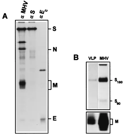
In the present study we have used RK13 cells instead of L cells for the production of VLPs since these cells are trans-fected much more efficiently (data not shown). All structural proteins were detected in the transfected cells by immunopre-cipitation with several antibodies directed against the different proteins (Fig. 1A). The VLPs from RK13 cells had a slightly lower density than MHV-A59 virions, which is in accordance with the data presented by Vennema et al. (50). Immunopre-cipitation of the sucrose gradient peak fractions with either anti-S MAbs or with rabbit polyclonal antiserum k134 showed that the S and the M proteins were present (Fig. 1B).
The VLP infectivity assay was performed as described pre-viously (3). When VLPs, formed in the transfected cells (P0), are infectious, they will transfer MIDI RNA to fresh L cells (P1; Fig. 2A). The P1 cells are coinfected with MHV-A59 helper virus to replicate the MIDI RNA. MIDI RNA is then amplified during subsequent undiluted passaging. VLPs that are not able to transfer the MIDI RNA to the P1 cells are by definition noninfectious. The presence of MIDI RNA in the intracellular RNA of P3 and P4 cells is used as a readout system for infectivity of the VLPs (3). Intracellular RNA of P3 and P4 cells was isolated and analyzed by gel hybridization with oligonucleotide 048MHV as the probe. This oligonucleotide binds to all viral RNAs, including MIDI RNA (Fig. 2B). When all the structural proteins of MHV-A59 were coexpressed in the P0 cells, MIDI RNA could be detected in the P3 and P4 intracellular RNA (Fig. 2B, lanes “all”). MIDI RNA was not detected when either one of the essential components of VLPs (M and E) was not expressed in the P0 cells (Fig. 2B, lanesDE andDM), demonstrating that the requirements for the produc-tion of infectious VLPs in RK13 cells are the same as those for production in L cells (3). When the plasmid encoding the spike protein was omitted in the P0 cells, the MIDI RNA was also
on November 9, 2019 by guest
http://jvi.asm.org/
absent in P3 and P4 cells (Fig. 2B, lanesDS). Similar amounts of RNA were analyzed in all lanes, as judged from the amounts of viral mRNA and rRNA. We thus showed that the spike protein must be expressed in the P0 cells to yield infectious VLPs.
Several other DI RNAs arose during passaging, which often occurs during passaging of MHV-A59 (47), but none was of the same size as MIDI (Fig. 2B). To specifically detect MIDI RNA, the gel was rehybridized with probe 010MHV, which binds to the specific ORF1a-ORF1b junction of MIDI. The endogenous DI RNAs did not hybridize to this probe; only MIDI RNA did. MIDI RNA was detected in P3 and P4 intra-cellular RNA when all proteins were expressed in the P0 cells but not when either E, M, or S was absent in the P0 cells (Fig. 2C). Since the probe discriminates between MIDI and novel DI RNAs, we have henceforth used probe 010MHV for RNA analysis.
VLPs are neutralized by S-specific MAbs.We went on to prove that the VLP infectivity assay is dependent on S by neutralizing the VLPs with spike-specific antibodies. MAbs A2.3 and A1.4 are both directed against the spike protein (17); however, MAb A2.3 neutralizes MHV-A59 virions, whereas MAb A1.4 does not. Prevention of VLP infection by MAb A2.3 but not by MAb A1.4 would thus be proof that S in VLPs is biologically similar to S in virions. However, a modified infec-tivity assay was used to avoid neutralization of the helper virus MHV-A59 by MAb A2.3. Murine L cells were first infected with helper virus MHV-A59 for 1 h. Two aliquots of VLP-containing medium were incubated with either MAb A2.3 or MAb A1.4 in a 1:1 ratio at 37°C for 1 h. Then the MHV-A59-infected cells were superMHV-A59-infected with this mixture of VLPs and MAbs for 1 h. To ensure S dependence in this experiment, spikeless VLPs were used as a negative control. As expected, MIDI RNA could not be detected in the P4 intracellular RNA when the VLPs were produced in the absence of the spike
[image:3.612.60.278.69.303.2]protein (Fig. 3A, lanesDS). VLPs that were incubated with MAb A1.4 could still infect the P1 cells, since MIDI was de-tected in the P4 cells (Fig. 3A, lanes A1.4). However, MIDI RNA was not detected in the P3 and P4 cells when the same VLPs were incubated with neutralizing MAb A2.3 (Fig. 3A, lanes A2.3). Oligonucleotide 010MHV has affinity for the 18S and 28S rRNAs (Fig. 3A) migrating below MIDI. We use these RNAs as an internal standard to show that similar amounts of intracellular RNA were analyzed in all lanes. We conclude that the VLPs, like MHV-A59 virions, could be neutralized by a neutralizing anti-S antibody.
FIG. 1. Expression of the structural proteins of MHV-A59 in RK13 cells. (A) Immunoprecipitation of cell lysates of vTF7.3-infected and DNA-transfected (M, E, N, and S) cells. (B) Immunoprecipitations on the peak fractions of sucrose gradient-purified VLPs from the medium of RK13 cells by using the anti-S MAbs (upper panel) and k134 (lower panel).aMHV, polyclonal antibody k134;aS, mixture of anti-S MAbs A2.3 and A1.4;aEc, polyclonal antibody anti-Ec.
FIG. 2. (A) Schematic outline of the VLP infectivity assay showing the in-fection with vTF7.3 and DNA transin-fection of the P0 cells. P0 RK13 cells were transfected with pM, N, E, S, and pMIDI-HD. P1 L cells were infected with VLPs and helper virus MHV-A59, and subsequently several passages were per-formed. (B and C) Analysis of intracellular RNAs from L-cell passages. As outlined for panel A, VLPs were produced and several undiluted passages were performed. Intracellular RNA from P3 and P4 cells was isolated and analyzed in a hybridization analysis. The gel was hybridized with oligonucleotide 048MHV (B) or with oligonucleotide 010MHV (C). Numbers on the left indicate the viral RNAs. MIDI-HD RNA is marked by an arrow. Ribosomal rRNAs are indicated by asterisks. Newly synthesized DI RNAs are indicated by open arrowheads.
on November 9, 2019 by guest
http://jvi.asm.org/
[image:3.612.344.505.69.486.2]Anti-MHV receptor antibody CC1 blocks VLP infection.The spike protein binds to the CEA receptor protein on the target cells as the initial step in the viral infectious cycle. It was shown previously that MHV-A59 infection of mouse fibroblasts could be blocked with MAb CC1 (7, 8, 51). This MAb is directed against the N-terminal part of the MHV receptor protein, which constitutes the binding site of S (8). Thus, if VLP infec-tivity could be blocked by MAb CC1, it would confirm the S dependency of the VLP infectivity assay. Again, a modified VLP superinfection assay was used. L cells were first infected with MHV-A59 for 1 h and subsequently kept at 37°C for an additional hour. Then, an aliquot of VLPs in P0 medium was used to superinfect the MHV-A59-infected cells. After several passages, MIDI RNA could be detected in the intracellular RNA (Fig. 3B, lanes mock). This control showed that the VLPs could infect the cells 2 h after MHV-A59 infection. In the receptor blockade experiment, L cells were incubated with MAb CC1 at 37°C in a 1:2 dilution for 1 h immediately after MHV-A59 infection. Subsequently, an aliquot of VLPs was mixed with the MAb CC1-containing medium, and this mixture was used to challenge the P1 cells for an additional hour. After passaging, MIDI was undetectable in P3 and P4 intracellular RNA, showing that VLP infection had been prevented by the MAb CC1 treatment of the target cells (Fig. 3B, lanes CC1). Approximately the same amounts of total RNA were analyzed, again indicated by the amounts of rRNAs (Fig. 3B). This experiment confirmed that the VLPs use the MHV receptor for entry into the target cells as do MHV-A59 virions, most likely through interaction of the spike protein with the receptor protein.
RT-PCR confirms the presence of MIDI RNA.We define the absence of VLP infectivity by the absence of MIDI RNA after passaging. Possibly, the detection limits of the hybridization assay prevented us from detecting small amounts of MIDI RNA. We therefore used a more sensitive detection method to analyze for the presence of MIDI RNA in P4 material. The intracellular P4 RNA was subjected to cDNA synthesis with an oligonucleotide that binds to the ORF1b region of MIDI (060MHV). A nested and MIDI-specific PCR amplification
[image:4.612.57.286.68.215.2]was subsequently performed on the cDNA with one oligonu-cleotide in the ORF1a (011MHV) and one in the ORF1b (055MHV) region of MIDI (Fig. 4A). This nested PCR should result in a MIDI-specific product of 771 bp. The PCR was also performed on pMIDI-HD DNA as a positive control (Fig. 4B, lane pMIDI-HD). PCR products of the predicted size were observed in the lanes of the samples in which we had previously detected the MIDI RNA in the hybridization assay (lanes mock and A1.4). No PCR products of this length were ob-served in the other samples (lanes bidest, CC1, deltaS, and A2.3) although aspecific PCR products of other sizes were observed in some samples. As an extra control, PCR products were transferred to nitrocellulose and hybridized to MIDI-specific probe 010MHV (Fig. 4C). The oligonucleotide hybrid-ized exclusively to the MIDI-shybrid-ized PCR products (lanes pMIDI-HD, mock, and A1.4). Even on an overexposed film we FIG. 3. Analysis of intracellular RNAs from the VLP infectivity assay. (A) L
[image:4.612.343.495.72.463.2]cells were infected with MHV-A59 2 h prior to VLP infection. VLPs were incubated for 1 h with MAb A2.3 or MAb A1.4 as indicated above the gel prior to infection. VLPs lacking S were included as controls (DS). A hybridization analysis was performed on the intracellular P3 and P4 RNA with oligonucleotide 010MHV. (B) L cells were infected with MHV-A59 and subsequently incubated for 1 h with MAb CC1 or with medium (mock). The cells were subsequently infected with VLPs carrying (CC1 and mock lanes) or not carrying (DS lanes) the spike protein. P3 and P4 intracellular RNA was analyzed with oligonucleotide 010MHV. MIDI-HD RNA is marked by an arrow. rRNAs are indicated by asterisks.
FIG. 4. RT-PCR of the P4 intracellular RNA. (A) Diagram of the PCR protocols. Oligonucleotides that were used for cDNA synthesis and for PCR are indicated. The orientations of these oligonucleotides are indicated by arrows. The size of the predicted PCR product is indicated. (B) Direct analysis of PCR products on agarose gel. (C) Hybridization analysis of PCR products. The DNA from the gel shown in panel B was blotted to nitrocellulose and subsequently hybridized to oligonucleotide 010MHV. The MIDI-specific PCR product is in-dicated by an arrow. bidest, negative control for the RT-PCR procedure; mock, nontreated P1 cells; delta S, VLPs lacking S.
on November 9, 2019 by guest
http://jvi.asm.org/
did not observe a signal with any of the other PCR products. Using this sensitive method we confirmed that MIDI RNA was absent in P4 cells when the P1 cells were blocked with MAb CC1, when the VLPs lacked the spike protein, and when S-containing VLPs were neutralized with MAb A2.3.
Absence of MIDI RNA in P4 is not due to the low titers of helper virus. The presence of MIDI RNA after passaging requires sufficiently high helper virus titers to carry along the DI viruses. To determine whether the absence of MIDI was a result of low helper virus titers due to the experimental con-ditions, we determined the plaque titer of the P1 virus. Only minor variations were observed in the helper virus titer in the different tests. Also, at later passages, the cytopathic effect did not differ drastically for the various experiments (data not shown). We can thus rule out the possibility that the absence of MIDI RNA in the P4 intracellular RNA is a result of a low helper virus titer during passaging.
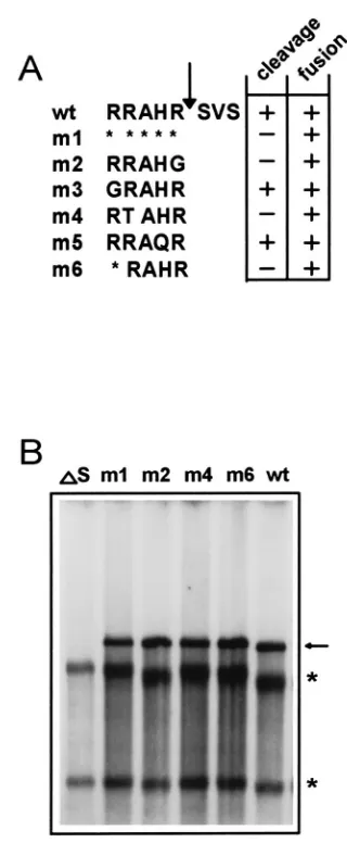
Cleavage of the spike protein is not required for infectivity. Having established that the VLP infectivity assay is dependent on the spike protein, we went on to express spike mutants on VLPs. We and others showed previously by expressing mutants that were altered in their cleavage recognition sequences that cleavage of the spike proteins of MHV-A59, -JHM, and -Wb1 was not required for syncytium formation (2, 41, 44). The S mutants that were cleavage negative (m1, m2, m4, and m6; Fig. 5A) (2) were introduced into the VLPs and tested for their effect on infectivity. As a control, spikeless VLPs were again produced to confirm that in the absence of S VLPs are not infectious (Fig. 5B, laneDS). Spike mutant m1 lacks the entire cleavage site and is not cleaved. However, its transport to the plasma membrane is not affected, and it induces syncytium formation, although with delayed kinetics (2). When mutant m1 was inserted into the VLPs, MIDI was detected in the P4 intracellular RNA (Fig. 5B, lane m1). Three other cleavage-negative spike mutants (m2, m4, and m6) have alterations in their cleavage recognition sequences and are all transported to the cell surface and induce delayed syncytium formation (2). Mutant m2 has the arginine residue at the P1 position replaced with a glycine. Mutant m4 has the arginine at P3 replaced with a threonine residue, and mutant m6 lacks an amino acid at the P5 position (Fig. 5A). All three spike mutants rendered the VLPs infectious, since MIDI was detected in the P4 intracel-lular RNA (Fig. 5B, lanes m2, m4, and m6). The presence of the mutant spike proteins on the VLPs can be inferred from their infectivity; spikeless VLPs are not infectious. With the data presented, it is not possible to determine whether cleav-age of S is advantcleav-ageous for infectivity. It should be noted that the VLP infectivity assay is qualitative and not quantitative. Any enhancing effects on infectivity of S cleavage cannot be determined. Nevertheless, this is the first report that deter-mines the role in infectivity of cleavage of MHV S by site-directed mutagenesis.
DISCUSSION
The spike protein has a crucial role during infection of cells by MHV-A59. When MHV-A59-infected cells were treated with tunicamycin, a drug that prevents N-glycosylation and thus proper transport of N-glycosylated proteins, or when hy-bridomas expressing anti-S antibodies were infected with MHV-A59, spikeless virus particles were formed and released into the medium (22, 23, 36). These particles were not infec-tious, but some residual infectivity remained (22, 36). In these experiments it was not possible to link the lack of infectivity of the virions to the absence of the spike protein unequivocally, since small amounts of S could still be inserted into the virions.
In this paper we describe a system in which we generate virus-like particles that completely lack S. We use it to show that the spike protein of MHV-A59 is absolutely required to yield in-fectious VLPs.
Our first objective was to ascertain that the function of S is the same in VLPs as it is in virions. Infection of cells by MHV-A59 virions can be blocked by treating the cells with MAb CC1. This antibody prevents the binding of the spike protein to the receptor, thereby inhibiting infection (7, 8, 51). VLP infection was also blocked by MAb CC1 treatment of the target cells (Fig. 3B). This indicates that the VLPs use the same receptor as do MHV-A59 virions. Furthermore, the VLPs reacted to treatment with anti-S antibodies A2.3 and A1.4 in a manner similar to that of MHV-A59 virions. This FIG. 5. (A) Schematic presentation of the mutations that were introduced near the cleavage site of MHV-A59 S (2). The Amino acid sequences just upstream of the cleavage sites of the different mutants and the wild type (wt) are shown. Cleavage and cell-to-cell fusion abilities of the mutants are indicated in the panel at the right. Arrow, cleavage site; asterisks, deleted amino acids. (B) Analysis of intracellular RNAs from the VLP infectivity assay. VLPs were pro-duced in RK13 cells. Either no S protein (DS), wild-type (wt) S protein, or mutant (m1, m2, m4 and m6) S protein was expressed in the P0 cells. The protocol used was the same as that described in the legend of Fig. 2. The intracellular RNA of P4 cells was subjected to a hybridization analysis using oligonucleotide 010MHV. Lanes:DS, VLPs lacking S; m1, m2, m4, and m6, VLPs carrying mutant S; wt, VLPs containing wild-type S. MIDI is marked by an arrow. rRNAs are indicated by asterisks.
on November 9, 2019 by guest
http://jvi.asm.org/
[image:5.612.341.501.63.451.2]indicates that the spike protein is exposed similarly on VLPs and virions and is functionally identical to S on MHV-A59 virions. Thus, although the VLP infectivity assay is an artificial system, it can be used as a tool to identify domains in S that are essential for infectivity. Also, mutations that block the infec-tivity of the virus can now be introduced into S. Such lethal mutations can never be detected with naturally occurring vari-ants.
The main goal of our studies is to understand the mechanism of fusion of the viral envelope with the host membrane and the role of the spike protein in this process. Our first focus was on the requirement of cleavage of S for fusogenicity, which we studied previously with transiently expressed spike mutants (2). Cleavage of the fusion protein is important for the infectivity and cell-fusing abilities of many enveloped viruses. Usually, cleavage allows exposure of a hydrophobic fusion peptide. In some proteins this peptide is located at the N terminus of the membrane-anchoring subunit (e.g., human immunodeficiency virus gp160 [10, 14], influenza virus hemagglutinin [16], and Sendai virus F [1]), but the peptide can also be located inter-nally as was shown for the Semliki Forest virus E1 protein (15). Since there is no obvious N-terminal hydrophobic amino acid stretch on the S2 subunit of MHV-A59 S (26), the fusion peptide must be located internally. Several regions in the spike protein have been implicated as being involved in the fusion process (2, 11, 13, 19), but the exact internal fusion peptide has yet to be identified.
Previous studies on the relation between cleavage of S and fusion have produced conflicting results. Rapid fusion of L cells induced by MHV-A59 virions purified from 17Cl1 cells required the addition of trypsin (42), and syncytium formation could be delayed by treating MHV-A59-infected cells with a protease inhibitor (9). From these studies it was concluded that cleavage of S of MHV-A59 was required for cell-to-cell fusion. Several groups have since studied the effect of cleavage of the spike protein on syncytium formation by transient expression of mutant spike proteins in the vaccinia T7 expression system (2, 41, 44). In these studies the opposite was found: cleavage of S of MHV-A59 and MHV-JHM was not required for syncy-tium formation, but cleavage-negative spike mutants were de-layed in syncytium induction. Only two studies were able to directly shed light on the relation between cleavage of S and infectivity. In one, MHV strain 2 was determined to have an uncleaved spike protein, implying that cleavage is not required for MHV-2 infectivity in DBT cells. Interestingly, cleavage of the spike protein of MHV-2 was required to induce syncytia (53). Yet, when in another study MHV-A59 viruses isolated from 17Cl1 cells were treated with trypsin in order to increase the amount of cleaved S on the virions, the infectivity increased twofold (42), thus indicating a relationship between cleavage and infectivity. Here we show unequivocally that cleavage of S is not required for infectivity of VLPs of strain A59. Why then do most murine coronaviruses maintain cleavage of the spike protein? Cleavage of S could be a codeterminant of the tissue tropism of the virus in the host. However, Gombold et al. (18, 21) showed that neither tissue tropism nor virulence depended solely on the spike protein but that other viral factors were involved. Another possibility is that cleavage of S enhances the cell-to-cell spread of the virus in the natural host: syncytium formation is more rapid in tissue culture when cleaved spikes are present (2, 41, 44).
In the present paper we show the feasibility of using the VLP infectivity assay of MHV-A59 to study the role of domains of S in infectivity by introducing mutant proteins. This system enables us to determine which regions of the spike protein are
required for incorporation of S into the VLPs, receptor bind-ing, and fusion during entry.
ACKNOWLEDGMENTS
E.C.W.B. was supported by grant 901-02-148 from the Dutch Orga-nization for Scientific Research (NWO-MW). W.L. is a fellow of the Royal Dutch Academy of Sciences (KNAW).
We thank J. Fleming for anti-S hybridoma cell lines A2.3 and A1.4 and K. Holmes for providing antibody CC1. We thank Guido van Marle, Richard Molenkamp, Jessika Dobbe, and Heleen Gerritsma for stimulating discussions.
REFERENCES
1. Blumberg, B. M., C. Giorgi, K. Rose, and D. Kolakofski. 1985. Sequence determination of the Sendai virus fusion protein gene. J. Gen. Virol. 66:317– 331.
2. Bos, E. C. W., L. Heijnen, W. Luytjes, and W. J. M. Spaan. 1995. Mutational analysis of the murine coronavirus spike protein: effect on cell-to-cell fusion. Virology 214:453–463.
3. Bos, E. C. W., W. Luytjes, H. Van der Meulen, H. K. Koerten, and W. J. M. Spaan.1996. The production of recombinant infectious DI-particles of a murine coronavirus in the absence of helper virus. Virology 218:52–60. 4. Bredenbeek, P. 1990. Nucleic acid domains and proteins involved in the
replication of coronaviruses. Ph.D. thesis. University of Utrecht, Utrecht, The Netherlands.
5. Chang, R. Y., M. A. Hofmann, P. B. Sethna, and D. A. Brian. 1994. A
cis-acting function for the coronavirus leader in defective interfering RNA
replication. J. Virol. 68:8223–8231.
6. Collins, A. R., R. L. Knobler, H. Powell, and M. J. Buchmeier. 1982. Mono-clonal antibodies to murine hepatitis virus-4 (strain JHM) define the viral glycoprotein responsible for attachment and cell-cell fusion. Virology 119: 358–371.
7. Dveksler, G. S., M. N. Pensiero, C. B. Cardelechio, R. K. Williams, G. S. Jiang, K. V. Holmes, and C. W. Dieffenbach.1991. Cloning of the mouse hepatitis virus (MHV) receptor: expression in human and hamster cell lines confers susceptibility to MHV. J. Virol. 65:6881–6891.
8. Dveksler, G. S., M. N. Pensiero, C. W. Dieffenbach, C. B. Cardelechio, A. A. Basile, P. E. Elia, and K. V. Holmes.1993. Mouse hepatitis virus strain A59 and blocking antireceptor monoclonal antibody bind to the N-terminal do-main of cellular receptor. Proc. Natl. Acad. Sci. USA 90:1716–1720. 9. Frana, M. F., J. N. Behnke, L. S. Sturman, and K. V. Holmes. 1985.
Pro-teolytic cleavage of the E2 glycoprotein of murine coronavirus: host-depen-dent differences in proteolytic cleavage and cell fusion. J. Virol. 56:912–920. 10. Freed, E. O., D. J. Myers, and R. Risser. 1990. Characterization of the fusion domain of the human immunodeficiency virus type 1 envelope glycoprotein. Proc. Natl. Acad. Sci. USA 87:4650–4654.
11. Gallagher, T. M., C. Escarmis, and M. J. Buchmeier. 1991. Alteration of the pH dependence of coronavirus-induced cell fusion: effect of mutations in the spike protein. J. Virol. 65:1916–1928.
12. Gallagher, T. M., M. J. Buchmeier, and S. Perlman. 1992. Cell receptor-independent infection by a neurotropic murine coronavirus. Virology 191: 517–522.
13. Gallagher, T. M. 1996. Murine coronavirus membrane fusion is blocked by modification of thiols buried within the spike protein. J. Virol. 70:4683–4690. 14. Gallaher, W. R. 1987. Detection of a fusion peptide sequence in the
trans-membrane protein of human immunodeficiency virus. Cell 50:327–328. 15. Garroff, H., A.-M. Frischauf, K. Simons, H. Lehrbach, and H. Delius. 1980.
Nucleotide sequence of cDNA coding for Semliki Forest virus membrane proteins. Nature 288:236–241.
16. Gething, M. J., R. W. Doms, D. York, and J. White. 1986. Studies on the mechanism of membrane fusion: site-specific mutagenesis of the hemagglu-tinin of influenza virus. J. Cell Biol. 102:11–23.
17. Gilmore, W., J. O. Fleming, S. A. Stohlman, and L. P. Weiner. 1987. Char-acterization of the structural proteins of the murine coronavirus strain A59 using monoclonal antibodies. Proc. Soc. Exp. Biol. Med. 185:177–186. 18. Gombold, J. L., S. T. Hingley, and S. R. Weiss. 1993. Fusion-defective
mutants of mouse hepatitis virus A59 contain a mutation in the spike cleav-age signal. J. Virol. 67:4504–4512.
19. Grosse, B., and S. G. Siddell. 1994. Single amino acid changes in the S2 subunit of the MHV surface glycoprotein confer resistance to neutralization by S1 subunit-specific monoclonal antibody. Virology 202:814–824. 20. Hernandez, L. D., L. R. Hoffman, T. G. Wolfsberg, and J. White. 1996.
Virus-cell and cell-cell fusion. Annu. Rev. Cell Dev. Biol. 12:627–661. 21. Hingley, S. T., J. L. Gombold, E. Lavi, and S. R. Weiss. 1994. MHV-A59
fusion mutants are attenuated and display altered hepatotropism. Virology 200:1–10.
22. Holmes, K. V., E. W. Doller, and L. S. Sturman. 1981. Tunicamycine resis-tant glycosylation of coronavirus glycoprotein: demonstration of a novel type of viral glycoprotein. Virology 115:334–344.
on November 9, 2019 by guest
http://jvi.asm.org/
23. Holmes, K. V., J. F. Boyle, R. K. Williams, C. B. Stephensen, S. G. Robbins, E. C. Bouer, C. S. Duchala, M. F. Frana, D. G. Weismiller, S. Compton, J. J. McGowan, and L. S. Sturman.1987. Processing of coronavirus proteins and assembly of virions, p. 339–349. In M. A. Brinton and R. R. Rueckert (ed.), Positive strand RNA viruses. Alan R. Liss, Inc., New York, N.Y. 24. Kim, K. H., K. Narayanan, and S. Makino. 1997. Assembled coronavirus
from complementation of two defective interfering RNAs. J. Virol. 71:3922– 3931.
25. Laemmli, U. K. 1970. Cleavage of structural proteins during the assembly of the head of bacteriophage T4. Nature 227:680–685.
26. Luytjes, W., L. S. Sturman, P. J. Bredenbeek, J. Charite, B. A. M. van der Zeijst, M. C. Horzinek, and W. J. M. Spaan.1987. Primary structure of the glycoprotein E2 of coronavirus MHV-A59 and identification of the trypsin cleavage site. Virology 161:479–487.
27. Luytjes, W., H. Gerritsma, E. Bos, and W. J. M. Spaan. 1997. Characteriza-tion of two temperature-sensitive mutants of coronavirus mouse hepatitis virus strain A59 with maturation defects in the spike protein. J. Virol. 71:949–955.
28. Makino, S., and M. M. C. Lai. 1989. High-frequency leader sequence switch-ing durswitch-ing coronavirus defective interferswitch-ing RNA replication. J. Virol. 63: 5285–5292.
29. Masters, P. S., C. A. Koetzner, C. A. Kerr, and Y. Heo. 1994. Optimization of targeted RNA recombination and mapping of a novel nucleocapsid gene mutation in the coronavirus mouse hepatitis virus. J. Virol. 68:328–337. 30. Meinkoth, J., and G. Wahl. 1984. Hybridization of nucleic acids immobilized
on solid supports. Anal. Biochem. 138:267–284.
31. Niemann, H., and H. D. Klenk. 1981. Coronavirus glycoprotein E1, a new type of viral glycoprotein. J. Mol. Biol. 153:993–1010.
32. Opstelten, D.-J. E., P. De Groote, M. C. Horzinek, H. Vennema, and P. J. M. Rottier.1993. Disulfide bonds in folding and transport of mouse hepatitis coronavirus glycoproteins. J. Virol. 67:7394–7401.
33. Peng, D., C. A. Koetzner, T. McMahon, Y. Zhu, and P. S. Masters. 1995. Construction of murine coronavirus mutants containing interspecies chi-meric nucleocapsid proteins. J. Virol. 69:5475–5484.
34. Ricard, C. S., and L. S. Sturman. 1985. Isolation of the subunits of the coronavirus envelope glycoprotein by hydroxyapatite high-performance liq-uid chromatography. J. Chromatogr. 326:191–197.
35. Ricard, C. S., C. A. Koetzner, L. S. Sturman, and P. S. Masters. 1995. A conditional-lethal coronavirus mutant that fails to incorporate the spike glycoprotein into assembled virions. Virus Res. 39:261–276.
36. Rottier, P. J. M., M. C. Horzinek, and B. A. M. Van der Zeijst. 1981. Viral protein synthesis in mouse hepatitis virus strain A59-infected cells: effect of tunicamycin. J. Virol. 40:350–357.
37. Sambrook, J., E. F. Fritsch, and T. Maniatis. 1989. Molecular cloning: a laboratory manual, 2nd ed. Cold Spring Harbor Laboratory, Cold Spring Harbor, N.Y.
38. Schmidt, M. F. G. 1982. Acylation of viral spike glycoproteins: a feature of enveloped RNA viruses. Virology 116:327–338.
39. Snijder, E. J., A. L. M. Wassenaar, and W. J. M. Spaan. 1994. Proteolytic processing of the replicase ORF1a protein of equine arteritis virus. J. Virol. 68:5755–5764.
40. Spaan, W. J. M., P. J. M. Rottier, M. C. Horzinek, and B. A. M. van der Zeijst.1981. Isolation and identification of virus-specific mRNAs in cells infected with mouse hepatitis virus (MHV-A59). Virology 108:424–434. 41. Stauber, R., M. Pfleiderer, and S. Siddell. 1993. Proteolytic cleavage of the
murine coronavirus surface glycoprotein is not required for fusion. J. Gen. Virol. 74:183–191.
42. Sturman, L. S., C. S. Ricard, and K. V. Holmes. 1985. Proteolytic cleavage of the E2 glycoprotein of murine coronavirus: activation of cell-fusing activ-ity of virions by trypsin and separation of two different 90K cleavage frag-ments. J. Virol. 56:904–911.
43. Taguchi, F., T. Ikeda, and H. Shida. 1992. Molecular cloning and expression of a spike protein of neurovirulent murine cornavirus JHMV variant cl-2. J. Gen. Virol. 73:1065–1072.
44. Taguchi, F. 1993. Fusion formation by the uncleaved spike protein of murine coronavirus JHM variant cl-2. J. Virol. 67:1195–1202.
45. Van Berlo, M. F., W. J. Van den Brink, M. C. Horzinek, and B. A. M. Van der Zeijst.1987. Fatty acid acylation of viral proteins in murine hepatitis virus-infected cells: brief report. Arch. Virol. 95:123–128.
46. Van der Most, R. G., P. J. Bredenbeek, and W. J. M. Spaan. 1991. A domain at the 39end of the polymerase gene is essential for the encapsidation of coronavirus defective interfering RNAs. J. Virol. 65:3219–3226.
47. Van der Most, R. G., L. Heijnen, W. J. M. Spaan, and R. J. de Groot. 1992. Homologous RNA recombination allows efficient introduction of site-spe-cific mutations into the genome of coronavirus MHV-A59 via synthetic co-replicating RNAs. Nucleic Acids Res. 20:3375–3381.
48. Vennema, H., L. Heijnen, A. Zijderveld, M. C. Horzinek, and W. J. M. Spaan.1990. Intracellular transport of recombinant coronavirus spike pro-teins: implications for virus assembly. J. Virol. 64:339–346.
49. Vennema, H., R. Rijnbrand, L. Heijnen, M. C. Horzinek, and W. J. M. Spaan.1991. Enhancement of the vaccinia virus/phage T7 RNA polymerase expression system with encephalomyocarditis virus 59untranslated region sequences. Gene 108:201–210.
50. Vennema, H., G. J. Godeke, J. W. A. Rossen, W. F. Voorhout, M. C. Hor-zinek, D. J. E. Opstelten, and P. J. M. Rottier.1996. Nucleocapsid-indepen-dent assembly of coronavirus-like particles by co-expression of viral envelope protein genes. EMBO J. 15:2020–2028.
51. Williams, R. K., G.-S. Jiang, S. W. Snyder, M. F. Frana, and K. V. Holmes. 1990. Purification of the 110-kilodalton glycoprotein receptor for mouse hepatitis virus (MHV)-A59 from mouse liver and identification of a non-functional, homologous protein in MHV-resistant SJL/J mice. J. Virol. 64: 3817–3823.
52. Williams, R. K., G.-S. Jiang, and K. V. Holmes. 1991. Receptor for mouse hepatitis virus is a member of the carcinoembryonic antigen family of gly-coproteins. Proc. Natl. Acad. Sci. USA 88:5533–5536.
53. Yamada, Y. K., K. Takimoto, M. Yabe, and F. Taguchi. 1997. Acquired fusion activity of a murine coronavirus MHV-2 variant with mutations in the proteolytic cleavage site and the signal sequence of the S protein. Virology 227:215–219.
54. Zhang, L., W. Luytjes, F. Homberger, and W. J. M. Spaan. 1997. Recombi-nant genomic RNA of coronavirus MHV-A59 after coreplication with a DI RNA containing the MHV RI spike gene. Virology 230:93–102.