0022-538X/00/$04.00⫹0
Copyright © 2000, American Society for Microbiology. All Rights Reserved.
Evolution of the Human Immunodeficiency Virus Type 1 Envelope
during Infection Reveals Molecular Corollaries of Specificity for
Coreceptor Utilization and AIDS Pathogenesis
QIN-XUE HU,1D† ASHLEY PERKINS BARRY,2‡ ZI-XUAN WANG,1SHANON M. CONNOLLY,2
STEPHEN C. PEIPER,1ANDMICHAEL L. GREENBERG2*
Henry Vogt Cancer Research Institute, University of Louisville, Louisville, Kentucky,1and Department of Surgery, Duke University Medical Center, Durham, North Carolina2
Received 10 December 1999/Accepted 15 September 2000
The evolution of human immunodeficiency virus type 1 infection is associated with a shift in the target cell
population, driven by variability in coreceptor utilization resulting from diversity in env. To elucidate the
potential consequences of these changes for Env-mediated fusion over the course of AIDS, we examined the
biological properties of serial viral isolates and determined coreceptor utilization by the products ofenvcloned
from two individuals, followed from the detection of seroconversion throughout the course of their infection. One had a typical course, and the other had an accelerated progression. Early isolates were non-syncytium inducing, and the corresponding Env exclusively utilized CCR5, whereas Env from late phases of infection showed restricted utilization of CXCR4 in both patients. Env from subject SC24, who had a standard progression, demonstrated multitropism, manifested by utilization of CCR3, CXCR4, and CCR5 in the intervening period. In contrast, Env from patient SC51, who experienced early conversion to the
syncytium-inducing phenotype, developed dualtropic coreceptor utilization of CCR5 and CXCR4. Genetic analysis ofenv
from each isolate revealed that those with an X4 phenotype formed a distinct subcluster within each subject.
Analysis of chimeras constructed from R5 and multispecificenvfrom patient SC24 demonstrated that while the
V3 domain played a dominant role in determining coreceptor utilization, sequences in the V4–V5 region also contributed to the latter phenotype. Immunoprecipitation experiments confirmed that the hybrid Env proteins were expressed at similar levels. These experiments demonstrate that progression from the R5 to X4 phenotype may occur through a multi- or dual-tropic intermediate and that multiple domains contribute to this process.
Human immunodeficiency virus type 1 (HIV-1) infection is initiated by entry of the viral genome following merging of the lipid bilayer that circumscribes the virus with the plasma mem-brane of the target cell. The product of theenvgene, which is processed into two noncovalently associated subunits, gp120 and gp41, mediates this membrane fusion process (25). This complex undergoes a series of conformational shifts that result from the interaction of gp120 with cellular receptors and cul-minate in membrane fusion (38, 53). The association of gp120 with the primary receptor, CD4, results in the exposure of cryptic protein surfaces in Env (40). The finding that human CD4 is necessary but not sufficient to confer sensitivity to infection led to the ultimate recognition that the role of core-ceptor is performed by a cadre of serpentine recore-ceptors that transmit the signals of chemoattractant cytokines, or chemo-kines (1, 13, 20, 23, 24, 28). The conformation of Env induced by engagement of CD4 is permissive for interaction with co-receptors, which triggers terminal events in the fusion process, presumably extension of the hydrophobic peptide of gp41 (38). The front-line coreceptor for commonly transmitted strains of HIV-1 is CCR5 (1, 13, 20, 23, 24), and the importance of this function is illustrated by the high degree of resistance to
in-fection of individuals homozygous for a 32-bp deletion in the gene encoding CCR5 (36, 52), who consequently lack a func-tional receptor.
Whereas the binding of gp120 to CD4 is virtually universal among HIV-1 isolates (18, 37, 39), there is variability in the interaction between coreceptors and this glycoprotein over the course of AIDS (6, 17). Selective pressures exerted by neutral-izing antibodies (3, 7, 44, 59, 63) and other immunological responses coupled with imperfect fidelity of reverse transcrip-tase (4, 47) are likely contributors to the genetic and biological diversification ofenv(56). This diversity in coreceptor utiliza-tion is manifested as the entry block associated with target cell tropism. Env derived from isolates obtained early in the evo-lution of HIV-1 infection demonstrate exclusive utilization of CCR5, while those arising later often use CXCR4 for mem-brane fusion, and correspond to macrophage (M)-tropic and T cell-tropic (T-tropic) strains, respectively. The designations of R5 and X4 for M- and T-tropic Env reflect this functional relationship (discussed previously by E. A. Berger, R. W. Doms, E. M. Fenyo, B. T. Korber, D. R. Littman, J. P. Moore, Q. J. Sattentau, H. Schuitemaker, J. Sodroski, and R. A. Weiss, Letter, Nature391:240, 1998). Although several chemokine receptors have been found to function as coreceptors in vitro, CCR5 and CXCR4 are recognized as the primary coreceptors for HIV-1 infection in vivo at this time, because all available cloned Env have been shown to use either one or both as coreceptors and simultaneous antagonism of these receptors blocks infection of primary cells (70; discussed previously by N. L. Michael and J. P. Moore, Letter, Nat. Med.5:740–742, 1999).
The molecular determinants specifying HIV-1 tropism have been studied extensively (11, 30, 31, 43, 57, 65, 66). Early
* Corresponding author. Mailing address: Center For AIDS Re-search, Box 2926, Rm. 113 SORF Bldg., Department of Surgery, Duke University Medical Center, Durham, NC 27710. Phone: (919) 681-5598. Fax: (919) 684-4288. E-mail: [email protected].
† Present address: Wuhan Institute of Virology, Chinese Academy of Sciences, Wuhan 430071, People’s Republic of China.
‡ Present address: Yerkes Regional Primate Center, Emory Univer-sity, Atlanta, GA 30329.
11858
on November 9, 2019 by guest
http://jvi.asm.org/
reports demonstrated that the V3 region of gp120 is a major, but not the sole, determinant of tropism that determines the target cell repertoire (11, 30, 43, 57, 65, 66). More recently, several laboratories have demonstrated that this region is a key factor differentiating coreceptor utilization as well (13, 15). There is emerging evidence that other regions of gp120 also have a critical role in programming tropism and coreceptor utilization (9, 31, 41, 58). Recent crystallographic studies have provided tremendous insight into the surface of the gp120 core structure that is involved in the association with CD4 and, coupled to mutagenesis experiments, have implicated struc-tures involved in the interaction with coreceptors (35, 46, 67, 68). However, these experiments were limited by the need to delete critical variable loop segments of gp120, including V3 itself. While analysis of serial isolates has provided some in-sight into changes in env that are associated with shifts in tropism, our understanding of the precise mechanism for the metamorphosis of coreceptor utilization from CCR5 to CXCR4 by Env is limited. In addition, characterization of the utilization of ancillary coreceptors by ENV in vivo or in vitro may further elucidate this process.
Thus far, most of the insights concerning virus tropism and coreceptor utilization have been gained from studies employ-ing laboratory strains and unrelated primary isolates as the starting points for analysis and genetic engineering of variants (5, 12, 13, 49). The present study addresses the question ofenv evolution during the course of AIDS through characterization of coreceptor utilization by the products of clonedenvgenes obtained from serial samples from two individuals identified during the earliest stages of primary HIV-1 infection. The subjects were members of an acute-infection cohort in Trin-idad and Tobago. The average time from infection to progres-sion to AIDS in this cohort is approximately 5 years, which may be more representative of rates of disease progression in de-veloping nations (2) than of those observed in North America and Europe (42, 48, 50). These patients exhibited a phenotypic switch in their virus isolates from non-syncytium-inducing (NSI) to syncytium-inducing (SI), demonstrating that there is an evolution of coreceptor utilization from an initial M-trop-ic/R5 strain to one that includes CXCR4. In one case, there was acquisition of multitropic coreceptor utilization (CCR3, CCR2b, and CXCR4 in addition to CCR5), followed by Env that had lost the ability to utilize coreceptors other than CXCR4. However, such multispecific coreceptor utilization was not apparent in infectivity studies with these Env. In the second case there was early conversion to the SI phenotype that was associated with emergence of a dual-tropic Env able to use CCR5 and CXCR4 in both fusion and infection exper-iments, but promiscuous utilization of other coreceptors was not apparent. Analysis of chimeric Env indicated that elements in the V3 loop made an important contribution to multitropic coreceptor utilization, but sequences within the V4–V5 region also impart this phenotype independent of the V3 sequences. These findings provide critical insights into the utilization of coreceptors during the course of AIDS required to understand the pathogenesis of target cell specificity that will be critical to the design of antagonists of viral entry.
(This work represents partial fulfillment of requirements for a Ph.D. in Biochemistry and Molecular Biology for Zi-xuan Wang.)
MATERIALS AND METHODS
Preparation and phenotypic characterization of low-passage primary virus isolate stocks.Peripheral blood mononuclear cells (PBMC) were prepared from venous blood obtained from HIV-1-positive study subjects and seronegative donors by standard Ficoll-Hypaque density separation. Low-passage primary
virus isolates were prepared by coculturing 5⫻106freshly prepared (or, in some instances, viably cryopreserved) PBMC from a study subject with phytohemag-glutinin-activated seronegative donor PBMC in RPMI 1640 medium supple-mented with 20% heat-inactivated fetal calf serum (FCS), 5% interleukin 2, and gentamicin. Twice weekly, half the culture supernatants were harvested and replaced with fresh medium alone (once per week) or medium containing 107 phytohemagglutinin-activated activated donor PBMC (once per week). HIV-1 p24 antigen levels were monitored in culture supernatants twice per week until two successive readings exceeded 30 pg/ml and the levels were increasing. The cells were then pelleted and cocultured in 20 to 30 ml with 1⫻107to 1.5⫻107 CD8⫹cell-depleted PBMC from a pool of 10 seronegative donors that had been
prepared and activated as follows. The PBMC from a pool of normal donors were activated for 3 days with a mixture of anti-CD3 (50 ng/ml) and anti-CD28 (100 ng/ml) antibodies in medium supplemented with 10% fetal calf serum, 20 U of recombinant interleukin 2 (Genzyme) per ml, and penicillin-streptomycin (pen/strep) at 37°C in a humidified incubator. The PBMC were depleted of CD8 T cells with anti-CD8-coated magnetic microspheres (Dynal) according to the manufacturer’s recommendations. At daily intervals, the cells were pelleted, supernatants were harvested and filtered, and the cells were resuspended in fresh medium. Supernatants were monitored for the presence and amount of viral reverse transcriptase (RT) activity with a micro-RT assay (10) to estimate virus replication. Supernatants containing significant amounts of viral RT activity were subsequently characterized with respect to infectious virus titer in a PBMC-based assay. Virus harvests possessing 103 50% tissue culture infective doses (TCID50)/ml were aliquoted and stored at⫺70°C until used in biological exper-iments.
SI phenotypic determinations of the virus isolates were performed in the MT-2 cell line according to established AIDS Clinical Trial Group protocols (33, 34). The MT-2 cell line was obtained from the National Institutes of Health (NIH) AIDS Research and Reference Reagent Program. The MT-2 cells were main-tained in log-phase growth by splitting the cultures 1:3 every 3 to 4 days using RPMI 1640 supplemented with 2 mML-glutamine, pen/strep (100 U/ml and 100 g/ml, respectively), and 10% fetal bovine serum. Briefly, the assay was per-formed in duplicate in 96-well flat-bottomed tissue culture plates. A total of 50,000 MT-2 cells were placed in assay wells in 150l of medium; 50l of virus stock was added to duplicate wells, and 50l of medium was added to duplicate control wells. Positive controls were included in each assay. On days 3, 6, 9, and 12, each well was examined microscopically for syncytium formation, and the day that syncytia were first observed was recorded. After inspection of wells, cells were resuspended, and 130l of the culture was removed and replaced with flesh medium. Assays continued through 14 days, when the cultures were terminated. Viruses that did not induce syncytium formation within this 14-day period were termed NSI.
Infectivity studies with primary virus isolates.Virus isolates were titrated on U87 cells and/or HeLa-CD4 cells to assess their ability to use CCR5 and/or CXCR4 to initiate productive infection. U87 cell lines stably expressing CD4 alone (obtained through the AIDS Reagent Program, DAIDS, NIAID, NIH, from HongKui Deng and Dan Littman), CD4 and CCR5, or CD4 and CXCR4 (both gifts from Dan Littman) were seeded in 48-well plates (Costar) 24 h prior to infection. Cells were seeded at 7.5⫻103cells/well in 150l of Dulbecco’s modified Eagle’s medium (DMEM) containing 10% heat-inactivated FCS. The U87-CD4 cells were maintained and initially seeded in medium containing G418 (300g/ml), whereas the U87-CD4-CCR5 cells and the U87-CD4-CXCR4 cells were maintained and initially seeded in medium containing G418 (300g/ml) and puromycin (1g/ml). Medium containing selective agents was removed 24 h later and replaced with 150l of DMEM containing 10% FCS. Serial dilutions of virus isolates (150-l volumes) were added to duplicate wells, bringing the volume of each test well to 300l. Cells were fed on days 2, 4, 7, and 10 with fresh DMEM containing 10% FCS. Aliquots of supernatants were taken on days 4, 7, and 11, adjusted to 1% Triton X-100, and assayed for RT activity as previously described (10). Infectious virus titers, expressed as TCID50per milliliter, were determined by the end-point dilution technique and calculated by the method of Reed and Muench (45).
In addition to the U87-based infectivity studies, we examined the ability of the primary virus isolates to replicate in the HeLa-CD4 indicator cell line (MAGI) originally described by Kimpton and Emerman (32) (obtained from the AIDS Reagent Program, DAIDS, NIAID, NIH, from Michael Emerman). As the HeLa cells express the CXCR4 coreceptor, this analysis scores specifically for the ability of virus isolates to infect via the CXCR4 coreceptor. MAGI cells were seeded 24 h prior to infection in a 48-well plate (Costar) at 2.5⫻104cells per well in 300l of DMEM containing 10% FCS, pen/strep (100 U and 100g/ml, respectively), 300 g ofL-glutamine per ml and 12.5g of DEAE per ml. Duplicate wells were infected with 100l of the appropriate virus. At 24 h after infection, DP178, an HR2 peptide mimetic and inhibitor of gp41-mediated fusion (C. Wild, T. Greenwell, and T. J. Matthews, AIDS Res. Hum. Retrovi-ruses9:1051–1053, 1993), was added to a final concentration of 5g/ml to ensure that infection readouts reflected a single round of replication. The cells were incubated for an additional 48 h and then fixed for 5 min with 1% formaldehyde and 0.2% glutaraldehyde in phosphate-buffered saline (PBS). Cells were stained in situ at 37°C for 50 min with X-Gal (5-bromo-4-chloro-3-indolyl--D -galacto-pyranoside), 0.4 mg/ml 2 mM MgCl2, 4 mM potassium ferricyanide, and 4 mM potassium ferrocyanide in PBS. Infection resulted in intense nuclear staining,
on November 9, 2019 by guest
http://jvi.asm.org/
and the number of stained nuclei was interpreted as being equal to the number of infectious virions in the challenge inoculum. Infected cells were enumerated using a charge-coupled device imager and Image Pro software (Georgia Instru-ments).
Direct sequencing of primaryenvgenes.The nucleotide sequence of a segment of the HIV-1envgene that includes the V3 loop of gpl20 was determined from amplification products obtained from proviral DNA templates using primers E100 (5⬘-ACACATGGAATTAAGCCAGT) and E130c (5⬘-CCTGCCACATG TTTATAATTTGT). The PCR products were excised from agarose gels, puri-fied, and concentrated. An oligonucleotide designed from sequences at the 5⬘ end of this segment, E110 (5⬘-CTGTTAAATGGCAGTCTAGCAGAA), was used to prime the sequencing reaction, which was performed using a Thermo Sequenase radiolabeled terminator cycle sequencing kit (Amersham).
Molecular cloning of primaryenvgenes.Proviral DNA templates were pre-pared from activated normal donor PBMC infected with the various primary isolates. Cell pellets were lysed in a buffer containing 10 mM Tris, 1 mM EDTA, 0.001% sodium dodecyl sulfate (SDS), and 0.5% Triton X-100 (pH 8.0). Pro-teinase K was added to a final concentration of 1 mg/ml, and the lysates were incubated at 60°C for 3 h. The proteinase K was heat inactivated by treating the lysates at 100°C for 10 min. In some cases the DNA was further purified by standard phenol-chloroform extraction techniques. The full-lengthenvgene con-taining the gp160 open reading frame was amplified by PCR using proviral DNA as templates. A region flankingenvwas amplified using upstream and down-stream primers designed from sequences intat(5⬘-TGCTGTTTATCCATTTC AGAATTGG) andnef(5⬘-TCCAGTCCCCCCTTTTCTTTTAAAAA), respec-tively. Standard conditions were employed for PCR amplification that included 1.5 mM MgCl2. PCR cycles were preceded by rapid heating to 95°C and con-sisted of the following intervals: denaturation, 30 s at 95°C; cooling ramp to 55°C, 60 s; annealing, 30 s at 55°C; heating ramp to 72°C; and synthesis, 7 min. Thirty-five amplification cycles were performed using the hot-start method. The 3.3-kb amplification product encompassedtat, rev, vpu, and part of thenefgene in addition toenv. The annealing temperature was optimized for each template. The amplification products were molecularly cloned into the pCR2.1-TOPO vector (Invitrogen Corp.).
The open reading frame encoding gp160 was amplified from this cloned template usingPfuDNA polymerase (Stratagene). Restriction sites forEcoRI andXhoI were incorporated into the upstream and downstream primers, respec-tively. Expression constructs encoding Env were prepared in pcDNA3.1 (Invitro-gen Corp.).
In some cases, full-lengthenvwas cloned from purified DNA templates as follows. Purified cellular DNA (0.5g) was used as the target for amplification of proviralenvDNA using a nested PCR approach. Briefly, first-round amplifi-cation was performed with the E0 5⬘-primer (5⬘-TAGAGCCCTGGAAGCATC CAGGAAGTCAGCCTA) and the Nef 9023 3⬘-primer (5⬘-CATTGGTCTTAA AGGTACCTGAGGT), each at 0.1M final concentration. Two units of the eLONGase enzyme mix (Gibco-BRL) per reaction was employed for PCR, with 200M deoxynucleoside triphosphates (dNTPs) and 1.5 mM MgSO4. An initial denaturation for 3 min at 94°C preceded 40 amplification cycles performed as follows: denaturation for 1 min at 94°C, annealing at 57°C for 1 min, and extension for 5 min at 70°C. These cycles were followed by a final 10-min extension at 70°C. One microliter of the first-round reaction was amplified for 25 cycles in a second-round reaction employing nested primers. Second-round prim-ers were the 5⬘-ENF primer (5⬘-AAAGAGCAGAAGACAGTGGCAATGAG AGTGAAGG) used in conjunction with either the 3⬘-ENR primer (5⬘-CAATC ACACTACTTTTTGACCACTTGCCACCCAT) or the 3⬘-NL4-38917R primer (5⬘-AGGTCTCGAGATACTGCTCCCA). The concentrations of all compo-nents and reaction conditions for the second-round amplifications were identical to those described above for the first-round PCR except for the number of cycles employed. Amplifiedenvwas gel purified and recovered using the QIAEX II gel extraction kit (Qiagen Inc.) and cloned into the pCDNA3.1/V5/His-TOPO ex-pression vector (Invitrogen Corp.).
Throughout the text and figures, individualenvclones may be designated either as c.X (for clone no. X) or simply as #X (for clone no. X). For example, QH1503c.1 (a designation appearing in Fig. 1A and B) refers to the sameenv
clone as designation QH1503#1 (a designation appearing in Table 2 and Fig. 2A).
Sequencing cloned full-lengthenvgenes and chimericenvconstructs.Clonedenv
genes were sequenced completely with an ABI 377 automated sequencer using the
TaqDye terminator cycle sequencing kit (ABI Prism) chemistries. All sequences were determined from both sense and antisense DNA strands to ensure the accuracy of the sequence determinations. Primers used to determine theenvsequences were as follows: ENF, 5⬘-AAAGAGCAGAAGACAGTGGCAATGAGAGTGAAGG; E155R, 5⬘-CTGTTCTACCATGTCATTTTTCCACATGT; E70, 5⬘-GGGATCAA AGCCTAAAGCCATGTGTAA; E90, 5⬘-CACAGTACAATGTACACATGGA AT; E145R, 5⬘-AGCAGTTGAGTTGATACTACTGG; NL437500, 5⬘-AAGTAG GAAAAGCAATGTATGCCCCTCCCAT; HXB21299R, 5⬘-ATGGGAGGGGC ATACATT; E180, 5⬘-GTCTGGTATAGTGCAGCA; E55R, 5⬘-GCCCCAGACT GTGAGTTGCAACAGATG; E220, 5⬘-TAACAAATTGGCTGTGGTATATAA; HXB28562R, 5⬘-CTCGTTACAATCAAGAGTAAGTCTCTCAA; ENR, 5⬘-CAA TCACACTACTTTTTGACCACTTGCCACCCAT; T7, 5⬘-TAATACGACTCAC TATAGGG; and PCR3.1, 5⬘-TAGAAGGCACAGTCGAGG. An R at the end of a primer name indicates a reverse primer. ENF and ENR were used only in direct
sequencing of PCR products. Primers residing in the cloning vector (T7 and PCR3.1) were used to sequence full-lengthenvclones in the immediate 5⬘and 3⬘ regions. Derived DNA sequences were initially analyzed and aligned using software packages from ABI. Gaps were inserted into the alignment as needed, and those regions were eliminated from the subsequent analysis. Phylogenetic analysis was performed, and consistency of the branching order of the phylogenetic trees was evaluated using the SEQBOOT, DNADIST, NEIGHBOR, CONSENSE, and DRAWGRAM modules of the Phylip Package (V3.572) (26, 27). Phylogenetic trees were constructed using neighbor-joining (51), and the stability of the nodes was assessed with the bootstrap value (26). Bootstrap values in excess of 70% were considered significant.
Reverse transcription, PCR amplification, and direct sequencing of plasma virusenvregions.Plasma was prepared from acid citrate dextrose-anticoagulated whole venous blood within 2 h of sample collection and stored at⫺70°C until analyzed. HIV-1 RNA was extracted with the Roche Amplicor HIV-1 Monitor Assay system (Roche Molecular Systems, Inc., Branchburg, N.J.) as specified by the manufacturer and stored at⫺70°C. A 25-l amount of the RNA extract was reverse transcribed and PCR amplified in a total volume of 75 l with the Perkin-Elmer GeneAmp Gold RNA PCR kit with the following reaction com-ponents and conditions: 1X RT PCR Buffer (Perkin-Elmer), 1.75 mM MgCl2, 0.35 mM each dNTP, 10 U of RNase inhibitor (Perkin-Elmer), 10 mM dithio-threitol, and 0.5M MG-12 3⬘-primer [5⬘-AA(T/C)(T/A)GTCTGGCCTGTAC CGTCAGCGT-3⬘] and 0.5M MG-6 5⬘-primer [5⬘-GGT(A/G)TCCTTTGA(G/ T)CCAATTCCCAT-3⬘], 2.5 U of AmpliTaq Gold, and 150 U of murine leuke-mia virus RT enzyme. Reverse transcription was carried out at 42°C for 60 min. Reaction components were heated to 95°C for 10 min, and then PCR was performed with 50 cycles of denaturation at 94°C for 1 min, annealing at 57°C for 1 min, and extension at 72°C for 2 min. A final extension at 72°C for 10 min was employed after the last cycle. The PCR mix (7.5l) was transferred to a separate tube for a second round of amplification with nested primers. Reaction condi-tions and components for the second-round PCR were 1X PCR buffer II (Perkin-Elmer), 1.5 mM MgCl2, 0.2 mM each dNTP, 0.2M MG-10 3⬘-primer [5⬘-CA CTT(C/T)TCCAATTGTCCCTCAT(A/G)TCTCCTCCT-3⬘] and 0.2M MG-7 5⬘-primer [5⬘-GTCAGCACAGTACAATGTACACAT-3⬘], and 2.5 U of Ampli-Taq Gold (Perkin-Elmer). Denaturation for 10 min at 95°C preceded 10 cycles of amplification using 95°C for 45 s for denaturation, 59°C for 1 min for anneal-ing, and 72°C for 1.5 min for extension. These were followed by 20 to 40 cycles of amplification using 94°C for 45 s for denaturation, 59°C for 1 min for anneal-ing, and 72°C for 1.5 min for extension. A final extension at 72°C for 10 min was employed. Direct sequencing and phylogenetic analysis of the amplified plasma viralenvregion were performed as described above for the full-lengthenvgenes using the MG-7, MG-10, E110, and HXB21129R (5⬘-AAATTCCCCTCCACA ATT-3⬘) primers for sequencing.
Genetic engineering of Env chimeras and mutants.Env chimeras between QH0648#1 and QH1520#2 and between QH0515#1 and QH1520#2 were gen-erated using sites that were present in theseenvgenes, includingEcoRI,AccI,
SauI, andXhoI. ADraI site was introduced at the 3⬘boundary of the V3 region as a silent mutation that did not alter the amino acid programmed by the codons at this position using the Chameleon double-stranded site-directed mutagenesis kit (Stratagene). The identity of all chimeras and mutants was confirmed by DNA sequencing.
Reporter gene assay for analysis of Env-mediated fusion.The coreceptor utilization of the products encoded by the primaryenvgenes, chimeras, and mutants was determined using a cell-cell fusion assay employing a luciferase reporter gene, as described previously (22, 61, 62), with modifications. Briefly, QT6 effector cells were prepared by infection with vTF1.1, which encodes T7 polymerase, for 1 h following transfection with pcDNA3 constructs containing the primaryenvclones. Target cells consisted of QT6 cells transfected with pcDNA3 constructs encoding CD4, coreceptors, and a luciferase reporter gene under the transcriptional control of the T7 promoter. The effector and target cell populations were mixed at 16 to 18 h following transfection, and the luciferase activity of cell lysates was determined approximately 9 h later using a LucLite luciferase reporter gene assay kit (Packard). Absolute light units emitted were measured using a Top Count luminometer (Packard).
Production of pseudotyped reporter viruses and infectivity assays.Stocks of pseudotyped reporter viruses were prepared as follows. 293T cells (a gift from Robert Doms) were maintained in DMEM containing 10% heat-inactivated FCS and 1% pen/strep (Gibco-BRL). 293T cells were seeded at 1.5⫻106cells per well in a six-well dish (Costar) 1 day prior to transfection. Cells were cotrans-fected with 10g of anenv expression vector and 5g of plasmid pNL4-3.Luc.R⫺E⫺(obtained from the AIDS Research and Reference Reagent
Pro-gram, DAIDS, NIAID, NIH, from Nathaniel Landau) by the calcium phosphate precipitation method essentially as described (16). Virus-containing supernatants were harvested 48 h posttransfection, mixed with heat-inactivated FCS to bring the concentration to 20% (vol/vol), filtered, and stored at⫺80°C until used for infection. The ability of clonedenvto mediate infection via the CCR5 and/or CXCR4 coreceptor was assessed in U87 astroglioma cells stably expressing the CD4 receptor and either the CCR5 or CXCR4 coreceptor or neither chemokine receptor. U87 cells were seeded at 1.5⫻104cells per well in a 96-well plate and infected with 100l of pseudotyped virus-containing supernatant. Cells were harvested 48 to 72 h postinfection and assayed with either the Promega luciferase
on November 9, 2019 by guest
http://jvi.asm.org/
assay system (Promega) in a Berthold Lumat 9501 (Wallac) or the LucLite luciferase assay kit (Packard) in a Top Count luminometer (Packard).
Metabolic labeling and immunoprecipitation of Env.The expression of wild-type and chimeric Env glycoproteins was analyzed by metabolic labeling and immunoprecipitation. Briefly, QT6 or HeLa cells (2⫻105) were seeded in six-well plates and grown to 80 to 90% confluence. Monolayers were infected with the vTF1.1 vaccinia virus, which directs the expression of T7 polymerase, for approximately 1 h and then transfected with theenvconstructs. These were prepared in the pcDNA3 vector, which contains a T7 promoter downstream of the cytomegalovirus promoter, which is typically employed in mammalian cells. The cells were allowed to incubate for 18 h at 37°C. The transfectants were then depleted of methionine and cysteine by growth in medium lacking these amino acids for 1 h. The cultures were then metabolically labeled in 1 ml of this medium supplemented with 100Ci of [35S]methionine/[35S]cysteine (NEN Life Science) for 1 h. Complete medium was then added, and the cells were incubated for an additional 1 h at 37°C. Lysates of metabolically labeled cells were clarified by centrifugation in a microcentrifuge and then incubated with HIVIG (obtained from the NIH AIDS Research and Reference Reagent Program) or normal immunoglobulin fractions, followed by protein G-Sepharose (Pharmacia) over-night at 4°C. Immune complexes bound by the protein G-Sepharose were washed, resuspended in SDS sample buffer, and resolved by SDS-polyacrylamide gel electrophoresis (PAGE), and the dried gels were analyzed by autoradiogra-phy.
Nucleotide sequence accession numbers.All sequences described were depos-ited in the GenBank database under accession no. AF310108 to AF310132.
RESULTS
envgenes from sequential primary isolates.To examine the
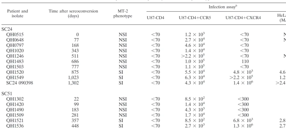
functional consequences of the diversification in envthat oc-curs during HIV-1 disease, we examined the biological and molecular characteristics of serial virus isolates obtained from two individuals in a cohort of acute seroconverters in Trinidad and Tobago. Cohort subjects were identified by virtue of ex-hibiting HIV-1 p24 antigenemia in the absence of a positive Western blot, and their status was confirmed by subsequent documentation of seroconversion to HIV-1 antigens. From a cohort of 22 HIV-positive individuals, we examined serial viral isolates andenvclones from two subjects whose isolates exhib-ited a phenotypic switch from NSI to SI during the period of observation. As shown in Table 1, the isolates studied spanned
the evolution from NSI to SI, which occurred over an interval of 875 days (⬃29 months) for patient SC24, but within a much shorter time frame, 12 months, for the second subject, SC51. Thus far, five subjects in the cohort have evidenced a switch in virus phenotype from an initial NSI isolate to a subsequent SI isolate. The mean time from infection to recovery of an SI isolate from the five subjects in the original cohort has been 31 months (range, 12 to 41 months), which appears to be consid-erably shorter than that observed in a recent study of nine individuals from the MACS cohort (56), and may reflect a more rapid pace of disease progression in developing versus developed countries. The NSI and SI designations in Table 1 are based on the ability of the virus isolates to induce syncytia in the MT-2 cell line.
We performed a parallel series of analyses using infection assays in U87-CD4 cells stably expressing either CCR5, CXCR4, or neither coreceptor and HeLa-CD4 cells (6, 32) to confirm that the conversion from NSI to SI corresponded to the emergence of viral species that utilize CXCR4 as a core-ceptor. For subject SC24, these studies indeed indicated that recovery of the SI phenotype was associated with the emer-gence of isolates with the ability to replicate in cells expressing CXCR4 (Table 1). For SC51, the results were somewhat less clear. Although the initial QH1302 isolate was restricted to replication in CCR5-expressing U87 cells, we observed very low levels of infection of U87-CD4 target cells expressing CXCR4 with the three successive NSI isolates (QH1420, QH1490, and QH1509) recovered from 99 to 281 days follow-ing seroconversion. However, titers of these isolates in CXCR4-expressing U87-CD4 cells never reached 300 TCID50/
[image:4.612.53.552.83.307.2]ml, and when the isolates were similarly evaluated on the MAGI target cells for infectivity, no evidence for infection of CXCR4-expressing cells was observed (Table 1). In contrast, we easily observed robust levels of replication in both the U87-based CXCR4 targets and the MAGI cells with the three SI isolates from this subject (Table 1), and their infection of
TABLE 1. Sequential HIV-1 primary isolates and phenotypesa
Patient and
isolate Time after sercoconversion(days) phenotypeMT-2
Infection assayb
U87-CD4 U87-CD4⫹CCR5 U87-CD4⫹CXCR4 HeLa-CD4(MAGI)
SC24
QH0515 0 NSI ⬍70 1.2⫻103 ⬍70 ND
QH0648 77 NSI ⬍70 2.7⫻104 ⬍70 ND
QH0797 168 NSI ⬍70 4.6⫻104 ⬍70 0
QH1020 343 NSI ⬍70 1.4⫻104 ⬍70 0
QH1246 511 NSI ⬍70 ⬎2.2⫻105 ⬍70 ND
QH1483 686 NSI ⬍70 1.0⫻105 110 0
QH1503 777 NSI ⬍70 1.1⫻105 ⬍70 0
QH1520 875 SI ⬍70 5.5⫻104 4.8⫻103 4.6⫻103
QH1549 1,023 SI ⬍70 6.3⫻104 ⬎2.2⫻105 1.2⫻104
SC24 090398 1,302 SI ⬍70 4.3⫻104 1.4⫻106 ⬎2.4⫻105
SC51
QH1302 22 NSI ⬍70 8.5⫻102 ⬍300 0
QH1420 99 NSI ⬍70 1.4⫻104 ⬍300 0
QH1490 183 NSI ⬍70 4.3⫻103 ⬍300 0
QH1509 281 NSI ⬍70 1.7⫻104 ⬍300 0
QH1521 357 SI ⬍70 8.5⫻102 6.8⫻103 2.8⫻103
QH1536 448 SI ⬍70 2.7⫻103 1.3⫻106 2.7⫻104
QH1558 539 SI ⬍70 4.7⫻103 8.5⫻105 1.9⫻104
aSubjects SC24 and SC51 were identified as experiencing acute HIV-1 infection on the basis of p24 antigenemia in the absence of a positive Western blot (defined as reactivity to two or more HIV-1 gene products).
bInfectious titers were determined by the end-point dilution technique, and TCID50per milliliter was calculated (45). For MAGI assay, titers represent number of infectious centers per milliliter. ND, not determined.
on November 9, 2019 by guest
http://jvi.asm.org/
MAGI cells was completely blocked by the CXCR4 inhibitor AMD3100 (data not shown). These results are consistent with the view that the SI phenotype is associated with the ability to productively infect via the CXCR4 coreceptor and document that the X4 character of virus isolates recovered from both subjects changed dramatically within a narrow time frame. The change in the X4 character of virus isolates was manifest in samples that were separated by only 98 days for subject SC24 and with samples separated by 76 days for subject SC51 (Table 1). After the initial recovery of virus isolates capable of using the CXCR4 coreceptor for entry, this characteristic became predominant in all subsequent isolates that were recovered from both subjects (Table 1).
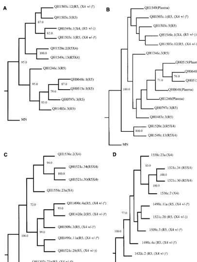
Primary env genes were amplified by PCR from proviral DNA templates derived from the successive isolates from these two individuals and molecularly cloned. A total of 128 env clones were isolated and tested for biological activity as de-scribed below. Comparison of the nucleotide sequence of mo-lecular clones active in Env-mediated fusion assays (see below) with that obtained from the direct analysis of proviral DNA templates confirmed that the former were representative of the predominant viral species in the isolate (Table 2). For SC24, a similar comparison with plasma virus sequences suggested that the fusion-competentenvclones were similar to variants cir-culating in vivo (Fig. 1B). Some variants showed ambiguities of the predicted primary structure of Env, which were interpreted to be the result of heterogeneity of integrated proviruses. Alignment of the nucleotide sequences of theenvclones and translation for the predicted primary structure from patients SC24 and SC51 confirmed that the sequential isolates from each patient were highly related to the others from that indi-vidual, consistent with being derived from similar (ancestral) viruses. Divergence at the level of primary structure was noted
most commonly in the V1, V2, V3, and V4 regions of gp120. Phylogenetic analysis of the successiveenv clones confirmed progression along an evolutionary tree (Fig. 1D). We noted a branching of the env sequences from both SC24 and SC51 when there was conversion of phenotype from NSI to SI, with envsequences associated with CXCR4 usage forming a distinct subcluster (Fig. 1A and C). The sequence of the QH1302c.23a envclone from SC51 was an outlier and did not group with either the CCR5- or CXCR4-associatedenv sequences from this subject (Fig. 1C). In one analysis, this sequence was used as an outgroup to root the phylogenetic analysis for SC51 (Fig. 1D). Inclusion of the QH1302c.23a sequence decreased the bootstrap values at all nodes except that between the 1521c.34 and 1521c.30 sequences, obscuring relationships among the otherenvclones from this individual.
Coreceptor utilization of primary Env during the evolution
of HIV-1 infection. The utilization of front-line HIV-1
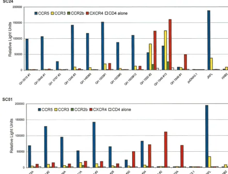
core-ceptors by the 128env-encoded glycoproteins was determined in cell fusion assays. Biological activity (Env-mediated cell-cell fusion) was detected with 40 of these clones (Table 3), and data from 21 representative clones are shown in Fig. 2.
[image:5.612.52.553.84.359.2]Env encoded by templates isolated from patient SC24 during the early stages of infection (QH0515#1 and QH0648#1 from 0 and 77 days postseroconversion, respectively) utilized CCR5 exclusively. An Env encoded by an NSI strain isolated 511 days following seroconversion, QH1246#3, showed minimal utiliza-tion of CCR3 that was above background levels observed in cells expressing CD4 alone. This Env lacked significant activity with CCR2b and CXCR4. Analysis of an Env glycoprotein from an SI isolate taken 875 days following the detection of seropositivity, QH1520#2, showed utilization of CXCR4, CCR3, CCR5, and CCR2b, in order of decreasing fusogenic activity. Two biologically activeenvwere obtained from an SI
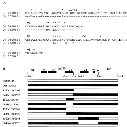
TABLE 2. Amino acid sequence alignment of HIV-1 envelope V3 loopsa
Patient and isolate V3 loop sequence by direct PCR sequencing envclone V3 loop sequence
SC24 ⴱ ⴱ ⴱⴱ ⴱⴱⴱ ⴱ ⴱ ⴱ ⴱ ⴱⴱⴱⴱⴱⴱ ⴱ ⴱ
QH0515 CTRPNNNTRKSIHIGAGKALYTGEIIGDIRQAHC QH0515#1 CTRPNNNTRKSIHIEAGKALYTGEIIGDIRQAHC
QH0648 ––––––N–R–––HI–AGK–L––G––––––––––– QH0648#1 ––––––N–R–––HIGAGK–L––G–––––––––––
QH0797 ––––––N–R–––HI–PGK–L––G––––––––––– QH0797#3 ––––––N–R–––HIGPGK–L––G–––––––––––
QH1020 ––––––N–R–––HI–PGK–L––G–––––––––––
QH1246 ––––––N–R–––HI–PGK–L––G––––––––––– QH1246#3 ––––––N–R–––HIGPGK–L––G–––––––––––
QH1483 ––––––N–R–––HI–PGK–L––G––––––––––– QH1483#3 ––––––N–R–––HIGPGK–L––G–––––––––––
QH1503#1 ––––––N–R–––HIGPGK–L––G–––––––––––
QH1503#5 ––––––N–R–––HIGPGK–L––G–––––––––––
QH1503#12 ––––––N–R–––HIGPGK–L––G–––––––––––
QH1520 ––––––N–R–––RM–IRR–F––R––––––––––– QH1520#2 ––––––N–R–––RMGIRR–F––R––––––––––– HI ?GK L G
QH1549 ––––––N–R–––RM–IRR–F––R––––––––––– QH1549#13 ––––––N–I–––RMGIRR–F––R–––––––––––
? HI ?GK L G QH1549#1 ––––––S–I–––RIGIRR–F––R–––––––––––
SC51 ⴱ ⴱ ⴱⴱ ⴱ ⴱ ⴱ ⴱ ⴱⴱ ⴱ ⴱ ⴱ
QH1302 CTRPNNNTRKSIHLGAGRALYTGEIIGDIRQAHC QH1302#23A CTRPNNNTRKSIHLGAGRALYTGEIIGDIRQAHC
QH1420 ––––––N–––S–––––GR–L–––––I–––––––– QH1420#2 ––––––N–––S–––––GR–L––G––I––––––––
QH1490 ––––––N–––S–––––GR–L–––––I–––––––– QH1490#4A ––––––N–––S–––––GK–L––G––I––––––––
? QH1490#11A ––––––N–––S–––––GR–L––R––I––––––––
QH1509 ––––––N–––S–––––GR–L–––––I–––––––– QH1509#3 ––––––N–––S–––––GR–L––G––I–––––––– ?
QH1521 ––––––N–––S–––––GK–L–––––I–––––––– QH1521#28 ––––––N–––S–––––GK–L––G––I––––––––
H R? F QH1521#30 ––––––N–––H–––––RK–F––G––I––––––––
QH1521#34 ––––––N–––H–––––GK–F––G––I––––––––
QH1536 ––––––Y–––H–––––RK–F–––––I–––––––– QH1536#2 ––––––Y–––H–––––RK–F––G––I––––––––
N L
QH1558 ––––––Y–––H–––––RK–F–––––V–––––––– QH1558#23A ––––––Y–––H–––––RK–F––G––V–––––––– aNonsynonymous sequence polymorphisms are indicated by assigning two amino acids or ? (if multiple codons are possible) to the same position. Dashes denote that an amino acid is identical to the residue in the first sequence; asterisks indicate the predicated residues are different.
on November 9, 2019 by guest
http://jvi.asm.org/
FIG. 1. Phylogenetic analyses ofenvnucleotide sequences cloned from SC24 and SC51 proviral isolate templates and direct sequences from SC24 plasma virusenv region. The complete nucleotide sequence of molecularenvclones was determined using theTaqDye terminator cycle sequencing kit analyzed with an ABI 377 automated sequencer. Direct sequences of SC24 plasma viralenvregions were similarly determined. All sequences were confirmed by the analysis of sense and antisense DNA strands. The DNA sequences were analyzed and aligned using ABI software. Gaps were inserted into the alignment as needed, and those regions were eliminated from the subsequent analysis. Phylogenetic analysis was performed, and consistency of the branching order of the phylogenetic trees was evaluated as described in Materials and Methods. Bootstrap values in excess of 70% were considered significant. (A) Phylogenetic analysis of full-length proviralenvgenes from subject SC24. Coreceptor usage of individualenvgene products is indicated for each clone. MN represents the sequence of the prototypic clade B HIV-1 MN isolate obtained from the Los Alamos data base and is used as an outgroup to root the phylogenetic tree. (B) Phylogenetic analysis of a 459-bp region that surrounds and includes the V3 loop from direct sequencing of PCR amplified plasma viralenvregion compared with cloned proviralenvgenes from subject SC24. (C) Phylogenetic analysis of full-length proviralenvgenes from subject SC51. Coreceptor usage of individualenvgene products is indicated for each clone. The HIV-1 MN isolate is used as an outgroup to root the phylogenetic tree. (D) Phylogenetic tree of the same clonedenvgenes from SC51 as in C but using the QH1302c.23a clonedenvas an outgroup to root the tree. A supercript a indicates that theenvclone is predominately R5 in the cell-cell fusion assay (see Fig. 2 and Table 3) but X4 activity was 5- to 10-fold above background levels in cell-cell fusion.
on November 9, 2019 by guest
http://jvi.asm.org/
strain isolated 1,023 days following seroconversion, designated QH1549#1 and QH1549#13. QH1549#13 utilized CXCR4, CCR3, and CCR5 at significant levels and CCR2b at low levels, with relative efficiencies similar to those observed for QH1520#2. In contrast, QH1549#1 showed significant fusogenic activity exclusively with CXCR4. It is noted that in addition to utiliza-tion of CXCR4, the HXB2 Env showed a low level of fusogenic activity with CCR3.
Parallel studies were performed withenvclones derived by amplification of proviral templates from isolates from patient SC51, who showed conversion from NSI to SI strains early in the course of infection (357 days postseroconversion) and ex-perienced a fulminant clinical course.
The predominant coreceptor utilized by Env encoded by early isolates, which were of the NSI phenotype, was CCR5, and the utilization of CCR2b and CCR3 by these glycoproteins was inconspicuous. They also demonstrated a low level of fu-sion with target cells expressing CXCR4 that was minimally above the background values established with CD4 alone. Pre-dominant CCR5 utilization with marginal CXCR4 utilization above background was also evident with one clone derived from day 99 (QH1420#2) and two clones from the 183-day isolate (QH1490#4A and QH1490#11A). Env encoded by the molecular clone from the QH1509 provirus used CCR5 in fusion experiments and also showed low-level utilization of CXCR4.
Threeenvclones were characterized from the isolate taken at 357 days (QH1521), which marked conversion to the SI phenotype (Table 1), although utilization of CCR5 and CXCR4 in infection experiments did not differ dramatically. Two of these clones demonstrated utilization of CCR5 and CXCR4, one in which the latter was the predominant corecep-tor (QH1521#30) and one that used them at equivalent
effi-ciencies (QH1521#34). CCR5 was the predominant corecep-tor for the thirdenvclone from this isolate (QH1521#28), but fusogenic activity with CXCR4 was minimally above back-ground levels. The SI isolates from the late phases of infection in this patient used CXCR4 in infection experiments, and the Env encoded by molecular clones derived from these templates (QH1536#2 and QH1558#23A) showed high-level, exclusive utilization of CXCR4.
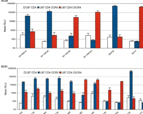
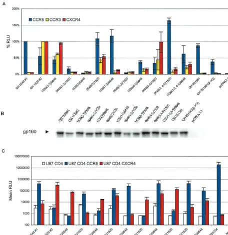
The findings in fusion assays were confirmed in experiments using pseudotyped viruses containing the Env glycoproteins and a luciferase reporter gene to infect target cells pro-grammed to express human CD4 and either CCR5 or CXCR4. Env encoded by proviral templates derived early in the course of infection from patient SC24 utilized CCR5 exclusively, as shown in Fig. 3. The Env encoded by QH1520#2 showed signif-icant utilization of CCR5 and CXCR4 in the cell-cell fusion model, but this was limited to the latter coreceptor in the infection experiments. The Env encoded by QH1549#13 also mediated viral entry only in cells expressing CXCR4. Several Env lacked activity in infection experiments (QH0515#1, QH0797#3, and QH1549#1) and are not depicted in the figure.
There was good agreement between the coreceptor utiliza-tion data from the cell-cell fusion model and infecutiliza-tion exper-iments for the Env encoded by isolates from patient SC51 (Fig. 3). CCR5 was the predominant coreceptor for products ofenv molecularly cloned from the first four isolates, which were NSI. The utilization of CXCR4 by these Env was consistently above baseline values established for CD4 alone. Subtle differences were evident in the utilization of CCR5 and CXCR4 by the three env clones from 357 days following seroconversion (QH1521), which were similar to those observed in fusion experiments. QH1521#28 showed predominant utilization of CCR5 in both assays, with low levels of fusion and viral entry with CXCR4. QH1521#34 was clearly dual tropic (R5X4) in both types of experiments, and QH1521#30 showed predom-inant X4 utilization with minimal R5 activity compared to background values established with CD4 alone in infection experiments. These approaches also revealed comparable ex-clusive utilization of CXCR4 for Env from late in this patient’s course, QH1536#2 and QH1558#23A. Viral isolates from the QH1521, QH1536, and QH1558 specimens had limited infec-tious activity with CCR5-expressing target cells.
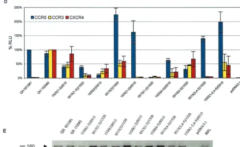
[image:7.612.51.294.83.291.2]Multiple domains of Env contribute to coreceptor specific-ity.The availability of genetically relatedenvthat were derived from successive viral isolates during the evolution of infection in a single patient offered a unique opportunity to study the domains that participate in determining the specificity of co-receptor utilization in vivo. To extend our initial observations, envclones from patient SC24 encoding R5 (QH0648#1) and multitropic (QH1520#1) products (Fig. 4A) were used to gen-erate chimeras, which are depicted in Fig. 4B. As shown in Fig. 5A, whereas the chimeric Env composed of the N-terminal segment of QH1520#2 through the V3 loop [1520(1–3)/0648] demonstrated significant utilization of CXCR4 and CCR3, the fusogenic activity of that extending to the region between the V2 and V3 loops [1520(1–2)/0648] was limited to CCR5. Re-placement of the V3 loop of QH1520#2 with that of QH0648#1 [0648(3)/1520] resulted in a multitropic Env with increased CCR5 utilization and mildly decreased use of CXCR4 and CCR3 in comparison to the wild-type QH1520#2 Env. Substitution of QH0648#1 sequences for the V3 loop and the segment containing the V4 and V5 domains of QH1520#2 [0648(3, 4–5)/1520] resulted in coreceptor utilization essen-tially limited to CCR5. The effect of the V4–V5 segment on coreceptor utilization is further illustrated by the ability of the hybrid Env containing the V1–V2 and V4–V5 domains of
TABLE 3. Env-mediated fusion activity of molecular clones
Patient and
isolate Total no. ofenvclonesa
No. of fusion-competent
envclones Description d
SC24 54 18
QH0515 3 1 R5
QH0648 5 1 R5
QH0797 4 1 R5
QH1020 4 0
QH1246 3 1 R5
QH1483 7 2 {1}R5, {1}R5 (X4⫾)b
QH1503 10 8 {3}R5, {5}R5 (X4⫾)b
QH1520 5 2 {2}R5X4
QH1549 13 2 R5X4, X4 (R5⫾)c
SC51 74 22
QH1302 18 2 R5 (X4⫾)b
QH1420 6 1 R5 (X4⫾)b
QH1490 8 5 R5 (X4⫾)b
QH1509 7 2 R5 (X4⫾)b
QH1521 17 6 {3}R5 (X4⫾)b, {3}R5X4
QH1536 3 1 X4
QH1558 15 5 X4
aAllenvgenes were amplified by PCR and cloned into pcDNA3.1 as described in Materials and Methods. Env constructs were tested for coreceptor utilization in cell-cell fusion assays. Primary Env showed a spectrum of M-topic (R5), dual-tropic (R5X4), and T-tropic (X4) activity. Representative clones are de-picted in Fig. 2.
bPredominantly R5, but X4 activity is 5- to 15-fold over background values with CD4 alone.
cPredominantly X4, but R5 activity is 5- to 15-fold over background values with CD4 alone.
dNumbers in braces refer to the number ofenvclones with the corresponding characteristic.
on November 9, 2019 by guest
http://jvi.asm.org/
QH1520#2 [1520(1–2, 4–5)/0648] to utilize CXCR4 and CCR3 as coreceptors in addition to CCR5. A similar effect was noted with 1520(4–5)/0648, although the total fusogenic activity of this hybrid was less than that observed with the parental Env. Several of the hybrid Env had limited fusogenic activity. These chimeras appeared to be expressed at intermediate [0648(1– 3)/1520 and 1520(3)/0648] or low [0648(1–2)/1520 and 1520(4– 5)/0648] levels in metabolic labeling experiments (Fig. 5B). All of the hybrids, including those with low levels of fusogenic activity, were expressed at levels similar to or greater than that of the parental QH1520#2 Env (Fig. 5B).
To confirm these observations, a parallel array of chimeras were genetically engineered using QH1520#2 and QH0515#1 as the R5 partner in place of QH0648#1. The hybrids 0515(1– 3)/1520 and 1520(3)/0515, which correspond to QH0648#1 chimeras that lacked significant fusogenic activity, showed in-termediate levels of fusogenic activity in the cell-cell fusion model (Fig. 5D) and expression in metabolic labeling experi-ments (Fig. 5E). The 0515(1–3)/1520 hybrid showed predom-inant utilization of CCR5 and activity with CXCR4 and CCR3
that was above background levels established with CD4 alone and greater than that of the parental Env, QH0515#1. The 1520(3)/0515 chimera demonstrated significant utilization of CXCR4 and CCR3 but minimal utilization of CCR5. The contribution of the V4–V5 segment of QH1520#2 to CXCR4 and CCR3 utilization is demonstrated by the 1520(4–5)/0515 and 1520(1–2, 4–5)/0515chimeric Env. In contrast to a previous report highlighting the contribution of an R3S conversion in the C4 region to coreceptor usage (9), both envelopes exam-ined in the current studies possessed an R in this position (Fig. 4). The V1–V2 region of QH1520#2 Env failed to contribute to the multitropic phenotype, as was noted in the panel of chimeras engineered with QH0648#1. All hybrid Env were expressed at levels similar to or greater than those observed for the wild-type parental Env in immunoprecipitation experi-ments (Fig. 5B and E).
[image:8.612.70.541.79.438.2]The coreceptor activity of hybrid Env derived from QH0648#1 and QH1520#2 was also tested in infection experiments (Fig. 5C). The 1520(1–3)/0648 Env used CXCR4, and 1520(1–2)/ 0648 used CCR5, as shown in the cell-cell fusion model. The FIG. 2. Fusogenic activity of Env encoded by molecular clones from sequential isolates from SC24 and SC51. The abilities of Env glycoproteins to mediate fusion with target cells expressing CD4 and candidate coreceptor was determined using a cell-cell fusion assay with a luciferase reporter gene. QT6 effector cells were prepared by infection with vTF1.1 (encoding T7 polymerase) for 1 h following transfection with Env constructs in pcDNA3.1. JRFL and HXB2 constructs in the same vector were used as positive controls. QT6 target cells were transfected with constructs encoding luciferase under the transcriptional control of a T7 promoter, CD4, and the indicated coreceptors. Effector and target cells were mixed and incubated for 9 h following culture overnight. Fusogenic activity was determined by analysis of luciferase activity in detergent lysates. Data shown are representative of at least three separate experiments yielding similar results, with each determination performed in duplicate.
on November 9, 2019 by guest
http://jvi.asm.org/
substitution of V3 sequences in QH0648#1 with those from QH1520#2 was sufficient to confer utilization of CXCR4. Con-versely, the substitution of V3 sequences in QH1520#2 with those from QH0648#1 did not significantly alter utilization of CXCR4 but markedly increased the utilization of CCR5. Swaps of the V1 and V2 regions did not modify the coreceptor utilization of QH0648#1 or QH1520#2. Replacement of the V4–V5 region of QH0648#1 with the corresponding sequence from QH1520#2 did not alter CCR5 utilization but was sufficient to confer the ability to use CXCR4. Similarly, the removal of V3 and V4–V5 segments from QH1520#2 and insertion of the analogous domains of QH0648#1 resulted in fusogenic activity with CCR5. The 1520(4–5)/0648 and 1520(1–2, 4–5) hybrids also support the contribution of the V4–V5 domains (of 1520#2) to the multitropic phenotype. These data provide evidence for the participation of the V3 and V4–V5 regions in determining the repertoire of coreceptor utilization for Env-mediated fusion through both gain- and loss-of-function
exper-iments. In contrast, they fail to establish a role for the V1–V2 segment in determining this phenotype.
DISCUSSION
[image:9.612.72.538.78.449.2]We examined the temporal relationships between the bio-logical, genetic, and functional characteristics of sequential HIV-1 isolates and clonedenvgenes from two individuals who exhibited evolution in their virus phenotype from an initial NSI isolate to subsequent SI isolates during the course of their disease. The conversion in virus phenotype occurred with dra-matically different kinetics in the two subjects and was paral-leled by a similar divergence in their clinical courses. Both subjects were enrolled in a study of acute HIV-1 infection and were followed from the earliest ascertainable stages of acute infection throughout their disease course. The initial virus iso-lates were obtained at a time when each individual exhibited p24 antigenemia but prior to conversion to positive Western FIG. 3. Analysis of coreceptor utilization by primary Env from patients SC24 and SC51 in infection experiments. The primaryenvclones were used to derive pseudotyped viruses containing the encoded glycoproteins and a luciferase reporter gene. The 293T cell line was cotransfected with 10g of a primary Env expression construct and 5g of plasmid pNL4-3.Luc.R⫺E⫺using the standard calcium phosphate precipitation method. Virus-containing supernatants were harvested 48 h
following transfection and filtered. The ability of clonedenvto mediate infection via CCR5 and CXCR4 was assessed in U87 astroglioma cells expressing the CD4 receptor alone or in combination with either coreceptor, as described in Materials and Methods. U87 target cells were infected with pseudotyped virus, harvested 48 to 72 h postinfection, and assayed for luciferase activity as described in Materials and Methods. The results are the means of three independent experiments, each performed in duplicate.
on November 9, 2019 by guest
http://jvi.asm.org/
blot status. Plasma virus load assessments documented that the initial viral samples from both subjects were obtained at or near the peak of plasma viremia that usually accompanies acute HIV-1 infection. Consistent with prior reports, the initial isolates from each individual were of the NSI phenotype and restricted to replication in target cells expressing CCR5 (6, 17, 54, 60). Subject SC24 exhibited a switch in virus phenotype to SI after approximately 2.4 years, while this switch occurred much more rapidly in subject SC51, as SI variants were recov-ered after about 1 year of infection. As documented in Table 1, infectivity studies of the isolates from both patients confirmed that recovery of SI isolates coincided with the emergence of strains that infect target cells expressing CXCR4, but also revealed that the SI isolates were composed of differing pro-portions of subpopulations that used CCR5. However, in both subjects there was a progression with time to populations that
were much more efficient in infecting through the CXCR4 receptor rather than CCR5. Characterization of coreceptor usage by the products of clonedenvgenes demonstrated that the SI phenotype represents the emergence of viral Env capa-ble of using both CXCR4 and CCR5, as well as individual Env restricted to CXCR4, and the persistence of species that use CCR5. These studies amplify the recent findings of Shan-karappa et al. (56) but also highlight the possibility that the time frame for the three distinct phases identified during pro-gression by those investigators may be significantly altered in cohorts from developing regions where rates of progression appear to be more rapid.
[image:10.612.64.474.72.479.2]In addition to the biological studies on the virus isolates, the genetic and functional studies of clonedenv genes have pro-vided novel insights on the nature of variants that make up these populations and the evolution of this gene in vivo. For
FIG. 4. Structure of chimeric Env created from R5 and multitropic Env from patient SC24. (A) Alignment of the variable domains and C4 region of Env encoded by QH0648#1 and QH1520#2 molecular clones from isolates obtained in the early (QH0648) and intermediate (QH1520) stages, respectively, of the evolution of AIDS in patient SC24. Dashes denote identical amino acid residues (⬎95% overall), and dots represent gaps introduced to optimize alignments. Asterisks indicate that the amino acids in that position are different. The R residue that appears in boldface type in the C4 region denotes the position of the R3S conversion highlighted previously (9). (B) Schematic representation of wild-type and chimericenvgenes showing restriction endonuclease cleavage sites that were used to genetically engineer the hybrids. Creation of theDraI site by site-directed mutagenesis resulted in the introduction of conservative nucleotide changes that did not alter the primary structure of Env.
on November 9, 2019 by guest
http://jvi.asm.org/
subject SC24, we did not recover individual envelopes that were capable of efficiently infecting via both CCR5 and CXCR4 coreceptors (i.e., dual-tropic Env). This result suggests that the virus quasispecies in these SI isolates are composed of subpopulations that mediate infection through either CXCR4
[image:11.612.73.530.73.544.2]or CCR5. In contrast to these findings, analysis in the cell-cell fusion model system revealed that the progression from R5 to X4 Env occurred via an intermediate that had multitropic coreceptor usage that included CCR5, CCR3, and CXCR4 as well as CCR2b at levels above background with CD4 alone. As FIG. 5. V3 and V4–V5 regions of multitropic Env contribute to coreceptor utilization. (A) The fusogenic activity of the panel of hybrid Env composed of complementary segments of the R5 Env QH0648#1 and the multitropic Env QH1520#2 was determined in the cell-cell fusion assay, as described in Materials and Methods, using assay conditions identical to those described in the legend to Fig. 2. Luciferase intensity is expressed as relative percentages normalizing CCR5 activity to that of QH0648#1 and CCR3 and CXCR4 activity to that of QH1520#2. The data are presented as averages of means of duplicate values from three independent experiments. The structures of the hybrid Env that were tested are depicted in Fig. 4. (B) Expression of wild-type and chimeric Env was analyzed by immunoprecipi-tation of metabolically labeled cells transiently expressing the clonedenvgene. Transfectants were starved for 1 h in medium lacking Met and Cys, labeled with [35S]Met/[35S]Cys for 1 h, and chased with complete medium for 1 h. Detergent lysates were immunoprecipitated with HIVIG or control reagents, as described in Materials and Methods. The components of the immune complexes were resolved by SDS-PAGE and detected by autoradiography. The gp160 precursor glycoprotein is marked with an arrowhead. (C) Ability of the chimeric Env to mediate virus entry was determined in infection experiments using pseudotyped viruses as described in Materials and Methods under conditions identical to those described in the legend to Fig. 3. (D) Contribution of the V3 and V4–V5 domains of QH1520#2, the multitropic Env, was confirmed in fusion assays with a parallel panel of chimeras using the earliest R5 Env from SC24, QH0515#1, which was active in the cell-cell fusion assay but inactive in infection experiments. The cell-cell fusion system was performed as described above and in Materials and Methods. (E) Metabolic labeling experiments confirmed the expression of the hybrid Env glycoproteins, as described above.
on November 9, 2019 by guest
http://jvi.asm.org/
they assess fusogenic activity in different contexts, the fusion assay may be a more sensitive measure of biochemical inter-actions that can occur between Env and coreceptors, although this surrogate may not predict productive Env-coreceptor in-teractions in the context of virus infection. In the second sub-ject (SC51), we observed a different picture for the 1-year transition from NSI R5 strains to the development of SI X4 variants. Molecular env clones from the QH1521 isolate of SC51 revealed a mixture in both fusion and infection assays that included R5X4 dual-tropic species in addition to an R5 Env variant. Utilization of CCR3 and CCR2b was not observed in the cell-cell fusion model. Taken together, the results sug-gest that there may be multiple pathways for HIV-1 to evolve in vivo from an initial CCR5 population to a CXCR4-permis-sive population during the course of disease. This interpreta-tion must be tempered by the fact that our examinainterpreta-tion ofenv clones was not exhaustive, and it is possible that dual-tropic or multitropic Env variants existed in both subjects but were not recovered.
A substantial proportion of HIV-1-infected individuals progress to AIDS without making the phenotypic switch to SI variants (21, 33, 55). In these subjects, theenvgenes may not evolve to use coreceptors other than CCR5, as suggested by a recent report (21). It is possible that in such individuals, subtle changes in the virus Env may alter the nature or efficiency of its interaction with the CCR5 coreceptor in a way that leads to increased virus replication. Indeed, we have observed an un-usual case of an individual who experienced an extremely rapid disease course (time from infection to death from AIDS of less than 7 months), characterized by persistently high virus loads without any evidence for use of CXCR4 (J. F. Demarest, N. Jack, F. R. Cleghorn, M. L. Greenberg, T. L. Hoffman, L. Fantry, J. Edwards, T. R. O’Brien, K. Cao, B. Muhabir, W. A.
Blattner, C. Bartholomew, and K. J. Weinhold, submitted for publication). Thus, our findings indicate that R5 NSI HIV variants, R5/X4 SI dual-tropic variants, and X4 SI variants are all capable of eliciting AIDS in the proper host. Moreover, they highlight the fact that CXCR4 usage per se is not a prerequisite for HIV-1 to cause disease and that CCR5 vari-ants can be quite pathogenic.
[image:12.612.68.537.79.366.2]The evolution of coreceptor utilization during the course of HIV-1 infection represents a key mechanism for extension of the spectrum of target cells. Multiple studies have implicated the V3 loop of gp120 as a major determinant of tropism (8, 11, 30, 31, 43, 57, 65, 66). These findings were extended to dem-onstrate that this domain plays a dominant role in governing coreceptor utilization (13, 15). Recent crystallographic analysis coupled with mutational evidence has identified amino acid residues in gp120 that are accessible to solvent following asso-ciation with CD4 and are critical to coreceptor utilization (35, 46, 67, 68). The indirect contribution of the V3 loop is evi-denced by the finding that only one of these amino acid resi-dues is located in this domain. Whereas truncation of the V1 and V2 loops of gp120 yielded a variant that retained the ability to bind CD4 and CCR5, further truncation of the V3 loop abolished the ability to bind this coreceptor. Thus, it is likely that the V3 loop controls the availability or conformation of residues that interact with coreceptor structures. Previous studies using genetically engineered hybrid env genes have confirmed the role of the V3 loop and also implicated the V1–V2 region (12, 49). These chimeric Env were typically composed of segments of different molecularly cloned, genet-ically unrelated laboratory strains of HIV-1. One study em-ployed a parental simian-HIV hybrid that showed exclusive utilization of CXCR4 and a pathogenic isolate that lacked utilization of both front-line coreceptors, CCR5 and CXCR4, FIG. 5—Continued.