0022-538X/94/$04.00+0
Copyright © 1994, American Society for Microbiology
Temporal
Patterns of Human
Immunodeficiency
Virus Type 1
Transcripts in
Human
Fetal
Astrocytes
CARLOTORNATORE,* KAREN MEYERS, WALTER ATWOOD, KATHERINE CONANT, AND EUGENE MAJOR
Laboratory of Molecular MedicineandNeluroscience, National Institute of Nellrological DisordersandStroke, Bethesda, Maryland20892
Received 3 August 1993/Accepted 5 October 1993
Human immunodeficiency virus type 1 (HIV-1) infection of the developing central nervous system results in adementing process inchildren, termedHIV-1-associated encephalopathy. Infection of astroglial elements of thepediatric nervous system has been demonstrated and suggests that directinfection of some astrocytes may contribute to theneurologic deficit. In this model, HIV-1 establishes a persistent state of infection in astrocytes, which can bereactivated by the cytokines tumor necrosis factor alpha(TNF-ex)andinterleukin 13
(IL-1p1).
To betterunderstand the natural history of viral persistence in astroglial cells, wecharacterized infection at the transcriptional level. The most abundant viral transcript during the establishment of persistence was the subgenomic multiply spliced 2-kb message, similar to mononuclear cell models of HIV-1 latency. Following reactivation with TNF-& or IL-11 the multiply spliced 2-kb message remained the most abundant viral transcript, in contrast to infected mononuclear cells in which reactivation leads to the reemergence of the 9-and 4-kb transcripts. Further characterization of the persistent 2-kb transcript by PCR amplification of in vitro-synthesized viral cDNA showed that, in the absence of cytokine stimulation, the most abundant multiply spliced transcripts were the Nef- and Rev-specific messages. However, following cytokine stimulation, double-and triple-spliced Tat-, Rev-, and Nef-specific messages could be identified. Immunohistochemical staining demonstrated that, during viral persistence, astrocytes expressed Nef protein but few or no viral structural proteins. These results demonstrate that viral persistence in astrocytes at the transcriptional level is fundamentally different from that seen in mononuclear cells and could account for the virtual absence of astroglial expression of viral structural antigens in vivo.Vertically transmittedhumanimmunodeficiencyvirustype 1 (HIV-1) infection isnowamongthe topfive leadingcausesof death in children <4 years of age (34). Of those children infected withHIV-1,approximately one-thirdtotwo-thirdswill go on to develop manifestations of central nervous system (CNS) dysfunction characterized by a loss of developmental milestones involvingboth motorand cognitive systems (4, 18). Thisconstellationofneurologic signsand symptoms hasbeen termed pediatric AIDS encephalopathy, which has as its parallel the dementia seen in adults infected with HIV-1 (32, 36). The pathogenesis of AIDS-related encephalopathy re-mains elusive. The neuropathological hallmarks of HIV-1 infection of the CNS have been well described, consisting of infiltrating macrophages, multinucleated giant cells, and reac-tive microglial cells associated with widespread astrogliosis, myelin pallor,andneuronalloss(31, 36, 44). Whilecellsof the mononuclear lineage haveconsistently been demonstrated to express viral antigens and to harborviral nucleic acids in the nervoussystem, thenumber of infectedcells is smallrelative to thewidespreadneuropathology.Toexplain this discrepancy, it has been proposed that the effects of mononuclear infection indirectly lead to the parenchymal changes.This is supported by data which demonstrate that infected monocytes can elab-orate HIV-1geneproductsand othercellular factors which are neurotoxic (5, 9, 15, 20, 21, 25, 37).
Direct infection of the neuronal and astroglial elements of the CNS has also been proposed to contribute to HIV-1-associateddementia. Anearlydescription ofpediatric HIV-1-associatedneuropathologydemonstrated the presenceofviral
* Correspondingauthor.
93
particles in astrocytes (17), suggesting that the immature elementsin thedevelopingnervoussystem weresusceptible to HIV-1 infectionduring vertical transmission. Invitroinfection of humanfetalastrocytes,aswellasastrocytomacelllines,has subsequently been demonstrated (8, 10-12, 14, 49). HIV-1 establishes a persistent state of infection in astroglial cells, whichcanbereactivated by the cytokinestumornecrosis factor alpha (TNF-ox) and interleukin 11
(IL-1p)
(49), an activation mediated by the induction ofNF-KB (47).To better understand the mechanisms which mayunderlie the establishment of persistent infection of astrocytes, we
examinedthetemporalappearanceof HIV-1mRNA inhuman fetal astrocytes. We report that during the establishment of persistence,attimesof littleornop24or
gp4I
expression,the predominantviraltranscript seenby Northern (RNA) hybrid-ization was the subgenomic, multiply spliced 2-kb message. Reactivationwith TNF-otorIL-I1 resulted inabriefphase of viralreplicationwithexpression of p24andgp4l;however,the multiply spliced message remained the most abundant viral transcript. Using reverse transcriptase (RT)-PCR technologyto identify the subpopulations represented in the 2-kb
tran-scripts, we found that during periods of viral persistence the most abundantregulatory transcriptswere those which coded for Nef and Rev (Nef- and Rev-specific transcripts) with a predominance of the Nef-specific transcripts. In contrast, following stimulation with TNF-otor
IL-11,
doubly and triply spliced Tat-, Rev-,and Nef-specificmessages could be identi-fied within 24 h. Immunohistochemical staining of astrocytes persistentlyinfectedwith HIV-1 further revealedexpressionof Nefprotein in these cells at times when little viral structural protein could be detected.on November 9, 2019 by guest
http://jvi.asm.org/
MATERIALS AND METHODS
Cell cultures from brain tissue. Cultures fromhuman fetal brain tissuewerepreparedasdescribedpreviously (49). Brain
tissue was dissected from 9- to 14-week-old human fetuses, mechanically disrupted by aspiration through a 19-gauge
nee-dle, washed in Eagle's minimum essential medium (E-MEM) and plated into poly-D-lysine (0.1 mg/ml in distilled water)-treated tissue culture flasks. Each brain specimenwasplated separately without pooling of tissues from fetusesof similaror
different gestational ages. Cultures were maintained and fed
every3to4days with E-MEM plus 10%fetal bovineserum.To prepare purecultures ofastrocytecells, thecultureswerefirst
refed with serum-free medium and placed inanorbital shaker (210 rpm)at37°C for 2 h. Cells released from the cultureswere
discarded while the adherent cells were refed with medium
with serum. For serial passage, cells were harvested with 0.025% trypsin and 0.005% EDTA,counted inan hemocytom-eter, and plated at 106 in 100-mm-diameter plates. Following two to four passages, the cultures were stained with an
antibodytoglial acidic fibrillary protein (DAKO)todetermine the percentage of cells which were astrocytes. Only those cultureswhichwere99%glial acidicfibrillary protein antibody positivewereused.
Transfection procedure. The calcium phosphate precipita-tion technique wasused for transfection. Five microgramsof uncleaved plasmid DNA per plate was used. The DNA was
precipitated in HEPES (N-2-hydroxyethylpiperazine-N'-2-ethanesulfonic acid [pH 7.1]) buffer (137 mM NaCl, 5 mM KCl, 0.7 mM Na2HPO4, 6mMdextrose,21 mMHEPES)with 125 mM CaCl2 for the final concentration. The DNA in HEPES bufferwas kept at room temperature for 30min for precipitate formation. The cell cultures were washed in HEPES buffer prior to addition of the DNAprecipitate. The HEPES-DNA precipitatewasplaced directly onthe cells for 30minatroomtemperature,and then the cellswererefed with E-MEM. Following a 4-h incubation at 37°C, the cells were
washed three times withE-MEM, shocked with 15% glycerol in E-MEM for 45 s, and then washed three more times with E-MEM.
Immunodetection assays.Cellswere plated onglass
cover-slips,fixed inacetoneand methanol for 10mineachat -20°C,
and washed in phosphate-buffered saline (PBS [pH 7.1]). All remaining incubations were performed at room temperature. Endogenous peroxidase activity was quenched for 30 min in 3% hydrogen peroxide in methanol. The cells were then
blocked with 3% normal goat serum for 20 min, followed by incubation with aprimary antibody (eithera mouse monoclo-nalantibodytoNefprotein diluted 1:250[AIDSResearch and Reference Reagent Program, catalogno. 1123],arabbit poly-clonal antibodytoglial acidic fibrillary protein [DAKO], or a mouse monoclonal antibody to HIV-1 gp4l [Genetic Sys-tems]). The incubation time for the glial acidic fibrillary protein and gp4l antibodies was 1 h,while the Nefantibody
was incubated for 24 h. Cells were then washed in PBS and incubated with either biotinylated anti-rabbit or anti-mouse
immunoglobulin G for1 h.Followingawashin PBS, the cells
wereincubated withperoxidase-conjugated streptavidin for 60
min. Color was then developed with a fresh solution of
diaminobenzidine tetrahydrochloride, resulting in a brown
precipitate. Slides were then washed, counterstained with hematoxylin, dehydratedingraded alcohols,andmounted with Permount(Fisher Scientific).
Cytokines. Allcytokines usedwere either human recombi-nants or werederived from a humancell source (Boehringer
Mannheim). Cytokines were stored in aliquots at -20°C
followingreconstitution inE-MEMwith10%fetal calfserum.
Aliquotswere thawed onlyonce. The concentrations of cyto-kines usedwere asfollows: TNF-u,10
ng/ml;
IL-11,
10U/ml.
Cytoplasmic RNA extraction.
Following transfection,
cyto-plasmic RNA was isolated from cells in 100-mm-diameter tissue culture dishes(1
x 106 to5 x 106cells).
Priorto this procedure, all of the reagentswere treated with 0.1% dieth-ylpyrocarbonate to inhibit RNases. The cells wereplaced
at4°C for 30 to 60 min, washed and harvested
by
mechanical scraping in cold 1x PBS, andpelleted by
centrifugation
at14,000rpmfor 30s at4°C.The cellswere
resuspended
in270p.I
of cold Iso-High (0.14MNaCl, 10 mM Tris[pH
8.4],
1.5mM
MgCI2)
and30 [L of cold5% NonidetP-40inH20.
Afterincubation on ice for 2 min, the nuclei were
pelleted by
centrifugation at 14,000 rpm for 30s at4°Cin an
Eppendorf
microcentrifuge. The supernatantwasextracted twice in
phe-nol-chloroform (1:1 ratio) and once in chloroform
alone,
followed by
precipitation
of the RNA in 70% ethanol at-20°C. RNAwas collectedby centrifugation at 14,000 rpm,
resuspended in TES buffer
(10
mM Tris[pH
7.5],
1 mMEDTA, 0.05% sodium
dodecyl
sulfate[SDS])
andreprecipi-tatedin1/10volume of3 Msodiumacetate
(pH 5.5)
and70% ethanol at -20°C
for several hours.Following
centrifugation
at 14,000rpm,the RNApelletwasair dried and
resuspended
inwater.
Total RNAextraction. Total RNAwas alsoextracted from parallel cultures of transfected astrocyte cultures
by
aprevi-ouslydescribedprotocol
(54).
Northernhybridization.RNAextraction
samples
wereana-lyzed by
electrophoresis
in 1x MOPS buffer {0.02M MOPS(3-[N-morpholino]propanesulfonic
acid),
0.005 MNaHAc,
0.001 M EDTA} on 1.0% horizontal agarose
gels
containing
0.62 Mformaldehyde.
Following
electrophoresis,
thegel
was rinsed once for 20 min with 0.05 NNaOH,
followedby
a 20-min wash in 20x SSPE(3.0
MNaCl,
0.2MNaH2PO4,
0.02M
EDTA-Na2).
Theremaining
stepsof theNorthern transferand
hybridization
are identical to those of the Southerntransfer and
hybridization
described below. Theprobe
usedfor Northern hybridization was an 8,088-bp fragment ofpNL4-3
(1) generated
by
endonucleaseAvaIdigestion
and nick trans-lation with a32P-labeled
dATP.cDNAsynthesis andPCRamplification.One
microgram
of either totalorcytoplasmic
RNAwasheatedat65°C
for 5 min andcooledon ice. First-strand synthesis ofcDNAwascarried outinatotal volumeof 20jlI consisting
of 50mMKCL,
10 mMTris-HCl, 5 mM MgCl2, 1 mMeach deoxynucleoside triphos-phate
(dNTP),
20UofRNaseinhibitor(Perkin-Elmer),
50Uof
Moloney
murine leukemia virusRT(Perkin-Elmer),
1Kgoftemplate RNA,
and 1jig
of an antisenseoligonucleotide
primer. For reverse
transcription
of themultiply
spliced
mRNAs, the
oligonucleotide
BamA located in exon 7 at the BamHI site was used. The sequence and location of BamA inpNL4-3 are5'-GCTAAGGATCCGTTCACTAATCGAAT GG-3' and nucleotides(nt)
8472 to8448,
respectively.
For reversetranscription
of the gagregion
of the 9-kb viral transcript, theoligonucleotide
SK431 wasused. Thesequence and location ofSK431 inpNL4-3
are 5'-TGCTATGTCACT TCCCCTTGGTTCTCT-3' and nt 1500 to 1474,respectively.
The reaction mixturewasincubated at45°Cfor 1h,
followed by incubation at 95°C for 5 min. PCRamplification
of thesingle-stranded
DNAwas carried outby
bringing
the cDNAreaction mixtureuptoafinalvolumeof 100
pl consisting
of50 mMKCL,10 mMTris-HCl,
2 mMMgCl2,
0.2 mMeachdNTP,
2.5 Uof
Ampli-taq
DNApolymerase
(Perkin-Elmer),
and1 Fg of a senseprimer.
Foramplification
of themultiply
spliced
transcripts, the
oligonucleotide
Bss, located in exon 1 at theon November 9, 2019 by guest
http://jvi.asm.org/
BssHII
site,
was added. The sequence and location of Bss inpNL4-3
are5'-GGCTTlGCTGAAGCGCGCACGGCAAG
AGG-3' andnt 700 to727, respectively.
Foramplificationof the gagregion
of the 9-kbtranscript,
theoligonucleotide
SK145 was used. The sequence and location of SK145 are
5'-AGTGGGGGGACATCAAGCAGCCATGCAAAT-3' and
nt 1359to
1388, respectively.
The reaction mixturewasdena-tured at
94°C
for 1min,primers
were annealed at55°C for 2min,and
primers
wereextendedat72°C
for3min foratotalof40
cycles
inaDNAthermalcycler
(Perkin-Elmer Cetus).
Tenmicroliters of each PCR
product
was run on a 1.5% agarose horizontalgel,
stained with ethidiumbromide,
visualized with UVlight,
andphotographed.
Southerntransfer and
hybridization.
Tofurtherensurethespecificity
ofthe PCRproducts,
all reaction mixtures under-went Southern blot transfer. The agarosegel
was rinsedoncewith distilledwaterandthen denatured twice in1 MNaCl-0.5 MNaOHfor 15 min. Thiswasfollowed
by
neutralization in0.5 MTris-1.5MNaClfor 15min,whichwasalsodone twice. The DNA was transferred to anylon
filterby capillary action,
cross-linked
by
UV radiation(Stratalinker;
Stratagene),
andprehybridized
in50% formamide-6x SSPE(0.9
MNaCl,
0.06 MNaH2PO4,
0.006 MEDTA-Na2)-5
x Denhardt's solution-0.5%SDS-100 ,ug of calfthymus
DNAper mlat42°C
for1h. The filterwashybridized
in an identical solution which also contained 106dpm
of a 32P-labeled nick-translated8,088-bp
AvaI
fragment
ofpNL4-3
perml(1).
Theprobe
wasallowedtohybridize
to the filterat42°C
for atleast 16h. The filterwaswashed twice in 6x SSPE-0.1% SDS for 30 min at room
temperature. Thiswasfollowed
by
two washes in 1x SSPE-0.5% SDS for 30 min at64°C
and a final wash in 0.1x SSPE-0.5% SDS for 30 minat64°C.
Thefilterwasdried and used forautoradiography.
Following autoradiography,
the filter wasstripped
of the radiolabeledprobe
in50% formamide-2x SSPE at65°C
for 90 min. The filter was thensequentially hybridized
with thefollowing
series ofoligonucleotide
probes
(labeled
with[-y-32P]ATP
with T4polynucleotide
kinase[New
England
Biolabs]): oligonucleotide
2,
located in exon 2 at nt 4962 to4945,
5'-CTITCCAGAGGAGCTITG;
oligonucleotide
3,
lo-cated inexon3at nt5446to5429,
5'GATATTCACACCTAGGAC;
oligonucleotide
4tat, locatedinthe 5'portion
ofexon4 at nt5803to5780,
5'-CCTATTCTGCTATGTCGACACCCA-3';
oligonucleotide
4a/b
tat/rev,
located inexon4a/b
at nt5981 to5959,
5'-TTCCTGCCATAGAGATGCC-3';
andoligonu-cleotide 5
tat/revlnef,
located in the5'portion
ofexon 5at nt 5997to5979,
5'-TCGCTGTCTCCGCTTCTTC-3'.
To detect the PCR
products generated by primers
SK145 andSK431,
the internaloligonucleotide
SK102 was used.SK102
is locatedinthe gag geneat nt1396to1428and hasthesequence 5'-GAGACCATCAATGAGGAAGCTGCAGAAT
GGGAT-3'.
RESULTS
Temporal
characteristics ofp24 production
inpNL4-3-transfected astrocytecultures. We have
previously
described the kinetics ofp24 production
in human fetal astrocytesfollowing
transfection with the infectious molecular clonepNL4-3
(1, 49).
The briefproductive phase
of viralreplication
seen
immediately posttransfection
is followedby
apersistent
phase during
which littlep24
isproduced.
AsseeninFig. 1,
theapplication
of thecytokine
TNF-ot results inbrief bursts ofp24
production (similar
resultswerefound forIL-1). Furthermore,
inthecurrent setof
experiments,
wefoundthat thelonger
the cellsremain inastateof viralpersistence,
themoreattenuated400
-300
-a.
co CL
200
100-I R \
0
DayT-1
10 * 20 *30 4CAddition of Addition of
Cells TNF lOng/ml TNF 1ong/ml
transfected
FIG. 1. Temporal characteristics of p24 production in pNL4-3-transfectedastrocytecultures stimulatedatdifferenttime pointswith TNF-ot. Two separate cultures of human astrocytes were established and transfectedin parallel.The concentration ofp24protein inthe supernatant fluid was determined every 24 h by using an antigen-capture enzyme-linked immunosorbent assay (ELISA[Coulter]).At eachtimepoint, the culture mediumwascollectedandreplacedwith fresh medium. Each datumpointrepresentsthe mean value of three experiments. Cytokines wereadded tothe medium onlyon the day noted on thegraph. At day15, TNFwas only added to one setof cultures (El1).Atday 27,TNFwasaddedtobothsets.
the effects ofcytokineadministration were.Asseen inFig. 1,if TNF-ot is added to the cultures 2 weeksposttransfection, the p24 levels rise to approximately 100 to 150 pg/ml from a baseline of approximately 15 pg/ml. However, if TNF-ot is added to thecultures 4weeksposttransfection, thep24 levels rise to ap24level ofapproximately50pg/mlfrom abaselineof <1 pg/ml. This phenomenon is independentofprior cytokine stimulation and appearsto be afunction of time from initial transfection.
As notedpreviously (49)andin asubsequent section of this paper (see section on temporal characteristics of Tat-, Rev-, and Nef-specific transcripts in astrocytes), supernatant from cytokine-treated cultures was infectious, as demonstrated by incubation of the supernatant with the CD4+ lymphoblastoid cellline A3.01 (19).
Temporal characteristics of HIV-1 mRNA transcripts in astrocytes.To betterunderstand themechanisms which under-lie the establishmentof viralpersistenceinastrocytes,we next characterized the temporal characteristics of viral mRNA
production during the establishment of persistence and in response to cytokine stimulation. As seen in Fig. 2, 2 days posttransfectionall three viraltranscriptscould be identified as unspliced (9 kb), singly spliced (4 kb), andmultiply spliced (2 kb), with the multiply spliced message beingthe most abun-dant. By day 15, when p24 levels were approaching the
baseline, only the multiply spliced transcriptwasevident.The addition of either IL-1orTNF-ot on day 15 led to a marked increase inthemultiply splicedtranscripts within24 h. While therewas not adetectable increase in theunspliced orsingly spliced transcripts, therewas a riseinthe levels ofp24in the culture supernatant (as seen in Fig. 1), suggesting that the numberofsingly splicedorunspliced transcriptswasbelow the
sensitivitylevels of Northernblotting.Northern blots done 48 and 72 h after cytokine administration, at times ofpeak p24 production,were no different from the blotseenin Fig. 2.
By day 27 posttransfection, viral transcripts could not be
on November 9, 2019 by guest
http://jvi.asm.org/
[image:3.612.323.559.77.245.2]DAYS POST-TRANSFECTION
16 16 28 28
+ + + +
2 15 IL-1 TNF 27 IL-1 TNF
9Kb
-4
[image:4.612.316.556.97.195.2]Kb-2Kb- _ s z
FIG. 2. Temporal characteristics of HIV-1 mRNA transcripts in astrocytes. RNA waspurifiedatdifferent time points from astrocytes transfected withpNL4-3.RNA wasextracted fromcytokine-stimulated astrocytes 24h after the addition of eitherTNF-cx orIL-11 onday 15 or27posttransfection. Northern transfer andhybridizationweredone asdescribed in the text.The approximate sizes of thetranscripts are givenonthe left.
detected inastrocyte cultureswhich had hadnoprior cytokine stimulation. Addition of either TNF-o. or IL-13 on day 27 posttransfection to astrocytes in a state ofviral persistence again led tothe inductionof themultiplyspliced2-kb message within24h,although itwas asignificantlyweaker signalthan that induced atday 16posttransfection.
Northern hybridization blots with total RNA from trans-fectedcells were nodifferentfromhybridizationwith cytoplas-mic RNA.
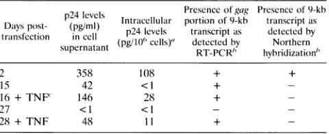
Giventhe absence of the9- and 4-kbtranscriptsattimesof cytokine stimulation, a series ofstudies were done to deter-mine whether thecytokine-induced riseinp24levelsin thecell supernatant resulted from de novo synthesis of p24 from nascent viral transcripts versus release of previously synthe-sized p24 inthe absence of viral transcripts. Inthe first setof experiments, five parallel astrocyte cultures were established and transfectedand thep24 levels in the cell supernatant and intracellularly were measured at different time points. As summarized in Table 1, thepresence orabsence ofp24 inthe supernatant was paralleled bythe presence of absenceofp24 intracellularly. Atday 27, nointracellularorextracellular p24 wasdetectable. However, 24 hfollowing cytokinestimulation, p24wasagaindetectable in both the supernatant and intracel-lularly, demonstrating that cytokine-induced rises in p24 re-sulted from de novosynthesis ofthe coreprotein.The second set of experiments attempted to identify the 9-kb viral tran-script byamoresensitive method than Northernhybridization. Forthis,weutilized an RT-PCR method in which RNA used in the Northernhybridizationwas reverse transcribed with the primer SK431 and then was PCR amplified with primers SK145 and SK431. The primersetSK145andSK431 amplified a 142-bp fragment of thegagregion ofthe 9-kb transcript, a
regionwhich issplicedout ofthe 4-and2-kb viraltranscripts. Following gel electrophoresis andSouthern transfer, the PCR productswere hybridized with an end-labeledinternal probe, SK102.Assummarizedin Table 1,RT-PCRwasabletodetect
TABLE 1. Comparison ofgag genetranscription and expression in
astrocytes atdifferent timepointsposttransfection
p24 levels Presenceof gag Presence of9-kb Dalys post-
(pg4ml)
Intracellular portion of 9-kb transcriptastransfection in cell p24 levels transcriptas detected by
supernatant (pg/10'cells)" detectedby Northern
RT-PCR"
hybridization"
2 358 108 + +
15 42 <1 +
-16 +TNF' 146 28 +
-27 <1 <1 -
-28 + TNF 48 11 +
-"Intracellular p24 levels weredetermined as follows. Cells(10')werewashed, resuspenided in 1 ml of serum-free medium, and vortexed for 5 min. The cellular debris waspelleted, and the supernatant was removed. The supernatant was mixed with t).5 ml of7t)% sorbitol and 0.5 ml of 1t)t)% fetal calfserum and assayed for p24antigenwith anlantigen-capture ELISA (Coulter).
b+, present; -, not present.
' + TNFdenotes administration of cytokines 24 h prior to the ELISA.
the9-kbtranscriptattimeswhen Northernhybridizationcould not. Furthermore, the presence or absence of the 9-kb tran-script as detected by RT-PCR paralleled the presence or absence of p24 in the culture system. In total, these experi-ments suggest that the cytokine-stimulated rise in p24 levels results fromthe expressionof nascent 9-kb transcripts.
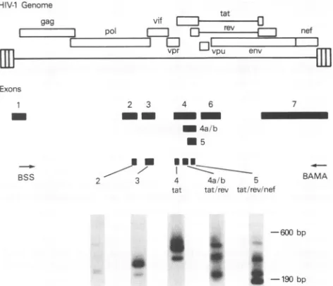
Characterization of the viral 2-kb message. Given that the 2-kbtranscriptwasbyfarthe mostabundanttranscriptseen in viral persistence and with cytokine stimulation, we character-ized the subpopulations ofthe multiply spliced transcriptsby utilizing a previously described RT-PCR method (42). This method uses a setofprimers in exon 1 (primer Bss)and exon 7 (primerBamA) to amplify all ofthe multiply spliced tran-scripts simultaneously because these exons are present in all HIV-1 viral transcripts. Because the same primer pair and PCR mixture are used to identify all of the multiply spliced transcripts,the messages can bequalitatively compared whena probeisused to detect all ofthe regulatorytranscripts.Given the positionof the primers, the expected sizes of the amplifi-cation products from multiply spliced transcripts would be between 190 and 600 bp, with the Tat-coding transcripts producing the largest of the PCR products, Nef transcripts producing the smallest products, and Rev transcripts produc-ing intermediate-sized products. The extension step of the cyclingparameter waskeptsufficientlyshort to prevent ampli-fication of the unspliced or singly spliced transcripts. cDNA was synthesized by RT and the antisense primer BamA, followed by PCR amplification with the addition of sense
primerBss. Amplification ofthe viraltranscripts 2days post-transfection gave a complex pattern of bandswhen the PCR products were run on a 1.5% agarose gel, transferred to a nylon membrane, and hybridizedwith an 8-kbAvaI fragment ofpNL4-3. Thisfragmentspansall ofthe exonsofHIV-1 and would be expected to hybridize to all multiply spliced mes-sages. As seen in Fig. 3, amplification of RNA 2 days post-transfectionconsistentlygave a patternof three sets of doublet bandsrangingin sizefrom200bptoalmost 600bp.Inthefirst lane, 10Ftlof the PCR mixture wasloaded,while in the second lane 20Illwasloaded.
To identify the genomic regions present in the amplified cDNAs,themembranewas sequentially hybridizedto aseries of probes located in exons 2, 3, 4, 4a/b, and 5. Probe 4 tat hybridizestothe 5' regionof exon 4 upstream of the tat AUG. As seeninFig. 4, probe4 tathybridizedtothe three uppermost cDNAbands,indicating that thereare atleast three
on November 9, 2019 by guest
http://jvi.asm.org/
HIV-1 Genome
gag vd tat
vpr vpU e
Exons
2 3 4 6
__ 4
_ 4a/b * 5
BSS
-600bp
-l190 bp FIG. 3. RT-PCRamplification of the viral 2-kb m of the figure illustrates the open reading frames ar HIV-1genome. Exons are numbered according to tU
Meusing et al. (30) and Schwartz et al. (42). Bss previously described primers (42) in exons 1 and 7,rc in the RT-PCRamplificationstep. The bottom of the resultsof RT-PCRamplificationand cDNA Southern RNAextractedfrom astrocytes 2 daysposttransfecti( In thefirst lane, 10 p.lof the PCRmixture wasload second lane 20,lIwasloaded.
sizedtranscripts with the Tat-coding exon in th The uppermost of these three transcripts alsoI probe for exon 2, identifying this band as a
tril
transcript which contains exon2inaddition to t
7. Probe 4a/b tat/rev hybridizes to a region spanning the rev AUG.Exon4a/b is nested
wit]
will therefore hybridize to the Tat- and Rev-( scriptsaswell.As seenin Fig. 4,probe 4a/btat,
to five transcripts, two more than the 4 tat pi smallest bands correspondto Rev-specific tran two Rev-specifictranscripts, the largerone alsc the probe for exon 3, identifying it as a trip transcriptwithexon3inadditiontoexons1,4a/1 5 tat/revlnefhybridizes to the 5' region of ex nested within exons 4 and 4a/b, which is presc Rev-,and Nef-encoding transcripts. This probe all six transcripts, demonstrating that the sma seenis Nefspecific.
Hybridization with the probes in exons2and these smallnoncodingexonswerealsopresent i transcripts which did not hybridize to the Nef-specificprobes.
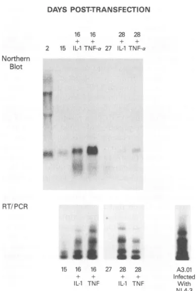
Temporal characteristics of Tat-, Rev-, an transcripts in astrocytes. We next analyzed characteristics of the regulatory transcripts id( RT-PCR methodology. RNA was extracted at
pointsposttransfection, reverse transcribed, an fiedasdescribed above.FollowingSoutherntrar
filter,the PCRproductswere hybridized withc probe 5
tatlrev/nef
to determine which of Itranscriptswerepresent.As seeninFig. 5, byda
fection, themostprominent regulatorytranscri which coded forNef and Rev. However, the ad(
TNF-ct
orIL-13
on day 15 posttransfection resulted in theaI
induction of Nef-, Rev-, and Tat-specific transcripts by day 16.4-1 nd By day 27 posttransfection, no transcripts were detectable,
either by Northern hybridization or RT-PCR, but again the ---fIJI addition of
cytokines
resulted in theinduction oftranscripts
for all three regulatory elements. In all cases, the Nef-specific transcriptwasthe strongest band present.7 Supernatant taken from the astrocyte cultures at day 17 posttransfection (treated with TNF-ax on day 15), which had a p24level of 105 pg/ml and presumably viable NL4-3, was used to infect A3.01cells, a CD4+ T-cell line(19). Fifteen days after the initial infection, the A3.01 cells produced 2 ng of p24 per BAMA
ml/day,
confirming
the presence of viable NL4-3 in theastro-cyte supernatant. Cytoplasmic RNA was extracted from 1 million of the A3.01 cells and reversetranscribed and amplified withprimers Bss and BamA as described above. The resulting banding pattern by Southern hybridization with the AvaI fragment of pNL4-3, as seen in Fig. 5, is more complex than that seenin astrocytes.
Immunohistochemistry for Nef protein in persistently in-fected astrocytes. Given that the Nef- and Rev-specific tran-essages. The top scriptswerethemostabundanttranscriptsatmosttimepoints nd exons of the posttransfection and given the prior observation that rev and ie numbering of nefgenetranscripts can code for Nef protein (42), we exam-and BamA are
ined
astrocytes at different time points for expression of Nef espectively, used protein as well as for expression of the structural proteingp4l.
hybridization
of Asseen in Table2,
Nefwasdetectableby
immunohistochem-on
withpNL4-3. istry of astrocytes through at least day 15 posttransfection, ied,while in the whilegp4l
expression was absent after 15 days. Cytokine administration at day 15 roughly doubled the number of astrocytes which expressed Nef 24 h later. By 27 days post-transfection, neither gp4l nor Nefexpression wasdetectable, but again, administration of cytokines increased Nef expres-iese astrocytes. sion and, to a lesser degree, gp4l expression within 24 h. hybridized toaple-spliced
Tatexons1,4, and HIV-1Genome
of exon 4a/b
gagta
tfhinexon 4and r Xpol
F
O 5l nfencoding tran-
vpr u n 1111 /revhybridizedrobe. The two
scripts. Of the Exons hybridized to
ile-spliced Rev b, and 7. Probe on 5, an exon znt in all Tat-, hybridized to
llest transcript
13 showed that inanumber of Tat-, Rev-, or
id Nef-specific the temporal entified by the different time id PCR ampli-nsferto anylon
)ligonucleotide
the regulatory
Ly
15 posttrans-iptswere those dition of eitherBSS
2 3 4 6 7
mm
-_ 4a/b * 5
BAMA
2 3 4 4a/b 5
tat tat/rev tat/rev/nef
-600bp
[image:5.612.59.295.81.280.2]1 I -190 bp
FIG. 4. Sequential Southern hybridization of RT-PCR-amplified 2-kbmessageswitholigonucleotide probestodifferentexons.RT-PCR amplification and Southern transferwere performed with RNA
ex-tracted from astrocytes 2daysposttransfectionwithpNL4-3.The filter wasthensequentially hybridizedto aseries ofoligonucleotide probes nested within exons 2 through 5. Beneath the exons of HIV-1 are
shown the relative positions of the oligonucleotide probes used to
identify the presence of the exons. The precise positions of the oligonucleotidesaregivenin thetext.
l~
on November 9, 2019 by guest
http://jvi.asm.org/
[image:5.612.317.553.435.638.2]DAYS POST-TRANSFECTION
16 16 28 28
+ + + +
2 15 IL-i TNF-a 27 IL-i TNF-a
Northern Blot
RT/PCR
15 16 16 27 28 28 A3.01
+ + + + Infected
IL-1 TNF IL-1 TNF With
[image:6.612.81.275.73.361.2]NL4-3
FIG. 5. Temporal characteristics ofTat-, Rev-, and Nef-specific transcripts in astrocytes. RNApreviously extracted atdifferent time pointsforuseinNorthernhybridization (see Fig. I andthetopof this
figure) was amplified with primers Bss and BamA in an RT-PCR
mixture and, following Southern transfer,washybridized to oligonu-cleotide probe 5 tat/rev/lnef, which is present in all Tat-, Rev-, and
Nef-encoding transcripts.RNAwasalsoextracted fromA3.0)1 cells 15 daysfollowing infcction with NL4-3. The results of RT-PCR of this RNA andSouthern hybridizationtothe AiwI fragmentofpNL4-3are
shownatthe bottom rightofthis figure.
Figure 6a and b demonstrate the perinuclearstaining pattern
of Nefproteinin astrocytes. Figure 6c demonstrates themore
diffusecytoplasmic staining patternofgp4l. DISCUSSION
The natural history of HIV-1 infection of human astrocytes and astrocyte cell lines is typified by a persistent state of infection in which few or no viral structural antigens are
expressed. Inthis series ofexperiments,we characterized the
infection atthe transcriptional level tobetter understand the viral mechanisms which mediate the establishment of viral persistence in astroglial cells. Models of HIV-1 latency in mononuclearcells and T cells havedemonstratedthat the viral mRNAexpressed in these cells consisted of singly and multiply spliced RNA species, with littleor nounspliced genomic RNA
during chronic infection (30, 35). In primary human fetal astrocytes,asimilarphenomenon isseenin which the
predom-inant viraltranscriptduring the establishment of persistence is multiply spliced. However, the astrocyte model is
fundamen-TABLE 2. Comparison of Nef and gp4l expression in astrocytes at different time points posttransfection
No. of astrocytespositive for
Days expressionby:
posttransfection
Nefstain gp4lstain
2 1 of 400 1 of100
15 1of200 0
16 + TNFa 1 of100 1 of 300
27 0 0
28 + TNF 1of400 1 of1,000
"+ TNFdenotes administration ofcytokines24 hpriorto
immunocytochem-istry.
tally different from the mononuclear cell model in one impor-tant regard; lipopolysaccharide stimulation of Ul cells results in amarkedincrease in the amountofmultiply splicedmRNA, which is then followed by an increase in the amount of unsplicedRNAspecies (30, 35).Whilecytokinestimulation of astrocytes leads to a clear increase in the amountofmultiply spliced mRNA, no detectable increase in the amount of unspliced RNA is seen. This suggests that there is a critical difference in the physiology of astrocytes which prevents the accumulation of theunsplicedHIV-1 viraltranscript.This is of particular interest giventhe recentobservation by Constantou-lakis et al. (13) that there are interferon-inducible proteinsin human cell types which bind to the responsive element, inhibiting function and resulting in an accumulation of the multiply spliced viral transcript.Thiswouldsuggestthathuman fetal astrocytes haveasimilarfactor ormechanism whichmay workatthe level of the responsiveelementtogivethe mRNA patterns seen in Fig.2. Work isbeing initiated in our labora-tory to investigate this possibility.
The subpopulations of the 2-kb message are strikingly similar in both mononuclear cells and astrocytes in which HIV-1 has established a state ofpersistence. In H9 cells and peripheralbloodmonocyte-macrophages infectedwithHIV-1, thepredominant multiply spliced transcriptwasthe one which coded forNef(23, 40). The Tat-specificmRNAs werethe least abundant, while the Rev transcripts were intermediate in number. In the astrocyte model outlined, at times of persis-tencewhenthe multiply splicedmessages werethemajorityof the viral transcripts, the RT-PCRmethodology found thatthe Nef- and Rev-encoding transcripts were the most abundant, withapredominanceof theNef-specificmessage,againsimilar to the mononuclear and lymphocytic cells (23, 40). Interest-ingly, Schwartz et al. (42) found that the transcripts for Rev and Nef both have open reading frames for Nef. Given the abundance of these transcripts and their increase during cytokine stimulation of astrocytes, the ability to detect Nef protein immunohistochemically is not surprising. The perinu-clear location of Nef is consistentwith findings from a persis-tently infected gliomacell line (24).
CouldNef, inaddition tocellularfactors, play arole in the establishment ofalatent orpersistent stateofviralinfection? This point remains controversial. In mononuclear cells, Nef wasinitially thoughttobe anegativeregulator ofviral expres-sion at the level of the long terminalrepeat. Similarly, in one astrocytoma model of HIV-1 infection,Nef also demonstrated a prominent suppression of the HIVlong terminal repeat at
FIG. 6. Immunohistochemical identification of Nef protein andgp4linastrocytes. Fig. 6a and b demonstrate the perinuclear localization of Nef protein in astrocytes. This is incontrast togp4l expression, which is more diffusely cytoplasmic asseen in panel c.
on November 9, 2019 by guest
http://jvi.asm.org/
[image:6.612.317.555.97.185.2]on November 9, 2019 by guest
http://jvi.asm.org/
the level of the negative regulatory element (8). However, other investigators have observed no effect of Nef protein on either gene expression or viral replication in mononuclearand glial cell models (2, 22). Other functions attributed to the nef gene product which support its role in the establishment of viral persistence include (i) the ability to reduce cell surface expression of gpl20 and CD4, presumably preventing the cytopathic effect of cell fusion (41), and (ii) inhibition of NF-KB induction, attenuating transcriptional upregulation at thelevelof the long terminal repeat(33). Interestingly, in the astrocytemodel, we found an attenuation of expression of p24 andgp4l as well as that of the RNA transcripts over time. This probably does not reflect cell death, since the number of Nef-expressing cells remained relatively constant over time. Instead, one could postulate that Nef expression downregu-lated the longterminal repeat as well as blocked expression of structural proteins in these cells. Whatever its role, the fact that the Nef open reading frame is conserved in all primate lentiviruses and in pathological tissue (6) suggests that this protein plays a critical, and as yet undetermined, role in the viral life cycle.
The abundance of the multiply spliced transcripts found in astrocytes in vitro and their ability to code for Nef has significance for the detection of HIV-1-infected astrocytes in vivo. Studies to identify infected cells of the CNS in vivo generallyuse methods that target the viral structural proteins orgenescoding for structuralproteins. However, if the natural history of HIV-1 infection of astrocytes in vivo is also one of viralpersistence in which the viral transcripts are restricted to the multiply spliced species, then detection methodology would have to be modified to reflect this. Specifically, immu-nohistochemical detection of infected astrocytes would have to target either Nef or Rev protein, since thestructural and core proteins would be almost nonexistent, given the lack of the unspliced and singly spliced message in these cells. In situ detection of infected astrocytes would also have to use a nucleic acid probe which targets the exons present in the multiply spliced transcripts, since these are the most abundant viral nucleic acids. Probes which target exons or sequences outside these transcripts will probably fail to hybridize to infected astrocytes, again because the nonspliced or singly spliced viral transcripts are so few in number. Furthermore, the sensitivity of the probe would need to bemaximized, given the restrictednatureof the infection. Both our laboratory and that of Blumberg et al. (7) have taken this approach to identify infected astrocytes in vivo. Using a 32P-labeled probe which contains the exons found in the HIV-1 multiply spliced tran-scripts and antibody to Nef protein, we were able to detect a small number of astrocytes which harbored HIV-1 nucleic acids in the subcortical white matter of 4 of 12 pediatric patients with AIDS encephalopathy (48). Blumberg et al., usingmonospecificnefprobesand an antibody to Nef protein, also foundevidence forHIV-1-infected astrocytes in postmor-tem CNS tissue from pediatric AIDS patients (7). With the advent ofmore sensitive detection techniques, such as in situ PCR (3, 16), the extent of glial infection may be further clarified.
The role infected astrocytes play in the pathogenesis of AIDS-associated dementia is unknown. While Nef protein has demonstratedfunctional similarities to scorpion peptides in its ability to interact with K+ channels of chicken dorsal root ganglia (55),it is unclearwhether infection disrupts the normal physiologic functions of astrocytes. Given the cytokine activa-tion seeninvivo in the CNS of patients with AIDS encepha-lopathy (50), astrocytes could act as low-level producers of virion when exposed to these monokines, infecting
surround-ing tissue and infiltrating mononuclear cells. In addition to
infecting the parenchymal elements, virions produced by as-troglial elements maybedirectly toxictothe neuronal constit-uents. The uninfected, reactive astrocyte may also contribute to the neuropathology of HIV by releasing TNF-o when exposed to viral antigens (28). TNF-ox is toxicto oligodendro-glial cells (39, 43) and could contribute to the myelin pallor seen at autopsy. Reactive astrocytes in patients with AIDS dementia also produce transforming growth factor ,3, which may act as a chemoattractant for mononuclear cells into the CNS, furtheraggravating theencephalitis caused byHIV(52). Finally, it appears that the natural history of HIV-1infection ofadult human astrocytes in vivo and in vitro may bedifferent thanthat of astrocytes of the developing nervous system. One in vitro attempt to infect adult human astrocytes was unsuc-cessful(45), while in vivodetection of infected adult astrocytes has been inconsistent (38, 46, 53, 56). The immaturity of the glial elements in utero, as well as the immaturity of the pediatric immune system in the perinatal period, could in theory make the developing CNS more susceptible to viral invasion (26, 27) than the mature nervous system.
ACKNOWLEDGMENTS
We thank Renee Traub and Blanche Curfman for technical and editorial assistance. The anti-Nef monoclonal antibody from James Hoxie was obtained through the AIDS Research and Reference Reagent Program, Division of AIDS, NIAID.
REFERENCES
1. Adachi, A., H. E. Gendelman, S. Koenig, T. Folks, R. Willey, A. Rabson, and M. A. Martin. 1986. Production of acquired immu-nodeficiency syndrome-associated retrovirus in human and non-human cells transfected with an infectious molecular clone. J. Virol.59:284-291.
2. Bachelerie, F., J. Alcami, U. Hazan, N. Israel, B. Goud, F. Arenzana-Seisdedos, and J.-L. Virelizer. 1990. Constitutive ex-pression of human immunodeficiency virus (HIV) nef protein in human astrocytes does not influence basal or induced HIV long terminal repeat activity. J. Virol. 64:3059-3062.
3. Bagasra,O., S. P. Hauptman, H. W. Lischner, M. Sachs, and R. J. Pomerantz. 1992. Detection of human immunodeficiency virus type 1 provirus in mononuclear cells by in situ polymerase chain reaction. N. Engl. J. Med. 326:1385-1391.
4. Belman, A. L., M. H. Ultmann, D. Horoupian, B. Novick, A. J. Spiro, A. Rubinstein, D. Kurtzberg, and B. Cone-Wesson. 1985. Neurological complications in infants and children with acquired immune deficiency syndrome. Ann. Neurol. 18:560-566. 5. Bernton, E., H.Bryant,M.Decoster, J. M. Ornestein, J. L. Ribas,
M. S. Meltzer, and H. E. Gendelman. 1992. No direct neurono-toxicity by HIV-1 virions or culture fluids from HIV-1 infected T-cells ormonocytes. AIDS Res. Hum. Retroviruses 8:495-501. 6. Blumberg, B. M., L. G.Epstein, Y. Saito, D. Chen, L. R. Sharer,
and R. Anand. 1992. Human immunodeficiency virus type I nef quasispecies in pathological tissue. J. Virol. 66:5256-5264. 7. Blumberg, B. M., L. R. Sharer, Y. Saito, J. Michaels, T. A.
Cvetkovich, M. Louder, K. Golding, and L. Epstein. Overexpres-sion of nef is a marker forrestricted HIV-1 infection of astrocytes in human CNStissue. Neurology, in press.
8. Brack-Werner, R., A.Kleinschmidt, A. Ludvigsen, W. Mellert, M. Neumann, R.Herrmann, M. C. L. Khim, A. Burny, N. Muller-Lantzsch, D.Stavrou, and V.Erfle.1992. Infection ofhuman brain cells by HIV-1: restricted virus production in chronically infected humanglial cell lines. AIDS 6:273-285.
9. Brenneman, D. E., G. L. Westbrook, S. P. Fitzgerald, D. L. Ennist, K.L.Elkins, M. R.Ruff, and C. B. Pert. 1988. Neuronal cell killing by the envelope protein of HIV and its prevention by vasoactive intestinal peptide. Nature(London) 335:639-642.
10. Cheng-Mayer, C., J. T. Rutka, M. L.Rosenblum, T. McHugh, D. P. Stites, and J. Levy. 1987. Human immunodeficiency virus can productively infect cultured human glial cells. Proc. Natl. Acad.
on November 9, 2019 by guest
http://jvi.asm.org/
Sci. USA 84:3526-3529.
11. Chiodi, F., S. Fuerstenberg, M. Gidlund,B.Asjo,and E. M.Fenyo. 1987. Infection of brain-derived cells with the human immunode-ficiency virus. J. Virol. 61:1244-1247.
12. Christofonis, G., L. Papadaki, Q. Sattentau, R. Ferns, and R. Tedder. 1987. HIV replicates in cultured human brain cells. AIDS 1:229-234.
13. Constantoulakis, P., M. Campbell, B. K. Felber, G. Nasioulas, E. Afonina, and G. M. Pavlakis. 1993. Inhibition of Rev-mediated HIV-1 expression by an RNA binding protein encoded by the interferon-inducible 9-27 gene. Science 259:1314-1317.
14. Dewhurst, S., K. Sakai, J. Bresser, M. Stevenson, M. J. Evinger-Hodges, and D. J. Volsky. 1987. Persistent productive infection of human glial cells by human immunodeficiency virus (HIV) and by infectious molecular clones of HIV. J. Virol. 61:3774-3782. 15. Dreyer, E. B., P. K. Kaiser, J. T.Offerman,and S. A. Lipton. 1990.
HIV-1 coat protein neurotoxicity prevented by calcium channel antagonists. Science 248:364-367.
16. Embretson, J., M. Zupancic, J. Beneke, M. Till, S. Wolinsky, J. L. Ribas, A. Burke, and A. T. Haase. 1993. Analysis of human immunodeficiency virus-infected tissues by amplification and in situ hybridization reveals latent and permissive infections at single-cell resolution. Proc. Natl. Acad. Sci. USA 90:357-361. 17. Epstein, L. G., L. R. Sharer, E.-S. Cho, M. Myenhofer, B. Navia,
and R.Price. 1984.HTLV-III/LAV-like retrovirus particles in the brains of patients with AIDS encephalopathy. AIDS Res. Hum. Retroviruses 1:447-454.
18. Epstein, L. G., L. R. Sharer, V. V. Joshi, M. M. Fojas, M. R. Koenigsberger, and J. M. Oleske. 1985. Progressive encephalop-athy inchildren withacquired immune deficiency syndrome. Ann. Neurol. 17:488-496.
19. Folks, T., S. Benn, A. Rabson, T. Theodore, M. Hoggan, M. Martin, M. Lightfoote, and K. Sell. 1985. Characterization of a continuous T cell line susceptible to the cytopathic effects of the acquiredimmunodeficiency syndrome (AIDS)-associated retrovi-rus.Proc. Natl. Acad. Sci. USA 82:4539-4543.
20. Genis, P., M. Jett, E. W. Bernton, T. Boyle, H. A. Gelbard, K. Dzenko, R. W. Keane, L. Resnick, Y. Mizrachi, D. J. Volsky, L. G. Epstein, and H. E.Gendelman. 1992. Cytokines and arachidonic acid metabolites produced during HIV-infected macrophage-as-troglial interactions: implications for the neuropathogenesis of HIVdisease. J. Exp. Med. 176:1703-1718.
21. Giulian, D., K. Vaca, and C. A. Noonan. 1990. Secretion of neurotoxins by mononuclear phagocytes infected with HIV-1. Science250:1593-1596.
22. Hammes, S. R., E. P. Dixon, M. H. Malim, B. R. Cullen, and W. C. Greene. 1989. Nefprotein of human immunodeficiencyvirus type 1: evidence against its role as a transcriptional inhibitor. Proc. Natl.Acad. Sci. USA86:9549-9553.
23. Klotman, M. E., K. Sunyoung, A. Buchbinder, A. DeRossi, D. Baltimore, and F. Wong-Staal. 1991. Kinetics of expression of multiplyspliced RNA in early humanimmunodeficiencyvirus type 1 infection oflymphocytes and monocytes. Proc. Natl.Acad. Sci. USA88:5011-5015.
24. Kohleisen, B., M. Neumann, R. Herrman, R. Brack-Werner, K. Krohn, V. Ovod, A. Ranki, and V.Erfle. 1992. Cellular localization of Nef expressed in persistently HIV-1-infected low-producer astrocytes. AIDS6:1427-1436.
25. Lipton, S. A., N. J. Sucher, P. K. Kaiser, and E. B. Dreyer. 1991. Synergistic effects of HIV coat protein and NMDA receptor-mediatedneurotoxicity. Neuron 7:111-118.
26. Lyman, W. D., Y. Kress, K. Kure, W.Rashbaum, A. Rubinstein, and R. Soeiro. 1990. Detection of HIV in fetal central nervous system tissue. AIDS 4:917-920.
27. Mano, H., andJ.-C. Chermann. 1991. Fetal human immunodefi-ciency virus type 1 infection of different organs in the second trimester. AIDS Res. Hum. Retroviruses 7:83-88.
28. Merrill, J. E., Y.Koyanagi, J. Zack, L. Thomas, F. Martin, and I.S. Y. Chen. 1992.Induction of interleukin-1 and tumor necrosis factor alpha in brain cultures by human immunodeficiencyvirus type 1. J. Virol. 66:2217-2225.
29. Michael, N. L., P.Morrow, J. Mosca, M. Vahey, D. S. Burke, and R.R.Redfield. 1991.Induction of humanimmunodeficiencyvirus
type 1 expression in chronically infected cells is associated primar-ily with a shift in RNA splicing patterns. J. Virol. 65:1291-1303. 30. Muesing, M. A., D. H. Smith, C. D.Cabradilla,C. V. Benton, L. A.
Lasky, and D. J. Capon. 1985. Nucleic acid structure and expres-sion of the human AIDS/lymphadenopathy retrovirus. Nature (London)313:450-458.
31. Navia, B. A., E.-S. Cho, C. K. Petito, and R. W. Price. 1986. The AIDS dementia complex. II. Neuropathology. Ann. Neurol. 19: 525-535.
32. Navia, B. A., B. D. Jordan, and R. W. Price. 1986. The AIDS dementia complex.I. Clinical features. Ann. Neurol. 19:517-524. 33. Niederman, T. M. J., J. V. Garcia, W. R. Hastings, S. Luria, and L. Ratner. 1992. Human immunodeficiency virus type 1 Nef protein inhibits NF-KB induction in human T cells. J. Virol. 66:6213-6219.
34. Novella, A. C., P. H. Wise, A. Willoughby, and P. A. Pizzo. 1989. Final report of the United States Department of Health and Human Services Secretary's work group on pediatric human immunodeficiency virus infection and disease: content and impli-cations. Pediatrics 84:547-555.
35. Pomerantz, R. J., T. Didier, M. B. Feinberg, and D. Baltimore. 1990. Cells nonproductively infected with HIV-1 exhibit an aber-rant pattern of viral RNA expression: a molecular model for latency. Cell 61:1271-1276.
36. Price, R. W., B. Brew, J. Sidtis, M. Rosenblum, A. C. Scheck, and P. Cleary. 1988. The brain in AIDS: central nervous system infection and AIDS dementia complex. Science239:586-593. 37. Pulliam, L., B. G. Herndier, N. M. Tang, and M. S. McGrath.
1991. Human immunodeficiency virus-infected macrophages pro-duce soluble factors that cause histological and neurochemical alterations in cultured human brains. J. Clin. Invest. 87:503-512. 38. Rhodes, R. H., and J. M. Ward. 1991. AIDS meningoencephalitis. Pathogenesis and changing neuropathologic findings. Pathol. Annu. 26:247-276.
39. Robbins, D. S., Y. Shirazi, B.-E. Drysdale, A. Lieberman, H. S. Shin, and M. L. Shin. 1987. Production of cytotoxic factors for oligodendrocytes by stimulated astrocytes. J. Immunol. 139:2593-2597.
40. Robert-Guroff, M., M. Popovic, S. Gartner, P. Markham, R. C. Gallo, and M. S. Reitz. 1990. Structure and expression of tat-, rev-, and nef-specific transcripts of human immunodeficiency virus type 1 in infected lymphocytes and macrophages. J. Virol. 64:3391-3398.
41. Schwartz, O., Y. Riviere, J.-M. Heard, and 0. Danos. 1993. Reduced cell surface expression of processed human immunode-ficiency virus type 1 envelope glycoprotein in the presence of Nef. J. Virol. 67:3274-3280.
42. Schwartz, S., B. K. Felber, D. M. Benko, E.-M. Fenyo,and G. N. Pavlakis. 1990. Cloning and functional analysis of multiply spliced mRNA species of human immunodeficiency virus type 1. J. Virol. 64:2519-2529.
43. Selmaj, K. W., and C. S. Raine. 1988. Tumor necrosis factor mediates myelin and oligodendrocyte damage in vitro. Ann. Neurol. 23:339-346.
44. Sharer, L. R., L. G. Epstein, E.-S. Cho, V. V. Joshi, M. F. Meyenhofer, L. F. Rankin, and C. K. Petito. 1986. Pathologic features of AIDS encephalopathy in children: evidence forLAV/ HTLV III infection of brain. Hum. Pathol. 17:271-284.
45. Sharpless, N., D. Gilbert, B. Vandercam, J. M. Zhou, E. Verdin, G. Ronnett, E. Friedman, and M. Dubois-Dalcq. 1992. The restricted nature of HIV-1 tropism for cultured neural cells. Virology 191:813-825.
46. Stoler, M. H., T. A. Eskin, S. Benn, R. C. Angerer, and L. M. Angerer. 1986. Human T-cell lympho-tropic virus type III infec-tion of the central nervous system-a preliminary in situ analysis. JAMA256:2360-2364.
47. Swingler, S., A. Easton, and A. Morris. 1992. Cytokine augmen-tation of HIV-1 LTR driven geneexpression in neural cells. AIDS Res.Hum. Retroviruses 8:487-493.
48. Tornatore, C. S., R. Chandra, J. Berger, and E.0. Major. HIV-1 infection of subcortical astrocytes in the pediatric central nervous system. Neurology, in press.
49. Tornatore, C., A. Nath, K. Amemiya, and E. 0. Major. 1991.
on November 9, 2019 by guest
http://jvi.asm.org/
Persistent human immunodeficiency virus type 1 infection in human fetal glial cells reactivated by T-cell factor(s) orby the
cytokines tumornecrosis factor alpha and interleukin-1 beta. J. Virol.65:6094-6100.
50. Tyor, W. R., J. D. Glass, J. W. Griffin, and P. S. Becker. 1992. Cytokineexpression inthe brainduring the acquired immunode-ficiency syndrome. Ann. Neurol. 31:349-360.
51. Vitkovic, L., G. P. Wood, E. 0. Major, and A. S. Fauci. 1991.
Human astrocytes stimulate HIV-1 expression in a chronically
infected promonocyte clone via interleukin-6. AIDS Res. Hum. Retroviruses 7:723-727.
52. Wahl,S. M., J. B. Allen, N. McCartney-Francis, M. C. Morganti-Kossmann, T. Kossman, L. Ellingsworth, U. E. H. Mai, S. E. Mergenhagen, and J. M. Orenstein. 1991. Macrophage- and astrocyte-derived transforming growth factor betaasamediator of
centralnervoussystemdysfunctioninacquired immune deficiency syndrome. J. Exp. Med. 173:981-991.
53. Ward, J. M., T. J.O'Leary, G. B. Baskin,R. Benveniste, C.A. Harris, P. L. Nara, and R. H. Rhodes.1987.Immunohistochemical localization of human and simian immunodeficiency viral antigens infixed tissue sections. Am. J. Pathol. 127:199-205.
54. WenQin,X., and L.I.Rothblum. 1991. Rapid, small-scale RNA
isolation from tissue culture cells. BioTechniques 11:324-327. 55. Werner, T., S. Ferroni, T. Saermark, R. Brack-Werner, R. B.
Banati, R. Mager, L. Steinaa, G. W. Kreutzberg, and V. Erfle. 1991. HIV-1 Nef proteinexhibits structural and functional simi-larityto scorpion peptides interacting with K+ channels. AIDS 5:1301-1308.
56. Wiley, C. A., R. D. Schrier, J. A. Nelson, P. W. Lampert, and M. B. A. Oldstone.1986.Cellular localization of human immuno-deficiencyvirus infection within the brains of acquired immune deficiency syndrome patients.Proc.Natl.Acad. Sci. USA 83:7089-7093.