0022-538X/94/$04.00+ 0
Copyright © 1994, AmericanSociety for Microbiology
Isolation and
Characterization of Simian T-Cell Leukemia Virus
Type
II from New World Monkeys
YI-MING A. CHEN,
1-2*
YNG-JUJANG,'
PHYLLIS J. KANKI,3 QIAN-CHUNYU,4 JAANG-JIUNWANG,'RICHARDJ. MONTALI,5 KENNETH P. SAMUEL,' ANDTAKIS S. PAPAS7t
Institutteof Biomedical Sciences, Academia Sinica, Nan-Kang, Taipei 11529,1 andAIDS Research Center, GraduiateInstitute ofPlublicHealthi, National Yang-Ming Medical College, Taipei11221,2 Taiwan, Republicof China;
DepartmentofCancerBiology, Harvard School ofPublicHealth, Boston, Massachusetts 021153;Department of
MolecularGenetics and CellBiology, University ofChicago, Chicago, Illinois606374; Departmentof Pathology, NationalZoological Park, Smithsonian Institution, Washington, D.C. 200085;Biotechnology Working Group,
LaboratoryofCelluilarBiochemistry, Program Resouirces, Inc./DynCorp., Frederick, Maryland217016;
and LaboratoryofMolecular Oncology, National CancerInstitute, Frederick, Maryland21702-12017
Received2September 1993/Accepted6November 1993
Since the description of human T-cell leukemia virus type I (HTLV-I) and its simian counterpart, simian T-cell leukemia virus type I (STLV-I), the possible existence of other related simian retroviruses has been raised. Here, we report a new retrovirus,STLV-II,which we haveidentified in spider monkeys (Atelesfusciceps), aNew Worldprimatespecies. Initially,arecombinantHTLV-IIenvelope protein (RP-IIB) was used toidentify anti-STLV-II antibodies in New World monkeys by Western blot (immunoblot) assays. Subsequently, the virus wascharacterized by Southern blot hybridization, which showed thatSTLV-IIandHTLV-II have a high degree of nucleotide sequence homology but havedifferent restriction enzyme patterns. Nucleotide sequenceanalysis of thepX-II region of STLV-II provirus revealed 3% variation with the corresponding region of HTLV-II. Electron micrographic studies revealedHTLV-like, typeC retrovirus particles outside the cell membranes of
STLV-II-infected cells. This study describes the first link between HTLV-II andasimian reservoir in the New World. Further molecular studies ofSTLV-II infection in differentspecies of New World monkeys, especially from the wild, may provide valuable information about the origin and intragroup relationships of South American monkeys. Spider monkeys infected with STLV-II may serve as an important animal model for
HTLV-II infection in humans.
Human T-cell leukemia viruses (HTLVs) are exogenous
retroviruses and belong tothe sameOncovirinae subfamily of the Retroviridae as bovine leukemia virus and simian T-cell leukemia virus (STLV) (53). HTLV type I(HTLV-I)wasfirst isolatedin 1980 from aT-lymphoblastoid cell lineestablished from a patient with cutaneous T-cell lymphoma (38). It has been found to be associated with adult T-cell leukemia/ lymphoma (55, 60) and HTLV-I-associated
myelopathy/tropi-cal spastic paraparesis (12, 37). HTLV-II was first isolated
fromapatient with atypical hairy cell leukemia (27). Although therehave been scattered casereports,the disease associated with HTLV-II infection is still unclear (43). On thebasis of
nucleic acid hybridization analysis, direct comparison of
pri-marynucleic acidsequencesoflong terminal repeats, and the extent ofserologic cross-reactivity between gaggene-encoded
products, HTLV-II has beenshown tobe related but distinct from HTLV-I (3, 48, 52). In general, HTLV-I and HTLV-II haveapproximately 65%genomic homology (46, 49).
In 1982, Miyoshi and colleagues identified HTLV-related
viruses in nonhuman primates by demonstratingthe presence of anti-HTLV antibodies in Japanese macaques (Macaca fuscata) (36). Since then, many studies have found antibody
reactivities toHTLVindifferent speciesof the genus Macaca
*Corresponding author. Mailing address: Institute of Biomedical Sciences,AcademiaSinica, Nan-Kang,Taipei11529, Taiwan,Republic of China. Phone: (886-2) 8744560. Fax: (886-2) 8743902. Electronic mail address:bmal91Caccvax.sinica.edu.tw.
tPresent address: Center for Molecular and Structural Biology,
Hollings Oncology Center, Medical University of South Carolina,
Charleston, SC 29425-2213.
aswellasin manyAfrican species of Old Worldprimates (17, 18, 22, 24, 26, 35, 59). Furthermore, a serological survey of captive macaques from Asia indicated strong association of lymphoma and lymphoproliferative disease with STLV
infec-tion (22). Nucleotide sequence analysis of an STLV isolate frompig-tailedmacaquesrevealed90% homology with cosmo-politan strainsof HTLV-I(57). Recently,serologicaltestshave also shown that STLV infections in Old World primates are
mainlydue to STLV type I (STLV-I) (44).
The recent identification of type-specific regions on the
envelopeglycoproteinsofHTLV-Iand HTLV-II (7, 8,30,31) anddevelopment ofserological tests complementaryto DNA
diagnosis by PCR have enabled a clarification of the
geo-graphic and racial distributions of the two viruses; HTLV-I infections are endemic in regions of Africa, the Caribbean islands, and southern islands of Japan (2, 19, 45), while
HTLV-II is endemic in several Indian tribes in Central and
South America (20, 29). Since it has been postulated that
HTLV-I infectionsinhumansmayoriginatefromcross-species
transmission through close contact with STLV-I-infected
pri-mates, we used an HTLV-II recombinant envelope protein
(RP-IIB) (5) to identify HTLV-II-related virus
(STLV-II)
infectioninvariousspeciesof New WorldmonkeysbyWestern blot (immunoblot) assays.MATERUILSAND METHODS
New World monkeys and blood samples. Serum samples from the following numbersofvarious speciesof New World monkeys were used in this study: 9 spider monkeys
(Ateles
fusciceps; designated SM-1 to SM-9), 47 owl monkeys (Aotus
1149
on November 9, 2019 by guest
http://jvi.asm.org/
1150 CHEN ET AL.
trivirgatus),
10capuchin monkeys (Cebus albifrons),
20 howlermonkeys (Alouatta villosa),
2squirrel monkeys (Saimiri
sciu-reus),
and 18 marmosets(2
Callithrixjacchus,
12Saguinus
oedipus,
and 4Saguinus
flaviceps).
All ofthemonkeys
werecaptive
and were housed atprimate
centers or zoos. Thehowler
monkeys
werecaught
inthewild,
and their bloodwas latersampled
in Venezuela.Cell lines and cultures. Forvirus
isolation,
several humancell
lines,
including
aT-cellline(Sup-Ti)
(32, 51),
anEpstein-Barr virus immortalized B-cell line
(BJAB) (15),
and a promonocyte line(THP-1) (54),
were used in thisstudy.
AnHTLV-I-producing
cell line(HUT-102) (40)
and anHTLV-II-producing
cell line[729(pH6neo)]
(4)
were also used aspositive
HTLVcontrols. The celllineswerepassaged
inRPMI1640 medium
(GIBCO-BRL, Gaithersburg, Md.)
supple-mented with 10% heat-inactivated fetal calf serum, 2 mML-glutamine,
100 IU ofpenicillin
perml,
and 100 IU ofstreptomycin
per ml.Virus isolation and cell immortalization.
Peripheral
blood mononuclear cells(PBMCs)
from anti-RP-IIBantibody-posi-tive
spider
monkeys
werecollected,
stimulated withphytohe-magglutinin-L,
and cultured in the presence of interleukin-2.Ten
days
later,
thecellswerecocultivated with human celllines for virus isolation as describedpreviously (34).
For the cell immortalizationstudy,
human cord bloodlymphocytes
werepurified
by lymphocyte separation
medium(Organon
TeknikaCorporation,
Durham,
N.C.)
and incubated in RPMI 1640medium
containing
20% fetal calfserumand 10 ,ug ofphyto-hemagglutinin-L (Sigma,
St.Louis, Mo.)
perml. Threedays
later,
recombinant human interleukin-2(Genzyme,
Cam-bridge, Mass.)
was addedtothecultureat aconcentration of100
U/ml.
The cordbloodlymphocytes
werethen cocultivated with STLV-II-infectedmonkey
PBMCs which had beensub-jected
to3,000
rad ofradiation.HTLV-I andHTLV-II recombinant
proteins.
Tworecombi-nant
proteins (RP-B1
andRP-IIB)
describedpreviously
wereused to
analyze
theantibody
reactivities ofmonkey
seraby
Westernblot assays
(5, 6).
RP-B1contains amino acid residues 166 to 201 of the HTLV-I exteriorglycoprotein
gp46, andRP-IIB contains amino acid residues 96 to 235 of HTLV-II
exterior
glycoprotein
gp46.
Western blot
analysis.
Detailedprocedures
have been de-scribedpreviously (8).
Todetectmonkey
antibodiesspecific
to HTLV-Iand/or
-II recombinantproteins,
a1/100
dilution ofmonkey
serumsamples
and a1/100
dilution ofbiotinylated
sheep
anti-humanimmunoglobulin (Amersham, Arlington
Heights, Ill.)
were used in the assay. For each testpanel,
HTLV-I- or
HTLV-II-positive
serum at a1/200
dilutionwasusedas the
positive
control.PCR. Forthe PCR assay, conditionswere asrecommended
by
the manufacturer(Perkin-Elmer
Cetus),
except that the concentration ofMgCl2
was 2 mM(1). Thirty-two cycles
ofamplification
withAmplitaq Taq
DNApolymerase
wereper-formed in a Perkin-Elmer Cetus DNA thermal
cycler.
Twomicrograms
oflymphocyte
DNAfrom eachsubject
wasusedastemplate.
Each PCRcycle employed
aprimer-annealing
stepat
54°C
for 1minandanextension stepat72°Cfor1 min.The nucleotide sequences ofprimers
used in the assay were asfollows:
SK43,
5'-CGGATACCCAGTCTACGTGT(7358
to 7377 of the HTLV-I taxgene);
SK44, 5'-GAGCCGATAACGCGTCATCG
(7516
to 7496 of the HTLV-I tax gene);SK58,
5'-ATCTACCTCCACCATGTCCG(4198
to 4217 ofthe HTLV-IIpol
gene);
SK59,
5'-TACGGGGAACAAGGGGAGCT
(antisense
of 4281to4300of the HTLV-IIpolgene);
SK110,
5'-CCCTACAATCCCACCAGCTCAG(4757
to4778of the HTLV-Ipol
gene);
SK111,
5'-GTGGTGGA'TTGCC
ATCGGGTlTTT7 (4942to4919 of theHTLV-Ipolgene). Both SK43-SK44 and SK110-111are HTLV-I and -IIgeneric prim-ers.SK58-SK59 is an HTLV-II-specific primer pair(10).
Dot blot assay. For the dot blot assay, 27
jil
of PCR-amplified product was added to a 96-well microtiter plate which contained 3 ,ul of 3 M NaOH perwell, and theplatewasincubatedat 55°C for 1 h. After denaturation, 30 ,ul of 2 M ammonium acetate was added to each well and the DNAwas
transferred to a nitrocellulose membraneby Hybri-Dot 96-well Filtration Manifold (Bio-Rad Laboratories,Hercules, Calif.).
32P-labeledoligoprobes prepared by T4 polynucleotide kinase
were used for the hybridization reaction. The nucleotide sequences of the probes used in this assay were as follows: SK45 (an HTLV-I- and/or -II-generic probe), 5'-AGCC CCTACTGGCCACCTGTCCAGAGCATCAGATCACCTG (7447 to7468 of the HTLV-I tax gene), SK60 (an HTLV-II-specific probe), 5'-TAAGGGAGTCTGTGTATTCATTGAA GG TGGAAATTGGGTC(4237to4276 of theHTLV-IIpol
gene), SK112 (an HTLV-I-specific probe), 5'-GTACTTTACT GACAAACCCGACCTAC(4825to 4850of the HTLV-Ipol
gene),SK188(anHTLV-II-specific probe), and 5'-TCATGAA CCCCAGTGGTAA(4880to4898of the HTLV-IIpol gene) (10).
Southern blot hybridization. For Southern blot
hybridiza-tion,20jigofgenomicDNAfrom BJAB cells cocultivated with SM-3 PBMCs, 729(pH6neo), orHUT-102 was digested with various restriction enzymes at 37°C overnight and then elec-trophoresed in a 0.8% agarose gel at 10 V. The gel was depurinatedwith 0.25 N HCl for 15 minat roomtemperature and then denatured (0.5 NNaOH, 1.5 M NaCl)for 1 h and neutralized(1 MTris-HCl [pH 8.0], 1.5MNaCl) for1 h. The
gel was then transferred to a nitrocellulose membrane with 10x SSC (1x SSC is 0.15 M NaCl plus 0.015 M sodium
citrate) for 36 h. After the transfer, the nitrocellulose paper
wascross-linked withaUV cross-linker(Spectrolinker X-1000;
Spectronics Co., Westbury, N.Y.). The nitrocellulose mem-branewas then prehybridizedwith 15 ml ofprehybridization solution(5x SSC,0.1% sodium dodecyl sulfate[SDS],10mM Tris-HCl[pH 7.5],2.5x Denhardt'ssolution, 100jigof salmon sperm DNA per ml, 50% deionized formamide, and 10% dextransulfate) in thehybridization bag and submerged in a
waterbath at 37°C for 2h. Twoplasmid DNAs, pH6neo (4) andpMT2(11),which containfull-length HTLV-II or HTLV-I
provirus, respectively,wereusedasprobes in the Southern blot
hybridization. 32P-labeled probes were prepared with a ran-dom primer labeling kit (Promega Co., Madison, Wis.). The
32P-labeledprobewas injected into the prehybridization bag, andthebagwasincubated ina37°C waterbath for 16 h. The nitrocellulose membranewasthen washed with thefollowing: Sx SSC-0.1% SDS at room temperature for 15 min (once)
andat 37°Cfor 15 min (once); 0.2 x SSC-0.1% SDS at 37°C for 15 min
(four
times),at42°Cfor 15 min(twice),at45°C for 15 min (twice), and at 50°C for 15 min (twice); and, finally, 0.1x SSC-0.1%SDSat55°Cfor 15 min(twice).The resultant nitrocellulose membranewasexposedtoX-ray filmat -80°C for3days.Theprobewasthenstripped off from the nitrocel-lulose paperwithboilingwater.The paper was then rehybrid-ized with the HTLV-I pMT-2 probe and washed under thesame stringencyconditions that were usedwith the pH6neo
probe.
DNAsequencing. SM-3 lymphocyte DNAprepared from a
12-week culture and genomic DNAs from HTLV-II cell line
729(pH6neo) and other control cell lines were amplified by HTLV-I and -IIgeneric primersSK43 and SK44(10) inaPCR hot-start reaction. Ten microliters of PCR product from the first round of amplificationwas used as the template for the J. VIROL.
on November 9, 2019 by guest
http://jvi.asm.org/
second-round PCR assay. Tenmicrolitersofdouble-amplified
DNA was used for automated direct DNA sequencing (Ap-pliedBiosystems, model373A, version 1.0.2). The procedures were asrecommendedby themanufacturer(50). ABio-spin 30
column (Bio-Rad) was used to separate free dye terminators
from the labeled DNA fragments.
IFA. The expression of STLV-II viral antigens in cells cocultured with PBMCs from spider monkeys was determined by an indirect immunofluorescent-antibody assay (IFA). The
acetone-fixed cells on microscope slides were incubated with
HTLV-II carrier serum (1:200dilution) or plasma from
anti-RP-IIB-seropositive spider monkeys (1:20 dilution) and were
exposed to fluorescein isothiocyanate-conjugated goat anti-humanimmunoglobulin GI orfluorescein isothiocyanate-con-jugated rabbit anti-monkey immunoglobulin G(1:64 dilution;
Sigma Chemical Co.). The assay was scored with a
fluores-cence microscope.
Electron microscopy examination. Fifty million BJAB cells
whichhad beencocultivatedwithlymphocytesfrom SM-3 were
pelletedandfixed in2.5% glutaraldehyde in 0.1 Mcacodylate
buffer with 8% sucrose, pH 7.4, at room temperature for 60 min.The cells were then washed in the same buffer overnight and postfixed in 1% buffered osmium tetroxide and 1.5%
potassium ferricyanide for 1 h at 4°C. After washing in
cacodylate buffer for 1 h and dehydration in graded ethanol,
the cellpelletswerestainedin 1%phosphotungstic acid for 30
min in absolute ethanol. The cells were embedded in
eponate-12 for morphological study with Zeiss EM-900 and JEM-1200 electron microscopes (56).
Sucrose gradients and RT assay. The viral particles were
collected from theculturemedium of either SM-3lymphocyte culture or the HTLV-II cell line 729(pH6neo) through
ultra-centrifugation.The viralparticleswerethenresuspendedin0.5 mlofphosphate-buffered salineand were loadedonto a20 to
60% (wt/vol) sucrose gradient solution containing 10 mM
Tris-HCl (pH 7.4),0.1 M NaCl, and 1 mM EDTA, and then
theywerecentrifugedat18°Cfor 18 h. Different fractions were
collected from the bottom ofthe gradient, and portionswere assayed for reverse transcriptase (RT) activity. For the RT assay, poly(rA)-
oligo(dT)121,8
(Boehringer Mannheim) wasused as the template primer in a standard Mg2+ or Mn2+
divalentcation-dependent RTassay (42).
Nucleotide sequence accession number. The nucleotide se-quence of SM-3STLV-II hasbeen deposited with the LANL data base under theaccession number L25271.
RESULTS
Among thedifferentspecies ofNewWorldmonkeys,5 of 9 (56%)spider monkeys and 1 of 47(2%) owlmonkeysshowed
antibody reactivity to HTLV-II recombinant envelope glyco-protein RP-IIB byWestern blot assay(Table 1). None ofthe
marmosets orthe capuchin, howler, orsquirrel monkeys that weretested showed antibodyreactivitytoRP-IIB. Inaddition,
noneof the New Worldmonkeysthat were tested hadantibody reactivitytoRP-B1,anHTLV-I-specificenvelope recombinant protein. Figure 1 shows the Western blot result ofsera from
five spider monkeys (SM-1 to SM-5) to RP-IIB and RP-B1. Accordingtothe Western blotanalysis, SM-2and hertwomale
offspring,SM-3 andSM-5,may have beeninfected withavirus related toHTLV-II.
PBMCs fromSM-2, SM-3,andSM-5werecollected,
stimu-lated with phytohemagglutinin-L, and cultured in RPMI 1640
medium with interleukin-2. Ten days later, the cells were cocultivated with several human cell lines including a T-cell line (SUP-TI), an Epstein-Barr virus-negative B-cell line
TABLE 1. Rates ofseropositivity toHTLV-I-orHTLV-II-specific recombinant proteins of different species of New World monkeys
(members of thefamilyPlatyrrhini)by Western blotassays No.with NewWorld Monkev No. of serum seropositivity to"
- ~~~samples
RP-BI RP-IIB(%) Spider monkey (A.
ftisciceps)
9 0 5(56) Owl monkey(A. trivirgatus) 47 0 1(2) MarmosetC.jacchus 2 0 0
S. oedipus 12 0 0
S. flaviceps 4 0 0
Capuchin monkey (C. albifrotns) 10 0 0
Howler monkey (A. villosa) 20 0 0
Squirrel monkey(S. scilureuts) 2 0 0
"RP-B1 contains amino acid residues 166 to2(01 of the HTLV-I envelope glycoprotein.and RP-IIBcontains amino acid residues96 to235of theHTLV-II envelopeglycoprotein.
(BJAB), and a promonocyte line (THP-1). It has been dem-onstrated that HTLV-II, but not HTLV-I, can infect and induce syncytium formationin BJABcells(15, 16).Judged by
RTactivity, IFA, PCR, andelectron microscopyexamination,
the virus present in the PBMCs from spider monkeys could
infectandreplicate inbothSUP-TI and BJAB cells but not in THP-1 cells (some of the data are shown below). However,
accordingtomicroscopic examination, thevirus did not cause any cytopathic effect in SUP-TI and BJAB cells. Several
attempts to immortalize human cord blood lymphocytes by cocultivation with the HTLV-II-related virus-infected PBMCs from spider monkeys have beenunsuccessful.
To confirm that the STLV infecting spider monkeys was
related to HTLV-II, genomic DNAs from the PBMCs of spider monkeys' or from different cocultures were extracted
and subjected to gene amplification with different HTLV-II-specificorgeneric primer pairs byPCR. As illustrated in Fig. 2, dot blots of PCR products amplified by HTLV-II-specific
primers SK58 and SK59 or HTLV-I and -II generic primers
SKI10 and
SKl
11 were hybridized with HTLV-II-specificprobes (SK60 and SK188) or an HTLV-I-specific probe
(SKI
12), respectively. In comparison with the control BJABcells (Fig. 2,lane 3),the BJABcells cocultivated with PBMCs frommonkey SM-2contained HTLV-II-specificsignals, which
1 2 3 4 5 6 7
20-U
iNw
trc '<-RP-IIB
14- __
32-2o- -RP-B1
FIG. 1. Antibody reactivities of serum samples from five spider monkeys, SM-1 to SM-5 (lanes 3 to 7, respectively) to HTLV-I1 envelope recombinantprotein(RP-BII)orHTLV-1envelope
recom-binantprotein(RP-BI)byWestern blot assay. Lanes: 1,HTLV-I- and -II-negative serum; 2, HTLV-II- or HTLV-I-positive serum for the RP-IIBorRP-B1 assay, respectively.Sizes(in kilodaltons)are atthe left.
on November 9, 2019 by guest
http://jvi.asm.org/
[image:3.612.317.560.106.224.2]1152 CHEN ET AL.
1 2 3 4 5 6
a 0
b O
c3
PRIMERS PROBE
SK58/59 SK60
SK110/111 SK112
* SK110/111 SK188
STLV-I1-SM3
HTLV-I1-729(pH6neo)
HTLV-I-HUT-1 02
STLV-I-PtM3 STLV-II-SM3
HTLV-I1-729(pH6neo)
HTLV-I-HUT-102 STLV-I-PtM3
TGG CGATTGTGT ACAGGCCGATTGGTGTCC C
---A----C--- A--G---
C---AA--C---
G---A--G---C---C---GTCTCAGGTGG TCTATG TTCCAC CCGCCT AC
A---T--- G---- A---
GG---A--- T---G---- A---C--G--- G--FIG. 2. Dotblotassayof PCRproductsoflymphocyte DNAs from
two spider monkeys. HTLV-II-specific primers (SK58-SK59) or HTLV-I and II generic primers (SKIIO-SKII) were used for the PCR. HTLV-II-specific probes (SK60 and SK188) or an HTLV-I-specific probe (SKI12)was used for the dot blotassay.The template DNAsusedwereprepared fromHTLV-I-positiveHUT-102cells(lane 1),HTLV-II-positive729(pH6neo) cells(lane 2), BJAB cells (lane 3), lymphocytes from SM-2 cocultivated with BJAB cells (lane4),Sup-TI cells(lane 5),orlymphocytes from SM-3 cocultivated withSup-TIcells (lane6).
wereamplified by SKI10-SKIll primers anddetectedwiththe SKI 88probe(lane 4,rowc). The HTLV-II-related viralsignals
were also detected in the coculture of SUP-TI cells with PBMCsfrom monkey SM-3(Fig. 2,lane 6, rows a andc)orin
the coculture of BJAB cells and SM-3 PBMCs (data not
shown). No HTLV-I-related viral signal was detected in the coculture ofBJABcells with SM-2 PBMCs(lane4, rowb)or thecoculture of SUP-TI cells with SM-3 PBMCs (lane6, row
b) byan HTLV-I-specific probe, SKI12.
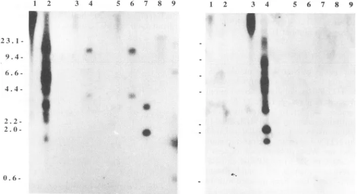
Southern hybridization analysis demonstrated the presence
ofthe HTLV-II-related provirus in the primary and cultured lymphocyte DNA of SM-3 (Fig. 3). After digestion with
different restriction endonucleases, genomic DNA from the coculture of BJAB cells with PBMCs from SM-3 contained fragments which hybridized with the HTLV-II full-length provirus probe pH6neo (4) under relatively high-stringency conditions (Fig. 3, left panel, lanes5 to9).Incontrast,when an
HTLV-I full-length provirus probe, pMT2 (11),wasusedas a
probe, very weak signal could be detected in the
HTLV-II-1 2 3 4 5 6 7 8 9
23.1
-9.4 -6.6 -4.4
-F
2. 2 -2. 0
-0:
.1a
STLV-II-SM3
HTLV-11-729(pH6neo) HTLV-I-HUT-1 02 STLV-I-PtM3
STLV-I1-SM3
HTLV-11-729(pH6neo)
HTLV-I-HUT-1 02 STLV-I-PtM3
ATCG ACA TGCCCT CCT GGC CAC CTG TCCAGA
---T----C---
A---C---- T---- C---
A---GCAGAAACTCAC CTGG#ACCC CA ---CC---
G---TC--- ---TC---
GA---G---FIG. 4. Partial nucleotide sequence of theX region of STLV-II isolated from SM-3; comparison with the X regions of HTLV-II 729(pH6neo), HTLV-I HUT-102, andSTLV-I PtM3. The sequences shown in the figure are equivalent to that of HTLV-II provirus sequence 7269 to7383(41). #,deletion.
positive cell line729(pH6neo) (Fig. 3, right panel, lanes I and
2) whereas no proviral signals could be detected in the
coculture of BJAB cells with SM-3 PBMCs(Fig. 3, right panel, lanes 5 to9). Furthermore, the restriction endonuclease pat-tern of theHTLV-II-related provirus(STLV-II)wasdifferent from that of HTLV-II; e.g., there was no BamHI site in
STLV-II, whereas there were three sites for HTLV-II, and there were twoHindIll-digested subgenomic fragmentsfrom
STLV-II,whereas thissite ismissing from HTLV-II.
The open reading frame (ORF) II within the pX region (ORF pX-II) of HTLV-I HUT-102 cells, HTLV-II
729(pH6neo) cells, and the corresponding regionofSTLV-II from SM-3PBMCs or the BJABcoculturewereamplifiedwith
SK43 and SK44primers bydoublePCR andwereanalyzedby DNAsequencing. Asshown in Fig. 4, STLV-II isolated from
SM-3 shares 97, 83, and 78% nucleotide homology with
HTLV-II, HTLV-I, and STLV-I, respectively. A published
1 2 3 4
'-1*
fW
t
5 6 7 8 9
0.6
-FIG. 3. Southern blot hybridization ofan HTLV-II probe,pH6neo (left panel), and an HTLV-I probe, pMT2 (right panel), torestriction enzyme-digested DNAs extracted from BJAB cells cocultivatedwith lymphocytes from SM-3 (BJAB-SM-3). Lanes for both panels: 1 and 2, undigested orPstI-digested genomicDNAsfromHTLV-II-positive 729(pH6neo) cells; lanes 3 and 4, undigested (lane 3) orPstI-digested(lane 4)genomicDNAsfromHTLV-I-positive HUT-102cells; lanes5 to 9,undigestedorPstl-,HindIII-,BamHI-,orEcoRI-digested genomic DNAs fromBJAB-SM-3, respectively. Sizes (in kilodaltons) are at the left.
J. VIROL.
on November 9, 2019 by guest
http://jvi.asm.org/
[image:4.612.70.284.73.142.2] [image:4.612.323.544.73.212.2] [image:4.612.130.481.476.667.2]FIG. 5. Electron micrographof BJAB cells cocultivated with lymphocytes from anSTLV-II-infected spider monkey (SM-3). Arrows, viral particles.
sequenceofasoutheast AsianSTLV-Iisolate(PtM3)wasalso includedforcomparison (58). Thesequence data of STLV-II
isolated either from fresh SM-3 PBMCs or from the
BJAB-SM-3 cocultured cells were identical and have also been
confirmedbygenecloninganddideoxynucleotide sequencing.
The deduced amino acid sequences of STLV-II Rex or Tax
proteins showed 95% homologywith the HTLV-II Rex and
Taxproteins, respectively.
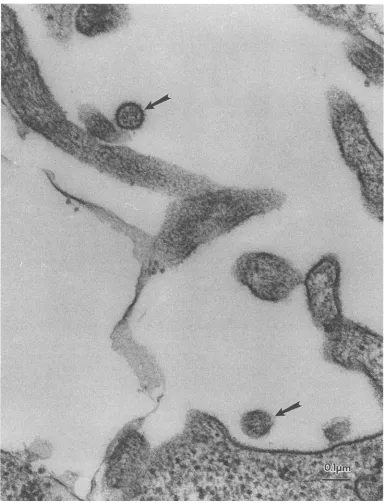
Electronmicrographic studies of SM-3 lymphocytesandthe
BJAB-SM-3 coculture revealed thepresenceoftypeC
retro-virus particles that were 100 to 110 nm in diameter and
possessed double envelope membranes andaround electron-densecore.Asshown inFig.5, the STLV-II virionswerefound outside the infected cell membrane.
STLV-II viralparticleswith arefractive index of1.393(cell
density of 1.19
g/cm3)
wereharvestedfromsucrosegradientsas indicatedbyMn2+-
orMg2+-dependent
RTactivity.Thevirallysatewasanalyzed withanHTLV-II-positiveserumsample by Westernblot assay(Fig.6).In contrast tothecontrolfractions witharefractive index of 1.383(Fig.6, lane 1),STLV-II from
on November 9, 2019 by guest
http://jvi.asm.org/
1154 CHEN ET AL.
Mn2+-
R.T.(cpm)
500
400
300
200
100
O L
92-
68-46-
3
32-20.-...
14 _S
1 2 3
1.375 1.380 1.385 1.390 1.395 1.400
[image:6.612.128.479.72.322.2]Refractive
Index
FIG. 6. RTandWesternblot assays ofdifferent fractions of thesucrosegradients ofSTLV-IIfromSM-3. Viralparticleswith refractiveindices of 1.383 and 1.393 (cell density of 1.19 g/cm3) were harvested from sucrose gradients separately, and their lysates were analyzed with an HTLV-II-positiveserum sampleby Western blot assays. Incontrast tothe controllysatewitharefractive index of1.383(lane 1), STLV-II from SM-3(lane 2) hadan antigenprofilesimilartothatofHTLV-II fromthe 729(pH6neo)cellline(lane3).
SM-3 (lane 2) had an antigen profile similar to that of
HTLV-II (Fig.6,lane3).TheSTLV-II Gag p55 precursorand
p15 proteins were reactive to the HTLV-II-positive human
serum.
DISCUSSION
Inthisstudy,5of 9spider monkeysand 1 of 47owlmonkeys
were found to have serologic evidence of STLV-II infection.
Among the 9 spider monkeys used in thisstudy, 8 originated
from Panama but were born in two metropolitanzoos in the
UnitedStates;only 1 spider monkey,whichalso hadantibody reactivity to RP-IIB, had been born in the wild. Recently, Kaplan et al. screened 75 spider monkeys by an HTLV-I
enzyme immunoassay (EIA) and found none of them to be
seropositive (28). It ispossible that the anti-STLV-II antibod-ies present in the monkey sera were missed by the HTLV-I antigen-based EIA since some HTLV-II-positive serum
sam-ples have been missed by such screening tests (5, 21). In
addition, the use of anti-human immunoglobulin G in the
HTLV-I EIAsmay raise concerns regarding the detectability of monkey antibodies in the serum samples. In the present
study, instead of using HTLV-I EIAs, we used an HTLV-II
recombinantenvelopeprotein, RP-IIB, in a Western blot assay todetectSTLV-II infection in New World monkeys. This assay has been proved to be able to detect virtually all HTLV-II carriers, including several intravenous drug users who were
seronegative to HTLV-I EIAs (5, 7). In order to cross-react
with monkey antibodies, the concentration of biotinylated sheep anti-human immunoglobulin used in the RP-IIB West-ern blot assay was raised from a dilution of 1/500 to one of 1/100. In addition, fluorescein isothiocyanate-conjugated
rab-bitanti-monkey immunoglobulin has been used in the IFA to
confirm the presence of anti-HTLV antibodies in the monkey sera.
One spider monkey, SM-4, which was not infected with
STLV-II, died of cardiomyopathy during the study. SM-4
spleen cells from autopsy had been used to cocultivate with different cell lines for virus isolation. Since multinucleated
giant foamy cells were found in SUP-Ti coculture, electron
microscopy examination was employed to rule out the pres-ence of simian foamy virus (SFV) infection (9, 23). Typical
intracellular particles, 35 to 50 nm in diameter, with an electron-lucent core surrounded by an electron-opaque shell were identified in the SUP-TI cocultures (data not shown).
SFVviruslysatesfrom SM-4have beenused inaWestern blot assay to detect anti-SFV antibody in serum samples from
SM-1, -2, -3, and -5. The analysis showed that none ofthem had anti-SFV antibodies (data not shown). In addition, no
intracellular viralparticles resembling SFV havebeenfoundin BJABcells cocultivated with STLV-II-positive SM-3 PBMCs
(Fig. 5).
There are several lines of evidence to suggest that the
STLV-II isolate described here is a new simian retrovirus
closelyrelated to butdistinct fromHTLV-II.Morphologically, itisatype C retrovirus with acondensed corethat resembles HTLV.Enzymatically,its RTactivityis bothMg2+ andMn2+
divalentcationdependent. Genetically, ithas ahighdegreeof
nucleotide homology with HTLV-II, but its restriction endo-nuclease patternis distinctfrom that ofHTLV-II. Ithas been
reported thatthere is 100% sequence homology of the ORF
pX-II region among different HTLV-II isolates (47).
There-fore, the difference in the nucleotide sequence of the ORF
pX-II region in STLV-II mayreflect differences important to thefunction ofitsgeneproducts,Tax andRex.The functional constraints of the gene products of the HTLV-I ORF pX
J. VIROL.
on November 9, 2019 by guest
http://jvi.asm.org/
regions
havebeenstudied,
andthere is96% sequence homol-ogy of the ORF pX-IIregion
between STLV-I PtM3 and HTLV-I HUT-102(Fig. 4).The deletion of nucleotide number 7378 intheXregion
of SM-3STLV-II will affect the ORFs of tax andrexgenes. FurtherDNAsequencingofthe remainingregion
of theXgene mayelucidate the functional domains of STLV-IITaxand Rexproteins.Inaddition,molecularcloningand
sequencing
of other regions of STLV-II are needed tounderstand its
phylogenetic
relationship with other primate T-cellleukemia viruses.STLV-II can infect andreplicatein BJABcells,but it does notinducesyncytiumformation upon cocultivation withBJAB
cells,
incontrasttoHTLV-II(15, 16).The syncytium-inducingcapability
of HTLV-II in BJAB cells has beenstudied,anditsfusogenic
domainwasfound tobelocated ina64-amino-acid stretch ingp2l
transmembraneprotein (39). Sequence analysisof the
analogous
domain of STLV-II may elucidate the deter-minants in HTLV-IIgp2l
that mediate cell fusion.It has been
reported
that HTLV-II infection may been-demic in certain New World
aboriginal
populations which includeanIndiantribe,theGuaymis,wholive in Panama(41).Since these Indian tribesare
relatively
isolated and donothavetypical
riskfactors for HTLV-IIinfection,
the endemicHTLVinfection may have arisen from closecontactwithprimates,as
some have
hypothesized regarding
the origin of HTLV-Iinfection
(25).
Recently, Goubauet al. reported thatpygmies from Zaire haveantibody
reactivities to an HTLV-II-specificrecombinant
protein,
K55(14).
Theconfirmation ofHTLV-II in thesepopulations requires
further investigation; however,more studies of STLV-II infection in Old World primates
appear necessary.
Since three of the five
STLV-II-seropositive monkeys
be-longed
to afamily grouping,
it seemslikely
that STLV-II infection canbe transmittedperinatally,
like STLV-I andthe HTLVs. Furtherserological
studies ofspider monkeys
andother New World
primate species
from thewild areneeded. Our data suggest that NewWorldprimates
may be infectedwith
STLV-II,
whereas Old Worldprimates
and apes arenaturally
infected with STLV-I(17, 59).
Further studies ofSTLV-LI may
help
us understand this curiousphylogenic
distribution. New fossil evidence suggests that the time oforigin
of simianprimates
may bepushed
back into thePaleocene
period,
which means that directmigration
ofsimi-ans between Africa and South America ismore
likely (33).
If STLV-I and STLV-II are ancientprimate retroviruses, they
maybe useful instudying
aspectsofprimate evolution,
suchas thedivergence
ofNewWorldprimates.
The lowgenetic
drift and limited horizontal transmission of the human counter-parts,HTLV-IandHTLV-II,
suggeststheutility
of STLVs for these types ofinvestigation
(13).
Finally,
since no disease isdefinitively
associated with HTLV-IIinfection, spider monkeys
infected withSTLV-II mayserve as auseful animal model to
study
thepathogenicity
of HTLV-II infection.ACKNOWLEDGMENTS
We thank R. Yanagihara, Y. Schulman, and M. Essex forhelpful
discussion; S. O'Brien for providing some of the serum samples of
spider monkeys;D. Hodge,X.-K.Zhang,J. Dobbs,and W.-H. Shiau for technical assistance; and Cathy Fletcher for correction of the manuscript.
This study was supported by the Fogarty International Center, NationalInstitutes of HealthVisitingProgram,Bethesda, Md.,and the National ScienceCouncil of theRepublicof China (NSC82-0203-1001-078A2-6).
REFERENCES
1. Abbott,M.A.,B.J. Poiesz,B.C.Byrne, S. Kwok, J. J. Sninsky, and G.D.Ehrlich.1988. Enzymaticgeneamplification: qualitative and quantitative methods for detecting proviral DNA amplified in vitro. J. Infect. Dis. 158:1158-1169.
2. Blattner, W. A., V. S. Kalyanaraman, M. Robert-Gurof, T. A. Lister, D.A.G.Galton,P. S.Sarin, M. H. Crawford,D.Catovsky, M.Greaves, and R. C. Gallo. 1982. The human type-C retrovirus, HTLV, in Blacks from the Caribbean region, and relationship to adult T-cellleukemia/lymphoma. Int. J. Cancer 30:257-264. 3. Chen,I.S. Y., J. McLaughlin, J. C. Gasson, S. C. Clark, and D.W.
Golde. 1983. Molecular characterization ofgenome of a novel human T-cell leukemia virus. Nature (London)305:502-505. 4. Chen, I. S. Y., J. McLaughlin, and D. W. Golde. 1984. Long
terminal repeats of human T-cell leukemia virus II genome determinetargetcellspecificity.Nature(London) 309:276-279. 5. Chen, Y.-M. A.,andM.Essex. 1991. Identification ofa
recombi-nant HTLV-II envelope protein for serological detection of HTLV-II carriers. AIDS Res. Hum. Retroviruses7:453-457. 6. Chen, Y.-M. A., T.-H. Lee, K. P. Samuel, A. Okayama, N.
Tachibana,I.Miyoshi,T.S.Papas, and M. Essex.1989.Antibody reactivitytodifferentregions of human T-cell leukemia virus type 1gp6l in infected people. J. Virol. 63:4952-4957.
7. Chen, Y.-M. A., T. H. Lee, S. Z. Wiktor, G.M.Shaw,E. L.Murphy, W. A. Blattner, and M. Essex. 1990. Type-specific antigens for serological discrimination of HTLV-I and HTLV-II infection. Lancet336:1153-1155.
8. Chen, Y.-M. A., X. Q. Zhang, C. E. Dahl, K.P. Samuel, R. T. Schooley, M. Essex, and T. S. Papas. 1991. Delineation of type-specific regions ontheenvelope glycoproteins of human T cell leukemia viruses.J.Immunol. 147:2368-2376.
9. Clarke, J. K, J. T. Attridge, D. S. Dane, and M. Briggs. 1967. A simian virus ofnewmorphology.J.Gen. Virol. 1:565-566. 10. Ehrlich, G. D., S. Greenberg,and M. A.Abbott.1990. Detection of
human T-cell lymphoma/leukemia viruses,p. 325-336. In M. A. Innis, D. H. Gelfand, J. J. Sninsky, and T. J. White (ed.), PCR protocols.AcademicPress, San Diego.
11. Gelmann, E. P., G. Franchini, V. Manzari, F. Wong-Staal, and R. C. Gallo. 1984. Molecular cloning of aunique human T-cell leukemia virus(HTLV-IIMo).Proc. Natl. Acad. Sci.USA 81:993-997.
12. Gessain, A., F. Barin, J. C. Vernant, 0. Gout, L. Maurs, A. Calender,andG. de The. 1985. Antibodiestohuman T-lympho-tropic virus type-I in patients with tropical spastic paraparesis. Lancetii:407-410.
13. Gessain, A., R. C. Gallo, and G. Franchini. 1992. Low degree of human T-cellleukemia/lymphomavirustypeIgenetic drift in vivo as a means of monitoring viral transmission and movement of ancient humanpopulations.J.Virol. 66:2288-2295.
14. Goubau, P., J. Desmyter, J. Ghesquiere, and B. Kasereka. 1992. HTLV-IIamongpygmies.Nature(London)359:201.
15. Hall,W.W., M.H.Kaplan, S.Z.Salahuddin,N.Oyaizu,C.Gurgo, M.Coronesi,K.Nagashima,and R.C.Gallo. 1990. Concomitant infections with human T-cell leukemia viruses (HTLVs) and humanimmunodeficiencyvirus(HIV):identification of HTLV-II infection in intravenous drug abusers (IVDAs), p. 115-129. In W. A.Blattner(ed.),Humanretrovirology,HTLV. RavenPress, New York.
16. Hall,W.W.,H.Takahashi, C. Liu,M. H.Kaplan, 0.Scheewind, S.Ijichi,K.Nagashima,andR.C. Gallo.1992. Multipleisolates andcharacteristics of human T-cell leukemia virustypeII.J.Virol. 66:2456-2463.
17. Hayami, M.,K.Ishikawa,A.Komuro,Y.Kawamoto, K.Nozawa, K.Yamamoto, T.Ishida,andY.Hinuma.1983.ATLVantibodyin cynomolgusmonkeysinthe wild. Lancet ii:620.
18. Hayami, M.,A.Kumuro,K.Nozawa,T.Shotake,K.Ishikawa,K. Yamamoto, T.Ishida,S.Honjo,andY.Hinuma. 1984. Prevalence of antibody to adult T-cell leukemia virus-associated antigens
on November 9, 2019 by guest
http://jvi.asm.org/
1156 CHEN ET AL.
(ATLA)
inJapanese monkeysand other non-humanprimates.Int. J.Cancer 33:179-183.19. Hinuma,Y., H.Komoda, T. Chosa,T. Kondo,M.Kohakura, T.
Takenaka, M. Kikuchi, M. Ichimaru, K. Yunoki, I. Sato, R.
Matsuo,Y.Takiuchi,H.Uchino,andM.Hanaoka. 1982. Antibod-ies to adultT-cell leukemia virus-associated antigen (ATLA) in
serafrom
patients
with ATL and controls inJapan:anation-widesero-epidemiologic
study.Int.J.Cancer29:631-635.20. Hjelle, B., R. Scalf, and S. Swenson. 1990. High frequency of human T-cell leukemia virus type II infection in New Mexico blood donors:determinationby sequence-specific oligonucleotide
hybridization.
Blood 76:450-454.21. Hjelle, B.,C.Wilson,S.Cyrus,P.Bradshaw,J.Lo,C.Schammel, T.Wiltbank,andS. Alexander.1993.Human T-cell leukemia virus typeIIinfectionfrequentlygoesundetected incontemporary US blooddonors.Blood81:1641-1644.
22. Homma, T.,P.J.Kanki,N. M.King,R. D.Hunt,M.J.O'Connell, N. L.Letvin,M. D.Daniel,R. C.Desrosiers,C. S.Yang,andM. Essex. 1984. Lymphoma in macaques: associationwith virus of human
T-lymphotropic family.
Science225:716-718.23. Hooks, J.,C.J.
Gibbs,
Jr.,S.Chou,R.Howk,M.Lewis,and D. C.Gajdusel. 1973. Isolation ofa new simian foamy virus from a
spider monkey
brain culture.Infect.Immun. 8:804-813. 24. Hunsmann, G.,J.Schneider,J.Schmitt,andN.Yamamoto. 1983.Detection ofserum antibodies to adult T-cellleukemia virus in non-human
primates
and in people from Africa. Int. J. Cancer 32:329-332.25. Ian, Y.,andT.J.Gojobori. 1990. Molecularevolutionofhuman T-cellleukemia virus. Mol. Evol. 31:493-499.
26. Ishida, T.,K.Yamamoto,R.Kaneko,E.Tokita,andY.Hinuma. 1983.
Seroepidemiological
study of antibodies to adult T-cell leukemiavirus-associated antigen (ATLA) infree-rangingJapa-nese
monkeys (Macaca
fuscata).Microbiol. Immunol. 27:297-301. 27. Kalyanaraman,V. S.,M. G.Sarngadharan,M.Robert-Guroff,I.Miyoshi,
D.Golde,and R. C.Gallo. 1982. Anewsubtypeofhuman T-cell leukemiavirus(HTLV-II)
associated withaT-cellvariantofhairy
cell leukemia.Science218:571-573.28. Kaplan,J.E.,M. U.Holland,D. B.Green,F.Gracia,andW.C. Reeves.FailuretodetecthumanT-lymphotropicvirusantibodyin
wild-caught
newworldprimates.
Am. J.Trop.Med.Hyg.,in press. 29.Lairmore,
M.D.,S.Jacobson,F.Gracia,B. K.De,L.Castillo,M.Larreategui,B. D.Roberts,P.H.Levine,W. A.Blattner,andJ.E.
Kaplan.1990.Isolation of human T-celllymphotropicvirustype 2 from
Guaymi
Indians in Panama. Proc. Natl. Acad. Sci. USA 87:8840-8844.30. Lal, R. B., D. L.Rudolph, M. D. Lairmore, R. F. Khabbaz, M.
Garfield, J. E. Coligan, and T. M. Folks. 1991. Serological
discrimination ofhuman T-cell lymphotropic virus infection by
using
asynthetic
peptide-based enzyme immunoassay. J.Infect. Dis. 163:41-46.31. Lipka,J.J.,K.Bui,G. R.Reyes,R.Moeckli,S. Z.Wiktor,W. A.
Blattner,E. L.Murphy,G. M.Shaw,C. V.Hanson,J. J.Sninsky, and S. K. H. Foung. 1990. Determination ofa unique immu-nodominant
epitope
ofhuman T-celllymphotropic
virustype-I.J. Infect. Dis. 162:353-357.32. Mann,D.L., S.J.
O'Brien,
D. A.Gilbert,Y.Reid,M.Popovic,E.Read-Connole,R. C.Gallo,and A.F. Gazdar.1989.Originof the
HIV-susceptible
human CD4+ cell line H9. AIDS Res. Hum. Retroviruses 5:253-255.33. Martin, R. D. 1993. Primate origins: pluggingthe gaps. Nature
(London)
363:223-234.34. Merl, S.,B.Kloster,J.Moore,C.Hubbell,R.Tomar,F.Davey,D.
Kalinow, A. Planas, G. Ehrlich, and D. Clark. 1984. Efficient transformationofpreviouslyactivatedanddividingT
lymphocytes
by
human T cellleukemia-lymphoma
virus. Blood64:967-974. 35.Miyoshi,
I.,M.Fujishita,H.Taguchi,K.Matsubayashi,N.Miwa,and Y. Tanioka. 1983. Natural infection in non-humanprimates
with adultT-cell leukemiavirusofacloselyrelatedcancer.Int. J. Cancer32:333-336.
36.
Miyoshi,
I.,S.Yoshimoto,M.Fujishita,H.Taguchi,I.Kuboishi, K.Niiya,andM.Minezawa.1982. Natural adult T-cell leukemia virus infectioninJapanese monkeys.Lancet ii:658.37. Osame, M.,K.Usuku,S.Izumo,N.Ijichi,H.Amitani,A.Igata,M.
Matsumoto, and M. Tara. 1986. HTLV-I associatedmyelopathy,a newclinical entity. Lancet i:1031-1032.
38. Poiesz, B. J., F. W. Ruscetti, A. F.Gazdar, P. A.Bunn,J. D.Minna, andR. C. Gallo. 1980. Detection and isolation of type C retrovirus particles from fresh and culturedlymphocytes ofapatient with cutaneousT-celllymploma.Proc.Natl.Acad. Sci. USA 77:7415-7419.
39. Poon, B., Q.-X. Li, and I. S. Y. Chen. 1993. Program Abstr. Retroviruses Meet., abstr. 12. Cold Spring Harbor Laboratory,
ColdSpringHarbor, N.Y.
40. Popovic,M., M. G. Sarngadharan, E.Read, and R. C. Gallo. 1984. Detection, isolation, and continuous production of cytopathic retroviruses(HTLV-III) frompatients with AIDS andpre-AIDS. Science224:497-500.
41. Reeves, W. C., P. H. Levine, M. Cuevas, E. Quiros, E.Maloney, and W. C. Saxinger. 1990. Seroepidemiology of human T cell lymphotropicvirus in the Republic of Panama. Am. J.Trop. Med. Hyg.42:374-379.
42. Rho, H. M., B. J. Poiesz, F. W. Ruscetti, and R. C. Gallo. 1981. Characterizationof the reversetranscriptase froma newretrovirus (HTLV) produced by a humancutaneous T-celllymphoma cell line.Virology 112:355-360.
43. Rosenblatt,J. D., J. C. Gasson, J. Glaspy, S. Bhuta, M.Aboud, I. S.Chen, and D. W. Golde. 1987.Relationship between human T cell leukemiavirus-II and atypical hairy cell leukemia. Leukemia 1:397-410.
44. Rudolph, D. L., S. S. Keesling, N. Lerche, J. A. Yee, and R.Lal. 1991.Dominance of HTLV typeI-specific antibody responsiveness in Old Worldmonkeys. AIDS Res. Hum. Retroviruses 7:721-722. 45. Saxinger, W., W. A.Blattner, P. H.Levine,J. Clark, R.Biggar,M. Hoh, J. Moghissi,P.Jacobs, L. Wilson, R. Jacobson, R.Crookes, M.Strong,A. A.Ansari, A. G. Dean, F. K. Nkrumah, N.Mourali, and R. C. Gallo. 1984. Human T-cellleukemia virus (HTLV-I) antibodies in Africa. Science225:1473-1476.
46. Seiki, M.,S.Hattori,Y.Hirayama, and M. Yoshida. 1983. Human adult T-cell leukemia virus:completenucleotide sequence of the provirus genome integrated in leukemia cell DNA. Proc. Natl. Acad. Sci. USA 80:3618-3622.
47. Sherman,M.P.,N. K.Saksena,D. K.Dube, R. Yanagihara, and B.J.Poiesz. 1992. Evolutionary insightsontheorigin of human T-cell lymphoma/leukemiavirus type I (HTLV-I) derived from sequence analysis of a new HTLV-I variant from Papua New Guinea. J. Virol. 66:2556-2563.
48. Shimotohno, K., D. W.Golde,M.Miwa,T. Sugimura,and I. S. Chen. 1984. Nucleotide sequence analysis of the long terminal repeat of human T-cell leukemiavirus type II. Proc. Natl. Acad. Sci. USA 81:1079-1083.
49. Shimotohno, K., Y. Takahashi,N. Shimizu,T. Gojobori, D. W. Golde,I. S.Chen, M. Miwa, and T. Sugimura. 1985. Complete nucleotide sequence of an infectious clone of human T-cell leukemiavirus type II: anopen readingframe for the protease gene. Proc. Natl. Acad. Sci. USA 82:3101-3105.
50. Smith, L.M., J. Z. Sanders, R.J. Kaiser,P. Hughes, C. Dodd, C. R. Connell, C. Heiner, S. B. Kent, and L. E. Hood. 1986. Fluorescence detection in automated DNA sequence analysis. Nature(London)321:674-679.
51. Smith,S.D.,M.Shatsky,P.S.Cohen,R.Warnke, M. P. Link, and B.E. Glader. 1984.Monoclonalantibodyandenzymatic profiles of humanmalignant T-lymphoidcells andderived cell lines. Cancer Res.44:5657-5660.
52. Sodroski,J.,M.Trus,D.Perkins, R. Patarca, F. Wong-Staal, E. Gelmann,R.Gallo,and W.A. Haseltine. 1984.Repetitive struc-turein thelong-terminal-repeatelement ofatype IIhuman T-cell leukemia virus. Proc.Natl. Acad. Sci. USA 81:4617-4621. 53. Teich,N.1985.Taxonomyofretroviruses,p. 1-16. In R.Weiss,N.
Teich,H.Varmus,and J.Coffin(ed.),RNA tumorviruses, 2nd ed. Supplements and appendixes. Cold Spring Harbor Laboratory, ColdSpring Harbor,N.Y.
54. Tsuchiya, S.,M.Yamabe,Y.Yamaguchi, Y. Kobayashi, T. Konno, and K. Tada. Establishment and characterization of a human acutemonocyticleukemia cell line(THP-1). Int. J.Cancer 26:171-176.
J.VIROL.
on November 9, 2019 by guest
http://jvi.asm.org/
55. Uchiyama,T.,J. Yodoi,K.Sagawa,K.Takatsuki,and H.Uchino. 1977.AdultT-cell leukemia: clinical and hematologic features of 16cases. Blood 50:481-492.
56. Wang, J. J., C. Hu, M. F. Shaio,and L.K.Chen. 1992. Internal-ization of CD4 molecules in human T-cells demonstrated by immuno-electron microscopy. Histochemistry97:51-59.
57. Watanabe, T., M. Seiki, Y. Hirayama, and M. Yoshida. 1986. HumanT-cellleukemiavirus type I isa member of theAfrican
subtype ofsimian viruses (STLV). Virology 148:385-388. 58. Watanabe, T., M. Seiki, H. Tsujimoto,I.Miyoshi, M. Hayami, and
M. Yoshida. 1985. Sequence homology of the simian retrovirus
genome with human T-cell leukemia virus type I. Virology 144:
59-65.
59. Yamamoto, N., Y. Hinuma, H.zurHausen, J. Schneider, and G.
Hunsmann. 1983. Africangreen monkeysare infected with adult T-cellleukaemia virusorclosely relatedagent. Lancet i:40. 60. Yoshida, M., I. Miyoshi, and Y. Hinuma. 1982. Isolation and
characterization of retrovirus from cell lines of human adult T-cell leukemia and itsimplicationinthe disease. Proc. Natl. Acad. Sci. USA79:2031-2035.