A
DISSERTATION ON
A STUDY OF RECURRENT STROKE
Submitted in partial fulfilment of
Requirements for the degree of
BRANCH - I D.M. (NEUROLOGY)
of
THE TAMILNADU Dr.M.G.R. MEDICAL UNIVERSITY CHENNAI
MADRAS MEDICAL COLLEGE CHENNAI – 600 003.
CERTIFICATE
This is to certify that this dissertation entitled “A study of Recurrent Stroke” submitted by Dr.P.V..KRISHNAN appearing for D.M. (NEUROLOGY) Degree examination in August 2008, is a bonafide record of work done by him under my direct guidance and supervision in partial fulfillment of regulations of the Tamil Nadu Dr. M.G.R. Medical University, Chennai. I forward this to the Tamil Nadu Dr.M.G.R. Medical University, Chennai, Tamil Nadu, India.
Prof. & Head of the Department Dr A.V.Srinivasan.
Institute of Neurology MMC ,Chennai – 600 003
Dr T.P.Kalanidhi, Dean
Madras Medical College Government General Hospital
DECLARATION
I, Dr.P.V.KRISHNAN solemnly declare that the dissertation titled "A study of Recurrent Stroke” is done by me at Madras Medical College & Govt. General Hospital, Chennai during Jan.2006 – Dec.2007.
The dissertation is submitted to The Tamilnadu Dr.M.G.R. Medical University towards the partial fulfilment of requirements for the degree of D.M.(NEUROLOGY). I also declare that this dissertation have not formed the basis of the award of any degree or diploma of any university.
Place: Chennai Date: 05-06-2008
Dr.P.V. KRISHNAN Postgraduate Student D.M. (NEUROLOGY) Institute of Neurology Madras Medical College
SPECIAL ACKNOWLEDGEMENT
ACKNOWLEDGEMENTS
I would like to express my sincere, respectful gratitude to
Prof.GEETHA LAKSHMIPATHY, former Professor and Head of the Institute of Neurology for her guidance and encouragement.
I would like to especially thank Prof.KAMAKSHI SHAMBOGUE,Prof.K.MUTHURAJ,Prof.A.V.SRINIVASAN,
Prof.V.NATARAJAN and Prof.R.M.BOOPATHY for their kind invaluable guidance and help.
I also extend my thanks to Dr C.Mutharasu ,Dr K.Bhanu, Dr V.Kamaraj , Dr S.Balasubramanian and Dr S.Velusamy Dr.S.Arunan,Dr.P.Muthukumar, Assistant Professors of Neurology for their co-operation and assistance.
Contents Page number
1. Introduction
1
2. Aim of the study. 6
3. Review of literature 7
4 Materials and methods. 25
5. Observation and Results 30
6. Discussion. 40 7. Conclusion. 43
8. References 44
9.Study Proforma 47
Introduction
Stroke was defined according to the World Health Organization criteria
as “rapidly developing symptoms and/or signs of focal, and at times global,
loss of cerebral function, with symptoms lasting more than 24 hours or
leading to death with no apparent cause other than that of vascular origin.”
The term “global” refers mainly to subarachnoid hemorrhage.
Recurrent stroke1, 31 is defined as a stroke, in which (1) there was clinical
evidence of the sudden onset of a new focal neurological deficit with no
apparent cause other than that of vascular origin (ie, the deficit could not be
ascribed to an intercurrent acute illness, epileptic seizure, or toxiceffect)
occurring at any time after the index stroke; or (2) there was clinical evidence
of the sudden onset of an exacerbation of a previous focal neurological deficit
with no apparent cause other than that of vascular origin occurring 21 days
after the index stroke.
Each recurrent stroke was classified as ischemic, hemorrhagic, or of
undetermined nature on the basis of a CT or MRI scan performed within 28
days of recurrence or autopsy examination of the brain. Etiologic subtypes of
ischemic stroke were defined according to Standardized criteria.
study .Recurrence represents the proportion of patients with stroke who had a
second stroke during a specified period of observation. For example
cumulative recurrence of stroke after surviving the first cerebral infarction in
a population based study of Rochester ,Minnesota residents was
approximately 6%.19%,29% at 1,5&10 years respectively.
If a stroke occurred duringtime interval 3-21 days, it often had to be in
a different vascular territory or anatomical site from the first event, of a
differentstroke subtype, or result in a different neurological deficit, in order
to be considered a recurrence.
A neurological worsening occurring at any time after the index event,
following a period of stability of 24 h should be considered a potential
recurrent stroke. This will not allow the very early recurrence risk to be
underestimated.
Stroke-in-progression has been defined by the European Stroke Database
collaboration as ‘neurological progression occurring within the first 3 days’.
There is a considerable variance of crude prevalence rates of
cerebrovascular diseases across India. Vellore & Rohtak gave rates of 13&
prevalence rate varied from 45-843 per 100000 with a high frequency of
diabetes & poorly controlled. Hypertension patients with stroke are seen very
frequently in daily Indian practice. Age adjusted mortality rates from stroke
are considerably higher among Indians as against Europeans.
The immediate period after stroke comes the greatest risk for
recurrence in stroke databank of 1273 patients with infarcty 3.3% had early
recurrence within 30 days. Long term stroke recurrence range from 4% to
14% with aggregate annual estimate of 6.1% for minor and 9% for major
stroke.
Based on WHO task force report on stroke prevention
diagnosis and therapy-1989, it appeared that for large artery hypertension &
smoking elevated blood lipid levels, obesity & diabetes are more important
modifiable risk factors. Cardio embolic stroke, RHD&IHD seem to be the
dominant risk factors among Indians. Broderick & Swanson of Rochester
study reported a 1% annual stroke recurrence in patients with a mean follow
up of 7 years(Broderick &Swanson 1987).This rate is significantly lower
than the 9% reported by Rothrock In This study the incidence of recurrent
effects of attempts at correction are analyzed.
The precise arterial pathology underlying lacunar infarcts, which are
presumed to result from the occlusion of single, small perforating arteries,
remains undetermined. It is often assumed to differ from the
atherothromboembolic processes that occlude large intracranial and
extracranial arteries and cause most other types of ischaemic stroke.
However, evidencefrom direct pathological studies is limited because lacunar
infarction has a low case fatality, autopsy rates are declining,and informative
pathological studies are expensive, technically demanding and
time-consuming.
Informative imaging studies are also scarce because of the difficulties in
imaging small arteries. Alternative, less direct methods have therefore been
used to study the pathology of lacunar infarction. These have included
observational studies comparing the risk factor profiles and prognosis of
patients with lacunar versus non-lacunar infarction, since differences might
suggest distinctarterial pathologies. Systematic review of studiescomparing
risk factor profiles in lacunar versus non-lacunar infarction found an excess
patients, but no clear difference in the frequency of any other risk factors,
including hypertension and diabetes.
Aim of the study
To study
*The clinical profile,
*Patterns of vascular involvement.
*Possible etiologies,
*Risk factors identified during the first episode of stroke.
*Risk factors persistent /corrected in the second or subsequent
stroke episodes.
*Effects of risk factor treatment on stroke free interval and the
Review of literature
Incidences of recurrent stroke are reported by prospective and retrospective
studies as hospital based and community based. Stroke during its first
occurrence and recurrence are analyzed on the basis of vascular territory,
clinico pathological types used as in Oxfordshire community based study,
Toast classification for the risk factors.
Graeme J. Hankey,etal’s1 community-based study aimed to determine the
absolute and relative risks of a first recurrent stroke over the first 5 years after
a first-ever stroke and the predictors of such recurrence in a population-based
series of people with first-ever stroke in Perth, Western Australia from 1989.
All people with a suspected acute stroke or transient ischemic attack of the
brain who were resident in a geographically defined region of Perth, with a
population of 138 708 people, were registered prospectively and assessed.
Patients were followed up prospectively at 4 months, 12 months, and 5 years
after the index event. Three hundred seventy patients with a first-ever stroke
cohort at 5 years, by which time 199 patients (58%) had died and 52 (15%)
had experienced a recurrent stroke, 12 (23%) of which were fatal within 28
days. The 5-year cumulative risk of first recurrent stroke was 22.5% (95%
confidence limits [CL], 16.8%, 28.1%). The risk of recurrent stroke was
8.8%- greatest in the first 6 months after stroke. (95% CL, 5.4%, 12.1%).
After adjustment for age and sex, the prognostic factors for recurrent stroke
were advanced, but not extreme, age (75 to 84 years) (hazard ratio [HR], 2.6;
95% CL, 1.1, 6.2), hemorrhagic index stroke (HR, 2.1; 95% CL, 0.98, 4.4),
and diabetes mellitus (HR, 2.1; 95% CL, 0.95, 4.4).Approximately 1 in 6
survivors (15%) of a first-ever stroke experience a recurrent stroke over the
next 5 years, of which 25% are fatal within 28 days. The pathological
subtype of the recurrent stroke is the same as that of the index stroke in 88%
of cases. The predictors of first recurrent stroke in this study were advanced
age, hemorrhagic index stroke, and diabetes mellitus, but numbers of
recurrent events were modest. Because the risk of recurrent stroke is highest
(8.8%) in the first 6 months after stroke, strategies for secondary prevention
should be initiated as soon as possible after the index event.
enrolled as acute ischemic stroke patients from Jan. 1998 to Dec. 2000
analyzed to identify the factors responsible for recurrent ischemic stroke.
Among 599 patients with ischemic stroke, 43 patients (7.2%) were had
recurrent stroke (27 men and 16 women; mean age=66.3 years).
Hypertension and hyperlipidemia were the risk factors which were
statistically significant in inducing recurrent ischemic stroke. According to
the TOAST classification, cardio embolism was more prevalent in recurrent
ischemic stroke.
Jean-Louis Mas 2 et al studied the risks of recurrent cerebrovascular
events associated with patent foramen ovale and atrial septal aneurysm . A
total of 581 patients (age, 18 to 55 years) who hadhad an ischemic stroke of
unknown origin within the preceding three months were consecutively
enrolled at 30 neurology centers. All patients received aspirin (300 mg per
day) for secondary prevention. After four years, the risk of recurrent stroke
was 2.3percent (95 percent confidence interval, 0.3 to 4.3 percent)among the
patients with patent foramen ovale alone, 15.2 percent(95 percent confidence
interval, 1.8 to 28.6 percent) among the patients with both patent foramen
interval, 1.8 to 6.6 percent) among the patients with neither of these cardiac
abnormalities. There were no recurrences among the patients with an atrial
septal aneurysm alone. The presence of both cardiac abnormalities was a
significant predictor of an increased risk of recurrent stroke (hazard ratio for
the comparison with the absence of these abnormalities, 4.17; 95 percent
confidence interval, 1.47 to 11.84), whereas isolated patent foramen ovale,
whether small or large, was not
Andy H.et al 3 reported Of the 678 patients in the cohort, 124
(18.3%) experienced repeated episodes of ischaemic stroke. Rural residence
and carotid endarterectomy procedure were positively associated with the
recurrence frequency, the adjusted incidence rate ratio being 1.66 (95% CI:
1.17–2.36) and 3.96 (95% CI: 2.30–6.82), respectively. Rural patients
contributed to 18% of the patients in the cohort yet they accounted for 27%
of those sustaining repeated episodes of stroke. Readmissions were also
related to the presence of diabetes at the index episode.
Goldstein et al 4 identified all patients admitted to Duke University
Hospital or the Durham Veterans Administration Medical Center during 1
Twelve randomly selected patients matched for age, sex, and race but having
only a single stroke served as controls. There were no significant differences
between the groups with respect to a variety of factors including the presence
of hypertension, diabetes, a history of transient ischemic attack, a history of
stroke, cerebral site of the index stroke, and subtype of the index stroke. A
potential cardioembolic source was more frequently identified in the patients
with early recurrent stroke (seven of the 12 case-control pairs were
discordant for a potential cardioembolic source.
Dong-wha kang et al 7 in a retrospective study reports that Prior
observations have shown that early recurrent ischemic lesions (ERILs) on
diffusion-weighted imaging occur frequently within the first week after an
index stroke. They included 133 patients who experienced an acuteischemic
stroke and who underwent initial diffusion-weightedimaging within 24 hours
and subsequent diffusion-weighted imaging within 7 days after onset, and
whose stroke subtype was Intra cranial large artery atherosclerosis(IC-LAA),
extra cranial LAA (EC-LAA), or cardio embolism (CE). Early recurrent
ischemic lesions were defined as new ischemic lesions on follow-up
recurrent ischemic lesions were observedin the following proportions: 50.9%
(28/55) in the IC-LAA group,47.4% (9/19) in the EC-LAA group, and 44.1%
(26/59) in the CE group. Early recurrent ischemic lesions in the IC-LAA
grouphad the following characteristics: (1) they occurred mostly(27 [96.4%]
of 28) in the pial area of the same vascular territory as the index stroke; (2)
they were more frequently observed in a higher grade of stenosis than in
milder stenosis (P<.001), whereas ERILs in the EC-LAA group were not
related to the degreeof stenosis; (3) they were not associated with subsequent
recanalization,whereas ERILs in the CE group were mostly associated with
subsequent recanalization (P<.001); and (4) they were more closely
associatedwith clinical recurrence than in the EC-LAA or CE group (P=.02).
They conclude that Early recurrent ischemic lesions in the IC-LAAgroup are
relatively frequent and have different patterns than in the EC-LAA or CE
group.
Secondary prevention of recurrent stroke by treating the modifiable
risk factors have been done in many landmark trials. The prevention of
stroke, with its attendant costs, both financial and personal, is the goal of
will reduce stroke risk. We’ll look at the evidence for the following
interventions: treatment of hyperlipidemia, smoking cessation, antiplatelet
therapy, treatment of hypertension and anticoagulation for atrial fibrillation.
Aggressive treatment of dyslipidemia decrease the risk of stroke.
This issue has been looked at indirectly in multiple placebo-controlled
trials, as a pre-specified secondary end point. Perhaps the best place to
demonstrate this is to look at the MRC/BHF study, commonly known as the
Heart Protection Study. This is the largest trial ever done on lipid lowering. It
included 20,536 people aged 40 to 80 with coronary disease, other occlusive
arterial disease or diabetes with a non fasting cholesterol >3.5 mm/l. There
were 6,793 subjects with a starting LDL < 3mm/l. They were randomized to
40 mg of simvastatin or a matching placebo. Follow up was for an average of
5 years. 4.3% of the simvastatin group vs. 5.7% of the placebo group suffered
a stroke. (RR 75%, RRR 25%, ARR 1.4% NNT 71). This small but
consistent effect is seen in multiple trials. There were no excess hemorrhagic
strokes in the treatment arm.
Smoking increases the risk of stroke.Clearly we do not have the best
this subject. In issues of harm, RCTs are impossible to do, as it is ethically
impossible to randomize people to a smoking arm! So we have to look at the
next best evidence, a prospective cohort trial, which systematically follows a
large group of healthy people until they get ill with the disease of interest,
and then tries to elucidate the risk factors for that outcome.
The Framingham Study 19 began soon after WWII, is still ongoing, and
is the longest running prospective study of this type. In 1988, it reported on
cigarette smoking as a risk factor for stroke. It looked at 4,255 men and
women, 36 to 68 years old and free of cerebrovascular disease. During a
26-year follow up 459 strokes occurred. Multivariate analysis using Cox
proportional hazard modeling, demonstrated cigarette smoking as an
independent risk factor for all strokes in general, and thrombotic strokes in
particular. The risk of stroke increased as the number of cigarettes smoked
increased. The RR was double in heavy smokers (> 40 cigarettes/ day)
compared to light smokers (< 10 cigarettes/day). Lapsed smokers retained
their increased risk for the first 2 years, which then gradually decreased over
the next 3 years. By 5 years the risk dropped to the level of non-smokers.
In 1989 a case controlled study20 looked at 621 patients with stroke
and 573 controls without stroke. The authors estimated an increased relative
risk of 1.5 for every 10 cigarettes consumed daily, both in men and women.
The use of antiplatelet medications in the secondary prevention of stroke has
long been established. Various drugs and combinations of drugs have been
studied over the last 20 years. We will look at the evidence for ASA,
clopidogrel, and ASA plus dipyridamole.
Acetyl salicylic acid is the best studied, cheapest, and most widely used
antiplatelet medication. In 1994 the Antiplatelet Trialists' Collaboration
published a meta-analysis of randomized trials21 of prolonged antiplatelet
therapy for prevention of stroke (along with various other endpoints). This
meta-analysis looked at trials published till 1990, the vast majority of the
patients having taken ASA. Amongst 10,000 patients with a past history of
stroke or TIA the net event rate for vascular events was estimated at 18%
with antiplatelet therapy vs 22% in the controls. (RR 82%, RRR 18%, ARR
4%, NNT 25).
Ticlopidine was the first antiplatelet drug demonstrated to be clearly better
The second drug in this class to be released was clopidogrel, along with the
results of the CAPRIE trial.
The CAPRIE Trial22 was a randomized, double blinded, international trial
designed to assess the relative efficacy of clopidogrel (75 mg. once daily) and
aspirin (325 mg. once daily), in reducing the risk of a composite outcome
cluster of ischemic stroke, myocardial infarction, or vascular death. The
population studied comprised subgroups of patients with atherosclerotic
vascular disease manifested as, either recent ischemic stroke, recent
myocardial infarction, or symptomatic peripheral arterial disease. 19,185
patients were followed for 1 to 3 years. The primary study end point was a
composite of ischemic stroke, MI or vascular death. Overall, in all 3
subgroups, there was an annual 5.32% event rate in the clopidogrel group vs
5.83% in the ASA group. (RR 91.3%, RRR 8.7%, ARR 0.51%, NNT 196/per
year, p=0.043).
This is not an impressive result and required a huge study to demonstrate it.
In the sub group of interest to us, the 6411 patients with a stroke as the
qualifying event to enter the trial, clopidogrel did not demonstrate a
argument can be made that this was just type 2 error and we just need a larger
study to demonstrate a significant difference - but this was already a huge
study). Also remember this data is for a composite end point whilst we are
most interested here in secondary stroke prevention. So we can conclude that
clopidogrel has not been demonstrated to be superior to ASA in the
secondary prevention of stroke. Further analysis of this trial supports the
view that clopidogrel is clearly superior to ASA in the prevention of MI in
the CAPRIE patient population. (RRR 16.6% NNT 119)
By the late 1980's dipyridamole and the combination with ASA was felt to be
no better than ASA alone. However, in 1987 ESPS-1 using a combination of
ASA 330 mg. plus dipyridamole TID for 2 years in patients with prior
ischemic stroke or TIA, showed a RRR of 38% for subsequent stroke. This
was far higher than expected from previous studies on ASA alone. There
were a lot of questions about the dosing of ASA and the conflicting data of
ESPS-1 vs previous trials, so a larger 2x2 factorial design study, ESPS-28
was undertaken in 1989 and published in 1996. Placebo was compared to
ASA 25 mg. BID, dipyridamole 200 mg. extended release, and the
ischemic CVA were enrolled and followed for 2 years in this international
multicenter randomized double blinded trial done in Europe.
The results were impressive. 15.78% of the placebo group, 13.21% of the
dipyridamole group, 12.93% of the ASA group and 9.95% of the
combination group suffered a recurrent stroke. There was no statistical
difference between the 2 active treatment groups and with a RRR of 18%, the
low dose ASA group was in line with previous trials on ASA. The surprise
was the additive effect seen in the combination group. (RR 63%, RRR 37%,
ARR 5.83%, p<0.001, NNT 17 over 2 years). There was no difference in the
death rate in the 4 groups and no excess of MI in the combination group.
From the evidence it is clear the first choice in secondary prevention should
be a combination of low dose ASA plus extended release dipyridamole. It is
superior to ASA or clopidogrel alone. The combination of ASA and
clopidogrel. There is good evidence to use it for ischemic heart disease but
the evidence is lacking for secondary prevention of stroke. This is an area of
active research and results of several ongoing trials are eagerly awaited.
Hypertension is a major risk factor for stroke and the treatment of
Numerous randomized placebo controlled trials, using primarily diuretics and
beta blockers, established this by the 1970's.
Concern about cerebral perfusion in patients with known cerebrovascular
disease and especially those with significant carotid disease, meant
extrapolation of primary prevention data regarding antihypertensive
treatment was resisted till well into the 1990's.
There is the evidence that treatment of hypertension will reduce the risk of
stroke in patients who have already suffered a stroke or TIA. This can be
reviewed most efficiently by looking at a recent systematic review of the
topic published in the Journal STROKE in November 2003. The authors,
through an exhaustive search of the world medical literature, identified 7
placebo controlled trials that assessed the effect of lowering blood pressure in
patients with prior stoke or TIA. They used meta-analytic techniques to
combine these results and came to several not unexpected conclusions. The
authors reported the results as odds ratios . The outcome, recurrent stroke,
occurred in 11.46% of the placebo group, and 8.86% of the treatment group.
(RR77%, RRR23%, ARR2.6%, NNT 38, p=0.005). Interestingly these results
interpreting the results, as the numbers are small in each drug class except
diuretics.
Overall diuretics decreased the risk of recurrent stroke, ACE-I reduced the
risk of MI and the combination of both drugs reduced both. Four of the seven
trials used fixed drug combinations and enrolled both hypertensive and non
hypertensive patients, bringing up a lively debate on whether the effects seen
are related to the drugs used, or the blood pressure drops seen, in the
treatment arms. If the effects seen were due to the blood pressure drop, this
brings in to play the question of the definitions of hypertension and target
blood pressures.
Atrial fibrillation is a potent risk factor for ischemic stroke. This dysrhythmia
affects 2-5% of the general population over the age of 60, but is found in
15% of all stroke patients. The use of anticoagulation with warfarin is now
well established as the preferred method for primary prevention of stroke in
this patient population, as long as the patient is not considered low risk. (Low
risk patients have no history of previous stroke or TIA, no treated or
disease.)17 In low risk patients there is no substantive advantage for
anticoagulation over antiplatelet agents.
Secondary prevention for a patient who has already suffered a stroke or a
TIA associated with atrial fibrillation- This was addressed in 1995 by a
Cochrane Review. This review looked at the world literature in a systematic
fashion, and performed a meta-analysis on the results. It combined the results
of the EAFT from 1993 and VA-SPINAF from 1992. Between the 2 trials
485 patients were included. Anticoagulation reduced the risk of recurrent
stroke by nearly 2/3 and the risk of all vascular events over 1/3. (RR 39.5%,
RRR 60.5%, p<0.0001, ARR 13.7%, NNT 7 for recurrent stroke).
In progress trial 6105 patients with stroke or TIA within 5 previous
years were randomized to perindopril+indapamide or placebo. At the end of 4
years they reported a reduction in recurrent stroke of 28% and total major
vascular events by 26% inpatients with or without hypertension. Combination
therapy with perindopril plus indapamide reduced recurrent stroke by 43%
perindopril alone had no significant impact.
WARSS15- 2206 patients with a non cardio embolic ischemic stroke
2years there was no difference between groups with respect to the primary
endpoint (warfarn 17.8% VS asprin 16%) or the rate of major hemorrhage.
In a similar multicentric perspective observational study in setting of
primary cause throughout India Sabash Kaul13 summarized the findings.
During 12 months perindopril based treatment it was found that incidence of
recurrent stroke was similar to that of progress.
SPARCL 12 investigators assigned 4731 patients who had a stroke or
TIA within 1-6 months before study entry had LDL level 100-190 and had no
known coronary artery disease to double blind treatment with 80 gm
atorvostatin per day or placebo .During 4-9 years absolute reduction in risk of
fatal / nonfatal stroke was2.2% & 3.5% reduction in cardiovascular events
.There was a small increase in incidence of haemorrhage stroke.
Graeme30Quotes that elevation of plasma homocysteine is associated
with laboratory evidence of atherogenesis and thombosis and epidemiological
evidence of an increased risk of ischemic stroke, independent of other
vascular risk factors. The vitamins in stroke prevention trial and the vitamins
to prevent stroke trial show insufficient evidence to recommend routine
Materials and methods
This study was conducted during January 2006 to December 2007 at
Madras institute of neurology, Government General Hospital, Chennai.
Patients with clinical features suggestive of second or subsequent
strokes were taken up for study. All were subjected to CT/MRI scan of brain.
Inclusion criteria
1. All the patients with clinical features suggestive of second or
subsequent stroke.
2. Imaging showing ischemic infarcts in the brain
Exclusion criteria
1. First ever episode of stroke
2. Evolving stroke.
3. Imaging showing evidence of hemorrhage.
4. Imaging showing evidence of venous infarct.
Patients’ details regarding age, sex, and family history, risk factors like
atrial fibrillation, trauma, smoking, and substance abuse were recorded. The
onset and details of the symptoms and clinical signs were noted during each
stroke episode from the patient or close relative and the old records. All
patients had detailed neurological examination.
All patients underwent a basic investigation protocol that included
complete blood counts , erythrocyte sedimentation rate, blood glucose,
urea ,creatinine, electrolytes, lipid profile (triglycerides, total cholesterol, and
fractions) CT brain, chest x-ray, electrocardiogram; transthoracic
echocardiogram.
MRI brain with MRA and DWI, B-mode carotid ultrasonography,
carotid and vertebral Doppler study had been done wherever indicated.
Other laboratory tests such as homocysteine, fibrinogen, prothrombin
time , partial activated thromboplastin time, antinuclear antibodies,
anticardiolipin antibody, lupus anticoagulant auto antibodies (SM,
anti-SSA, and anti-RNA), were done in selected patients where clinical features
indicated their need
Specific studies for the detection of natural anticoagulant deficiency,
such as measurement of protein C, protein S, and antithrombin III, were
family history indicated a prothrombotic disorder. Investigations done
during previous stoke episodes were also recorded.
Observation and Results
A total of 52 patients admitted in Government General Hospital between January 2005 and December 2006 with clinical features and neuroimaging suggestive of second or subsequent stroke were included in the study.
Sex
Distribution (n=52)(Males: 61.54%, Females: 38.46%)
males
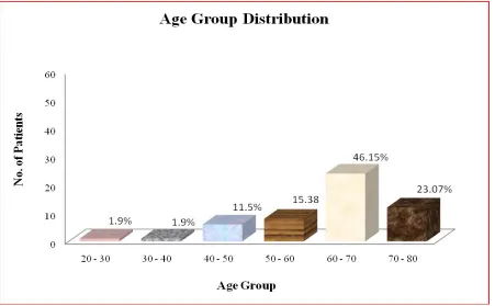
Age Distribution
Table-1 (n:52)
Age group Number of patients Percentage
20-30 1 1.9
30-40 1 1.9
40-50 6 11.5
50-60 8 15.38
60-70 24 46.15
Mortality in recurrent stroke
Table-2 (n:52)
Death No of patients Percentage
Within one month 2 4
Within one year 4 8
At the end of two years 7 14
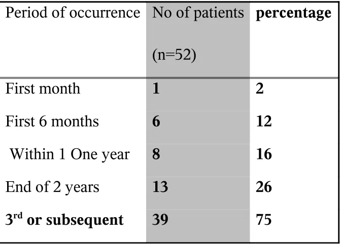
Incidence of stroke recurrence
Table-3
Period of occurrence No of patients
(n=52)
percentage
First month 1 2
First 6 months 6 12
Within 1 One year 8 16
End of 2 years 13 26
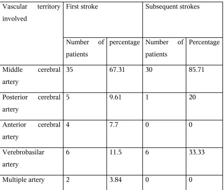
[image:32.612.51.386.388.631.2]Vascular territory involved in Ischemic strokes
Table-4
Vascular territory involved
First stroke Subsequent strokes
Number of patients
percentage Number of patients
Percentage
Middle cerebral artery
35 67.31 30 85.71
Posterior cerebral artery
5 9.61 1 20
Anterior cerebral artery
4 7.7 0 0
Verebrobasilar artery
6 11.5 6 33.33
Multiple artery 2 3.84 0 0
(Only ischemic strokes were included in this study)
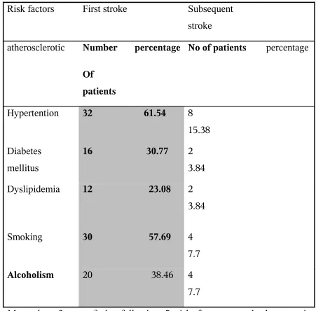
Table-5
Risk factors First stroke Subsequent stroke
atherosclerotic Number
Of patients
percentage No of patients percentage
Hypertention 32 61.54 8 15.38
Diabetes mellitus
16 30.77 2 3.84
Dyslipidemia 12 23.08 2 3.84
Smoking 30 57.69 4 7.7
Alcoholism 20 38.46 4 7.7
More than 3 out of the following 5 risk factors namely hypertension diabetes, dyslipidemia, smoking and alcoholism was present in 40 cases, (76.92%) as persistent causes, 2 out of 5 in 12 (23.08%).
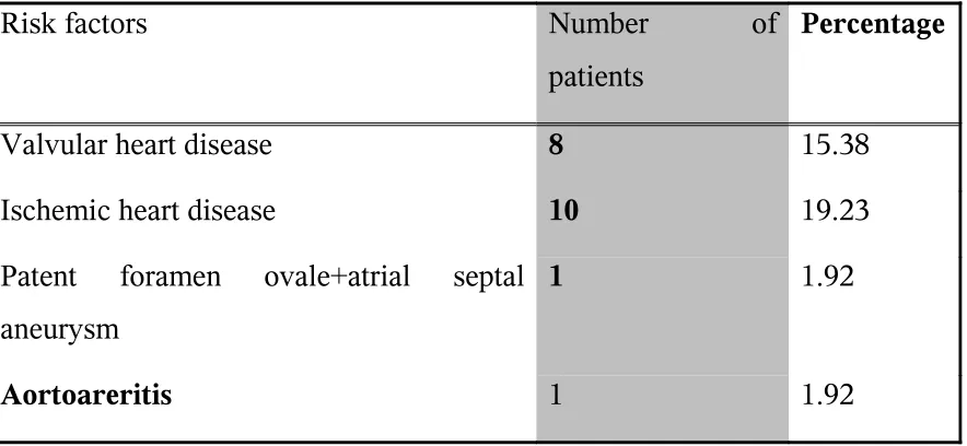
Table-6
Risk factors Number of patients
Percentage
Valvular heart disease 8 15.38
Ischemic heart disease 10 19.23
Patent foramen ovale+atrial septal aneurysm
1 1.92
Aortoareritis 1 1.92
Among the valvular heart disease 6 out of 8 were rheumatic heart disease- mitral stenosis with atrial fibrillation, one was Aortic stenosis with atrial fibrillation and one was infective endocarditis. All the 10 (19.23%) ischemic heart disease patients were in congestive heart failure during the recurrent stroke episode.
Patent foramen ovale with atrial septum aneurysm was detected during the first recurrent stroke. The aortic arteritis patient was on irregular therapy – steroids. The first recurrence occurred during drug withdrawal.
Treatment and its effect on stoke free interval & recurrences
Table-7
Treatment & number of cases
(years) Number percentage
Hypertension (32) 1.5 15 46.87
Asprin (52) 2 10 19.23
Dyslipidemia(12) 2 0 0
All the 32 cases of hypertension were on anti hypertensive and the recurrent stroke occurred during the non compliance. The mean stroke free interval was 1.5 years while on regular treatment recurrence occurred on drug withdrawal.
Ten of the cases had stopped asprin for more than a month accounting for subsequent episode of stroke. The mean stroke free interval was 2 years with good drug compliance.
Discussion
Only ischemic type of stroke patients was included in this study. 61% of them were males showing a male preponderance. Age group 60-70 had the maximum recurrence stroke (46.15%).This was similar to Jung B et al's 5 report.
The one month mortality rate was 4%, one month stroke recurrence rate 2%, and 1 to 12 month stroke recurrence as 16% in this study. In Carolene jackson's e al’s 16 study
they were 3.81%,2.11%, and 1.24% respectively. This study shows similar one month mortality rate and more incidences of late recurrence.
Middle cerebral artery was the commonest vascular territory involved(67.31%) in this study as it was in all other recurrent stroke studies .Most of the recurrence was of the same arterial territory -the middle cerebral artery region (85.71%).
Hypertension was found to be the risk factor in majority of cases (61.54%) and diabetes 30.77% in this study. In Jung B et al's 5 study hyperlipidemia was the major
associated risk factor. In Perth community study1 diabetes mellitus was reported in 95%
of cases while if is 30.77% in this study.
In this study an attempt was made to analyze the cumulative effect of risk factors on the recurrence of stroke. It was clearly shown that addition of one or more risk factors to the existing one increases the chance of developing recurrence. For example when a hypertensive develops diabetes and dyslipidemia he has more likelihood of developing the next stroke.
This study showed an incidence of 15.38% of valvular heart diseases and 19.23% of ischemic heart disease – showing a marked increase in the incidence of valvular heart disease as a cause recurrent stroke. While Gold stein et al 4 report 30 day recurrence rate
of 4.39% with cardio embolic source of other than valvular heart disease.
atrial septal aneurysm. Masse et al’s32 recommendation that all cryptogenic stokes
should be reevaluated proved fruitful.
This study shows stroke free interval of 1.5 years (mean) while on Antihyperensive therapy, 2 years each for patients who were an aspirin and atorvostatin. Progress study 14 reports a reduction in recurrent stroke of 28% with perindopril and
indapamide. The event rate was estimated to be 18% with Asprin therapy by antiplatelet
trialists’ collaboration 21.The reduction in risk of fatal and non fatal stroke was 2.2%
with use of statin by Sparcl investigators 12.
Comparison between near similar studies had been made. Other observations made were: Hypertension was the large single contributor and patients on Anti hypertensives had a comparable stroke free interval. Asprin was effective in prolonging the stroke interval. Statin had an added on effect in prolonging stroke free interval.
Biomarkers like high sensitivity C-reactive protein (hs-CRP) and Liporotein associated phospholipase A2 (LP-PLA2) are found to predict recurrent stroke risk. Statins
appear to lower hs-CRP levels33. A better understanding of these biomarkers may lead to
use of prophylactic treatments to reduce risk of people suffering debilitating strokes.
Early recurrent ischemic lesions detected by diffusion weighted image study can predict (1) the presence of higher grade of stenosis in intracranial large artery atherosclerosis subtype of stroke (2) possibility of good recanalization in cardioembolic – ischemic strokes 7
As a stroke survivor or a caregiver for a stroke survivor, one should
know that having a stroke puts the patient at greater risk for getting another
Conclusion
Recurrent strokes account for considerable amount of mortality and morbidity.
Pathological subtype breeds true to the index stroke.
Middle cerebral artery territory is the commonest site involved in recurrent stroke.
Hypertension was the risk factor found in majority of the recurrent episodes.
Presence of three risk factors confers the maximum risk of development of subsequent strokes.
All patients deserve a repeat clinical examination and relevant investigations .This enables to identify new risk factors in cryptogenic strokes and confirm existing and persisting / corrected risk factors.
Aggressive treatment of risk factors will prolong the stroke free interval and prevent the occurrence of recurrent stroke.
References
:1)Graeme J. Hankey, Robyn J. Broad Hurst, Craig S. Anderson, Long-Term Risk of First Recurrent Stroke in the Perth Community Stroke Study. (Stroke. 1998;29:2491-2500.)
(2).Jean-Louis Mas, M.D., Caroline Arquizan, M.D.,., for the Patent Foramen Ovale and Atrial Septal Aneurysm Study Group December 2001 NEJM.
(3)Andy H. Lee, Kelvin K.W. Yau and Kui) Recurrent ischemic stroke hospitalizations: A retrospective cohort study using Western Australia linked patient records December 2001 NEJM
(4) Goldstein, Larry B. MD; Perry, April RN Early Recurrent Ischemic Stroke: A Case-Control Study. Stroke.23(7):1010-1013,July1992.
(5)Jung B, Yoon OY, Park KH, Lee KY, Lee YJ, Kim HT, Kim SH, Kim J, Kim MH.)Analysis of Risk Factors for Recurrent Ischemic Stroke: Based on Data of Outpatient Clinic in a University Hospital. Department of Neurology, Han yang University College of Medicine, Guri,
(6)Stroke prevention guidelines-National strike association/htm
(7)Dong-Wha Kang, MD, PhD; Sun U. Kwon, MD, PhD; Sung-Hee Yoo, RN, MS; Kyum-Yil Kwon, MD; et al Imaging in
Symptomatic Intracranial Atherosclerosis Arch Neurol. 2007;64(1):50-54.
(8) NHWadia-K.prasad-V.goyal introduction to Indian neurology.Cerebrovascular diseases neurological practice pg 7-8&309-331.
(9) Neurology protocol seminar in stroke1998 Madras institute of neurology Chennai.
(10) WHO task force in stroke and other cerebrovascular disorders (1989)
(11) CJ estol, Louis R stroke syndromes 2nd edition. pg 63-64
(12) The stroke precaution by aggressive reduction in cholesterol levels (SPARCL) investigators New England journal of medicine aug2006 355.6 549-559
(13) MV Padma , Subash Kaul –incidence of recurrent stroke in primary care during preventive treatment based on perindopril with or without indapamide Neurology India April 2007.
(14) Perindopril protection against recurrent stroke study (PROGRESS) lancet 2001; 358:1033-41
(15) warfarin asprin recurrent stoke study (WARSS) NEJM2001 345,1444-51.
(16)Caroline Jackson and Cathie Sudlow -Comparing risks of death and recurrent vascular events between lacunar and non-lacunar infarction- Brain 2005 128(11):2507-2517
(17)Farokh Buhariwalla -Stroke Prevention Show Me The Evidence .a review article, Archives -the Berries ns ca. homepage.1-3.
(18).MRC/BHF Heart Protection Study of cholesterol lowering with simvastatin in 20,536 high-risk individuals: a randomized placebo-controlled trial. Lancet. 2002 Jul 6; 360(9326): 7-22. nbsp;
Study.JAMA.1988Feb19;259(7):1025-9
(20) Gill, JS et al, Cigarette Smoking. A Risk Factor for Hemorrhagic and NonhemorrhagicStroke.ArchInternMed.1989Sept;149(9):2053-7
(21) Collaborative Overview of Randomized Trials of Antiplatelet Therapy-I: Prevention of Death, Myocardial Infarction, and Stroke by Prolongs Antiplatelet Therapy in Various Categories of Patients. Antiplatelet Trialists' Collaboration. BMJ 1994 Jan 8; 308(6921):81-106.
(22) CAPRIE Steering Committee. A randomized, blinded, Trail of Clopidogrel versus aspirin in patients at risk of ischemic events (CAPRIE), Lancet 1996; 348:1329-39.
(23) Cannon, PC, Effectiveness of Clopidogrel Versus Aspirin in Preventing Acute Mycocardial Infarction in Patients with Symptomatic Atherothrombosis (CAPRIE Trial). AmJCardiology2002Oct1;90:760-762
(24)European Stroke Prevention Study. ESPS Group. Stroke. 1990 Aug; 21(8): 1122-30
(25)Diener HC et al, European Stroke Prevention Study. 2. Dipyridamole and acetylsalicylic acid in the secondary prevention of stroke. J Neurol Sci. 1996 Nov;143(1-2):1-13.
(26)Yusuf S et al, Effects of clopidogrel in addition to aspirin in patients with acute coronary syndromes without ST-segment elevation. N Engl J Med.2001Aug16;345(7):494-502.
(27)Rashid P, Leonardi-Bee J, Bath P. Blood pressure reduction and secondary prevention of stroke and other vascular events:a systematic review. Stroke. 2003 Nov, 34(11):2741-8.
(28)Koudstaal PJ. Anticoagulants for preventing stroke in patients with nonrheumatic atrial fibrillation and a history of stroke or transient ischemic attacks (Cochrane Review). In: The Cochrane Library, Issue 4, 2003. Chichester, UK: John Wiley & Sons, Ltd.
(29) Van Walraven C, et al, A clinical prediction rule to identify patients with Atrial Fibrillation and a low risk for stroke while taking aspirin. Arch Intern Med. 2003 Apr 28; 163(8): 936-43.
(30)Graeme j.Hankey, Secondary prevention of recurrent stroke.Stroke.2005;36:218-221.
(31) Burn J, Dennis MS, Bamford J, Sandercock PAG, Wade D, Warlow CP. Long-term risk of recurrent stroke after a first-ever stroke: the
(32)S.R. Messé, MD; I.E. Silverman, MD; J.R. Kizer, MD, MSc; Recurrent stroke
with patent foramen ovale and atrial septal aneurysm. American Academy of Neurology Neurology 2004;62;1042-1050
(33)Mitchell.s.v,elkind;Biomarkers found to predict risk of recurrent stroke and mortality;Archives of nerology.oct,2003.
RECURRENT STROKE - PROFORMA
Name Age: Sex : DOA: DOD: MIN/IP no : Address/Phone no:
Occupation:
Stroke Episodes First Second third fourth
Admission delay (0-3h,3-6,6-12,12-24,24-48,>48h)
Interval between strokes Symptoms
1.Headache
2.Nausea/Vomiting 3.Weakness of limbs 4.Numbness face/limbs 5.Mental changes 6.Neck pain 7.Dysphasia 8.Articulation disturbed 9.Impaired vision 10.Diplopia 11.Giddiness/vertigo 12.Dysphagia
13.Imbalance of gait/ in coordination
14.Convulsions-focal / Generalized
15.Retention of urine 16.Fever
17.Others 18.Altered consciousness
CLINICAL EXAMINATION:
Obesity Pallor Jaundice Cyanosis Lymph nodes Skin rash Purpura Xanthelasma
Pulses : carotid vertebral radial dorsalis pedis Blood pressure :
HMF : conscious drowsy comatose MMSE :
Lobar functions :
Visual acuity visual field pupils fundus EOM
Facial palsy
Other cranial nerves : Bulk:
Tone:
Power : UL : LL :
Reflexes: Abdominal reflex : cremastric : BJ KJ
TJ AJ SJ Clonus Plantar : Sensory : Cerebellar : EPS : Bladder &bowel: CVS :
RS :
National Institute of Health Stroke scale(NIHSS)-score: Modified Rankin Scale-score:
RISK FACTORS-PRESENT STROKE
1.BP on admission: RUL LUL at discharge FH 2.Diabetes mellitus; Blood sugar HbA1c
3.Cardiac diseases;a)IHD/MI b)Atrial fibrillation c)VHD:Rhematic/Prosthetic d)Congenital heart disease
4.Dyslipideamia:TC LDL HDL TGL VLDL (mg/dl) 5.Tobacco:
6.Anemia:
7.Homocystinemia(>20/dl) 8.Alcohol:
9.TIA/Previos stroke:
10.Peripheral vascular disease: 11.Obesity:
12.Oral contraceptive use: 13.Drug abuse:
14.Family history of stroke: 15.Trauma:
16.Recent MI(<6weeks): 17.Deep venous thrombosis: 18.Polycythemia:
INVESTIGATIONS:
Blood : a) Hb : TC : DC : ESR: Sugar
Urea Creatinine
Total cholesterol Triglyceride LDL VLDL HDL b) BT CT PT PTT Platelet count FDP Peripheral smea
c) CRP Homocysteine
ANA aCL LAC p-ANCA c-ANCA AT-III protein C /
CXR –PA : ECG : ECHO :
CT scan : Distribution of hemispheric leision 0.normal 1.cortical
2.subcortical
3.cortical&subcortical 4.brainstem/cerebellum 5.not applicable
Location of leision:
Frontal/parietal/temporal/occipital/basal ganglia /thalamus/sub cortical white matter /cerebellum /midbrain /pons/medulla
MRI : Distribution of hemispheric leis ion 0. Normal 1. Cortical 2. Subcortical
3. Cortical&subcortical 4. brainstem/cerebellum 5. Not applicable
Location of leis ion:
Frontal/parietal/temporal/occipital/basal ganglia
/thalamus/sub cortical white matter/
/cerebellum/midbrain/pons/medulla DW
Perfusion MRA
Doppler studies: Carotid & Vertebral Angiography DIAGNOSIS:
Clinical Radiological
Stroke first second thir d
fourt h Date
Type of stroke Vascular territory Subtype of stroke Hypertension Diabetes Smoking Alcohol Dyslipidemia
Valvular heart disease Coronary artery disease Others Asprin Other drugs Drug compliance Modified Rankin scale
A. Type of Stroke:
1.Ischemic 2.Hamorrhage 3.TIA 4. Others 5.Unknown
B.Vascular Territory:
1.Right MCA 2.Left MCA 3.Right ACA
4. Left ACA 5.Right PCA 6.Left PCA 7.Vertebrobasilar C.Subtype of ischemic stroke:
1a.Large artery extra cranial atherosclerosis 1b.Large artery intracranial atherosclerosis 2. Cardio embolism
3. Small vessel occlusion
4. Stroke of other determined etiology 5. Stroke of other undetermined etiology