A DISSERTATION ON THE ISOLATION, CULTURE,
SUB-CULTURE AND SPECIES IDENTIFICATION
OF CANDIDA FROM THE MUCOSAL LESIONS OF
IMMUNOCOMPROMISED INDIVIDUALS
Dissertation Submitted to THE TAMILNADU
DR.M.G.R. MEDICAL UNIVERSITY
in partial fulfillment of the regulations for the award of the degree of
M.D. (Dermatology, Venereology and Leprology)
BRANCH - XII A
MADRAS MEDICAL COLLEGE
THE TAMILNADU DR.M.G.R. MEDICAL UNIVERSITY
CHENNAI, INDIA.
DECLARATION
I, Dr.K.RAJKUMAR, solemnly declare that dissertation titled,
“A DISSERTATION ON THE ISOLATION, CULTURE,
SUB-CULTURE AND SPECIES IDENTIFICATION OF
CANDIDA FROM THE MUCOSAL LESIONS OF
IMMUNOCOMPROMISED INDIVIDUALS” is a bonafide work
done by me at Madras medical college during 2006-2008 under the
guidance and supervision of Prof. Dr. B. PARVEEN, M.D., D.D.,
Professor and Head, Department of Dermatology, Madras Medical
College, Chennai - 600 003.
The dissertation is submitted to The Tamilnadu, Dr.M.G.R.
Medical University, towards partial fulfillment of requirement for the
award of M.D. Degree in Dermatology, Venereology and Leprology
(BRANCH - XII A).
Place : Chennai
Date :
SPECIAL ACKNOWLEDGEMENT
I gratefully acknowledge and sincerely thank
Prof. Dr. T. P. KALANITI, M.D.,
DEAN,
Madras Medical College and Government General Hospital,
Chennai,
I would like to express my most sincere thanks to the following
persons who went the extra mile to help me complete this dissertation.
I express my heartiest thanks to the Professor and Head of the
Department of Dermatology Dr.B.Parveen, M.D., D.D., for her kind
help and advice. I express my sincere thanks to Dr.S.Jayakumar, M.D.,
D.D., Additional Professor of Dermatology for his kind help and
support. I sincerely thank Dr.C.Janaki, M.D., D.D., Additional
Professor of Mycology for her invaluable advice, guidance and
encouragement throughout the study.
I sincerely thank the Professor and Head of the Department of
Occupational Dermatology and Contact Dermatitis Dr.D.Prabhavathy,
M.D., D.D., for her help rendered, constant motivation and support.
I also thank Dr.S.V.Somasundaram, M.D., D.D., Additional
Professor, Department of Occupational Dermatology and Contact
Dermatitis for his encouragement and timely support.
I would like to thank my Assistant Professors, Dr.V.Anandan,
M.D., D.C.H., D.N.B. (Pediatrics), Dr.R.Priyavathani, M.D., D.N.B.,
(Dermatology), Dr. G.K. Tharini, M.D., Dr. N. Hema, M.D.,
Dr. S. Anupama Roshan, D.D.V.L., for the immense interest shown by
D.D., Dr.S. Kumaravel, M.D., D.D., Dr.J.Manjula, M.D., D.N.B.,
(Dermatology) and Dr.Afthab Jameela Wahab, M.D., D.D., for the
encouragement given by them in doing my dissertation.
I sincerely thank the Director and Professor, Institute of
Venereology, Dr.V.S.Dorairaj, M.D., D.V., for his kind help and
guidance. I thank Additional Professor Dr.N.Kumar, M.D., D.V.,
D.M.R.D., Institute of Venereology for his guidelines.
I also extend my sincere hearty thanks to Assistant Professors,
Dr.V.Thirunavukkarasu, M.D.,D.V., Dr.K.Venkateswaran, M.D., D.V.,
Dr.S.Thilagavathy, M.D., D.V., Dr.P.Mohan, M.D., D.V., Dr.S.Kalaivani,
M.D., D.V., Dr.S.Arunkumar, M.D., D.V., Dr.P.Prabahar, M.D.,
(DVL), of Institute of Venereology for their able guidance.
I express my sincere gratitude to Dr.K.Rathinavelu, M.D., D.D.,
Former Professor of Leprosy and Dr.R.Arunadevi, M.D., D.D.,
Lecturer / Registrar, Department of Dermatology for their support.
I extend my heartfelt thanks to Mr. P. Kannan, Research Scholar
Entomological Institute, Loyola College for his timely help all through
my study.
I am also grateful to my colleagues and paramedical workers for
their kind support and timely help.
A special mention of thanks to the patients for their co-operation
CONTENTS
Sl.No. Title Page No.
1. INTRODUCTION 1
2. REVIEW OF LITERATURE 3
3. AIM OF THE STUDY 47
4. MATERIALS AND METHODS 49
5. OBSERVATION AND RESULTS 55
6. DISCUSSION 60
7. CONCLUSION 67
BIBLIOGRAPHY
PROFORMA
INTRODUCTION
Infections caused by the yeasts of the genus candida are common
in clinical practice. They may involve the mucous membranes and / or
skin or spread internally to produce systemic infections.
Superficial infections of the mucous membranes and skin are
numerically most important but more serious involvement of the internal
organs as in septicemia, endocarditis, meningitis, can also occur.
Candida albicans is an oval yeast 2-6 x 3-9 µm in size, which can produce budding cells, pseudohyphae and true hyphae. The ability to
simultaneously display several morphological forms is known as
polymorphism. Although hyphae are likely to be produced during the
process of tissue invasion, yeasts without hyphae may also occur in
invasive disease, particularly in infections caused by non-albicans
candida species.
Candida species are normal commensals of the human beings.
They are commonly found throughout the gastrointestinal tract, female
genital tract and anterior urethra. They cause opportunistic infections in
man. They have many functions inside our digestive tract and one of
There are more than 200 species (1) of candida but the following
are considered to be important pathogens for humans : Candida albicans, Candida tropicalis, Candida krusei, Candida guillermondii, Candida glabrata (now classified as Torulopsis glabrata) Candida parapsilosis. Candida albicans is the most commonly isolated species and accounts for 90% of cases of invasive Candidiasis. Trichosporon
species which resembles Candida krusei have recently been isolated from clinical specimens. Candida species are the fifth most common
primary blood stream invaders and the seventh most common pathogen
to cause nosocomial infections (1).
Candida species grow easily on Sabouraud's Dextrose Agar at
37oC at room temperature within 24-48 hrs. The colonies are white or
cream coloured, smooth with a yeasty odour that are naturally present as
a part of the normal commensal in the digestive system. A problem can
occur when there is an abnormal overgrowth of the yeast, that is
normally controlled by the "Beneficial bacteria" in the intestines. When
factors such as antibiotics, steroids, and refined sugar are used in excess,
since bacteria and specific nutrients in the gut are destroyed imbalanced
yeast can overgrow. The immune system and therefore our body's
resistance is then lowered and the yeasts begin to invade and colonise
the tissues. These yeasts release toxic chemicals into the blood that
causes varying symptoms. These toxins attack the immune system,
permitting the fungi to continue their tissue invasion and to cause more
REVIEW OF LITERATURE
The history of Candidiasis dates back to the 4th century B.C. when
Hippocrates, described oral aphthae (thrush) in two patients with severe
underlying disease. Rosen Von Rosenstein in 1771 and Underwood in
1784 recognized thrush as a condition in infants and described oral and
gastrointestinal thrush. In 1835, Veron first described a case of
esophageal candidiasis and postulated that new borns acquire the disease
during the passage through the vagina. The initial discovery of the
thrush organism was made in 1839 by Langenbeck, who observed a
fungus in scrapings of buccal thrush from a patient with typhus. In 1842,
Gruby, described the thrush fungus before the academy of sciences at
Paris as "Levrai muguet des enfants" and placed it in the genus
Sporotrichum.
Two years after Gruby's report, Bennett illustrated the
microscopic characteristics of a fungus that appeared to be Candida albicans in the sputum and lung of a patient who had tuberculous pneumothorax. In 1849, wilkinson first described vaginal Candidiasis
and its mycotic origin, the dimorphic nature of thrush fungi was noticed
by Grawitz in 1877. He described the budding yeast form, the mycelial
form, and chlamydospores, although he did not name these structures.
Ten years later, Audrey proved that the diverse morphologic forms are
produced by the same strain, depending on environmental conditions. In
many organs. During the following 50 years, reports followed on the
various different forms of Candidiasis including Onychomycosis,
Chronic mucocutaneous disease, Cystitis, Endocarditis, Osteomyelitis
and Endophthalmitis. In 1890, Zopf named the thrush fungus Monilia albicans,, from which Moniliasis, the early name of Candidiasis, originated. As defined by Saccardo in 1886, the genus "monilia"
included certain filamentous fungi isolated from rotting fruits and
leaves. In 1923, Berkhout noted that the medical "Monilia" species
differed physiologically and morphologically from the fruit-rotting
Monilia. He established the genus Candida to accommodate "Monilia"
and defined it to include anascosporogenous yeast species that develop
pseudohyphae. During the first two decades of this century, Castellani
made extensive studies on mycoses caused by yeasts. His report of
"Tea-Taster's cough" in 1912 has been considered to be an early account
of broncho-pulmonary candidiasis. He suggested that yeast species other
than Monilia albicans might be involved in candidiasis and made the first description of the species currently known C.guillermondii, C.krusei, C.kefyr (C.pseudotropicalis) & C.tropicalis (2).
Fungal infection in human being ranges from those involving only
the most superficial areas of the body to those in which subcutaneous
tissue is attacked, to those deep and systemic infections that often
involved most of the major organs of the body. The last decade has seen
a marked increase in systemic mycoses, an increase that has been
CHARACTERIZATION OF CANDIDA SPECIES
Candida Species are budding yeast like fungus and they produce
pseudohyphae also.
Candida albicans
On Sabouraud's Dextrose Agar (SDA), the colonies are cream
coloured, pasty and smooth. In cornmeal agar at 25oC for 72 hrs,
pseudohyphae are seen in clusters. Large thick-walled, terminal,
chlamydospores are the characteristic of this species. They form germ
tube on incubation with human serum. They do not produce a pellicle on
Sabouraud's dextrose broth.
Candida tropicalis
On Sabouraud's dextrose agar, the colonies are cream-coloured,
smooth, wrinkled with mycelial fringe. On corn-meal agar, it forms
blastospores singly or in small groups. It produces narrow surface film
with bubbles on Sabouraud's dextrose broth.
Candida krusei
On Sabouraud's dextrose agar, the colonies are dull, dry, wrinkled
and smooth. The elongated cells have tree like arrangement or crossed
matchsticks appearance. It produces wide pellicles on the sides of tube
on Sabouraud's dextrose broth. Urease test is a positive reaction and is
indicated by a purple-pink or red colour of Christensen's urease agar
Candida glabrata
The colonies on Sabouraud's Dextrose Agar are cream coloured,
smooth, soft and they produce no pseudohyphae. These are small round
or ovoid yeast cells. They are also called Torulopsis glabrata. They do
not produce pellicles on Sabouraud's dextrose broth.
Candida parapsilosis
On Sabouraud's Dextrose Agar the colonies are cream coloured,
shiny, smooth. It produces blastospores singly or in small clusters that
are seen along the pseudomycelium. They do not produce pellicles on
Sabouraud's dextrose broth.
Candida gulliermondii
On Sabouraud's Dextrose Agar, the colonies are thin flat, glossy,
cream to pink, smooth or dull, wrinkled. Variable development of
pseudomycelium that are fine and short with small cells, some times
bearing ramified chains of small ovoid blastospores.
Trichosporon species
On Sabouraud's Dextrose Agar, the colonies are creamy coloured
moist, soft, wrinkled. They produce arthrospores, budding yeast like
cells, and pseudohyphae are also seen. They produce wide pellicles on
Rhodotorula species
On Sabouraud's dextrose agar, the colonies are pink to red soft,
smooth. On cornmeal - Tween 80 agar at 25oC for 72 hrs budding cells
that are round or oval and pseudohyphae are seen. Urease test is positive
and the reaction is indicated by a purple-pink or red colour of the
medium. They do not produce pellicles on Sabouraud's dextrose broth.
CANDIDA ECOLOGY AND THE COMMENSAL OR PARASITE
ROLE (3)
Gastrointestinal tract carriage
Many species of animals and birds carry yeasts, often species of
candida in their gut as part of the normal commensal flora and the
human is no exception. Candida albicans is a frequent but not invariable
inhabitant of the gastrointestinal tract. Colonization occurs during birth
directly from the birth canal, at sometime during infancy or perhaps later
in life.
In his review Odds (4) concludes that fewer than 26% normal
subjects carry yeasts in the mouth and that the figure for C.albicans
carriage is about 18%. If specialized techniques or repeated sampling
are used, the proportion of healthy adults carrying candida in the
Vaginal carriage (5)
The healthy vagina may be colonized by yeasts - most commonly
C.albicans, sometimes C.glabrata, but only in a minority of women(6). The percentage of vaginal carriers differs widely in different surveys,
but a figure of 12.7% for C.albicans is probably accurate. Higher rates are found in hospital patients, even without vaginal disease [4].
Cutaneous carriage
Neither C.albicans nor any other species of Candida is a permanent member of the normal flora of the skin. At the same time, it
is clear from numerous surveys that skin adjacent to the body orifices
and the skin of the fingers, which are in frequent contact with the mouth,
often yield C.albicans and sometimes other species, particularly
C.parapsilosis and C.guilliermondii(7). In moist intertriginous sites, candida may be a persistent colonizer in a few individuals. Age and
climate are important in this connection. Samples from the very young
and the very old are more likely to yield candida.
Carriage in other sites
The bronchial tree is not normally colonized by Candida and
whenever the organism is isolated from sputum specimens, at least in
low amounts, it can be assumed that it has come from the mouth or
environment, usually in situations where there are heavily infected
subjects(4). Normally however, candida is not part of the air-borne
microflora. Except for the neonatal and conjugal infections, most of the
cases of candidiasis probably result from infection of the host by his or
her own commensal yeasts. Candida albicans may also demonstrate an
unusual phenomenon known as “phenotypic switching”, whereby a
strain may change morphology or another phenotypic character such as
drug sensitivity in response to a change in growth conditions. Such
changes are reversible and not associated with genetic variation (8).
PATHOGENESIS
Fungal virulence
The most common pathogen in skin disease is C.albicans, although increasingly other species are isolated in vaginal infections and
from AIDS patients.
Enzymes and Toxins
Factors such as the production of an acid proteinase by certain
strains of C.albicans are also known to affect pathogenicity. Proteinase - negative strains are known to be less virulent (9). Laboratory - generated
gene defective strains have not been shown to be less virulent.
Yeast-mycelial shift (10)
In oral and cutaneous candidiasis, scrapings examined
forms. In histopathology of invasive candidiasis hyphae are usually
present. This suggests that the production of hyphae may contribute to
fungal virulence.
Adherence
The ability of yeast forms to adhere to the underlying epithelium
is also an important prerequisite for tissue invasion [11-13]. Adherence
to epithelial surface is mediated through a number of receptor
interactions. Candida adhesins are either based on cell wall mannan or
protein components. Among the latter is a candida surface C3d - binding
protein [6]. It has also been shown that proteinase, production is
necessary for adherence.
HOST FACTORS
Host factors involved in mucocutaneous candidiasis are
numerous. Any form of local tissue damage may be important in the
pathogenesis of candidiasis. Experimental removal of the stratum
corneum facilitates the establishment of cutaneous candidiasis and with
FACTORS PREDISPOSING TO CANDIDA INFECTION
• Mechanical factors : Trauma, local occlusion, moisture,
maceration dentures, occlusive garments
and obesity.
• Nutritional factors : Avitaminosis, iron deficiency, generalised malnutrition.
• Physiologic alteration : Extremes of age, pregnancy and menstruation.
• Systemic illness : Down's syndrome, Acrodermatitis enteropathica, Diabetes mellitus and
certain other endocrinopathies viz.,
Cushing's syndrome, hypoadrenalism,
hypothyroidism, hypoparathyroidism.
• Intrinsic immunodeficiency
States : DiGeorge's syndrome, Nezelof syndrome, severe combined
immunodeficiency syndrome,
myeloperoxidase deficiency, Chediak -
Higashi syndrome, Hyperimmuno
globulinemia E syndrome.
Chronic Granulomatous Disease - AIDS
Iatrogenic causes
Barrier weakening factors, indwelling catheters, I.V. drug
Drugs
Corticosteroids and other immunosuppressive agents, antibiotics,
oral-contraceptives, especially estrogen dominant, colchicine,
phenylbutazone.
Local tissue damage
The pathogenesis of candidiasis is favoured by any form of local
tissue damage. Use of artificial dentures, favours oral candidiasis.
Maceration and moisture favour the growth of candida on the skin.
Studies have shown an increased incidence of candidal paronychia in
psoriatic patients, also increased levels of candida carriage in psoriatic
or eczematous skin.
Role of iron
Iron deficiency has been found to be associated with chronic
mucocutaneous candidiasis. However in vitro studies have shown that
unsaturated transferrin acts as an inhibitor of Candida albicans. This
inhibitory action is mediated via direct binding of the molecule to the
Serum Factors
A serum factor has been described that reduces the number of
colony forming units by clumping but in practice it may increase the
susceptibility to infection. Persistent candidiasis sometimes leads to the
production of a serum factor that causes T cell inhibition.
Endocrine Factors
Like diabetes mellitus, Cushing's syndrome also increases the
susceptibility to candidiasis by suppressing the immune system. Apart
from these, other endocrinopathies associated with candidiasis are
Addison's disease, hypoparathyroidism and hypothyroidism that occur
in candidal endocrinopathy syndrome.
Immunological factors
Cell-mediated immunity (CMI) plays a major role though
humoral immunity may also have some role. Phagocytosis by
polymorphs and macrophages is also important (14, 15). T-cell function
is depressed in chronic mucocutaneous candidiasis, which is associated
with the absence of specific anti-candida secretory IgA antibody (16).
Systemic steroids increase the susceptibility to candidiasis by
suppressing the T-cell function though topical steroids reduce the
or living organism. Decreased CMI in the elderly and debilitated
persons, in patients with Malignancies and with AIDS makes them more
prone to mucosal or cutaneous candidiasis, but not to systemic
infections, whereas patients with defective neutrophil / macrophage
function are susceptible to systemic candidiasis.
Patients with defective T-lymphocyte function, such as those with
AIDS, appear to be particularly susceptible to mucosal or cutaneous
candidiasis, but not systemic infections (17). Congenitally T-cell
deficient - mice (nu / nu ) do not show reproducible increased
susceptibility to systemic infection by candida. In fact some
investigators have found heightened resistance, suggesting that
T-lymphocyte activity alone does not account for resistance to systemic
invasion. In contrast, in patients with chronic mucocutaneous
candidiasis, the most consistent abnormalities have been those of
T-lymphocyte function, particularly cytokine expression (18), even though
some of these are now thought to be secondary to immunoregulation
induced by the infection.
Patients with defective neutrophil or macrophage function are
susceptible to both superficial and systemic candidiasis. The activity of
neutrophils and macrophages in phagocytosis and killing of candida
interferon-γ appear to interact with these cells to enhance killing of the
organism. It appears that there is therefore substantial interplay between
different immune mechanisms in defence against candidiasis.
CANDIDIASIS AND AIDS
In the untreated HIV - Positive population, oral candida carriage
rates are generally high and this has been confirmed by the finding that
carriage rates are, for instance, higher in HIV positive homosexual
males than in a control group of HIV-negative homosexual men (1).
Colonization rates were higher in intravenous drug abusers, Center for
Disease Control (CDC) group IV and in those with lymphopenia. In
addition, patients with CD4 cell depletion and those with elevated β2
microglobulin levels were more likely to be carriers (19).
Oral thrush does appear to reflect viral load (20). Both hairy
leukoplakia and oral candidiasis are markers for increased rate of
progression to AIDS. The presence of oral candidiasis may also be a
marker of survival in some patients. For instance, HIV positive patients
with oral candidiasis but who have no other features of AIDS have a
poorer survival rate than those without (21). An important factor
implicated as a possible predisposing cause of oral candida infection in
CLINICAL SYNDROMES OF CANDIDOSIS
Oral Candidosis (22)
Acute pseudomembranous candidosis
(synonym - oral thrush) (23,24).
Clinically characterised by a sharply defined patch of creamy,
crumbly, curd-like white pseudomembrane, which when removed,
leaves an underlying eyrthematous base. This membrane consists of
desquamated epithelial cells, fibrin, leukocytes and fungal mycelium
that attaches it to the inflamed epithelium. There may be one or many
patches. The buccal epithelium on the cheeks, the gums or the palate
may be affected; Tongue is also involved in the immunocompromised
patients. It may be present in the neutropenic patient or those with AIDS
(25-27).
In both cases, the clinical changes are often erosive with severe
symptoms resulting in inadequate food intake because of pain.
Extension of erosions to the buccal mucosa, tongue and esophagus is
common.
Acute erythematous candidosis (Acute atrophic oral candidiasis) (28)
In this condition there is marked soreness and denuded atrophic
tongue. It may follow pseudomembranous candidosis when traces of the
residual membrane will often be found. It is especially associated with
antibacterial antibiotic therapy and can also occur in HIV positive
subjects.
Chronic Pseudomembranous Candidosis
This doesn't differ clinically from the acute pseudomembranous
variety but as the name suggests lesions are very prominent and
persistent. It occurs principally in immunocompromised patients.
Chronic Erythematous Candidosis (Chronic atrophic candidiasis) (29)
Denture sore, denture stomatitis.
Some soreness in the epithelium in the denture - bearing area is
said to affect nearly one quarter of all denture wearers. A similar
problem may also occur in children wearing orthodontic appliances.
Chronic mechanical irritation and bacterial colonisation have a
role in the pathogenesis of this condition.
Chronic plaque like candidosis (Chronic hyperplastic candidiasis) (30)
Very persistent, firm, irregular white plaques occur in the mouth,
there may be a margin of erythema. Unlike the pseudomembrane of oral
thrush, this plaque cannot be easily removed. Serious predisposing
factors are not present. This type has to be differentiated from other
types of leukoplakia. Although the affected areas may undergo
malignant (31, 32) change it may eventually clear with prolonged
anti-candida therapy.
Chronic nodular candidosis
This is a rare form, where the clinical appearance that usually
affects the tongue is of a cobbled appearance. It is most often seen in
certain patients with chronic mucocutaneous candidosis.
Angular cheilitis (Angular Stomatitis; Perleche ) [33]
Soreness at the angles of the mouth extending outwards in the
folds of the facial skin is a well known syndrome. Nutritional status and
mechanical factors (eg. the depth of the fold), the presence of moisture
from persistent salivation or licking the lips may also be important. The
yeasts involved clearly come from the mouth, and the association with
denture stomatitis is important. Although the condition may present
acutely, it is common to find a long history of soreness and cracking at
Median rhomboid glossitis (34)
This condition characterised by a more or less, diamond shaped
area on the dorsum of the tongue with loss of papillae, occurs as an
acquired condition. It has been regarded in the past as a developmental
abnormality, but current opinion suggests that it is simply a variant of
chronic plaque - like candidosis.
Candidosis, steroids and the mouth
Apart from systemic steroid therapy, local applications of steroids
in the form of steroid creams, mouth washes and lozenges for the
treatment of aphthosis or lichen planus of the mouth may predispose to
candidosis, sometimes occurring as a secondary invasion of the primary
pathology. Similarly, steroid aerosols, for asthma must be considered as
at least a potential cause of diminished local immunity in this area.
Candidosis of the skin and genital mucous membranes
Most cases of cutaneous candidosis occur in the skin folds or
where occlusion from clothing or medical dressings produces
abnormally moist conditions. Areas close to the body orifices and the
Candida intertrigo (Flexural candidosis)
In obese subjects, any skin fold may be affected. Signs are
typically erythema and a little moist exudation starting deep in the fold.
As the condition develops, it spreads beyond the area of contact, usually
developing the typical features of candidosis with a fringed irregular
edge and subcorneal pustules rupturing to give tiny erosions and further
peeling of the stratum corneum. Satellite lesions, pustular or papular are
classical. Soreness and itching on occasions may be intense. In case of
hands some abnormality including wide fat fingers, appears to
predispose to infection. In this particular syndrome, known as Erosio
interdigitalis blastomycetica or interdigital candidosis, candida and
gram negative bacteria are often co-pathogens (35). Apart from skin
folds, macerated skin under rings and dressings may become infected
with candida.
Differential diagnosis of intertriginous candiosis includes, tinea
infection, seborrhoeic dermatitis, bacterial intertrigo, flexural psoriasis,
Hailey-Hailey disease and flexural Darier's disease.
Vulvovaginitis (Vulvo-vaginal thrush) (36, 37)
Common condition, presents with itching and soreness, and with a
thick creamy white discharge. Most women with vaginal candidosis
More common in pregnancy. In the non-pregnant it is said to be
more prevalent in the premenstrual phase, but a fluctuating course not
clearly related to the menstrual cycle is frequent. Although largely
confined to sexually active subjects it has been described in childhood,
sexually inexperienced and elderly people. Typically there is dusky red
erythema of the vaginal mucosa and the vulval skin with curdy white
flakes of discharge but on occasions the only sign is erythema. Candidial
vulvo vaginitis may recur and in some it appears to be a chronic
condition (38). Management of the recurrent or chronic case is difficult;
the condition causes considerable distress.
Candidial balanitis (39)
Skin of the glans penis especially in the uncircumcised, may
sometimes be colonized by candida asymptomatically (4).
When candidial balanitis develops, it is usual to find either
abundant vaginal candida carriage or frank vulvo-vaginitis in the sexual
partner although this is variable. In the mildest cases transient tiny
papules or pustules develop on the glans penis, a few hours after
intercourse, and rupture leaving a peeling edge. This mild form is
usually associated with a little soreness and irritation. In the male,
failure to find the organism does not exclude the diagnosis if swabs or
scrapings were not taken during the acute phase. It is wise to consider
Perianal and scrotal candidosis
May occur with or independently of genital involvement.
Although usually starting around the anal margin with non-specific
erythema, soreness and irritation subsequently spreading along the natal
cleft is common with classical features developing as it extends.
Candidosis must be included in the differential diagnosis of unexplained
erythema of the scrotal skin. Secondary infection of flexural psoriasis
with candida may have to be considered.
Napkin candidosis (Diaper candidiasis)
Candida albicans is commonly isolated from the moist skin of the
buttocks and genitalia of the infant but is more prevalent where the skin
is affected by napkin rash (40). In some instances, the classical
subcorneal pustules, a fringed irregular border and satellite lesions are
found. If the bacterial flora has been suppressed by a topical antibiotic
this will also favour the yeast.
Nodular or granulomatous candidosis of the napkin area
(granuloma gluteale infantum) (41, 42)
Clinical picture is that of a napkin eruption over the buttocks,
sometimes as large as 2 cm across, bluish or brownish in colour,
reminiscent of Kaposi's Sarcoma. The primary napkin dermatitis may
clear leaving only the nodules.
Candidial paronychia
Candidial species can be isolated from the majority of the cases of
chronic paronychia (43, 44). The yeast is thought to have an etiological
role in this condition, but bacteria and irritant or allergic contact
dermatitis also play a part, although the contribution of each varies from
patient to patient. Condition is common among those whose hands are
frequently immersed in water, but in chefs and pastry cooks the presence
of organic debris such as flour and other carbohydrates may equally be
important.
Clinical features
Typically several fingers are chronically infected, but one or all
may be involved. The nail fold is red and swollen and there is loss of the
cuticle, and detachment of the nail fold from the dorsal surface of the
Onychomycosis resulting from candida
Erosion of the Distal and lateral nail plate of the fingernails
not usually progressing to total nail dystrophy has been associated with
C-albicans invasion of the nail (DLSO) (45). Two important
predisposing conditions are Raynaud's Phenomenon or disease and
Cushing's syndrome. Rarely candida may invade the nail plate in the
neonatal period, sometimes causing an isolated nail dystrophy with
evidence of penetration of the superior aspect of the nail plate (SWO).
Deep - seated candidiasis
The term 'deep-seated candidiasis' (Odds 1988, Bodey 1993,
Edwards 1995) refers to infection of visceral organs and possibly to
multiple organ or disseminated disease. Clinical entities include,
candidiasis of the gastrointestinal tract, respiratory system, central
nervous system, renal and urinary tract, cardiovascular system'
hepatosplenic candidiasis, haematogenous disseminated disease and
CANDIDIASIS OF THE GASTROINTESTINAL TRACT
Oesophagitis
This syndrome (Musial, Cockerill and Roberts, 1988) includes
odynophagia and chest pains. Additional symptoms may include nausea
and / or vomiting. White patches, which resemble those of oral
candidiasis, can be noted by endoscopy on the esophageal mucosa.
Oesophagitis may be associated with the presence of oral candidiasis,
but it may also present as a separate clinical entity. The frequency of this
syndrome has increased since the emergence of AIDS. It is estimated
that 10-30% of AIDS patients with oral candidiasis may also have
candidial esophagitis. This infection is also seen in cancer patients after
anti-cancer therapy.
GIT candidiasis
Candida species primarily C.albicans, C.glabrata and C.tropicalis
colonise the gastrintestinal tract in a significant proportion of normal
individuals (Stone et al., 1973, Bolivar and Bodey 1985, Odds 1988) as
judged by fecal fungal isolation. An increase in percentage of colonized
individuals is noted among the hospitalized patients. (Kusne et al.,
1994).
Candidal gastrointestinal colonization and infection are believed
to play a role in the pathogenesis of disseminated candidiasis. The
gastrointestinal tract can serve as a reservoir for the fungus, from where
Candidiasis of the liver, spleen and other organs
Hepatosplenic candidiasis is seen primarily in individuals with
leukemia, with hepatosplenic involvement becoming apparent during
recovery from the neutropenic state. This clinical entity is difficult to
diagnose and manage. Diagnosis can be assisted by Computed
Tomography (CT) demonstrating lesions in the liver and spleen.
Candidiasis of the respiratory system
Respiratory candidiasis involving the lungs or bronchial system
appears predominantly in patients with underlying primary diseases.
(Masur, Rosen and Armstrong 1977, Gueteau et al., 1991).
Bronchopneumonia can originate from haematogenous spread of the
fungus as part of a disseminated infection or from introduction of the
pathogen into the lung. Diagnosis is difficult because candida species
are found in the sputa of individuals without candidiasis, so that it is
pertinent to demonstrate the presence of the fungus in
bronchopulmonary tissues.
Candidiasis of the Cardiovascular system
Candida species can cause clinical manifestations in various
organs of the cardiovascular system. (Musial, Cockerill and Roberts,
myocardium and the endocardium can be involved with endocarditis
being the best known clinical entity.
Endocarditis (Hallum and Williams 1993, Wilson et al., 1993.a) is
seen primarily in intravenous drug users and in individuals with
impaired or prosthetic heart valves. It has also been described in patients
after various cardiac surgery procedures or as sequelae of anti-cancer
therapy. As a result of the increase in drug addiction and frequency of
cardiac surgery an increase in the prevalence of candida endocarditic
was noted. Endocarditis can be caused by C.albicans as well as
C.parapsilosis (Cancelas et al 1994) and C.tropicalis.
RENAL AND URINARY TRACT CANDIDIASIS
Lower urinary tract infection
Candidal lower urinary tract infection (UTI) is quite frequently
seen in association with indwelling catheters. It is seen more frequently
in women. The infection is also found in diabetic patients (Musial,
Cockerill and Roberts, 1988, Gentry and Price, 1993). An interesting
clinical feature is the possible formation of fungal masses (Fungus
balls). (Scerpella and Alhalel, 1994) which may cause obstruction and
Renal Infection
Renal candidiasis can theoretically, originate from
haematogenous dissemination of candida spp. or as an ascending UTI.
Renal candidiasis is characterised by microabscess formation; this is
primarily evident in the cortex of the kidneys. The kidneys are a target
organ for the fungus in infections induced by intraperitoneal or
intravenous inoculations.
Central Nervous System Candidiasis
Involvement of the Central Nervous System (CNS) by Candida
species is uncommon and limited predominantly to C.albicans. The most
susceptible individuals are AIDS patients and pre-term infants. (Fakes,
1984, Baley et al., 1986, Hughes, Lepow and Hill 1993, Edwards 1995).
The central nervous system is generally seen as part of disseminated
candidiasis involving, primarily the meninges, although the abscess
formation in brain tissue has also been reported. The cells of Candida
species may be detected microscopically in direct smears from
Cerebrospinal Fluid (CSF). In addition abnormal protein and / or sugar
values may be found in CSF.
Disseminated candidiasis and candidemia
Can be defined as a multi-organ infection including possible
Disseminated candidiasis caused by C.albicans and other species is
associated with debilitation. Candidemia may present with the
non-specific symptoms of a septic state, including fever, presence of pustules
and muscle tenderness.
Ocular candidal infections
Candida species can affect both the outer and inner eye. Infection
may originate from haematogenous dissemination or from direct fungal
introduction. Eye involvement is caused by the following species viz.,
C.albicans, C.glabrata, C.krusei and C.parapsilosis. Endophthalmitis is
generally a result of fungal haematogenous spread, although it may also
result from exogenous sources, can cause total loss of vision and is
characterised by the presence of typical white cotton-like lesions which
as indicated are an important diagnostic criteria for disseminated
candidiasis.
CHRONIC MUCOCUTANEOUS CANDIDIASIS
Persistent candida infection of the mouth, the skin and the nails
refractory to conventional topical therapy is a distinct syndrome
occurring as a more or less, isolated feature. Sometimes it is associated
with a variety of other infections, both cutaneous and systemic (46, 47).
In the latter case, it may represent a manifestation of a primary defect in
immune function, for example severe combined deficiency. (Swiss-type
Clinical Features (46)
But for minor variations, the syndrome consists of the following
features, usually starting in infancy or early childhood.
1. Persistent oral thrush responding only partially to
conventional therapy or relapsing promptly after apparently successful
treatment. Chronic hypertrophic changes may follow.
2. Cutaneous Candidosis : Often intertriginous skin is
involved, but also the face and the hands and sometimes it is widespread
over the trunk and limbs. In long standing lesions, the cutaneous
changes are often atypical suggesting ringworm. Dermatophytosis in
such patients may present in a similar manner.
3. Paronychia is commonly a feature, often with serious nail
plate invasion and total dystrophic onychomycosis (48). The important
findings are nail invasion at an early age often proceeding to complete
nail involvement.
Patients with this syndrome comprise a heterogenous group. It is
probably best to exclude from the syndrome of chronic mucocutaneous
candidosis, those patients who present with a well-documented
underlying immune defect, such as severe combined immunodeficiency
or agammaglobulinemia, where severe candidosis may form a minor
serious infections such as recurrent pneumonia or aspergillosis may
occur.
Within the childhood onset group, there are a number of different
variants that show features in common. Such features should not be
taken as inflexible markers of a particular type, as there is probably
considerable overlap in clinical expression between the different groups.
The different types are as follows :
1. Autosomal recessive CMC [49]
Usually starts in the first decade with persistent oral and nail plate
infections. They do not develop endocrine defects and also tend to
improve with increasing age.
2. Autosomal dominant CMC [50]
Usually more severely affected, than those with the recessive
variety and other infections such as dermatophytosis may be particularly
troublesome.
3. Idiopathic CMC [51]
This form was named diffuse CMC by Higgs and Wells in
children who had no evidence of genetic predisposition. Their
candidosis is also very severe with esophageal involvement and
The term candida granuloma was originally used to describe these
severely affected patients, who may produce sheets of hyperkeratosis
caused by candida infection on the skin and scalp.
4. CMC associated with endocrinopathy
Majority of these patients appear to have the Familial
polyendocrinopathy syndrome [52,53]. Usually seen in early childhood,
and occassionally the onset of the infection may predate the appearance
of endocrine disease by as much as 10 years. Main cluster of endocrine
abnormalities are hypoparathyroidism with hypoadrenocorticalism.
Other autoimmunal abnormalities like pernicious anemia, vitiligo (54)
and ovarian failure can occur. Condition is also inherited as an
autosomal recessive condition. Another group of CMC patients with
associated hypothyroidism has also been noted.
5. Late onset CMC (55, 56)
Occasionally adult patients are found to have the syndrome of
CMC. Cases have been associated with a thymoma, but the occasional
sporadic infection in a patient with no detectable abnormality may be
IMMUNOLOGICAL CLASSIFICATION
It is still not possible to correlate precisely defects of immune
function, with different clinical variants of the CMC syndrome and
indeed with current investigative techniques, a substantial minority of
cases have no demonstrable defect of immune function at all. Certain
antigenic components of C.albicans, such as mannan as well as some
glycoproteins are immunomodulatory [57]. Reversal of immune defects,
such as absent delayed - type hypersensitivity to candida antigens, has
been with successful clearance of candidosis in CMC patients. It is
therefore possible that some of the immunological changes may be
secondary to the infection itself.
DIAGNOSIS
The diagnosis of CMC normally requires, the elapse of time and
repeated failure to respond to conservative treatment. Confusion may
occur with persistent ringworm infections. A family history is of
obvious importance and special note of full endocrine investigation is
indicated.
ANTIGENICITY, IMMUNE RESPONSE AND IMMUNITY
In defining the antigenicity of candida species, primarily that of
candida albicans most of the studies, particularly the earlier
The mannan - protein complex, particularly the polysaccharide
moiety of the cell wall, is the major antigenic component. Antigens both
heat labile and heat stable are present.
IMMUNE RESPONSE
Exposure to candida species, stimulates, both humoral and cell
mediated immune responses. The antibodies represent the different
immunoglobulin types IgG, IgA, IgM or IgE. IgG and IgM are generally
found in sera of patients with deep seated candidiasis except in highly
immunosuppressed patients, who are unable to mount an immune
response. IgG and IgM antibodies can also be found in the
muco-cutaneous forms of candidiasis, so that they cannot be used effectively,
for differentiating between mucocutaneous candidiasis and deep seated
candidiasis.
LABORATORY DIAGNOSIS
Direct Examination
Clinical specimens from diseased skin or nails can be collected by
scraping the affected area or by the use of swabs.
Preferred method for direct examination of clinical specimens
from cutaneous and nail candidiasis is the wet mount technique.
substance, generally 10-30% KOH, which facilitates microscopic
examination of the specimen.
GRAM STAIN
This is the most commonly used differential stain. Candida
species are said to be Gram positive and hence they retain the colour of
crystal violet after being treated with iodine and alcohol and hence
candida appear purple or bluish purple.
Addition of Parker's ink or the lactophenol cotton blue stain,
enhances the demonstration of fungal elements. Recent development
involves use of calcofluor white, a fluorochrome with an affinity for
chitin and glucan which makes demonstration of fungal elements with a
fluorescent microscope relatively simple.
Direct examination of skin or nail material, reveals the oval
thin-walled yeasts bud on a narrow base, and are usually accompanied by
filaments, either true hyphae or pseudohyphae. Presence of mycelial
form shows colonisation and tissue invasion and therefore their
demonstration is significant.
CULTURE
The routine medium used for isolation of fungi in culture from
mucocutaneous infections is Sabouraud's Dextrose Agar (SDA)
prevent bacterial overgrowth. Candida species from mucocutaneous
sources are relatively easily cultured. Cultures can be incubated at 37oC
and candida colonies will be apparent within 2-3 days. In some cases
growth will be noted after 24 hours, but it may take more than 3 days.
The colonies are cream coloured, smooth, pasty, dry and
wrinkled.
Sub-culture on indicator media
Chromogenic agars have now been developed that allow the
identification of candida species. On Albicans ID agar (Bio Merieux),
the colonies of candida albicans are blue and all other yeasts creamy or
white. On chromagar (Becton & Dickinson), colonies of C.albicans,
C.tropicalis and C.krusei are green, blue and pink respectively.
On Himedia CHROM agar various species produce the following
colony colours.
Candida species Colony colour
Candida albicans - Green
Candida tropicalis - Blue Grey or Purple
C.glabrata - Light Pink
C.parapsilosis - White, Pale Pink
C.guillermondii - Pale Pink, Purple
C.krusei - Pink , dry
GERM TUBE TEST
The culture of candida species is inoculated into 0.5ml of sterile
human serum and incubated at 37oC for 2-4 hours. A drop of suspension
is examined on a slide under the microscope. The germ tubes are seen as
long tube - like projections extending from the yeast cells (hand mirror)
appearance. There is no constriction at the point of attachment to the
yeast cell.
Germ tubes are nothing but rudimentary true hyphae.
Demonstration of the germ tube is also known as Reynolds -
Braude phenomenon. The germ tube formation commonly occurs with
candida albicans. The only other species that is germ-tube positive and
produces vesicles on depleted media is C.dubliniensis, a yeast associated
predominantly, with oral infections in HIV positive patients.
CHLAMYDOSPORE FORMATION
The strain of candida isolate is grown on the corn meal agar with
1% Tween 80. It shows the formation of large, highly rounded refractile
vesicles termed chlamydospores (8-12 µm diameter), at the sides and
ends of the filaments. These are produced within 24-96 hours of
BIOCHEMICAL TESTS
• The biochemical tests like sugar fermentation and assimilation are
important for the identification of yeast isolates.
• Sugar fermentation is being carried out with sugars like Glucose,
maltose, Sucrose and Lactose.
• Sugar Assimilation - Glucose, Sucrose, Maltose, Lactose,
Galactose, Raffinose and Xylose can be used.
SEROLOGY
Mannan is a major structural component of the cell wall of the
yeasts and the principal surface antigen that is available for immune
interaction with colonised or infected host. It is a large molecular weight
protein polysaccharide whose carbohydrate portion consists of repeating
mannose units in (1,6) linkage and numerous (1,2) and (1,3).
Oligomannoside side chains. The whole cell agglutination by specific
antisera identifies two major serotypes of candida albicans, type A and
B and mannan comprises the type specific antigen. The serological tests
available currently lack specificity and sensitivity. Serum antibodies and
cell-mediated immunity are demonstrable in most people as a result of
life long exposure to candida. In systemic candidiasis, antibody titres to
various candidal antigens may be elevated, but there are no clear criteria
ANIMAL PATHOGENECITY
Rabbits and mice are the most commonly used experimental
animals in the mycology laboratory. They are susceptible to different
species of candida.
INDICATOR MEDIA - Himedia CHROMagar
CHROM agar was founded by Dr.Alain Rambach an
Internationally recognised microbiologist and pioneer in the field of
Chromogenic media for the detection of pathogenic micro-organisms.
With its headquarters and laboratory located in Paris near the Pasteur
Institute, CHROMagar is currently developing an international sales and
distribution network.
The products of Dr.Rambach's current research are "New First
generation Chromogenic media and second generation (multicolor)
Chromogenic media". CHROMagar Candida is a chromogenic media
for the isolation, detection and differentiation of different Candida
species. Yeasts produce coloured colonies within 24-48 hrs.
Over 95% of Candida albicans, Candida tropicalis, Candida krusei, Candida glabrata were correctly identified on the basis of colony morphology and pigmentation on CHROMagar Candida.
CHROMagar Candida contains chloramphenicol to inhibit bacterial
medium, and colonies of different pathogenic yeast species that grow on
this agar cannot be easily distinguished from each other. The high over
all prevalence of yeast isolations in clinical laboratories has led earlier to
the design of atleast three isolation media intended to differentiate
Candida albicans from other yeasts on the basis of colony colour (58).
Nickerson's medium (59) which is essentially the same as the
commercially available Biggy Agar, relies on the differential reduction
of complex bismuth salts to give light and dark-coloured colonies.
Pagano et al., (60) added triphenyl tetrazolium chloride as an
indicator to Sabouraud agar. On this medium, Candida albicans isolated give pale-coloured colonies, while other yeast species develop various
shades of pink. The medium has been used successfully to reveal mixed
yeast species in clinical isolates.
Costa and de Lourdes branco (61) devised a phosphomolybdate
agar on which Candida albicans colonies are green and those of other species are blue. Despite the independent studies confirming the value
of these differential isolation media, none of them has achieved wide
acceptance for use in the routine isolation of clinically important yeasts.
The Pagano - Levin and phosphomolybdate agar are not currently
available from commercial sources, and Pagano - levin medium (62) in
results when used to differentiate species. Bismuth - based media do not
adequately differentiate yeast species from each other or from bacteria
since most organisms form colonies with a brown to black colour on this
substrate.
CHROM agar Candida medium is used for the routine isolation
and presumptive differentiation of yeasts. An indicator medium should
exhibit several properties. It should support the growth of yeasts but not
of bacteria. The differential property of the medium should allow
unambiguous presumptive discrimination between the yeast species
most commonly encountered in clinical samples and it should facilitate
the recognition of specimens containing mixture of yeast species and
exposure of the fungi to the differential indicator substances should not
affect their viabilities for subsequent subculture. CHROMagar Candida
appears to fulfill all of these requirements.
The new medium supported the growth of clinically isolated
yeasts and most of the moulds (with exception being chiefly among the
dermatophytes) but evidently retards the growth of bacteria. The
viability of fungi grown on CHROMagar candida is not affected by
exposure to the medium, and the medium affords an extremely high
level of discrimination among the most commonly isolated yeast
with previous differential yeast media (Nickerson's medium,
Pango-Levin medium) with Himedia CHROMagar. The performance of
CHROMagar Candida exactly paralleled that of Sabouraud glucose agar
in terms of its ability to support the isolation of yeasts from clinical
samples.
TREATMENT
The fungal infections these days are becoming very common not
only as primary diseases but secondary to various predisposing factors.
Moreover, it is very difficult to treat these infections particularly among
the immunocompromised individuals. Though there have been multiple
antifungal agents developed so for, only a few are clinically effective.
There are many obstacles in the management of fungal diseases by these
agents. Due to the eukaryotic nature of both fungal and human cells,
their metabolism resembles significantly with each other. Therefore any
antifungal drug inhibiting particular metabolic activity will be toxic for
use by human beings also. There is poor penetration of the drug in the
tissue because the fungi infect relatively poorly vascularised areas.
Moreover, slow growth of fungi and the granulomatous response of host
tissue also make the drug less penetrable to reach the target sites. The
efficacy of the treatment of fungal infections is dependent on several
1. Drug factors such as potency of the drug tissue penetration and
distribution within the body.
2. Organism factors such as virulence, susceptibility to a given drug
and development of resistance and
3. Host factors such as underlying immune status including the
alteration of the normal mucosal flora, neutropenia and humoral
and cellular immunity.
Based on the source, the antifungal agents are divided into the
following groups.
1. Antifungal Antibiotics
2. Synthetic Antifungal Agents
ANTIFUNGAL ANTIBIOTICS
Polyenes
a. Amphotericin B
b. Nystatin
SYNTHETIC ANTIFUNGAL AGENTS
Azoles
a. Fluconazole
b. Itraconazole
Thrush and other mucocutaneous forms of candidiasis are usually
treated with topical or systemic Nystatin, Fluconazole and
Ketoconazole. Systemic candidiasis is treated with Amphotericin B,
sometimes in conjunction with oral flucytosine. Chronic mucocutaneous
candidiasis responds well to Ketoconazole and other azoles, but patients
who have a genetic defect often require life - long treatment (63 & 64).
ANTIFUNGAL AGENTS
Amphotericin B
This is a broad - spectrum polyene antibiotic which is obtained
from Streptomyces nodosus. It has greater affinity for ergosterol than cholesterol, the latter being the predominant sterol in the mammalian
cell wall. The pores formed by Amphotericin B increases permeability
so that essential molecules leak from the cytoplasm and fungal growth is
inhibited. It is insoluble in water and unstable at 37oC. It is a potentially
effective fungicidal drug.
The intravenous route of Amphotericin B remains the gold
standard of therapy for most of the fungal diseases. Amphotericin B is
available in 50mg vial as deoxycholate Amphotericin B suspension. It is
not absorbed after oral intake and hence, given through intravenous
infusions in 5% dextrose over 2-4 hours. Intrathecal injections may also
day. The total dose should not exceed 2.0-2.5gm. The side effects of this
drug restricts its uses, it is known for its nephrotoxicity. There may be
headache, chills, fever, severe hemolytic anaemia, hypokalemia and
nephritis.
NYSTATIN
Like Amphotericin B, Nystatin also belongs to the group of
polyene antibiotic and it is obtained from Streptomyces noursei. It combines with the fungal cell membrane and interferes with vital
cellular processes like respiration and glucose utilisation. Nystatin
exhibits both fungistatic and fungicidal activity depending on the drug
concentration, the susceptibility of the fungus, presence of blood.
TRIAZOLES
Fluconazole
This triazole derivative has proper water solubility, oral
absorption, extensive bioavailability independent of food or pH, least
protein binding and a sufficiently long half-life to allow once-a-day
administration. It penetrates readily into CSF and is excreted unchanged
in urine and feces being metabolically stable with recovery of over most
Itraconazole
This triazole compound has been found superior to other azoles in
several respects. It was first synthesized in 1980. It has better
distribution in tissues and its expanded half - life is 15-24 hours. This is
a lipophilic compound characterized by good absorption in all tissues
except CSF.
NEW ANTIFUNGAL AGENTS
The new antifungal agents fall into several groups or drug classes.
There are the triazoles, which include Posaconazole, Ravuconazole,
and Voriconazole. Another class of new agents is the Candins,
the beta - Glucan synthase inhibitors. Included are : (1)
Caspofungin, (2) Echinocandin and (3) FK463 (micafungin);
Liposomal Nystatin (Nyotran) belongs to the polyene class of antifungal
drugs.
AIM AND OBJECTIVE OF THE STUDY
BACKGROUND
The term immunocompromised host is used to define a patient
with impaired host defences who is at the risk of developing an
opportunistic infection. This includes patients with immunodeficiency
because of the disease perse such as patients with ‘Acquired Immune
Deficiency Syndrome’ (AIDS) or induced iatrogenically as a result of
chemotherapy. Susceptibility to infection is increased when normal host
defence mechanisms are compromised by underlying disease states,
therapeutic interventions or iatrogenic manipulations. Frequently all the
three factors play a role in creating an immunocompromised state, in
which infection is likely. Candida species are the most common cause of
systemic fungal infections in the immunocompromised patients
(Hawkins, 1984).
This study has been designed,
1. To isolate, culture and subculture thereby identify the subspecies
of candida from the mucosal lesions of immunocompromised
individuals.
2. To study the age and sex distribution of immunosuppressed
3. To study the morphological pattern of lesions in the background
of immunosuppression.
4. To identify the subspecies of candida causing various types of
lesions in immunosuppressed patients.
5. To study the dose and duration of the immunosuppressive therapy
MATERIALS AND METHODS
This study was conducted at the Government General Hospital,
Chennai.
One hundred and Twenty (120) patients who were either on
immunosuppressive therapy or suffering from acquired
immunodeficiency states, attending, The Department of Dermatology
were screened for the present study. Study was carried out from 1st July
2006 to 30th September 2007. Cases were included in the study after
confirming their diagnosis through appropriate investigations including
histopathological studies and serological studies.
INCLUSION CRITERIA
1. Patients should have cutaneous manifestations related to the
disease with which they are suffering from
2. Patients should have either been instituted therapy with systemic
immunosuppressive drugs for the management of their disorders
or should suffer from immunodeficiency because of the disease
perse.
3. Development of candidiasis should be after the institution of
EXCLUSION CRITERIA
1. Patients below 12 years of age.
2. Pregnant women
3. Patients who have taken Anti-fungal treatment (topical / systemic)
within the past 6 months.
4. Patients with biological false positive serological tests.
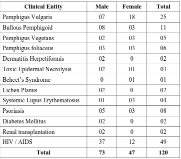
BREAK-UP DETAILS OF PATIENTS ARE AS GIVEN BELOW
[image:56.612.133.495.394.711.2]Break-Up details of patients Diagnosis-Wise and Gender-Wise Screened for Candidiasis
Table - 1
Clinical Entity Male Female Total
Pemphigus Vulgaris 07 18 25
Bullous Pemphigoid 08 03 11
Pemphigus Vegetans 02 03 05
Pemphigus foliaceus 03 03 06
Dermatitis Herpetiformis 02 0 02
Toxic Epidermal Necrolysis 02 01 03
Behcet’s Syndrome 0 01 01
Lichen Planus 02 0 02
Systemic Lupus Erythematosus 01 03 04
Psoriasis 05 03 08
Diabetes Mellitus 02 0 02
Renal transplantation 02 0 02
HIV / AIDS 37 12 49
Detailed case history of each patient was collected with reference
to the duration of primary disease for which the immunosuppressive
drug was given; appropriate note was also made regarding the dose and
duration of each drug in their immunosuppressive regime.
Site, symptomatology and the duration of candidial infection that
appeared after immunosuppressive therapy were recorded.
Detailed examination was also done to note down the other
dermatological lesions.
MYCOLOGICAL EXAMINATION
Specimens such as mucosal scrapings were collected under
aseptic precautions and examined microscopically in 10% potassium
hydroxide (KOH) solution for the presence of fungal elements.
Whenever the scraping was positive for budding yeast cells,
hyphae or pseudohyphae, inoculation was done on Sabouraud’s
Dextrose Agar (SDA) (Media of pH 6.5) with chloramphenicol
(0.05mg/ml). Duplicate slants were maintained for all specimen.
All the inoculated slants were duly numbered and incubated at
37oC for a period of 24-48 hours, with everyday observation. Candida
colonies appeared as white or cream coloured, smooth with a yeasty
SABOURAUD’S DEXTROSE AGAR
MEDIA PREPARATION
Ingredients :
Dextrose - 2g
Peptone - 1g
Agar - 2g
Distilled water - 100 ml
pH - 6.5
The ingredients are weighed accurately and then dissolved in
100ml of distilled water. Its then warmed gently till the boiling point of
water so that agar dissolves completely and the solution becomes
homogenous. When the media has become transparent warming up is
stopped.
The media is sterilized by autoclaving at 15 lbs pressure (121oC)
for 15 mts.
Antibiotic chloramphenicol is added (0.05mg/ml); sterilized
media is then allowed to cool to 50oC and then poured into sterile test
tubes and then kept in appropriate inclination for the bud and slant
formation. After a time lag of 4-6 hours the media is ready for
SUB-CULTURE MEDIA → INDICATOR MEDIA
Hicrome Candida Agar
Hicrome candida agar is recommended for rapid isolation and
identification of candida species from mixed cultures.
Composition
Ingredients Grams / Litre
Peptic Digest of animal tissue 15.0
Agar 15.0
Chloramphenicol 0.5
Chromogenic mixture 11.22
Dipotassium hydrogen phosphate 1.0
Final pH (at 25oC) 6.3 ± 0.2
Constitution of Media
21.36 gms of Hicrome candida agar is dissolved in 500 ml of
distilled water. It is allowed to boil to dissolve the medium completely.
Media should not be autoclaved. Media is then cooled to 50oC and
aseptically poured into sterile petri plates.
Subculture
Subculture of candida from SDA in Hicrome candida agar is done
Principle and interpretation
Perry and Miller (65) reported that candida albicans produces an
enzyme β-N-acetylgalactosaminidase and according to Rousselle et al.,
(66) incorporation of chromogenic or fluorogenic hexosaminidase
substrates into the growth media helps in identification of candida
albicans isolates directly on primary isolation. Hicrome candida agar
media are selective and differential medium which facilitates rapid
isolation of yeasts from mixed cultures and allows differentiation on the
basis of colouration and colony morphology.
Peptic digest of animal tissue, yeast extract, malt extract and
glucose provides nitrogenous, carbonaceous, compounds and other
essential growth nutrients. Chloramphenicol suppresses bacteria.
Cultural response
Organisms Colour
C.albicans - Light green
C.tropicalis - Light blue
C.krusei - White fuzzy
C.glabrata - Light pink