0022-538X/95/$04.0010
Copyrightq1995, American Society for Microbiology
A Human Recombinant Fab Identifies a Human Immunodeficiency
Virus Type 1-Induced Conformational Change in
Cell Surface-Expressed CD4
ROBIN E. BACHELDER,* JOANNA BILANCIERI, WENYU LIN,
ANDNORMAN L. LETVIN
Harvard Medical School, Beth Israel Hospital, Boston, Massachusetts 02215
Received 17 March 1995/Accepted 31 May 1995
To explore the role of the CD4 molecule in human immunodeficiency virus (HIV) infection following initial
virus-CD4 binding, we have characterized CD4-specific antibodies raised by immunizing an HIV-1-infected
human with human recombinant soluble CD4 (rsCD4). Fabs were selected from a human recombinant Fab
library constructed from the bone marrow of this immunized individual. Here, we describe a human
rsCD4-specific recombinant Fab clone selected by panning the library over complexes of human rsCD4 and
recom-binant HIV-1 envelope protein. While this Fab does not bind to CD4-positive T-cell lines or to human T
lymphocytes, it recognizes cell surface-expressed CD4 following the incubation of these cells with a
recombi-nant form of HIV-1 gp120 or with HIV-1 virions. The Fab is not HIV-1 envelope specific, since it does not bind
to recombinant gp120 or to native cell surface-expressed HIV-1 envelope proteins. As confirmation of its CD4
specificity, we show that this Fab immunoprecipitates a 55-kDa protein, corresponding to the molecular mass
of cellular CD4, from an H9 cell lysate. The specificity of this human Fab provides evidence for a virus-induced
conformational change in cell surface-expressed CD4. The characterization of this altered CD4 conformation
and its effects on the host cell will be important in defining postbinding events in HIV infection.
In serving as the human immunodeficiency virus (HIV)
re-ceptor, CD4 plays an essential role in HIV infection of
lym-phocytes and macrophages. The CD4 molecule is a member of
the immunoglobulin (Ig) superfamily, consisting of four
extra-cellular domains, a membrane-spanning region, and a
cytoplas-mic tail (17). The first of the extracellular domains consists of
three segments, resembling the complementarity determining
regions (CDRs) of an Ig variable domain. Mapping studies
have identified the second of these Ig CDR-like domains
(CDR2) in domain 1 as the HIV-binding site (1). It has been
suggested that other regions of the CD4 molecule may also be
important in HIV infection. While HIV is able to infect cells
expressing hybrid CD4 molecules composed of CD4 domains 1
and 2 fused to CD8 hinge, transmembrane, and cytoplasmic
sequences, HIV infection of these cells is significantly delayed
(9). Cells expressing a CD4 molecule with a truncated
cyto-plasmic domain similarly demonstrate a delay in the
produc-tion of HIV virions (4).
Monoclonal antibodies have proven critical in clarifying the
role of CD4 in HIV infection. Murine monoclonal anti-CD4
antibodies were used to demonstrate that CD4 acts as the HIV
receptor and that the virus binds to domain 1 of CD4 (7, 14).
Moreover, CD4-specific antibodies generated by immunizing
mice with a recombinant soluble form of the human CD4
molecule (rsCD4) have been shown to inhibit HIV infection in
vitro without blocking virus binding to CD4 (3, 10, 11, 19). The
epitope specificities of these antibodies have been mapped to
domains 2, 3, and 4 of the CD4 molecule. The existence of such
antibodies provides further evidence that CD4 may be involved
in critical postbinding steps of HIV infection.
The CD4-specific antibodies used in these studies were
gen-erated by immunizing mice with human CD4. It has been
assumed that the immunogenic determinants of human CD4
in these mice were defined by sequence differences between
human and murine CD4. However, we have previously
dem-onstrated that CD4-specific antibodies which inhibit simian
immunodeficiency virus infection of lymphocytes and
macro-phages in vitro can be elicited by human rsCD4 immunization
of rhesus monkeys (25). The elicitation of these antibodies was
somewhat surprising, since the human and rhesus CD4
mole-cules share 92% sequence homology (25). We have further
demonstrated that simian immunodeficiency
virus-neutraliz-ing, CD4-specific antibodies can be generated by immunizing
rhesus monkeys with rhesus rsCD4 (24). The elicitation of
these antibodies by immunization with a self protein
demon-strates that epitopes to which these monkeys were not tolerant
were presented to the immune system by rhesus monkey
rsCD4.
We were interested in determining whether CD4-specific
antibodies could be raised by immunizing humans with human
rsCD4. Here, we describe the characterization of a human
specific Fab clone generated from a human
rsCD4-immunized, HIV-1-infected human. This clone was selected
from a recombinant Fab library constructed from the bone
marrow of this immunized individual. The binding
character-istics of this recombinant Fab provide definitive evidence for
an HIV-1-induced conformational change in cell
surface-ex-pressed CD4.
MATERIALS AND METHODS
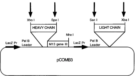
Vector.The pCOMB3 vector (Fig. 1) was provided by the Scripps Research Institute, La Jolla, Calif.
Recombinant HIV-1 envelope protein.HIV-1SF2recombinant gp120 (rgp 120)
was provided by Chiron Corp., Emeryville, Calif.
Monoclonal antibodies.The monoclonal antibodies used in these studies in-cluded 5A8 and humanized 5A8 (Hu5A8), which recognize domain 2 of CD4 (Biogen, Inc., Cambridge, Mass.); OKT4, specific for CD4 domain 3 (American Type Culture Collection, Rockville, Md.); 5D4 (10), specific for an epitope in the carboxy-terminal half (domain 3/domain 4) of the CD4 molecule; M-T807, a humanized antibody specific for CD8 (Centocor, Inc., Malvern, Pa.); and L736523, a human monoclonal antibody specific for the V3 loop of HIV-1MN
gp120 (Merck Research Laboratories, West Point, Pa.).
* Corresponding author. Mailing address: Harvard Medical School, Beth Israel Hospital, 330 Brookline Ave., Boston, MA 02215. Phone: (617) 667-4746. Fax: (617) 667-8210.
5734
on November 9, 2019 by guest
http://jvi.asm.org/
Cells.H9 and COS cells were obtained from the National Institutes of Health AIDS Research and Reference Reagent Program, Rockville, Md. Human pe-ripheral blood lymphocytes (PBL) were isolated from heparinized human blood by Ficoll-diatrizoate density gradient centrifugation. Chronically infected H9 cells were produced by infecting 53106
H9 cells with cell-free supernatants containing 32 infectious doses of HIVIIIB(1 infectious dose is defined as the
minimum dilution of this stock virus-containing supernatant that infects H9 cells under these culture conditions). Staining experiments were performed after these cells had been maintained in culture for 3 weeks, at which time no surface CD4 could be detected by flow cytometry.
Viruses.The production of recombinant HIV-1 virions capable of expressing chloramphenicol acetyltransferase has been described previously (13). Briefly, COS cells were cotransfected with plasmid pHXBDenvCAT, which encodes an HIV-1 genome with deletions in the envelope protein and an inserted chloram-phenicol acetyltransferase gene, and with plasmid pSVIIIenvMN, which encodes
a full-length HIV-1MNenvelope protein. After 3 days, recombinant virions were
collected in the supernatant of these transfected cells. These virus preparations were used for the virus-binding studies (see Fig. 5).
Human rsCD4 immunization of an HIV-1-infected individual.An HIV-1-infected human with an absolute CD4 count greater than 500/ml was immunized and given intramuscular booster doses five times with 1 mg of human rsCD4 (Biogen, Inc.) formulated in an emulsion of 85% mineral oil (Drakeol-6VR; Penreco, Butler, Pa.) and 15% Arlacel A (Montamide-80; Seppic, S.A., Fairfield, N.J.).
Determination of rsCD4-specific antibody titers in the plasma of a human rsCD4-immunized, HIV-1-infected individual. Human rsCD4 was adsorbed overnight at 48C onto Nunc Maxisorp Immunoplates at a final concentration of 0.3mg/ml. The wells were washed three times with phosphate-buffered saline (PBS) and blocked for 2 h at room temperature with 0.5% nonfat dry milk–PBS. The wells were then washed three times with 0.5% nonfat dry milk–0.05% Tween 20–PBS. A 1-ml sample of patient plasma was heat inactivated at 568C for 30 min and diluted 1:60, 1:180, 1:540, and 1:1,620 with 0.5% nonfat dry milk–0.05% Tween 20–PBS. A 50-ml sample of diluted plasma was then added to the appro-priate wells and incubated for 2 h at room temperature. The wells were washed three times with 0.5% nonfat dry milk–0.05% Tween 20–PBS, and 50ml of 1:50,000-diluted horseradish peroxidase-conjugated F(ab9)2 goat anti-human
IgG was added. After 1 h at room temperature, the wells were washed three times with 0.5% nonfat dry milk–0.05% Tween 20–PBS. Color was developed by adding 100ml of TMB one-component substrate solution (KPL, Gaithersburg, Md.). Color development was stopped by adding 100ml of 1 N H2SO4. Plates
were read on a Dynatech plate reader at an optical density of 450 nm (OD450).
Harvesting and storage of bone marrow samples.Heparinized bone marrow cells (20 ml) were harvested from a human rsCD4-immunized, HIV-1-infected individual 200 days after the initial immunization, at the time that the peak rsCD4-specific antibody titer was measured in the serum of this patient. Lym-phocytes were isolated from these samples by Ficoll-diatrizoate density gradient centrifugation, immediately frozen on dry ice, and stored at2708C prior to RNA extraction.
Amplification of Ig DNA.mRNA was isolated from 23107
bone marrow lymphocytes with the Quickprep Micro-mRNA Purification Kit (Pharmacia). Ig heavy- and light-chain cDNA was then synthesized with primers specific for the first constant domain of human Ig heavy chains, kappa light chains, and lambda light chains (Fig. 2). Specifically, 400 ng of the appropriate constant region primer was added to 30ml of isolated mRNA. This mixture was heated to 658C
for 5 min and cooled slowly to room temperature in a water bath. Reverse transcription was initiated by adding reverse transcriptase buffer (Gibco-BRL), 80 U of rRNasin (Promega), 0.8 mM deoxynucleoside triphosphates (dNTPs; Promega), 200 U of Moloney murine leukemia virus reverse transcriptase (Gibco-BRL), and 16.7 mM dithiothreitol. After this reaction had proceeded for 2 h at 378C, the reverse transcriptase was inactivated by heating the reaction mixture to 658C for 20 min. The resulting cDNA was stored at2208C prior to PCR amplification.
Two rounds of PCR amplification were used to obtain sufficient quantities of Ig heavy-chain material for cloning. The first-round PCR mixture contained 20 ml of heavy-chain-specific cDNA, 2.5 U of Pfu polymerase (Stratagene), Pfu buffer II, 0.2 mM dNTPs (Promega), 10 ng of heavy-chain constant-region primer (Operon), 10 ng of one of six variable-region primers corresponding to six heavy-chain families (VHA through VHF) (Operon) (Fig. 2), and RNase-free water to make a final volume of 100ml. Twenty-five amplification cycles, followed by a 10-min final extension at 728C, were performed by the hot-start technique under the following conditions: 948C for 1.5 min, 528C for 2.5 min, and 728C for 3 min. A 1-ml sample of the first-round PCR product was then exposed to 25 additional cycles of amplification under the same conditions.
Ig light-chain DNA was amplified under identical conditions, with primers corresponding to the first constant domain of kappa or lambda light chains, as well as one of four variable-region primers specific for different kappa (VK1 and VK2) and lambda (VL1 and VL2) light-chain families (Fig. 2).
Cloning of Ig heavy- and light-chain DNAs into the M13 phagemid vector, pCOMB3.PCR-amplified heavy- and light-chain DNAs were gel purified. Am-plified DNA from each light-chain family was then pooled, digested with the enzymes XbaI and SacI (Boehringer Mannheim), and ligated into the pCOMB3 vector.
Electrocompetent XL1-blue cells (Stratagene) were transformed with these recombinant plasmids by electroporation. Specifically, phenol-chloroform-ex-tracted, ethanol-precipitated ligation products were added to 300ml of electro-competent XL1-blue cells in prechilled 0.2-cm gene pulser cuvettes (Bio-Rad). These cells were pulsed in a Bio-Rad gene pulser apparatus set at 25mF, 2.5 kV, and 200V. SOC (0.5% yeast extract, 2% tryptone, 10 mM NaCl, 2.5 mM KCl, 10 mM MgCl2, 10 mM MgSO4, 20 mM glucose) (3 ml) was then added, and the cells
were grown at 378C for 1 h in a shaking incubator. Transformed cells were selected for 1 h at 378C in 10 ml of superbroth containing 20mg of carbenicillin per ml and 10mg of tetracycline per ml. Finally, transformed cells were amplified overnight in a 378C shaker in 100 ml of superbroth containing 50mg of carben-icillin per ml and 10mg of tetracycline per ml.
Recombinant plasmid DNA was then digested with the restriction enzymes
XhoI and SpeI (Boehringer Mannheim), gel purified, and ligated with pooled XhoI-SpeI-digested, gel-purified heavy-chain PCR products. The combinatorial
ligation products were then electroporated into XL1-blue cells, as described above. A restriction analysis of the resulting clones was performed to ensure that this library consisted of clones with both light- and heavy-chain inserts.
Conversion of the combinatorial library to an Fab-expressing M13 bacterio-phage format.The initial combinatorial library was amplified at 378C in a shaking incubator for 1 h in 100 ml of superbroth containing 50mg of carbenicillin per ml and 10mg of tetracycline per ml. The M13 helper phage VCSM13 (Stratagene) (1012PFU) was then added to direct the assembly of M13 combinatorial
bacte-riophage. After 2 h of growth at 378C, 70mg of kanamycin per ml was added, and helper phage-infected cells were grown overnight at 378C.
Precipitation of Fab-expressing M13 bacteriophage.Recombinant M13 bac-teriophage in the supernatant of helper phage infected cultures were precipitated on ice for 1 h in the presence of 4% polyethylene glycol 8000 and 3% NaCl. Precipitated phage were pelleted at 9,000 rpm for 20 min at 48C in a JA10 rotor. Finally, the phage pellet was resuspended in 2 ml of PBS and stored at2208C.
Determination of the titer of M13 combinatorial bacteriophage.Various di-lutions of bacteriophage were incubated for 15 min at room temperature with 50 ml of an XL1-blue culture grown to an OD600/ml of 1.0. The infected cells were
then plated onto Luria broth–50mg of carbenicillin per ml agar plates.
Panning.Four wells of a Maxisorp 96 well plate (Nunc) were coated overnight at 48C with 2mg of the CD4-specific monoclonal antibody 5D4 in 100ml of PBS per well. Unbound antibody was removed by washing three times with 350ml of Tris-buffered saline (TBS). The wells were then blocked with 350ml of 2% nonfat dry milk–TBS for 30 min at room temperature. After shaking out the block solution, the wells were incubated with preformed rsCD4-gp120 complexes for 2 h at room temperature. The complexes were formed by incubating rgp120 and human rsCD4 in a 1.4:1 molar ratio at 378C for 1.5 h. Unbound proteins were removed by performing three washes with 350ml of sterile double-distilled water. The wells were again blocked at 378C for 1 h with 350ml of 3% bovine serum albumin (BSA) PBS. After shaking out the block solution, 100 ml of M13 combinatorial phage (1012
[image:2.612.60.296.70.204.2]PFU) was added to each well and the mixtures were incubated for 2 h at 378C. Nonadherent phage were removed, and each well was washed once with sterile double-distilled water. Each well was then washed 10 times over a period of 1 h at room temperature with 350ml of TBS–0.5% Tween 20. Detergent was removed by washing once with sterile double-distilled water, and phage were eluted by adding 100ml of phage elution buffer (0.1 M HCl, 1 mg of BSA [pH 2.2] per ml; adjusted to pH 2.2 with glycine). The elution was allowed to proceed for 10 min at room temperature. The solution was then pipetted up and down several times, transferred to a sterile tube, and neutralized with 6ml FIG. 1. Restriction site map of the phagemid pCOMB3 used for constructing
a combinatorial Ig library from a human rsCD4-immunized, HIV-1-infected individual. Arrows indicate the restriction sites used for cloning. Fab expression is induced through the lacZ promoter with IPTG. The pelB leader sequence directs heavy and light chains to the periplasmic space of induced bacterial cells, where Fab assembly occurs. The heavy chain is expressed as a fusion protein with the M13 coat protein encoded by gene III, which allows for the incorporation of recombinant Fab molecules on the virion surface.
VOL. 69, 1995 HIV-INDUCED CONFORMATIONAL CHANGE IN CELL SURFACE CD4 5735
on November 9, 2019 by guest
http://jvi.asm.org/
of 2 M Tris base per 100ml of eluted phage. The titer of the panned phage was determined, and the phage were stored at2208C.
Amplification of panned bacteriophage.Eluted phage were incubated with 2 ml of an XL1-Blue culture (OD600/ml, 1.0) for 15 min at room temperature. The
cells were then grown in a 378C shaking incubator for 1 h in 10 ml of superbroth containing 20mg of carbenicillin per ml and 10mg of tetracycline per ml. These cells were then grown for an additional 1 h in 100 ml of superbroth plus 50mg of carbenicillin per ml and 10mg of tetracycline per ml. At this time, the cells were infected with 1012PFU of the M13 helper phage VCSM13 (Stratagene) to
direct the assembly of Fab-expressing M13 bacteriophage. Finally, cells which had been infected with the helper phage were selected by growing the culture overnight in the presence of 70mg of kanamycin per ml. These amplification and panning procedures were repeated until enrichment for antigen-specific clones was achieved, as determined by the percent yield of phage (the number of phage eluted divided by the number of phage applied, multiplied by 100) after each amplification and panning cycle.
Conversion of clones to a soluble Fab expression format.To obtain soluble Fab, it was necessary to remove M13 gene III, the gene encoding the coat protein responsible for anchoring Fab molecules on the phage surface, from the combi-natorial plasmids. Plasmid DNA was isolated from panned clones, digested with
SpeI and NheI to remove gene III, and gel purified. Since the SpeI and NheI
restriction sites are compatible, recircularized plasmid was made by religating this digested vector. The ligation product was transformed into XL1-Blue cells in preparation for the induction of Fab expression.
Induction of soluble Fab expression.Combinatorial clones were inoculated into 500 ml of superbroth containing 50mg of carbenicillin per ml and 20 mM MgCl2and grown at 378C in a shaking incubator to an OD600/ml of 1.0. Soluble
Fab expression was then induced by growing these cultures at 308C overnight in the presence of 1 mM isopropyl-b-D-thiogalactopyranoside (IPTG; Stratagene) and 4 nM dibutyryl cyclic AMP (Sigma).
Isolation of soluble Fab from induced bacterial cultures.Since the pelB leader sequence of the pCOMB3 vector directs Fab molecules to the periplasm of induced bacterial cells, an osmotic shock procedure was performed to obtain a
periplasmic extract from these cells. Specifically, induced bacterial cells were pelleted at 4000 rpm in an HS-4 rotor for 30 min at 48C. The supernatant was discarded, and the induced bacterial cells were resuspended on ice in 20 ml of osmotic shock solution A (100 mM Tris-HCl [pH 8.6], 500 mM sucrose, 0.5 mM EDTA). The cell wall was lysed by adding 1 ml of 4-mg/ml lysozyme immediately followed by 80 ml of osmotic shock solution B (50 mM Tris-HCl [pH 8.6], 250 mM sucrose, 0.25 mM EDTA, 2.5 mM MgCl2). After a 10-min incubation on ice,
bacterial debris was pelleted by centrifuging the lysate in Oakridge 35-ml tubes at 12,500 rpm in an SS-34 rotor for 5 min at 48C. The supernatant was transferred to a new Oakridge tube, and the protease inhibitor aminoethylbenzene-sulfo-nylfluoride (AEBSF; Calbiochem) was added to a final concentration of 1 mM. Remaining bacterial debris was removed by centrifuging the extract again at 12,500 rpm for 15 min at 48C. Finally, the supernatant was filtered through a 0.2-mm-pore-size bottle filter system (Costar).
Purification of soluble Fab.To obtain a pure source of Fab for characteriza-tion, the periplasmic extracts of induced cells were applied to goat anti-human F(ab9)2affinity columns. The affinity columns were prepared by incubating 4 mg
of goat anti-human F(ab9)2(Jackson Immunoresearch) with 2 ml of Gammabind
G Sepharose beads (Pharmacia) for 1 h at room temperature on a rocking platform. After the conjugated beads were washed four times with 10 ml of 0.2 M sodium borate (pH 9.0), they were incubated on a rocker for 1.5 h with the coupling reagent dimethyl pimelimidatez2HCl (DMP; Pierce) at a concentra-tion of 20 mM in 0.2 M sodium borate (pH 9.0). The coupling reacconcentra-tion was terminated by washing once with 0.2 M ethanolamine (pH 8.0) and incubating the beads in ethanolamine for 2 h at room temperature on a rocker. The beads were washed three times in 0.2 M sodium borate (pH 9.0), resuspended in 10 ml of PBS–0.05% sodium azide, and stored at 48C.
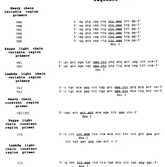
[image:3.612.144.467.77.404.2]The coupled beads were loaded onto Econo-Pac columns (Bio-Rad), and sodium azide was removed by washing the column several times with PBS. The column was then equilibrated with 10 column volumes of elution buffer (3.5 M sodium thiocyanate) to remove uncoupled IgG. The columns were washed sev-eral times with PBS, and periplasmic extract isolated from a 500-ml induced culture was applied to the column at 48C. Nonspecifically bound proteins were FIG. 2. Sequences of the primers used for amplification of immunoglobulin heavy- and light-chain DNA. Restriction sites incorporated into primers for cloning purposes are underlined. These primers map to the following regions of the indicated human Ig chains: VHA to VHF, heavy-chain amino acids 1 to 8; VK1 and VK2, kappa light-chain signal sequence24 to amino acid 8; VL1 and VL2, lambda light-chain signal sequence27 to amino acid 8; CH1, heavy-chain amino acids 226 to 232; CLK, kappa light-chain amino acids 201 to 214; CLL, lambda light-chain amino acids 210 to 215.
on November 9, 2019 by guest
http://jvi.asm.org/
washed off with 200 ml of PBS–1 mM AEBSF. Finally, bound Fab was eluted with 8 column volumes of 3.5 M NaSCN and desalted with Centriprep 30 ultrafiltration devices (Amicon).
Determination of soluble Fab concentration.Wells of a 96-well Maxisorp plate were coated overnight at 48C with 1mg of F(ab9)2goat anti-human F(ab9)2
(Jackson Immunoresearch). After eight washes with TBS, the wells were blocked as described above. Various dilutions of each Fab preparation, as well as known concentrations of a purified human Fab standard (Biodesign International), were then loaded. Bound Fab was detected as described above.
Screening soluble Fabs for antigen specificity.Wells of a 96-well Maxisorp plate were coated overnight at 48C with 1mg of the antigen of interest in a 150-ml volume. After being washed eight times with TBS, the wells were blocked for 1 h at 378C with 0.5% nonfat dry milk–0.5% Tween 20–TBS. Blocking solution was then removed, and the Fabs were incubated in the appropriate wells for 1 h at 378C. Unbound Fab was removed by eight washes with TBS. Bound Fab was then detected by incubating the wells for 1 h at 378C with a 1:50,000 dilution of horseradish peroxidase-conjugated F(ab9)2goat anti-human F(ab9)2(Jackson
Immunoresearch). After eight TBS washes, TMB one-component substrate so-lution (KPL) was added. The reactions were terminated after 20 min by the addition of 2/3 N H2SO4, and the A450 was read on a Dynatech MR5000
enzyme-linked immunosorbent assay (ELISA) reader.
Cell staining.For the HIV-1 rgp120 pulsing experiments, cells were preincu-bated at 378C for 1 h with either PBS or 100mg of recombinant gp120 (rgp120; HIV-1SF2strain) per ml. After being washed with PBS, these cells were
incu-bated for 20 min on ice with the indicated Fabs or monoclonal antibodies at a concentration of 2mg/ml and washed again with PBS. The cells were then incubated at 48C for 20 min with a 1:100 dilution of either fluorescein isothio-cyanate (FITC)-conjugated F(ab9)2goat anti-human F(ab9)2(Jackson
Immu-noresearch) or FITC-conjugated F(ab9)2goat anti-mouse F(ab9)2(Jackson
Im-munoresearch). Nonspecifically bound antibodies were removed by washing the cells with PBS.
In the virus-binding experiments, H9 cells were incubated overnight at 378C with infectious recombinant HIV-1MN(20 ng of p24 per 1.5310
6
cells) in the presence of the indicated Fab preparations or monoclonal antibodies at a con-centration of 2mg/ml. After these cells were washed with PBS, bound antibodies or Fabs were detected by incubating the cells with FITC-labelled secondary antibodies, as described above.
The cross-competition studies were performed by incubating freshly isolated human PBL with medium alone or with HIV-1SF2rgp120, as described above.
These cells were then washed with PBS and incubated with PBS or Fab clone 3-47 at 10mg/ml for 20 min on ice. The cells were washed with PBS, and the samples were incubated with the indicated monoclonal antibodies at 10mg/ml on ice for 20 min. These cells were then washed with PBS. Bound antibodies were detected as described above.
For each of these experiments, stained cells were fixed in 1% formalin–PBS and analyzed on an EPICS XL-MCL flow cytometer (Coulter Corp.). As a negative control, an indirect stain substituting PBS for the primary antibody was performed.
Biotin labelling of H9 surface proteins.H9 cells (33107) were washed three
times with PBS-Plus (PBS, 0.1 mM CaCl2, 0.1 mM MgCl2) and resuspended in
3 ml of 1-mg/ml Sulfo-NHS-Biotin (Pierce)/PBS-Plus. After being incubated for 1 h at 48C with gentle agitation, the cells were washed once with RPMI 1640 and then three times with PBS-Plus.
Immunoprecipitation of biotinylated H9 proteins.The biotinylated H9 cell pellet was lysed in Triton X-100 lysis buffer (300 mM NaCl, 50 mM Tris-HCl [pH 7.6], 0.5% Triton X-100, 10mg of leupeptin per ml, 10mg of aprotinin per ml, 1 mM phenylmethylsulfonyl fluoride, 1.8 mg of iodoacetamide per ml) on ice for 45 min with occasional mixing. After cell debris was pelleted in a microcentrifuge for 15 min at 48C, the supernatant was transferred to a 1.5-ml Eppendorf tube. This lysate was then incubated for 1 h at 48C with 300ml of a 50% suspension of Gammabind G beads (Pharmacia)–PBS with gentle agitation. These samples were centrifuged in a microcentrifuge for 1 min. The precleared lysate was then incubated overnight with the indicated antibody or Fab preparation at 15mg/ml. These samples were incubated with 200ml of a 75% suspension of goat anti-human IgG, F(ab9)2-conjugated Gammabind G beads–PBS for 1 h at 48C with
shaking. The beads were then pelleted as described above, washed three times with high-salt wash buffer (0.5 M NaCl, 20 mM Tris-HCl, 1 mM EDTA, 1% sodium deoxycholate, 0.5% Nonidet P-40, 30% sucrose), and then washed twice with low-salt wash buffer (10 mM NaCl, 10 mM Tris-HCl [pH 7.6]).
Immunoprecipitated proteins were eluted from the beads in nonreducing sample buffer (60 mM Tris-HCl [pH 6.8], 25% glycerol, 2% sodium dodecyl sulfate [SDS], 0.1% bromophenol blue) at 958C for 10 min and loaded onto an SDS–10% polyacrylamide gel. After electrophoresis these proteins were trans-ferred overnight to nitrocellulose. The membrane was blocked with 5% BSA– TBST (100 mM Tris-HCl [pH 7.5], 0.9% NaCl, 0.1% Tween 20) and probed with 2mg of horseradish peroxidase-conjugated avidin (Pierce)–0.3% BSA–TBST per ml to detect biotinylated proteins. After extensive washing with TBST, biotiny-lated proteins were visualized by the enhanced chemiluminescence detection system (Amersham).
RESULTS
Generation of a human recombinant Fab library from the
bone marrow of a human rsCD4-immunized, HIV-1-infected
individual.
To characterize the antibodies elicited by human
rsCD4 immunization of an HIV-1-infected human, we
pro-duced a human combinatorial Fab library. Ig heavy- and
light-chain fragments, encompassing the entire variable domain and
first constant region of each chain, were amplified by PCR
from the bone marrow of this immunized individual and cloned
into the phagemid pCOMB3 (Fig. 1). This vector directs the
expression of Ig heavy- and light-chain fragments in the
periplasmic space of a bacterial cell, where these chains
asso-ciate to form recombinant Fabs. Since the heavy chain is
ex-pressed as a fusion protein with the M13 bacteriophage coat
protein encoded by gene III, infection of the transformed
bac-terial cells with an M13 helper phage results in the assembly of
bacteriophage particles that express recombinant Fab
mole-cules on their surface. Antigen-specific Fab clones can be
se-lected from this library by panning the recombinant
Fab-ex-pressing bacteriophage over the antigen of interest. The
combinatorial library constructed from the bone marrow of
this human rsCD4-immunized individual consisted of 3
3
10
6members.
Selection of human rsCD4-specific recombinant Fab clones
from the combinatorial Fab library.
To select for novel human
rsCD4-specific Fab clones, the library was panned against
com-plexes of human rsCD4 and HIV-1
SF2rgp120. After three
rounds of panning, we achieved a twofold enrichment for
hu-man rsCD4-specific Fab clones as determined by the percent
yield of phage. Thirty-five recombinant Fab clones from this
enriched library were selected for further analysis. Soluble
Fab-expressing clones were generated by removing the M13
bacteriophage gene III, which encodes the coat protein
re-sponsible for retention of the recombinant Fab on the phage
surface. Soluble Fab expression was then induced from
trans-formed bacterial cells by the addition of IPTG. Soluble Fab
was isolated from the induced bacterial cells by osmotic shock
and purified on goat anti-human IgG, F(ab
9
)
2affinity columns.
The purified Fabs were then tested for ELISA reactivity with
both human rsCD4 and HIV-1
SF2rgp120. Of the 35 panned
clones studied, 7 which bound to human rsCD4 but not to
HIV-1
SF2gp120 (selected data are given in Table 1) were
identified. Three of these human rsCD4-specific Fabs were
selected for further analysis.
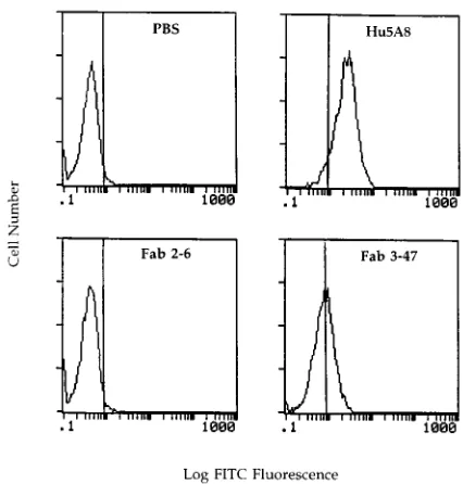
[image:4.612.314.555.91.147.2]Fab clone 3-47 binds to HIV-1 rgp120-preincubated cells but
not to untreated CD4-positive cells.
We next sought to
deter-mine whether we could identify human rsCD4-specific Fab
clones which recognized cell surface-expressed CD4.
Interest-ingly, none of the three human rsCD4-specific Fab clones
tested bound to native CD4 expressed on the surface of H9
cells (Fig. 3). In an attempt to identify a biologically relevant
form of CD4 which these Fab clones might recognize, we next
assessed whether any of these Fabs, selected by panning the
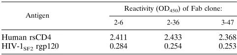
TABLE 1. ELISA reactivity of selected Fab clones with recombinant antigensa
Antigen
Reactivity (OD450) of Fab clone:
2-6 2-36 3-47
Human rsCD4 2.411 2.433 2.368
HIV-1SF2rgp120 0.284 0.254 0.253
a
Human rsCD4 or HIV-1SF2rgp120 (1mg) was adsorbed onto ELISA wells.
Affinity-purified Fab preparations were incubated in these wells and detected with a horseradish peroxidase-labelled secondary antibody.
VOL. 69, 1995 HIV-INDUCED CONFORMATIONAL CHANGE IN CELL SURFACE CD4 5737
on November 9, 2019 by guest
http://jvi.asm.org/
library over human rsCD4/HIV-1 envelope complexes,
recog-nized cell surface-expressed CD4 complexed with recombinant
HIV-1 envelope protein. As shown in Fig. 3, one of these
human rsCD4-specific Fab clones (Fab clone 3-47) bound to
H9 cells after these cells had been incubated for 1 h at 37
8
C
with HIV-1
SF2rgp120. In fact, Fab clone 3-47 exhibited the
same binding specificity on primary cells, recognizing freshly
isolated human PBL only after preincubation of these cells
with HIV-1
SF2rgp120 (Fig. 4).
Fab clone 3-47 binds to H9 cells preincubated with HIV-1
virions.
On the basis of these observed binding characteristics,
we postulated that Fab clone 3-47 recognized an epitope of
CD4 which is not exposed on the native cell surface-expressed
molecule but is revealed on the cell surface after HIV-1
enve-lope protein binding. While we had demonstrated that this
CD4 epitope was exposed upon binding of a recombinant form
of the HIV-1 envelope protein, it was important to show that
the exposure of the same epitope could be induced upon
bind-ing of HIV-1 particles to cell surface-expressed CD4. H9 cells
were incubated overnight at 37
8
C with HIV-1
MNvirions in the
presence or absence of purified Fabs. Bound Fabs were
de-tected by incubating these cells with a FITC-labelled goat
anti-human IgG, F(ab
9
)
2. As demonstrated in Fig. 5, Fab clone 3-47
specifically bound to the virus-preincubated cells.
Fab clone 3-47 does not recognize native HIV-1 envelope
proteins expressed on the surface of chronically infected H9
cells.
While the above observations suggested that Fab clone
3-47 recognized a conformationally altered CD4 molecule
in-duced upon HIV-1 binding, it was important to confirm that
this Fab, generated from an HIV-1-infected human, did not
react with the HIV-1 envelope protein. Although this Fab
clone did not recognize HIV-1
SF2rgp120, as determined by
ELISA, it was possible that the Fab recognized cell
surface-expressed HIV envelope proteins. The CD4 molecule is
down-modulated off the surface of cells productively infected with
HIV-1 (7). Therefore, we reasoned that Fab clone 3-47, if
specific for the CD4 molecule, should not bind to cells
chron-ically infected with HIV-1. By flow cytometry, we
demon-strated that Fab clone 3-47 does not recognize native HIV-1
envelope proteins expressed on the surface of chronically
in-fected H9 cells (data not shown).
Fab clone 3-47 immunoprecipitates cellular CD4.
Since Fab
clone 3-47 had been selected by panning the combinatorial Fab
library over human rsCD4/HIV-1
SF2rgp120 complexes, it was
possible that this Fab was specific for a neoepitope defined by
domains of both human CD4 and HIV-1 envelope protein. To
rule out this possibility, we attempted to identify a form of
cellular CD4 which is recognized by Fab clone 3-47.
[image:5.612.123.494.70.373.2]Since Fab clone 3-47 had been generated from an individual
immunized with rsCD4, which probably assumes a tertiary
structure different from that of membrane-expressed CD4,
we were interested in determining whether the epitope
recog-nized by Fab clone 3-47 was exposed on
detergent-solubi-lized cellular CD4. As shown in Fig. 6, Fab clone 3-47
specif-ically immunoprecipitated a 55-kDa protein, corresponding
to the molecular mass of cellular CD4, from H9 cells. Thus,
although Fab 3-47 bound to cell surface-expressed CD4 only
following HIV-1 envelope binding, this Fab did recognize
solubilized cellular CD4 in the absence of added HIV-1
gp120.
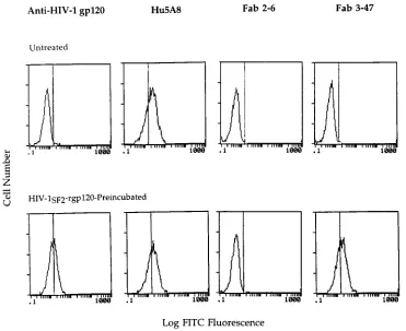
FIG. 3. Fab clone 3-47 reactivity with cell surface-expressed CD4 on untreated and HIV-1SF2rgp120-pretreated H9 cells. The H9 cells were incubated with medium
alone or with HIV-1SF2rgp120 and then with the indicated antibodies or Fab fragments. Bound antibodies were detected by incubating these cells with a
FITC-conjugated goat anti-human IgG, F(ab9)2. The cells were analyzed by flow cytometry. The data are also expressed as the percent HIV-1 rgp120-preincubated cells
specifically bound by antibody: Hu5A8, 65%; anti-HIV-1 gp120, 46%; Fab clone 3-47, 60%.
on November 9, 2019 by guest
http://jvi.asm.org/
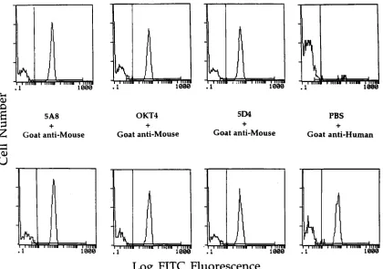
Competition studies do not identify the domain of CD4
recognized by Fab clone 3-47.
Having confirmed that Fab clone
3-47 was CD4 specific, we were interested in mapping the
location of this epitope within the four-domain structure of
CD4. Therefore, we performed competition studies between
Fab clone 3-47 and a panel of monoclonal antibodies specific
for different domains of CD4. Human PBL were pulsed with
HIV-1 recombinant envelope protein and incubated
sequen-tially with Fab clone 3-47 followed by a CD4-specific murine
monoclonal antibody. Fab clone 3-47 did not compete with
monoclonal antibodies specific for CD4 domains 2, 3, and 4 for
binding to HIV-1
SF2rgp120-preincubated human PBL (Fig. 7).
DISCUSSION
To access and characterize novel CD4-specific monoclonal
antibodies, we have constructed a recombinant Fab library
from the bone marrow of a human rsCD4-immunized,
HIV-1-infected individual. Although the production of hybridomas
has proven useful in the generation of murine monoclonal
antibodies, this technology has not been perfected for the
synthesis of human monoclonal antibodies. The construction
of combinatorial Fab libraries is an important alternative to the
generation of hybridomas, providing a powerful tool for the
selection of numerous antibody clones with high affinity for an
antigen of interest. This approach has proven valuable in the
characterization of antibody responses to various antigens (2,
22, 26).
The pCOMB3 phagemid has become the favored cloning
vehicle for the production of combinatorial Fab libraries. This
vector directs the periplasmic expression of an Ig light-chain
fragment and a protein consisting of Ig heavy-chain sequences
fused to the membrane-spanning region of a bacteriophage
coat protein. This fusion protein allows Fab molecules to be
incorporated onto the surface of M13 bacteriophage particles
[image:6.612.328.541.453.676.2]which are produced after these transformed cells are infected
with an M13 helper phage. One can select for a subpopulation
of clones with high affinity for an antigen of interest from such
libraries by performing repeated rounds of bacteriophage
pan-ning over this antigen.
FIG. 4. Reactivity of Fab clone 3-47 with cell surface-expressed CD4 on untreated and HIV-1SF2rgp120-pretreated human PBL. Cells were incubated with either
medium alone or HIV-1SF2rgp120, followed by the indicated primary antibodies or Fab preparations. Bound antibodies were detected with a FITC-labelled secondary
antibody. The cells were analyzed by flow cytometry. The data are also expressed as the percent HIV-1 rgp120-preincubated cells specifically bound by antibody: Hu5A8, 58%; anti-HIV-1 gp120, 49%; Fab 3-47, 54%.
FIG. 5. Fab clone 3-47 reactivity with H9 cells preincubated with HIV-1 virions. H9 cells were incubated with live HIV-1MN. The cells were then washed
with PBS and incubated with the indicated monoclonal antibodies or Fab clones. Bound antibodies were detected with a FITC-labelled secondary antibody, and the cells were analyzed by flow cytometry. The data are also expressed as the percent virus-preincubated cells bound by antibody: Hu5A8, 86%; Fab 3-47, 52%.
VOL. 69, 1995 HIV-INDUCED CONFORMATIONAL CHANGE IN CELL SURFACE CD4 5739
on November 9, 2019 by guest
http://jvi.asm.org/
In addition to allowing for the rapid generation of numerous
antigen-specific Fab clones, the production of large Fab
librar-ies in the pCOMB3 vector increases the probability of
ac-cessing antibodies represented at a low frequency in the Ig
repertoire of an individual. Although human rsCD4-specific
antibodies were elicited in this study by immunization of a
human with a recombinant self protein, the titer of these
an-tibodies was lower than might be expected following
immuni-zation with a foreign antigen. Because of the low frequency of
B cells secreting CD4-specific antibodies in this immunized
individual, a monoclonal antibody with the specificity of Fab
clone 3-47 may never have been identified by B-cell
immortal-ization and/or fusion techniques. We were interested in
deter-mining whether antibodies specific for a conformationally
al-tered form of the CD4 molecule, induced upon HIV binding,
were represented in the Ig repertoire of this human
rsCD4-immunized individual. To optimize the likelihood of accessing
Fab fragments which demonstrate a greater affinity for CD4
complexed with the HIV envelope protein than for CD4 alone,
we panned this recombinant Fab library over complexes of the
HIV-1 envelope protein and human rsCD4. The generation of
Fab clone 3-47 from this panned library demonstrates the
power of this cloning technology in gaining access to
high-affinity, low-frequency antibodies.
[image:7.612.118.236.70.227.2]Ig heavy- and light-chain fragments combine randomly in
the pCOMB3 vector. Therefore, one potential drawback of
using the recombinant Fab cloning technology to characterize
the antibody specificities existing in an individual is that Fab
specificities which do not exist in the immunized subject may
be represented in the library. Thus, we cannot be certain that
the CD4-specific Fab clone 3-47 is truly representative of the
antibody specificities present in the Ig repertoire of this human
rsCD4-immunized individual. However, others have reported
that Fabs with high affinity for a given antigen can be accessed
FIG. 6. Reactivity of Fab clone 3-47 with solubilized cellular CD4. H9 cells were labelled with Sulfo-NHS biotin. These cells were then lysed in Triton X-100 lysis buffer. Cellular proteins were immunoprecipitated with the indicated anti-body or Fab clone. The humanized CD8-specific antianti-body M-T807 was used as a negative control antibody (Neg. Control Ab) in these experiments. Immunopre-cipitated proteins were transferred to nitrocellulose and probed with horseradish peroxidase-conjugated avidin. Biotinylated proteins were then detected by chemiluminescence.
FIG. 7. Cross competition between Fab clone 3-47 and a panel of CD4-specific monoclonal antibodies for binding to HIV-1SF2rgp120-preincubated human PBL.
Freshly isolated human PBL were incubated with HIV-1 rgp120SF2. These cells were then incubated with PBS (top four panels) or Fab clone 3-47 (bottom four panels).
Finally, the indicated monoclonal antibodies were incubated with these cells. Bound antibodies were detected with the indicated FITC-conjugated secondary antibodies. These samples were analyzed by flow cytometry.
on November 9, 2019 by guest
http://jvi.asm.org/
[image:7.612.100.520.391.686.2]from combinatorial libraries only if a measurable antibody
response against this antigen is elicited in the immunized
in-dividual (18, 20). These studies argue against the probability of
accessing Fab clones consisting of artifactual heavy- and
light-chain combinations. Moreover, Fab clones with binding
spec-ificities similar to that of clone 3-47 were represented at a high
frequency in the panned library generated from this individual
(unpublished results). These observations suggest that B cells
secreting an antibody with the same specificity as Fab clone
3-47 were probably present in this immunized individual at the
time of library construction.
We believe that this antibody specificity was raised upon
human rsCD4 immunization of this HIV-1-infected individual.
Although CD4-specific autoantibodies have been reported to
exist in the serum of approximately 10% of AIDS patients (15),
the serum antibodies of this human rsCD4-immunized
individ-ual demonstrated reactivity with human rsCD4 only after
im-munization (unpublished results). This observation suggests
that Fab clone 3-47 is not representative of an autoantibody
produced by this patient prior to rsCD4 immunization. The
elicitation of this antibody specificity by immunization with a
recombinant soluble form of a self protein suggests that the
rsCD4 molecule assumes a tertiary structure different from
that of cell surface-expressed CD4.
The binding specificity of Fab clone 3-47 provides
compel-ling evidence that HIV-1 induces a conformational change in
cell surface-expressed CD4. This conformational change is not
induced upon the binding of other CD4 domain one ligands,
since antibodies specific for this domain do not induce the
exposure of the Fab 3-47 epitope (data not shown). Previous
studies have raised the possibility that a conformational change
in the HIV receptor occurs following virus binding. Healey et
al. (11) described an antibody raised in a mouse by human
rsCD4 immunization that exhibited a greater affinity for
hu-man rsCD4–HIV-1 rgp120 complexes than for huhu-man rsCD4
alone. Monoclonal antibodies with a similar binding specificity
have also been generated by immunizing mice with human
rsCD4–HIV-1 rgp120 complexes (6, 8). While these studies
demonstrated that recombinant gp120 binding to recombinant
CD4 can induce rsCD4 conformational alterations, they did
not address whether such changes occur in cell
surface-ex-pressed CD4 following HIV binding.
On the basis of our knowledge of cell surface receptors
which undergo conformational changes upon ligand binding, it
is interesting to speculate on the physical nature of the CD4
conformational change defined by Fab clone 3-47.
Ligand-induced alterations in the oligomeric state of their respective
receptors have been described for numerous receptors,
includ-ing the platelet-derived growth factor receptor (12). Such
changes are often accompanied by conformational alterations
in these receptors. Although X-ray crystallography studies of
rsCD4 have not indicated that this molecule tends to
oligomer-ize (5, 16, 23), proteins which bind to cell surface-expressed
CD4, such as the tyrosine kinase lck, may facilitate the
forma-tion of CD4 multimers. Direct evidence for the existence of
CD4 oligomers was recently provided by the observation that
the interaction of major histocompatibility complex class
II-expressing cells with CD4 is dependent on the expression of
CD4 oligomers on the cell membrane (21a). It is possible that
the binding of HIV to CD4-positive cells alters the oligomeric
state of CD4 on the cell membrane. The conformationally
altered form of cell surface-expressed CD4 which is recognized
by Fab clone 3-47 may represent an intermediate or final
con-figuration of CD4 as it undergoes such alterations in its
oligo-meric state. Alternatively, this HIV-induced CD4
conforma-tional change may reflect changes in the interaction of CD4
with other cell surface proteins, such as the T-cell receptor.
Numerous growth factor-induced conformational changes in
receptor molecules have been shown to result in the activation
of signal transduction cascades. Since the CD4 molecule is
known to play a critical role in T-cell receptor-mediated signal
transduction and cellular activation (21), it is possible that this
HIV-induced conformational change alters the activation state
of a cell during viral entry. In fact, it has been demonstrated
that HIV binding to cell surface-expressed CD4 results in the
translocation of NF-
k
B to the nucleus of a cell (4). This
cel-lular activation was shown to be a direct consequence of virus
binding to CD4 and was not dependent on virus replication. It
is possible that the virus-induced CD4 conformational change
described in the present article plays a role in such signalling
through the CD4 molecule.
These studies indicate that the CD4 receptor undergoes a
conformational change upon HIV-1 binding. The elucidation
of the consequences of this conformational change on the host
cell and its importance in postbinding steps of HIV infection
may provide novel targets for the design of antiviral
therapeu-tic agents.
ACKNOWLEDGMENTS
We thank B. Paul and other members of the nursing staff of the Dana-Farber Cancer Institute for assistance in the care of the rsCD4-immunized, HIV-1-infected subject who took part in this study; J. Ritz for performing the bone marrow aspirate on the immunized subject; L. Fichter and S. Tadesse for technical assistance in purifying Fabs for this study; the Scripps Research Institute for providing the pCOMB3 vector; Biogen, Inc., for providing the human rsCD4; Chiron for pro-viding HIV-1SF2rgp120; V. Sato for helpful conversations; H. Saito for technical advice on Fab library construction; J. Lambert, A. Profy, M. R. van Schravendijk, and S. Khandekar for valuable advice on Fab purification; K. Reimann for help in the analysis and presentation of the flow cytometry data; and A. Williamson and D. Burton for gener-ous advice on the construction of this Fab library.
This work was supported by NIH grants AI-20729 and CA-50139, CFAR grant AI-93-14, and funds from Biogen, Inc.
REFERENCES
1. Arthos, J., K. C. Deen, M. A. Chaikin, J. A. Fornwald, G. Sathe, Q. J.
Sattentau, P. R. Clapham, R. A. Weiss, J. S. McDougal, C. Pietropaolo, R. Axel, A. Truneh, P. J. Maddon, and R. W. Sweet.1989. Identification of the residues in human CD4 critical for the binding of HIV. Cell 57:469–481. 2. Barbas, C. F., J. E. Crowe, D. Cababa, T. M. Jones, S. L. Zebedee, B. R.
Murphy, R. M. Chanock, and D. R. Burton.1992. Human monoclonal Fab fragments derived from a combinatorial library bind to respiratory syncytial virus F glycoprotein and neutralize infectivity. Proc. Natl. Acad. Sci. USA
89:10164–10168.
3. Benkirane, M., P. Corbeau, V. Housset, and C. Devaux. 1993. An antibody that binds the immunoglobulin CDR3-like region of the CD4 molecule inhibits provirus transcription in HIV-infected T cells. EMBO J. 12:4909– 4921.
4. Benkirane, M., K. Jeang, and C. Devaux. 1994. The cytoplasmic domain of CD4 plays a critical role during the early stages of HIV infection in T-cells. EMBO J. 13:5559–5569.
5. Brady, R. L., E. J. Dodson, G. G. Dodson, G. Lange, S. J. Davis, A. F.
Williams, and A. N. Barclay.1993. Crystal structure of domains 3 and 4 of rat CD4: relation to the NH2-terminal domains. Science 260:979–983.
6. Celada, F., C. Cambiaggi, J. Maccari, S. Burastero, T. Gregory, E. Patzer, J.
Porter, C. McDanal, and T. Matthews.1990. Antibody raised against soluble CD4-rgp120 complex recognizes the CD4 moiety and blocks membrane fusion without inhibiting CD4-gp120 binding. J. Exp. Med. 172:1143–1150. 7. Dalgeish, A. G., P. C. Beverly, P. R. Clapham, D. H. Crawford, M. F.
Greaves, and R. A. Weiss.1984. The CD4 (T4) antigen is an essential component of the receptor for the AIDS retrovirus. Nature (London) 312: 763–767.
8. Gershoni, J. M., G. Denisova, D. Raviv, N. I. Smorodinsky, and D. Buyaner. 1993. HIV binding to its receptor creates specific epitopes for the CD4/gp120 complex. FASEB J. 7:1185–1187.
9. Golding, H., R. Blumenthal, J. Manischewitz, D. R. Littman, and D. S.
Dimitrov.1993. Cell fusion mediated by interaction of a hybrid CD4.CD8
VOL. 69, 1995 HIV-INDUCED CONFORMATIONAL CHANGE IN CELL SURFACE CD4 5741
on November 9, 2019 by guest
http://jvi.asm.org/
molecule with the human immunodeficiency virus type 1 envelope glycop-rotein does occur after a long lag time. J. Virol. 67:6469–6475.
10. Hasunuma, T., H. Tsubota, M. Watanabe, Z. Chen, C. Lord, L. Burkly, J.
Daley, and N. L. Letvin.1992. Regions of the CD4 molecule not involved in virus binding or syncytia formation are required for HIV-1 infection of lymphocytes. J. Immunol. 148:1841–1846.
11. Healey, D., L. Dianda, J. P. Moore, J. S. McDougal, M. J. Moore, P. Estess,
D. Buck, P. D. Kwong, P. C. Beverly, and Q. J. Sattentau.1990. Novel anti-CD4 monoclonal antibodies separate human immunodeficiency virus infection and fusion of CD41cells from virus binding. J. Exp. Med. 172: 1233–1242.
12. Heldin, C. H., A. Ernlund, C. Rorsman, and L. Ronnstrand. 1989. Dimer-ization of B-type platelet-derived growth factor receptors occurs after ligand binding and is closely associated with receptor kinase activation. J. Biol. Chem. 264:8905–8912.
13. Helseth, E., M. Kowalski, D. Gabuzda, U. Olshevsky, W. Haseltine, and J.
Sodroski.1990. Rapid complementation assays measuring replicative poten-tial of human immunodeficiency virus type 1 envelope glycoprotein mutants. J. Virol. 64:2416–2420.
14. Klatzmann, D., E. Champagne, S. Chamaret, J. Gruest, D. Guetard, T.
Hercend, J. C. Gluckman, and L. Montagnier.1984. T-lymphocyte T4 mol-ecule behaves as the receptor for human retrovirus LAV. Nature (London)
312:767–768.
15. Kowalski, M., B. Ardman, L. Basiripour, Y. C. Lu, D. Blohm, W. Haseltine,
and J. Sodroski.1989. Antibodies to CD4 in individuals infected with human immunodeficiency virus type 1. Proc. Natl. Acad. Sci. USA 86:3346–3350. 16. Kwong, P. D., S. E. Ryu, W. A. Hendrickson, R. Axel, R. M. Sweet, G. F.
Wasserman, P. Hensley, and R. W. Sweet.1990. Molecular characteristics of recombinant human CD4 as deduced from polymorphic crystals. Proc. Natl. Acad. Sci. USA 87:6423–6427.
17. Maddon, P. J., D. R. Littman, M. Godfrey, D. E. Maddon, L. Chess, and R.
Axel.1985. The isolation and nucleotide sequence of a cDNA encoding the T cell surface protein T4: a new member of the immunoglobulin gene family. Cell 42:93–104.
18. Marks, J. D., H. R. Hoogenboom, T. P. Bonnert, J. McCafferty, A. D.
Grif-fiths, and G. Winter.1991. By-passing immunization. Human antibodies from V-gene libraries displayed on phage. J. Mol. Biol. 222:581–597. 19. Moore, J. P., Q. J. Sattentau, P. J. Klasse, and L. C. Burkly. 1992. A
monoclonal antibody to CD4 domain 2 blocks soluble CD4-induced confor-mational changes in the envelope glycoproteins of human immunodeficiency virus type 1 (HIV-1) and HIV-1 infection of CD41cells. J. Virol. 66:4784– 4793.
20. Persson, M. A., R. H. Caothien, and D. R. Burton. 1991. Generation of diverse high-affinity human monoclonal antibodies by repertoire cloning. Proc. Natl. Acad. Sci. USA 88:2432–2436.
21. Portoles, P., J. Rojo, A. Golby, M. Bonneville, S. Gromkowski, L.
Green-baum, C. A. Janeway, Jr., D. B. Murphy, and K. Bottomly.1989. Monoclonal antibodies to murine CD3 epsilon define distinct epitopes, one of which may interact with CD4 during T cell activation. J. Immunol. 142:4169–4175. 21a.Reinherz, E. Personal communication.
22. Roben, P., J. P. Moore, M. Thali, J. Sodroski, C. F. Barbas, and D. R.
Burton.1994. Recognition properties of a panel of human recombinant Fab fragments to the CD4 binding site of gp120 show differing abilities to neu-tralize human immunodeficiency virus type 1. J. Virol. 68:4821–4828. 23. Wang, J., Y. Yan, T. P. J. Garrett, J. Liu, D. W. Rodgers, R. L. Garlick, G. E.
Tarr, Y. Husain, E. L. Reinherz, and S. C. Harrison.1990. Atomic structure of a fragment of human CD4 containing two immunoglobulin-like domains. Nature (London) 348:411.
24. Watanabe, M., J. E. Boyson, C. I. Lord, and N. L. Letvin. 1992. Chimpanzees immunized with recombinant soluble CD4 develop anti-self CD4 antibody responses with anti-human immunodeficiency virus activity. Proc. Natl. Acad. Sci. USA 89:5103–5107.
25. Watanabe, M., Z. W. Chen, H. Tsubota, C. I. Lord, C. G. Levine, and N. L.
Letvin.1991. Soluble human CD4 elicits an antibody response in rhesus monkeys that inhibits simian immunodeficiency virus replication. Proc. Natl. Acad. Sci. USA 88:120–124.
26. Williamson, R. A., R. Burioni, P. P. Sanna, L. J. Partridge, C. F. Barbas, and
D. R. Burton.1993. Human monoclonal antibodies against a plethora of viral pathogens from single combinatorial libraries. Proc. Natl. Acad. Sci. USA
90:4141–4145.