0022-538X/95/$04.0010
Copyrightq1995, American Society for Microbiology
The Human Immunodeficiency Virus Type 1 vpr Gene Arrests
Infected T Cells in the G
2
1
M Phase of the Cell Cycle
JEREMY B. M. JOWETT, VICENTE PLANELLES, BETTY POON, NEIL P. SHAH,
MENG-LIANG CHEN,
ANDIRVIN S. Y. CHEN*
Department of Microbiology & Immunology,
1Division of Hematology-Oncology, Department of
Medicine,
2and Jonsson Comprehensive Cancer Center,
3UCLA School of
Medicine, Los Angeles, California 90095-1678
Received 1 May 1995/Accepted 22 June 1995
Human immunodeficiency virus type 1 (HIV-1) infection causes profound immunological defects in afflicted
patients. Various mechanisms have been proposed to account for the immune dysfunction in AIDS ultimately
leading to loss of CD4
1T cells, including HIV-1 envelope-mediated syncytium formation, apoptosis, and
cytokine modulation. Here we present results which suggest a novel hypothesis for T-cell dysfunction. We show,
using HIV-1 bearing a novel cell surface reporter gene, that infected cells are unable to progress normally
through the cell cycle and became arrested in the G
21
M phase. Furthermore, we identify the HIV-1
vpr
gene
product as being both necessary and sufficient for eliciting this cell cycle arrest. Cell cycle arrest induced by
Vpr correlates with an increase in the hyperphosphorylated (inactive) form of the cyclin-dependent serine/
threonine kinase CDC2, consistent with an arrest of cells at the boundary of G
2and M.
Human immunodeficiency virus type 1 (HIV-1), the
etiolog-ical agent of AIDS, can infect and ultimately incapacitate the
immune system. The immunopathogenesis associated with
HIV-1 infection has been closely related to the functional
abnormalities and quantitative depletion of CD4 T
lympho-cytes, the primary target of the virus (11). Both virological and
immunological mechanisms have been proposed to account for
this immune dysfunction (10, 43, 52, 53). Early studies focused
on cytopathogenesis by direct killing of target cells following
infection. Among the virally mediated mechanisms
investi-gated were toxicity as a result of accumulation of unintegrated
viral DNA following reverse transcription (51), lethal
mem-brane permeability changes resulting from virus particles
bud-ding at the surface of the infected cell (11), and terminal
differentiation causing a shortened life span of the CD4 T
lymphocyte (66). Another possible cause of cell death was
noted in in vitro studies in which HIV-1-infected cells fused
with neighboring cells by a viral envelope/receptor-mediated
interaction to form multinucleated cells (syncytia) that die
within 48 h (29, 30, 56). The emergence of the
syncytium-inducing strains coincided with the decline in CD4 cell number
and loss of immune function, but the cause and effect have yet
to be established (5, 62). Furthermore, evidence of syncytium
formation has not been found in vivo; therefore, its role in
immunopathogenesis remains unclear (53). A type of
grammed cell death known as apoptosis has also been
pro-posed as a cause of cell death, which occurs through
inap-propriate receptor signaling (2, 4). Fresh peripheral blood
lymphocytes (PBL) from HIV-1-infected patients had a
greater propensity to undergo apoptosis following stimulation
in vitro than those from healthy individuals (15, 32). HIV-1
infection in vitro has also been observed to induce apoptosis in
certain T-cell lines (14, 25, 61), although this type of death was
linked to envelope/receptor-mediated cell fusion.
In an effort to learn more about the consequences of HIV-1
infection in individual cells, we designed a system to examine
the effect of HIV-1 early after infection. To distinguish
in-fected from uninin-fected cells, we used a novel cell surface
marker to tag HIV-1-infected cells (47). We demonstrate that
individual cells infected with HIV-1 are impaired in their
abil-ity to progress through the cell cycle. More specifically, HIV-1
infection results in arrest of the target cell in the second gap
(G
2) phase or early in mitosis (M). Furthermore, we
demon-strate that the virally encoded vpr gene product is both
neces-sary and sufficient for the observed cell cycle perturbation.
From these results, we propose an alternative model for
HIV-1-induced immunodeficiency.
MATERIALS AND METHODS
Cells.SupT1 cells (human CD41T cells), derived from a non-Hodgkin’s lymphoma (AIDS Research and Reference Reagent Program, National Insti-tutes of Health), were passaged in RPMI 1640 medium supplemented with 10% fetal calf serum (FCS; Gemini, Calabasas, Calif.), penicillin (100 U/ml), strep-tomycin (100mg/ml), and 2 mM glutamine (Irvine Scientific, Santa Ana, Calif.). MT-2 cells, a human T-cell lymphotrophic virus type 1 (HTLV-1)-transformed T-cell line (AIDS Research and Reference Reagent Program), were propagated in Iscove’s medium (Gibco-BRL) supplemented with 10% FCS and antibiotics, as described above. HeLa (human epithelial), COS, CV-1 (African green mon-key kidney), and NIH 3T3 (murine embryonic) cells were grown in Dulbecco’s modified Eagle’s medium (DMEM) with 10% calf serum (Gemini).
PBL were obtained from normal donors by venipuncture, isolated by centrif-ugation over Ficoll-Hypaque (Pharmacia, Sweden), and depleted of macro-phages by adherence to plastic for 4 h. The lymphocytes were then cultured in the presence of phytohemagglutinin (PHA; 5mg/ml; Sigma Chemical Co., St. Louis, Mo.) for 3 days prior to infection. The culture was enriched for the CD41 population by negative selection panning (64) with CD8 (OKT8) and anti-CD11b (OKM1) antibodies. These antibodies were prepared from hybridoma cell lines obtained from the American Type Culture Collection (OKT8 and OKM1). Levels of CD4 cells were determined pre- and postpanning by staining with anti-CD4 antibodies (Becton Dickinson, San Jose, Calif.) conjugated to fluorescein isothiocyanate (FITC), and flow cytometry was carried out as de-scribed below. Following infection, these cells were cultured in RPMI supple-mented with 10% FCS and 30 U of recombinant interleukin 2 (Amgen, Thou-sand Oaks, Calif.) per ml with antibiotics.
Virus and infections.Viral stocks of HIV-1NL4-3and NL-Thy were generated
by electroporation of MT-2 cells. Briefly, 53106
cells were collected at mid-log phase, pelleted at 3003g, and resuspended in electroporation medium (RPMI with 20% FCS). Plasmid pNL4-3 (1) or pNL-Thy (10mg) (see below) was added, incubated on ice for 5 min, electroporated at 960mF and 300 V, incubated on ice for a further 5 min, and finally resuspended in 20 ml of growth medium. At 6 to 8 days posttransfection, culture supernatant was harvested and assayed for in-* Corresponding author. Mailing address: 11-934 Factor, Division of
Hematology-Oncology, Department of Medicine, UCLA School of Medicine, Los Angeles, CA 90095-1678. Phone: (310) 825-4793. Fax: (310) 825-6192.
6304
on November 9, 2019 by guest
http://jvi.asm.org/
fectious virus titer by limiting dilution assay and for HIV-1 core antigen (p24) by enzyme-linked immunosorbent assay (ELISA; Coulter, Hialeah, Fla.). Cultures of MT-2 cells were also mock transfected with sterile TE buffer, and supernatant was harvested at the same time for use in mock infections. Viral stocks were stored at2708C until use. Target cells (53106
) were infected by suspension in viral stock and 10mg of Polybrene (Sigma) per ml at 378C for 1 h, with gentle agitation. Cells were washed and resuspended at 53105/ml in growth medium.
Mock-infected cultures were generated as above except that the inoculum was derived from mock-transfected MT-2 cells, as described above.
The Vpr-X mutant virus was based on NL-Thy except that plasmid pNL-Thy was digested with EcoRI (nucleotide position 132 of the vpr open reading frame), blunt-ended by filling in with the DNA polymerase I Klenow fragment, and religated according to standard procedures (49). The resulting insertion/frame-shift replaced the carboxy-terminal 33 residues of the 96-amino-acid Vpr protein with the sequence NSATTAVYPFQNWVST (shown in single-letter amino acid code). Viral stocks were prepared as for NL-Thy.
Inhibitors of viral replication were added to culture media where stated in the text as follows: soluble CD4 (sCD4) was used at 10mg/ml, azidothymidine (AZT) was used at 5 mM, and the protease inhibitor A77003 (kind gift of Andrew Kaplan, UCLA) was used at 3mM. sCD4 and A77003 were added at 3 h post-infection, while AZT was added at 12 h to allow completion of reverse tran-scription.
Plasmids.The construction of pNL-Thy has been described in detail elsewhere (HIV-Thy-1) (47). Briefly, the nef open reading frame of pNL4-3 (1) was deleted from the XhoI to KpnI sites (nucleotide positions 8888 and 9006, respectively) and replaced with the coding sequence for the murine thymocyte surface antigen Thy 1.2 (13). This surface antigen was selected for its small size (600 bp), its efficient surface expression, and the availability of specific antibodies suitable for detection by flow cytometry (no cross-reactivity with the human counterpart).
The expression plasmids were constructed to contain the thy1.2 (13) and the vpr (derived from HIV-1NL4-3) open reading frames, both driven by tandem
cytomegalovirus (CMV) immediate-early promoter transcription units. Briefly, an SpeI-XhoI fragment of pCDM8 (Invitrogen, San Diego, Calif.) containing the CMV immediate-early promoter was transferred to the SpeI and XhoI sites of pNL-Thy (47) to create plasmid NLCMVThy. A PstI digest of NLCMVThy yielded a fragment containing the CMV promoter, the thy1.2 open reading frame, and the HIV-1NL4-339long terminal repeat (LTR) sequences, which was
transferred into Bluescript II KS Plus (Stratagene, La Jolla, Calif.) cut with PstI to create the intermediate plasmid BSCMVThy. A ScaI-SacI fragment that contained the vpr open reading frame was recovered from the digest of pNL4-3 (1). This was first cloned into the SmaI and SacI sites of plasmid pGEM 7Zf(2) (Promega, Madison, Wis.) to create plasmid pGEMVpr. The vpr open reading frame (pGEMVpr cut with XhoI and NsiI) was then subcloned into pCDM8 digested with XhoI and PstI to give plasmid CDM8Vpr.
To create the dual expression plasmid BSVprThy and the control plasmid BSThy, an NruI-BamHI fragment of either CDM8Vpr or pCDM8 containing the CMV promoter and simian virus 40 transcription termination sequences with or without the vpr open reading frame was cloned into the BSCMVThy intermedi-ate plasmid cut with NotI (blunt-ended by filling in with Klenow) and BamHI. The plasmid containing the mutant vpr (BSVprXThy) was constructed by diges-tion of the plasmid with EcoRI and filling in. The vectors used in some of the experiments shown in Fig. 4 and 5 contained an untranslated intron of the CMV immediate-early promoter (derived from pCMV-Thy-1 [47]) that was found to increase expression efficiency in some cell types (data not shown). The dual expression vector carrying the human cdc2 gene (BSCDC2Thy) was constructed from pOB231 (kindly provided by Paul Nurse) (27), digested with EcoRI, and blunt-ended with Klenow and BamHI. This cdc2-containing fragment was cloned into the EcoRV and BamHI sites of Bluescript II KS Plus to create BSCDC2. A ClaI-NotI fragment of BSCDC2 was then cloned into the dual expression vector (carrying CMV introns, as described above) in place of vpr. The single vpr expression vector BSVpr was created from BSVprThy; the Thy 1.2 expression cassette was deleted by MunI digestion and religation. All cloning steps described followed standard procedures described elsewhere (49).
Plasmid DNA was prepared for transfection by purification on an anion-exchange resin (Qiagen, Chatsworth, Calif.), in accordance with the manufac-turer’s protocol. Plasmid DNA (10mg) was added to 53106cells in
electropo-ration medium. The electropoelectropo-ration conditions for SupT1 cells were as described for MT-2 cells. For COS and HeLa cells, the electroporation parameters were 250 V at 960mF. Culture medium was replaced at 24 h posttransfection. Cells were harvested at 48 h posttransfection and stained for expression of Thy 1.2 and DNA content.
Flow cytometry.Detection of cells bearing the surface marker Thy 1.2 was done as follows. Cells (106
) were harvested and stained in 100ml of Thy 1.2 FITC-conjugated monoclonal antibody (Caltag) diluted 1:200 in fluorescence-activated cell sorting (FACS) buffer (phosphate-buffered saline [PBS] with 2% FCS and 0.01% sodium azide) and incubated for 20 min on ice. An additional sample of cells was stained with the isotype antibody immunoglobulin G2b (IgG2b)-FITC (Caltag) to control for nonspecific background antibody binding. The cells were then washed and resuspended in FACS buffer containing pro-pidium iodide (PI) (1mg/ml) and acquired on a FACScan flow cytometer (Bec-ton Dickinson) equipped with a 15-mW air-cooled 488-nm argon ion laser. Green FITC fluorescence was collected after a 530/30-nm band pass filter and
displayed on a four-decade log scale. Orange PI fluorescence was collected after a 585/42-nm band pass filter and displayed similarly. Electronic compensation was used among the fluorescence channels to remove residual spectral overlap. A minimum of 5,000 events were collected for each sample. Data analysis was performed with Lysis II software (Becton Dickinson). Since dead cells cannot exclude PI, they were removed from the calculation of percent Thy 1.21cells by gating on the low angle (forward scatter) versus orange fluorescence intensity.
The method of double staining for surface antigen and DNA content of cells was adapted from the technique of Schmid et al. (50). Briefly, 106cells were
harvested and stained as described above for Thy 1.2 surface antigen. After washing, the cells were fixed in PBS with 0.3% paraformaldehyde for 1 h on ice and permeabilized in 0.2% Tween 20 (Bio-Rad) in PBS on ice for 15 min, and the DNA was finally stained in FACS buffer containing propidium iodide (10mg/ml) and RNase A (11.25 Kunitz units) for 30 min at 48C. At least 5,000 events were collected, as described above, except that orange PI fluorescence was displayed on a linear scale.
Data analysis and acquisition were performed with both Lysis II and Cellfit software (Becton Dickinson). Samples were gated to exclude debris and clumps, and electronic compensation was used to remove residual spectral overlap. Gates for distinction between Thy 1.22and Thy 1.21cells were set based on control stained populations to channel 15 to 20 of FITC relative fluorescence intensity. The mathematical model SOBR (sum of broadened rectangles) was used to calculate the proportions of cells in the G1, S, and G21M phases of the cell cycle
by using Cellfit. These values were in agreement with those obtained with Lysis II software when the S-phase events were divided equally between the G1and G2
1M phases, based on peak channels of fluorescence intensity. For simplicity, the G1and G21M values and calculated G1/G21M ratios from Lysis II have been
provided.
Cell lysis and protein analysis.HeLa cells were lysed at 48C in lysis buffer (Hanks’ balanced salt solution with 1% Nonidet P-40, 50 mMb -glycerophos-phate, 10 mM NaF, 1% aprotinin, 1 mM sodium orthovanadate, and 1mg of leupeptin per ml), and protein concentrations were estimated by the Bio-Rad protein assay according to the manufacturer’s protocol. Equivalent amounts of protein per sample were subjected to electrophoresis on sodium dodecyl sulfate– 15% polyacrylamide gels (SDS–15% PAGE), and Western blotting (immuno-blotting) was performed with a monoclonal antibody specific for CDC2 kinase (CDC2 p34 [17]; Santa Cruz Biotechnology) and developed with the enhanced chemiluminescence assay (Amersham).
RESULTS
HIV-1 infection of SupT1 cells causes cell cycle
perturba-tion.
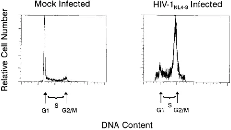
We analyzed cell cycle kinetics in initial experiments with
HIV-1
NL4-3used to infect SupT1 cells at relatively high
mul-tiplicities of infection (MOIs). Analysis by hypotonic
pro-pidium iodide staining (35) and flow cytometry indicated an
abnormal cell cycle profile, with a greater number of cells
containing a 4n amount of DNA (G
21
M) than in
[image:2.612.318.551.73.204.2]mock-infected cultures (Fig. 1). However, precise quantitative
anal-ysis was complicated by the presence of uninfected cells in the
culture. To examine more precisely the properties of infected
cells, it was necessary to distinguish infected from uninfected
cells in the population in a manner that would still allow cell
FIG. 1. Infection of SupT1 cells with HIV-1NL4-3causes cell cycleperturba-tion. DNA content of nuclei from mock-infected and HIV-1NL4-3-infected SupT1
cells (6.9mg of p24; day 4 postinfection) stained with propidium iodide and analyzed by flow cytometry. The histogram indicates relative proportions of cells in G1, S, and G21M phases of the cell cycle by relative fluorescence intensity
of the stained nuclei (x axis).
on November 9, 2019 by guest
http://jvi.asm.org/
cycle characterization by flow cytometry. The use of
HIV-1-specific surface markers such as gp160 has been technically
problematic, so we chose to use instead a
replication-compe-tent HIV-1
NL4-3vector (47) bearing the gene for the murine
thymocyte surface antigen, thy1.2 (13). Other marker genes
have been inserted into the HIV-1 nef gene to generate HIV-1
vectors effective in monitoring HIV-1 infection in vitro, so we
substituted thy1.2 for a part of nef.
We first demonstrated that thy1.2 expression alone did not
alter cell cycle profiles by transfection of the thy1.2 vector
under the control of the CMV immediate-early promoter. The
resulting cell cycle profile of the transfected Thy 1.2
1subpopu-lation was identical to that of the Thy 1.2
2subpopulation and
mock-transfected cells (data not shown; also see Fig. 4). The
kinetics of cell cycle perturbation were determined by a time
course experiment with SupT1 cells as the viral targets. After
infection (MOI,
'
0.05), cells were harvested from both
mock-and NL-Thy-infected cultures. A portion was stained for Thy
1.2 expression combined with a fluorescent dye to allow
exclu-sion of dead cells, and another was doubly stained for Thy 1.2
and DNA content. On day 1 postinfection, 4% of the cells were
positive for the Thy 1.2 marker; this population increased over
the subsequent days to 6, 15, and 52%. Mock-infected cultures
were similarly stained and found to be negative for the Thy 1.2
antigen (Fig. 2). HIV-1 core antigen (p24) levels increased
over time and correlated with Thy 1.2 expression (data not
shown).
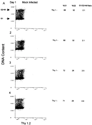
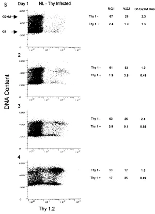
Cell cycle analysis of the cultures at day 1 postinfection
showed that the G
1/G
21
M ratio was 2.3 for the Thy 1.2
2 [image:3.612.144.466.69.504.2]subpopulation and remained at a similar ratio for the duration
FIG. 2. Thy 1.21SupT1 cells in an NL-Thy-infected culture accumulate in the G21M phase of the cell cycle over time. SupT1 cells (53106) were infected withNL-Thy (1.8mg of p24). Aliquots were taken at indicated days and analyzed for cell cycle profile. (A) Mock-infected SupT1 cells were harvested at 24-h intervals postinfection, doubly stained for Thy 1.2 antigen and DNA content, and analyzed by flow cytometry. Fluorescence intensity of the propidium iodide DNA stain is plotted on the y axis, and Thy 1.2-FITC fluorescence intensity is plotted on the x axis. Arrows indicate G1and G2peaks. Percentages of cells in the G1and G21M phases
and calculated G1/G21M ratios are shown to the right of the dot plots. (B) NL-Thy-infected culture analyzed as for panel A. Percentages of cells in G1and G21
M for both Thy 1.22and Thy 1.21subpopulations and calculated G1/G21M ratios are shown to the right of the dot plots.
on November 9, 2019 by guest
http://jvi.asm.org/
of the experiment (Fig. 2). Similar ratios were observed in the
mock-infected culture. However, a different pattern was
ob-served for the Thy 1.2
1subpopulation. Initially, the G
1/G
21
M ratio was 1.3 on day 1 postinfection and then decreased to
approximately 0.5 on the following days. This indicated an
accumulation of cells in G
21
M relative to those in G
1. It was
noteworthy on day 4 postinfection that although the majority
of the culture was infected and cell cycle perturbation was
evident, the noninfected subpopulation exhibited a normal cell
cycle profile. This indicated that the uninfected cells, despite
being in the same culture, were not adversely affected by the
presence of the HIV-1-infected cells.
HIV-1 infection causes G
21
M arrest in SupT1 cells.
The
observed inversion of the G
1/G
21
M ratio could be explained
either by the arrest and accumulation of HIV-1-infected cells
in the G
21
M phase of the cell cycle or by the death and loss
of infected cells from the G
1compartment. If the former were
true, the initial number of HIV-1-infected cells in G
21
M
should stay the same or increase. However, if the increase in
the proportion of G
21
M cells were due to the death of cells
in G
1, the total number of cells in G
21
M should decrease as
cells progress to G
1and are deleted from the population. In
the above experiment, these two possibilities cannot be
distin-guished, since the spread of HIV-1 infection through the
cul-ture can replenish the HIV-1-infected cell numbers in the G
1and G
21
M phases. To clarify the mechanism of the apparent
G
21
M accumulation, an experiment similar to that described
[image:4.612.143.463.71.512.2]above was performed, with the modification that the
subse-quent spread of virus within the culture was inhibited by the
addition of three well-characterized HIV-1 antagonists that act
at different points in the viral life cycle: AZT, which blocks
reverse transcription (33); a protease inhibitor, A77003, that
prevents maturation of the virion (19, 21); and sCD4, which
binds to virion-associated gp120 and prevents the virion from
adhering to the cell surface CD4 receptor (8, 12, 18, 40, 55, 63).
The addition of sCD4 also inhibits the formation of envelope/
FIG. 2—Continued.on November 9, 2019 by guest
http://jvi.asm.org/
receptor-mediated syncytia by binding to gp120 expressed on
the surface of infected cells (18).
For this experiment, the absolute number of infected cells
was calculated by multiplying the total cell number in the
culture by the percentage of Thy 1.2
1cells. At day 2
postin-fection, the number of HIV-1-infected cells was approximately
4
3
10
5and remained constant over days 3 and 4 postinfection
(Table 1), although the total cell number (infected plus
unin-fected) increased fourfold. In the absence of inhibition, the
infected cell population increased 10-fold over the same period
(data not shown). The mock-infected culture was found to
exhibit a cell cycle profile and growth kinetics similar to those
in other experiments, demonstrating that the presence of the
antagonists did not affect the growth characteristics of the cells.
Likewise, analysis of the Thy 1.2
2subpopulation of the
in-fected culture showed G
1/G
21
M ratios similar to that of the
mock-infected culture. However, the ratio for the Thy 1.2
1subpopulation decreased from 2.0 on day 1 postinfection to
approximately 0.15 from days 2 to 4 of the time course. The
observations that a constant number of HIV-1-infected cells
persisted in the culture over a period of 3 days and that an
increasing proportion of those cells were found to be in G
21
M over time argue strongly in favor of arrest of these cells in
the G
21
M phase of the cell cycle. As expected for the use of
sCD4 in this experiment, no syncytia were observed, indicating
that cytopathic effects due to Env-mediated syncytia do not
play a role in the G
21
M arrest.
HIV-1 infection causes G
21
M arrest in stimulated PBL.
We next tested whether HIV-1 could induce G
21
M arrest in
primary rather than transformed T cells. This experiment was
carried out with CD4
1cells enriched from PHA- and
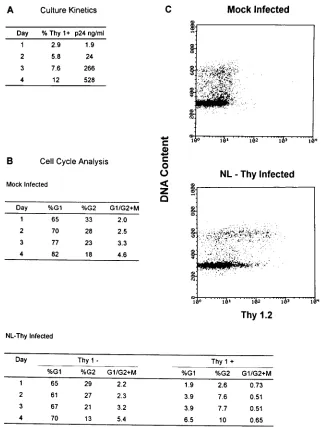
inter-leukin-2-stimulated PBL as targets. The proportion of Thy
1.2
1cells increased from 2.9% on day 1 to 12% on day 4
postinfection, and the HIV-1 p24 antigen levels present in the
culture supernatant also increased (Fig. 3). For PBL, the
cal-culated proportion of cells seen by flow cytometry in the G
1peak also includes quiescent cells in G
0. Cell cycle analysis
revealed that the G
01
G
1/G
21
M ratio of both the
mock-infected culture and the Thy 1.2
2subpopulation of the
NL-Thy-infected culture remained similar throughout the time
course. The ratio was observed to increase over the time
pe-riod, most likely because of a relative increase in the number of
T cells arresting in G
1or reentering a quiescent G
0state
following initial activation by PHA. The Thy 1.2
1subpopula-tion showed an inverted G
01
G
1/G
21
M ratio of 0.51 to 0.73,
consistent with the data presented for infection of SupT1
cells.
Determination of viral proteins necessary for G
21
M
ar-rest.
We determined whether the G
21
M arrest could be
attributed to specific HIV-1 genes. Since the NL-Thy virus
contained a deletion in the nef gene, we were able to rule out
the role of nef in G
21
M arrest. We next tested a vpr insertion/
frameshift mutant of the NL-Thy virus that produced a protein
lacking the carboxy-terminal 33 residues of the 96-amino-acid
protein. The vpr mutant virus, termed Vpr-X, was used to
infect a culture of SupT1 cells in parallel with NL-Thy and at
an equivalent MOI (approximately 0.01). Aliquots of cells were
harvested from mock-, NL-Thy-, and Vpr-X-infected cultures
at 24-h intervals and assayed for Thy 1.2 expression and DNA
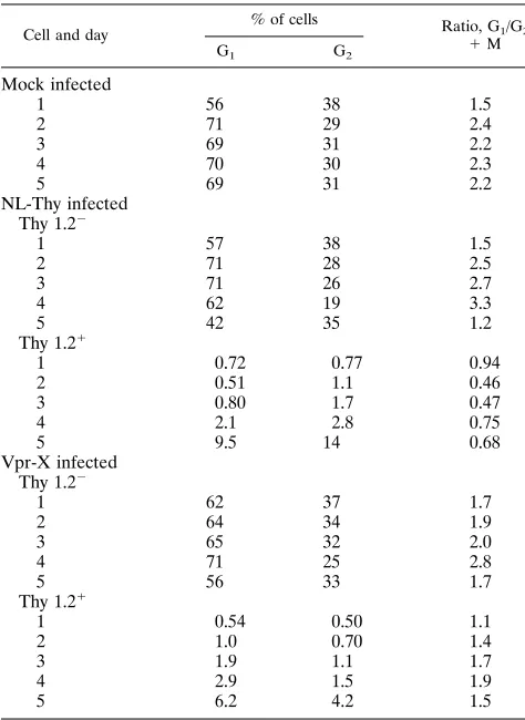
content. As expected, the Thy 1.2
2subpopulations of both the
NL-Thy- and Vpr-X-infected cultures had ratios similar to that
of the mock-infected culture throughout the experiment
(Ta-bles 2 and 3). Cell cycle arrest occurred in the Thy 1.2
1sub-population of the NL-Thy-infected culture, consistent with
ear-lier experiments. However, the cells infected with the Vpr-X
virus showed reduced cell cycle perturbation. Thus, mutation
of the vpr gene impaired the ability of the virus to elicit G
21
M arrest.
HIV-1 Vpr is sufficient to cause G
21
M arrest.
We
[image:5.612.60.296.84.155.2]dem-onstrated above that the vpr gene product contributes to
in-duction of cell cycle arrest following infection of lymphoid cells
by HIV-1. We next determined whether the vpr gene product
was sufficient to elicit arrest in the absence of all other HIV-1
proteins. To address this question, we developed a
transient-transfection and expression system to produce Vpr in target
cells and examine their cell cycle profile. To allow differential
analysis of transfected and nontransfected cells in the same
population, a dual expression vector (called BSVprThy) was
constructed; it contains the vpr and thy1.2 open reading frames,
TABLE 1. NL-Thy-infected SupT1 cells arrest in G21Ma
Day Thy 1.2
1cells (103
) Ratio, G
1/G2
1M Total G1 G21M
1 130 88 45 2.0
2 350 55 291 0.19
3 450 41 410 0.10
4 460 55 402 0.13
aSupT1 cells (53106) were infected with NL-Thy (5mg of p24), grown in the
presence of sCD4, AZT, and a protease inhibitor, and analyzed by flow cytom-etry at indicated intervals for Thy 1.2 expression and DNA content. The actual cell numbers (percent Thy 1.21times the number of cells in culture) are shown for the G1and G21M populations, together with the calculated G1/G21M
ratios.
TABLE 2. NL-Thy and Vpr-X infection of SupT1 cells: cell cycle analysis
Cell and day
% of cells Ratio, G
1/G2
1M G1 G2
Mock infected
1 56 38 1.5
2 71 29 2.4
3 69 31 2.2
4 70 30 2.3
5 69 31 2.2
NL-Thy infected Thy 1.22
1 57 38 1.5
2 71 28 2.5
3 71 26 2.7
4 62 19 3.3
5 42 35 1.2
Thy 1.21
1 0.72 0.77 0.94
2 0.51 1.1 0.46
3 0.80 1.7 0.47
4 2.1 2.8 0.75
5 9.5 14 0.68
Vpr-X infected Thy 1.22
1 62 37 1.7
2 64 34 1.9
3 65 32 2.0
4 71 25 2.8
5 56 33 1.7
Thy 1.21
1 0.54 0.50 1.1
2 1.0 0.70 1.4
3 1.9 1.1 1.7
4 2.9 1.5 1.9
5 6.2 4.2 1.5
on November 9, 2019 by guest
http://jvi.asm.org/
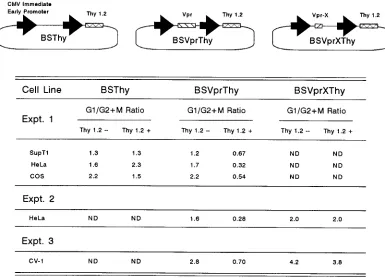
[image:5.612.315.552.402.727.2]each under the control of a separate CMV immediate-early
promoter (Fig. 4). As a control, an otherwise identical
expres-sion plasmid lacking the vpr open reading frame was also
constructed (BSThy). The control vector had no effect on cell
cycle profiles. Furthermore, a dual expression vector
contain-ing the same mutant vpr open readcontain-ing frame as that present in
the Vpr-X virus did not cause cell cycle perturbation (Fig. 4,
experiments 2 and 3). The Thy 1.2
1subpopulation of the
Vpr-containing BSVprThy-transfected culture showed an
in-verted ratio of 0.67, consistent with that seen in virus-infected
cells, and the Thy 1.2
2subpopulation was normal. We
there-fore conclude that expression of the HIV-1 vpr gene product
alone is sufficient to cause G
21
M arrest. Cell cycle arrest was
[image:6.612.146.468.68.495.2]also observed in COS, CV-1 (African green monkey kidney),
and HeLa cells (human cervical carcinoma), but not in NIH
3T3 (murine embryonic) cells (data not shown).
FIG. 3. NL-Thy infection of CD41primary T lymphocytes causes cell cycle accumulation in the G21M phase. PHA-stimulated primary CD41T lymphocytes (6
3106) were mock infected or infected with NL-Thy (1.8mg of p24) and analyzed at 24-h intervals. (A) Analysis of infected cultures for expression of Thy 1.2 antigen
and HIV-1 p24 core antigen in the culture medium. (B) Cell cycle analysis of the NL-Thy- and mock-infected cultures by double staining for Thy 1.2 antigen and DNA content. Percentages of cells in G1and G21M and G1/G21M ratios for Thy 1.22and Thy 1.21subpopulations are shown. (C) Dot plot analysis of mock- and
NL-Thy-infected cultures at 4 days postinfection. DNA content is plotted on the y axis, and Thy 1.2 antigen expression is plotted on the x axis.
TABLE 3. NL-Thy and Vpr-X infection of SupT1 cells: culture kineticsa
Day
% Thy 1.21 p24 (ng/ml)
NL-Thy Vpr-X NL-Thy Vpr-X
1 1.0 1.0 1.0 0.6
2 1.4 1.4 8.0 3.0
3 2.5 2.2 50 26
4 5.7 3.7 460 320
5 26 12 2,540 1,290
aSupT1 cells (53106) were infected with NL-Thy and Vpr-X viruses (1mg of
p24) and analyzed at the indicated intervals. Infected cultures were analyzed for expression of Thy 1.2 antigen and HIV-1 p24 core antigen in the culture medium.
on November 9, 2019 by guest
http://jvi.asm.org/
[image:6.612.57.300.604.685.2]Alteration of CDC2 kinase.
One of the key regulatory factors
involved in progression of cells from the G
2phase into M is the
serine/threonine kinase CDC2. It has previously been
demon-strated that dephosphorylation of this kinase at two critical
residues, Thr-14 and Tyr-15, is necessary for activation of
CDC2 kinase (23, 57, 58) and subsequent phosphorylation
events mediated by CDC2 that lead to progression into mitosis.
Chemical agents that block DNA synthesis or that induce
DNA damage can arrest cells in G
2through activation of
pathways that lead to an accumulation of hyperphosphorylated
CDC2 kinase (34, 41). The hyperphosphorylated form of
CDC2 kinase has previously been shown to be distinguishable
from other forms by decreased mobility on gels (9, 37).
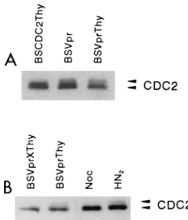
There-fore, we tested by SDS-PAGE whether cells arrested by Vpr
show evidence of alterations in the phosphorylation state of
CDC2 kinase. Two different Vpr-expressing constructs, BSVpr
and BSVprThy, showed increased levels of the
hyperphospho-rylated form of CDC2 relative to a control transfection with a
vector expressing the cdc2 gene (Fig. 5A). In a second
exper-iment, we used nocodazole and nitrogen mustard (HN
2) as
controls for different forms of CDC2. Nocodazole is an
inhib-itor of mitotic spindle formation and arrests cells in mitosis,
when the majority of the CDC2 is in an active
nonhyperphos-phorylated form (20, 37, 41). Consistent with previously
pub-lished reports, nocodazole-treated cells contained little or no
hyperphosphorylated CDC2 (Fig. 5B, Noc). Nitrogen mustard,
in contrast, induces DNA damage, which delays cells in G
2,
resulting in greater levels of hyperphosphorylated CDC2 (41).
As expected, nitrogen mustard-treated cells showed greater
levels of the hyperphosphorylated form of CDC2 (Fig. 5B,
HN
2). Cells transfected with Vpr-expressing constructs showed
increased levels of the hyperphosphorylated form of CDC2
kinase, whereas those transfected with Vpr-X-containing
vec-tors did not (Fig. 5B, BSVprThy and BSVprXThy). Thus, these
results are consistent with the known properties of CDC2
kinase in cells blocked at the G
2/M transition and provide
further support for our conclusion that Vpr acts to block cells
in the G
21
M phase of the cell cycle.
DISCUSSION
Here we report that HIV-1 infection of CD4 T lymphocytes
and T lymphoblastoid cell lines causes cell cycle perturbation,
resulting in an apparent accumulation of cells in the second
gap (G
2) or mitosis (M) phase of the cell cycle in an
asynchro-nously dividing population of cells. With a marked retrovirus
vector and by inhibiting the spread of the virus through the
culture, it was apparent that cells infected with HIV-1 were
unable to transit normally from the G
2phase through mitosis
to complete the cell cycle, thus demonstrating G
21
M arrest
as opposed to G
21
M accumulation due to death of infected
cells in G
1. Cell cycle arrest in the G
21
M phase occurred in
the absence of syncytium formation or other visible cytopathic
effect. The cause of cell cycle arrest was attributed to the
HIV-1 vpr gene product. Mutant viruses lacking the full-length
[image:7.612.111.496.73.351.2]vpr open reading frame failed to inhibit cell cycle progression.
FIG. 4. HIV-1 Vpr protein causes G21M arrest in various cell types. In experiment 1, SupT1 (53106), HeLa (107), and COS (107) cells were transfected with
BSVprThy (10mg), a plasmid that expresses both the Thy 1.2 antigen and the HIV-1 Vpr protein, and a control plasmid (10mg), BSThy, that expresses Thy 1.2 alone. Cells were harvested at 48 h posttransfection and doubly stained for Thy 1.2 expression and DNA content. In two additional experiments (2 and 3), HeLa and CV-1 cells were transfected with BSVprThy (10mg) and another plasmid carrying a mutated vpr open reading frame, BSVprXThy (10mg), and analyzed as for experiment 1. These plasmids also contained an untranslated CMV intron, as described in Materials and Methods. Transfection efficiencies monitored by Thy 1.2 expression were as follows: experiment 1, SupT1 cells, 3%; HeLa cells, 45%; and COS cells, 19%; experiment 2, HeLa cells, 50%; and experiment 3, CV-1 cells, 10%. (Top) Schematic structure of plasmids. The bold arrow indicates the CMV immediate-early promoter, the hatched bar indicates the position of thy1.2, and the long and short shaded bars indicate the vpr and vpr-X genes, respectively. (Bottom) The ratios of cells in G1to cells in G21M for the Thy 1.22and Thy 1.21populations are given. ND,
not done.
on November 9, 2019 by guest
http://jvi.asm.org/
Transient transfection of vpr expression constructs in cells
re-vealed that expression of Vpr alone was sufficient to elicit cell
cycle arrest. Cell cycle arrest correlates with an increase in the
level of hyperphosphorylated CDC2, indicating an arrest of
cells at the G
2/M boundary. The inhibition of cellular
prolif-eration in a number of cell types, including primary T cells,
suggests a novel mechanism for HIV-1-induced immune
dys-function.
Recent studies in our laboratory (46) and others (48) have
identified a role for HIV-1 Vpr in preventing the establishment
of chronic viral infection of the host cell. Those results
sug-gested that Vpr altered the growth properties of
HIV-1-in-fected cells in a population by either causing the death of cells
or arresting cell proliferation. Rogel et al. (48) further
dem-onstrated that the cell cycle profiles of cells transfected with
Vpr were altered, leading to a greater proportion of cells in the
G
2or M phase. However, those studies did not distinguish
between cytolytic and cytostatic mechanisms. The data
pre-sented here are concordant with these findings and expand on
them by directly demonstrating that cell cycle arrest occurs in
HIV-1-infected T cells. We also established that the cell cycle
arrest of infected cells occurs in the G
2or M phase of the cell
cycle.
Vpr is known to be incorporated into the virion via a direct
interaction with the HIV-1 Gag protein (6, 22, 26, 45, 65). Its
presence in the virion particle facilitates nuclear localization of
viral nucleic acids following entry in nondividing
monocyte-derived macrophages and mimosine growth-arrested lymphoid
cell lines (17). In support of the latter hypothesis, Vpr was
found to be required for efficient replication of HIV in human
macrophages but not PBL or an immortalized T-cell line (3,
16).
Transfection of Vpr has been observed to promote
differ-entiation and growth inhibition of a human
rhabdomyosar-coma cell line previously used to study terminal differentiation
and tumorigenesis of muscle cells (28). Although the cause and
effect of growth arrest versus differentiation was not
estab-lished, the observed inhibition of growth is consistent with our
data showing G
21
M arrest in multiple cell types. These
include COS, CV-1, HeLa, and SupT1 cells; however, G
21
M
arrest was not observed in the murine cell line NIH 3T3. Other
experiments show similar arrest in MT-2 and HUT 78 cell lines
(20a).
Vpr has also been described as a weak transcriptional
acti-vator and can increase both the viral replication rate and
cy-topathic effect of HIV-1 in vitro (7, 42). We noted that there
was an approximately threefold increase in surface levels of
Thy 1.2 antigen on infected cells in G
21
M compared with
that on those in G
1(Fig. 2) and that cultures infected with virus
capable of producing Vpr yielded higher levels of p24 core
antigen (Table 3). It is possible that Vpr exerts a
trans-activat-ing effect in the G
21
M phase of the cell cycle. Alternatively,
the G
21
M phase of the cell cycle may be more conducive to
the function of certain promoters, including the HIV-1 LTR.
Vpr is likely to act upon one or more of the steps necessary
for transition through mitosis into G
1. One critical event that
drives mammalian cells from G
2into mitosis is the activation of
the maturation-promoting factor, whose components include
CDC2 kinase and cyclin B. The CDC2 kinase is highly
regu-lated, requiring dephosphorylation by CDC25 for its
activa-tion. Antagonizing this reaction is the Wee 1 kinase, which
phosphorylates CDC2 and thus inhibits the transition into
mi-tosis (36, 38, 39, 57). Additional control elements may regulate
these enzymes; for example, the Nim 1 kinase has been shown
to inhibit the Wee 1 homolog in fission yeast (44), although a
mammalian counterpart has not yet been described. Our
re-sults show an increase in the inactive hyperphosphorylated
form of the CDC2 kinase. This suggests that the Vpr protein
acts to antagonize the activation of CDC2 kinase either directly
or indirectly through modulation of its regulatory cascades. By
analogy with other factors, such as DNA-damaging agents that
are known to block the progression of cells from G
2to M
through inhibition of the activation of CDC2 kinase (41),
de-tection of increased levels of hyperphosphorylated CDC2
ki-nase in our experiments suggests but does not prove that the
cell cycle arrest is more likely in late G
2phase than in mitosis.
We consider a number of possibilities for the role that G
21
M arrest plays in HIV-1 replication. First, an activated T cell
can differentiate into a G
0-phase memory cell, having passed
through mitosis (59, 60). Such a cell would exhibit minimal
metabolic and transcriptional activity and, as such, be unable
to efficiently produce viral proteins. An activated T cell
har-boring HIV-1 and arrested in the G
21
M phase of the cell
cycle would be prevented from continuing the cell cycle and
potentially reentering G
0. Thus, by prolonging the time that a
host cell spends in the activated state, the virus may maximize
the output of progeny virus. Second, in addition to reentering
G
0, an activated T cell can undergo apoptosis; G
21
M arrest
[image:8.612.106.243.70.230.2]may prevent this. A recent report indicated that activation of
the CDC2 kinase was required for certain cell types to undergo
apoptosis (54). Our results indicated that HIV-1 Vpr could
prevent or delay the host cell from initiating apoptosis through
inhibition of this kinase. It has been suggested, however, that
HIV-1 can kill cells by apoptosis (14, 25, 61). These hypotheses
FIG. 5. HIV-1 Vpr protein induces increased levels of thehyperphosphory-lated form of CDC2 kinase. HeLa cells were lysed at 48 h posttransfection and analyzed by Western blotting with a CDC2-specific monoclonal antibody. The hyperphosphorylated form of CDC2 kinase is detected as a more slowly migrat-ing form of CDC2 (9, 37, 41). (A) HeLa cells (107
) were transfected, as described in Materials and Methods, with BSCDC2Thy, a plasmid that expresses both human CDC2 and the Thy 1.2 antigen, or with expression plasmids that express Vpr alone (BSVpr) or both Vpr and Thy 1.2 antigen (BSVprThy). In this experiment, the expression vectors for Vpr also contained a nonapeptide epitope derived from the influenza virus hemagglutination protein at the amino terminus of Vpr, which does not affect the ability of Vpr to cause G21M arrest in HeLa
cells (data not shown). Ten micrograms of total protein was loaded per lane. (B) HeLa cells (107) were transfected with BSVprThy or BSVprXThy, as described
in the legend to Fig. 4. Transfection efficiencies monitored by Thy 1.2 expression were 40% for BSVprThy and 53% for BSVprXThy. G1/G2 1M ratios for
BSVprThy-transfected cells were 2.2 for the Thy 1.22population and 0.5 for the Thy 1.21population; for BSVprXThy-transfected cells, they were 1.5 and 2.3 for the Thy 1.22and Thy 1.21populations, respectively. Nocodazole (Noc)-treated HeLa cells were incubated with 0.5mg of nocodazole per ml in the medium for 12 h before harvesting. HeLa cells were treated with nitrogen mustard (HN2) at
0.5mM for 30 min, resuspended in medium without HN2, and analyzed 12 h
later. The G1/G21M ratios for untreated and nocodazole-, and HN2-treated
cells were 2.9, 0.02, and 1.0, respectively. Five micrograms of total protein was loaded per lane.
on November 9, 2019 by guest
http://jvi.asm.org/
may not necessarily be incompatible. HIV-1-infected
immor-talized SupT1 cells were observed in our experiments to
re-main in the G
21
M state for at least 3 days, approximately
three times longer than the natural duration of the typical
mammalian cell cycle. The subsequent fate of these cells is
unknown, and it is possible that they eventually die by
apop-tosis. Third, as indicated above, we have observed that
NL-Thy-infected cells in G
21
M express threefold more Thy 1.2
surface antigen than those in G
1. This suggests that cells in G
21
M may be more efficient in producing proteins under the
control of the HIV-1 LTR. The observation that Vpr can act as
a weak transcriptional activator (7) may be related to this
phenomenon, although the mechanism is not clear. Cell
cycle-dependent protein production has been well documented for
the cyclins, a class of proteins that regulate various events in
cell cycle progression (38). HIV-1 LTR promoter
responsive-ness to cell cycle phase has not been reported; however, it is
possible that the availability of transcription factors known to
interact with the promoter fluctuates during cell replication.
For example, it has been reported that the cell surface
replace-ment rate of CD4 is highest during late DNA synthesis phase
(S) and in G
2(31). Fourth, another advantage of inhibition of
T-cell proliferation for HIV-1 is in directly limiting the extent
and breadth of the immune response directed against it (see
below).
The implications of Vpr-induced G
21
M arrest on the
immunopathogenesis of AIDS are potentially significant. In
the lymphoid organs, a successful immune response depends
on activation by antigen and subsequent clonal expansion and
migration of antigen-specific T cells into the circulation. We
propose here a novel mechanism for immune dysfunction, in
which an activated T-cell population could be infected with
HIV-1 and, as a consequence, be unable to proliferate further,
thus aborting clonal expansion. Limiting this critical phase of
the cellular immune response would likely result in additional
disruption of downstream events dependent on the clonal
ex-pansion of the CD4 T-lymphocyte subpopulation. Such events
may include T-cell helper activity via release of cytokines,
stimulation and modulation of the humoral immune response,
differentiation resulting in the generation of memory cells, and
CD4
1cytotoxic T-cell activity.
The targeting of the Vpr protein or its mode of action may
offer a new avenue for therapeutic intervention in the
patho-genesis of AIDS. Notably, in an animal model system,
muta-tion of the vpr open reading frame from the related retrovirus
simian immunodeficiency virus (SIV) dramatically reduced
vi-rus load and persistence in macaques compared with wild-type
SIV (24). Monkeys in which the vpr mutation did not revert
showed no signs of disease development during the
observa-tion period. Therapy directed against Vpr funcobserva-tion may offer a
novel twofold approach for ameliorating disease progression.
In the first instance, blocking Vpr action would likely reduce
the efficiency of viral replication. However, as with other virally
targeted drugs, the development of resistance is cause for
con-cern. Second, blocking Vpr-mediated cell cycle arrest would
allow proliferation of infected cells and would therefore be
likely to augment the immune response. A putative therapeutic
operating by this latter mechanism would be particularly
at-tractive, as it could circumvent the problem of virally acquired
drug resistance by acting on cellular components to overcome
the block to cell cycle progression.
ACKNOWLEDGMENTS
We thank J. Zack and M. Emerman for helpful discussions, E. Withers-Ward and D. Camerini for critical review of the manuscript, D. Camerini for OKT4 and OKM1 antibodies, P. Nurse for the cdc2
gene, A. Kaplan for protease inhibitor A77003, and W. Aft for prep-aration of the manuscript.
This work was supported by a grant from Amgen Inc. and grants from the National Institutes of Health (AI31822) and Center for AIDS Research (CFAR; AI28697). V.P. is supported by a training grant from the National Institutes of Health (AI07388); I.S.Y.C. is a Scholar of the Leukemia Society of America; and J.B.M.J is supported by an American Cancer Society fellowship.
REFERENCES
1. Adachi, A., H. E. Gendelman, S. Koenig, T. Folks, R. Willey, A. Rabson, and
M. A. Martin.1986. Production of acquired immunodeficiency syndrome-associated retrovirus in human and nonhuman cells transfected with an infectious molecular clone. J. Virol. 59:284–291.
2. Ameisen, J. C., and A. Capron. 1991. Cell dysfunction and depletion in AIDS: the programmed cell death hypothesis. Immunol. Today 12:102–105. 3. Balotta, C., P. Lusso, R. Crowley, R. C. Gallo, and G. Franchini. 1993. Antisense phosphorothioate oligodeoxynucleotides targeted to the vpr gene inhibit human immunodeficiency virus type 1 replication in primary human macrophages. J. Virol. 67:4409–4414.
4. Banda, N. K., J. Berrier, D. K. Kurahara, R. Kurrle, N. Haigwood, R. P.
Sekaly, and T. H. Finkel.1992. Crosslinking CD4 by human immunodefi-ciency virus gp120 primes T cells for activation induced apoptosis. J. Exp. Med. 176:1099–1106.
5. Cheng-Mayer, C., D. Seto, M. Tateno, and J. A. Levy. 1988. Biologic features of HIV-1 that correlate with virulence in the host. Science 240:80–82. 6. Cohen, E. A., G. Dehni, J. G. Sodroski, and W. A. Haseltine. 1990. Human
immunodeficiency virus vpr product is a virion-associated regulatory protein. J. Virol. 64:3097–3099.
7. Cohen, E. A., E. F. Terwilliger, Y. Jalinoos, J. Proulx, J. G. Sodroski, and
W. A. Haseltine.1990. Identification of HIV-1 vpr product and function. J. Acquired Immune Defic. Syndr. 3:11–18.
8. Deen, K. C., J. S. McDougal, R. Inacker, G. Golena-Wasserman, J. Arthos,
J. Rosenberg, P. J. Maddon, R. Axel, and R. W. Sweet.1988. A soluble form of CD4 (T4) protein inhibits AIDS virus infection. Nature (London) 331: 82–84.
9. Draetta, G., and D. Beach. 1988. Activation of cdc2 protein kinase during mitosis in human cells: cell-cycle-dependent phosphorylation and subunit rearrangement. Cell 54:17–26.
10. Fauci, A. S. 1993. Multifactorial nature of human immunodeficiency virus disease: implications for therapy. Science 262:1011–1018.
11. Fauci, A. S. 1988. The human immunodeficiency virus: infectivity and mech-anisms of pathogenesis. Science 239:617–622.
12. Fisher, R. A., J. M. Bertonis, W. Meier, V. A. Johnson, D. S. Costopoulos, T.
Liu, R. Tizard, B. D. Walker, M. S. Hirsh, R. T. Schooley, and R. A. Flavell.
1988. HIV infection is blocked in vitro by recombinant soluble CD4. Nature (London) 331:76–78.
13. Giguere, V., K. I. Isobe, and F. Grosveld. 1985. Structure of the murine Thy-1 gene. EMBO J. 4:2017–2024.
14. Gougeon, M. L., A. G. Laurent-Crawford, A. Hovanessian, and L.
Montag-nier.1993. Direct and indirect mechanisms mediating apoptosis during HIV infection: contribution to in vivo CD4 T cell depletion. Semin. Immunol.
5:187–194.
15. Groux, H., G. Torpier, D. Monte, Y. Mouton, A. Capron, and J. C. Ameison. 1992. Activation-induced death by apoptosis in CD41T cells from human immunodeficiency virus-infected asymptomatic individuals. J. Exp. Med.
175:331–340.
16. Hattori, N., F. Michaels, K. Fargnoli, L. Marcon, R. C. Gallo, and G.
Franchini.1990. The human immunodeficiency virus type 2 vpr gene is essential for productive infection of human macrophages. Proc. Natl. Acad. Sci. USA 87:8080–8084.
17. Heinzinger, N. K., M. I. Bukrinsky, S. A. Haggerty, A. M. Ragland, V.
Kewalramani, M.-A. Lee, H. E. Gendelman, L. Ratner, M. Stevenson, and M. Emerman.1994. The Vpr protein of human immunodeficiency virus type 1 influences nuclear localization of viral nucleic acids in nondividing host cells. Proc. Natl. Acad. Sci. USA 91:7311–7315.
18. Hussey, R. E., N. E. Richardson, M. Kowalski, N. R. Brown, H.-C. Chang,
R. F. Siliciano, T. Dorfman, B. Walker, J. Sodroski, and E. L. Reinhartz.
1988. A soluble CD4 protein selectively inhibits HIV replication and syncy-tium formation. Nature (London) 331:78–81.
19. Jaskolski, M., A. G. Tamasselli, T. K. Sawyer, D. G. Staples, R. L.
Heinrik-son, J. Schneider, S. B. Kent, and A. Wlodawer.1991. Structure at 2.5-Å resolution of chemically synthesized human immunodeficiency virus type 1 protease complexed with a hydroxyethylene-based inhibitor. Biochemistry
30:1600–1609.
20. Jordan, M. A., D. Thrower, and L. Wilson. 1992. Effects of vinblastine, podophyllotoxin and nocodazole on mitotic spindles: implications for the role of microtubule dynamics in mitosis. J. Cell Sci. 102:401–416. 20a.Jowett, J. B. M., and V. Planelles. Unpublished data.
21. Kaplan, A. H., J. A. Zack, M. Knigge, D. A. Paul, D. J. Kempf, D. W.
Norbeck, and R. Swanstrom.1993. Partial inhibition of human
on November 9, 2019 by guest
http://jvi.asm.org/
ficiency virus type 1 protease results in aberrant virus assembly and the formation of noninfectious particles. J. Virol. 67:4050–4055.
22. Kondo, E., F. Mammano, E. A. Cohen, and H. G. Gottlinger. 1995. The p6gag
domain of human immunodeficiency virus type 1 is sufficient for the incor-poration of Vpr into heterologous viral particles. J. Virol. 69:2759–2764. 23. Krek, W., and E. A. Nigg. 1991. Differential phosphorylation of vertebrate
p34cdc2kinase at the G1/S and G2/M transitions of the cell cycle:
identifi-cation of major phosphorylation sites. EMBO J. 10:305–316.
24. Lang, S. M., M. Weeger, C. Stahl-Hennig, C. Coulibaly, G. Hunsmann, J.
Muller, H. Muller-Hermelink, D. Fuchs, H. Wachter, M. M. Daniel, R. C. Desrosiers, and B. Fleckenstein.1993. Importance of vpr for infection of rhesus monkeys with simian immunodeficiency virus. J. Virol. 67:902–912. 25. Laurent-Crawford, A. G., B. Drust, S. Muller, Y. Riviere, M. A. Rey-Cuille,
J. M. Bechet, L. Montagnier, and A. G. Hovanessian.1991. The cytopathic effect of HIV is associated with apoptosis. Virology 185:829–839. 26. Lavallee, C., X. J. Yao, A. Ladha, H. Gottlinger, W. A. Haseltine, and E. A.
Cohen.1994. Requirement of the Pr55gag
precursor for incorporation of the Vpr product into human immunodeficiency virus type 1 viral particles. J. Virol. 68:1926–1934.
27. Lee, M. G., and P. Nurse. 1987. Complementation used to clone a human homologue of the fission yeast cell cycle control gene cdc2. Nature (London)
327:31–35.
28. Levy, D. N., L. S. Fernandes, W. V. Williams, and D. B. Weiner. 1993. Induction of cell differentiation by human immunodeficiency virus 1 vpr. Cell
72:541–550.
29. Lifson, J. D., M. B. Feinberg, G. R. Reyes, L. Rabin, B. Banapour, S.
Chakrabarti, B. Moss, F. Wong-Staal, K. S. Steimer, and E. G. Engleman.
1986. Induction of CD4-dependent cell fusion by the HTLV-III/LAV enve-lope glycoprotein. Nature (London) 323:725–728.
30. Lifson, J. D., G. R. Reyes, M. S. McGrath, B. S. Stein, and E. G. Engleman. 1986. AIDS retrovirus induced cytopathology: giant cell formation and in-volvement of CD4 antigen. Science 232:1123–1127.
31. Martin, C. F., J. Correale, S. Kam-Hansen, and A. Ehrnst. 1992. Cell cycle-dependent expression of CD4 antigen in a monocytoid cell line. Scand. J. Immunol. 34:483–489.
32. Meyaard, L., S. A. Otto, R. R. Jonker, M. J. Mijnster, R. P. Keet, and F.
Miedema.1992. Programmed death of T cells in HIV-1 infection. Science
257:217–219.
33. Mitsuya, H., K. J. Weinhold, P. A. Furman, M. H. St. Clair, S. N. Lehrman,
R. C. Gallo, D. Bolognesi, D. W. Barry, and S. Broder.1985. 39-Azido-39 -deoxythymidine (BW A509U): an antiviral agent that inhibits the infectivity and cytopathic effect of human T-lymphotropic virus type III/lymphadenop-athy-associated virus in vitro. Proc. Natl. Acad. Sci. USA 82:7096–7100. 34. Murray, A. W. 1992. Creative blocks: cell-cycle checkpoints and feedback
controls. Nature (London) 359:599–604.
35. Nicoletti, I., G. Migliorati, M. C. Pagliacci, F. Grignani, and C. Riccardi. 1991. A rapid and simple method for measuring thymocyte apoptosis by propidium iodide staining and flow cytometry. J. Immunol. Methods 139: 271–279.
36. Nigg, E. A. 1993. Targets of cyclin-dependent protein kinases. Curr. Opin. Cell Biol. 5:187–193.
37. Norbury, C., J. Blow, and P. Nurse. 1991. Regulatory phosphorylation of the p34cdc2
protein kinase in vertebrates. EMBO J. 10:3321–3329.
38. Norbury, C., and P. Nurse. 1992. Animal cell cycles and their control. Annu. Rev. Biochem. 61:441–470.
39. Nurse, P. 1990. Universal control mechanism regulating onset of M-phase. Nature (London) 344:503–508.
40. O’Brien, W. A., E. S. Daar, D. D. Ho, and I. S. Y. Chen. 1992. Mapping genetic determinants for human immunodeficiency virus type 1 resistance to soluble CD4. J. Virol. 66:3125–3130.
41. O’Connor, P. M., D. K. Ferris, M. Pagano, G. Draetta, J. Pines, T. Hunter,
D. L. Longo, and K. W. Kohn.1993. G2delay induced by nitrogen mustard
in human cells affects cyclin A/cdk2 and cyclin B1/cdc2-kinase complexes differently. J. Biol. Chem. 268:8298–8308.
42. Ogawa, K., R. Shibata, T. Kiyomasu, I. Higuchi, Y. Kishida, A. Ishimoto,
and A. Adachi.1989. Mutational analysis of the human immunodeficiency virus vpr open reading frame. J. Virol. 63:4110–4114.
43. Pantaleo, G., C. Graziosi, and A. S. Fauci. 1993. New concepts in the immunopathogenesis of human immunodeficiency. N. Engl. J. Med. 328: 327–335.
44. Parker, L. L., S. A. Walter, P. G. Young, and H. Piwnica-Worms. 1993. Phosphorylation and inactivation of the mitotic inhibitor Wee 1 by the nim1/cdr1 kinase. Nature (London) 363:736–738.
45. Paxton, W., R. I. Connor, and N. R. Landau. 1993. Incorporation of Vpr into human immunodeficiency virus type 1 virions: requirement for the p6 region of gag and mutational analysis. J. Virol. 67:7229–7237.
46. Planelles, V., F. Bachelerie, J. B. M. Jowett, A. Haislip, Y. Xie, P. Banooni,
T. Masuda, and I. S. Y. Chen.Unpublished data.
47. Planelles, V., A. Haislip, E. S. Withers-Ward, S. A. Stewart, Y. Xie, N. P.
Shah, and I. S. Y. Chen.1995. A new reporter system for detection of viral infection. Gene Ther. 2:369–376.
48. Rogel, M. E., L. I. Wu, and M. Emerman. 1995. The human immunodefi-ciency virus type 1 vpr gene prevents cell proliferation during chronic infec-tion. J. Virol. 69:882–888.
49. Sambrook, J., E. F. Fritsch, and T. Maniatis. 1989. Molecular cloning: a laboratory manual, 2nd ed. Cold Spring Harbor Laboratory, Cold Spring Harbor, N.Y.
50. Schmid, I., C. H. Uittenbogaart, and J. V. Giorgi. 1991. A gentle fixation and permeabilization method for combined cell surface and intracellular staining with improved precision in DNA quantification. Cytometry 12:279–285. 51. Shaw, G. M., B. H. Hahn, S. K. Arya, J. E. Groopman, R. C. Gallo, and F.
Wong-Staal.1984. Molecular characterization of human T-cell leukemia (lymphotropic) virus type III in the acquired immune deficiency syndrome. Science 226:1165–1170.
52. Shearer, G. M., and M. Clerici. 1993. How human immunodeficiency virus ravages the immune system. Curr. Opin. Immunol. 4:463–465.
53. Sheppard, H. W., and M. S. Ascher. 1992. The natural history and patho-genesis of HIV infection. Annu. Rev. Microbiol. 46:533–564.
54. Shi, L., W. K. Nishioka, J. Th’ng, E. M. Bradbury, D. W. Litchfield, and A. H.
Greenberg.1994. Premature p34cdc2
activation required for apoptosis. Sci-ence 263:1143–1145.
55. Smith, D. H., R. A. Byrn, S. A. Marsters, T. Gregory, J. E. Groopman, and
D. J. Capon.1987. Blocking of HIV-1 infectivity by a soluble, secreted form of the CD4 antigen. Science 238:1704–1707.
56. Sodroski, J., W. C. Goh, C. Rosen, K. Campbell, and W. Haseltine. 1986. Role of the HTLV-III/LAV envelope in syncytium formation and cytopatho-genicity. Nature (London) 322:470–474.
57. Solomon, M. J. 1993. Activation of the various cyclin/cdc2 protein kinases. Curr. Opin. Cell Biol. 5:180–186.
58. Solomon, M. J., M. Glotzer, T. H. Lee, M. Philippe, and M. W. Kirschner. 1990. Cyclin activation of p34cdc2. Cell 63:1013–1024.
59. Sprent, J. 1994. T and B memory cells. Cell 76:315–322.
60. Sprent, J., and D. F. Tough. 1994. Lymphocyte life-span and memory. Sci-ence 265:1395–1400.
61. Terai, C., R. S. Kornbluth, C. D. Pauza, D. D. Richmann, and D. A. Carson. 1991. Apoptosis as a mechanism of cell death in cultured lymphoblasts acutely infected with HIV-1. J. Clin. Invest. 87:1710–1715.
62. Tersmette, M., R. A. Gruters, F. de Wolf, R. E. Y. de Goede, J. M. A. Lange,
P. T. A. Schellekens, J. Goudsmit, H. G. Huisman, and F. Miedema.1989. Evidence for a role of virulent human immunodeficiency virus (HIV) vari-ants in the pathogenesis of acquired immunodeficiency syndrome: studies on sequential HIV isolates. J. Virol. 63:2118–2125.
63. Traunecker, A., W. Luke, and K. Karjalainen. 1988. Soluble CD4 molecules neutralize human immunodeficiency virus type I. Nature (London) 331:84– 86.
64. Wysocki, L. J., and V. L. Sato. 1978. ‘‘Panning’’ for lymphocytes: a method for cell selection. Proc. Natl. Acad. Sci. USA 75:2844–2848.
65. Yuan, X., Z. Matsuda, M. Matsuda, M. Essex, and T. H. Lee. 1990. Human immunodeficiency virus vpr gene encodes a virion-associated protein. AIDS Res. Hum. Retroviruses 6:1265–1271.
66. Zagury, D., J. Bernard, R. Leonard, R. Cheynier, M. Feldman, P. S. Sarin,
and R. C. Gallo.1986. Long term cultures of HTLV-III-infected cells: a model of cytopathology of T cell depletion in AIDS. Science 231:850–853.