0022-538X/95/$04.0010
Copyrightq1995, American Society for Microbiology
Intermediates of Adeno-Associated Virus Type 2 Assembly:
Identification of Soluble Complexes Containing Rep and Cap Proteins
ANDREAS WISTUBA, STEFAN WEGER, ANDREA KERN,ANDJU¨ RGEN A. KLEINSCHMIDT*
Deutsches Krebsforschungszentrum, Forschungsschwerpunkt Angewandte Tumorvirologie, D-69120 Heidelberg, Germany
Received 23 February 1995/Accepted 5 May 1995
The proteins encoded by the adeno-associated virus type 2 (AAV-2)rep andcapgenes obtained during a productive infection of HeLa cells with AAV-2 and adenovirus type 2 were fractionated according to solubility, cellular localization, and sedimentation properties. The majority of Rep and Cap proteins accumulated in the nucleus, where they distributed into a soluble and an insoluble fraction. Analysis of the soluble nuclear fraction of capsid proteins by sucrose density gradients showed that they formed at least three steady-state pools: a monomer pool sedimenting at about 6S, a pool of oligomeric intermediates sedimenting between 10 and 15S, and a broad pool of assembly products with a peak between 60 and 110S, the known sedimentation positions of empty and full capsids. While the soluble nuclear monomer and oligomer pool contained predominantly only two capsid proteins, the 30 to 180S assembly products contained VP1, VP2, and VP3 in a stoichiometry similar to that of purified virions. They probably represent different intermediates in capsid assembly, DNA encap-sidation, and capsid maturation. In contrast, the cytoplasmic fraction of capsid proteins showed a pattern of oligomers continuously increasing in size without a defined peak, suggesting that assembly of 60S particles occurs in the nucleus. Soluble nuclear Rep proteins were distributed over the whole sedimentation range, probably as a result of association with AAV DNA. Subfractions of the Rep proteins with defined sedimentation values were obtained in the soluble nuclear and cytoplasmic fractions. We were able to coimmunoprecipitate capsid proteins sedimenting between 60 and 110S with antibodies against Rep proteins, suggesting that they exist in common complexes possibly involved in AAV DNA packaging. Antibodies against the capsid proteins, however, precipitated Rep78 and Rep68 predominantly with a peak around 30S representing a second complex containing Rep and Cap proteins.
Adeno-associated virus type 2 (AAV-2) is a human parvo-virus with a single-stranded DNA (ssDNA) genome of approx-imately 4.6 kb encapsidated in an icosahedral virion 20 to 24 nm in diameter (5, 37). Generally, AAV depends on coinfec-tion with a helper virus (either adenovirus or herpesvirus) for efficient DNA replication (2, 4, 7). In the absence of helper virus coinfection, the AAV genome integrates into the host cell genome to establish a latent infection (21, 32). The AAV genome contains two major open reading frames flanked by the 145-bp inverted terminal repeats which serve as origins of replication (37). The right half of the genome codes for the three overlapping structural proteins VP1, VP2, and VP3, with relative molecular weights of 87,000, 72,000, and 62,000. The capsid proteins are produced from a 2.6-kb transcript of the AAV p40 promoter, which is spliced into two 2.3-kb mRNAs by using alternate splice acceptor sites (3, 9, 42). While VP1 can be expressed only from a translation initiation codon at nucleotide (nt) 2203 of one mRNA species, VP2 and VP3 can be expressed from both 2.3-kb RNAs by use of an ACG initi-ation site at nt 2615 or a downstream AUG at nt 2810. This means that the larger capsid proteins share most of their amino acid sequence with the smaller ones but contain additional amino-terminal sequences. They terminate 27 amino acids downstream of the originally proposed C terminus (30). The stoichiometry of capsid proteins in the mature virion of 1:1:10 for VP1:VP2:VP3 (8, 29) is believed to be generated by the relative abundance of alternatively spliced mRNAs and by the
reduced translation initiation frequency at the ACG initiation codon for VP2 (23). The left half of the genome encodes four overlapping nonstructural proteins, Rep78, Rep68, Rep52, and Rep40. The mRNAs for the two larger proteins (Rep78 and Rep68) are transcribed from the p5 promoter and are trans-lated from unspliced and spliced transcripts. Likewise, Rep52 and Rep40 are translated from unspliced and spliced tran-scripts which are generated from the p19 promoter located further downstream. The Rep proteins are required for AAV DNA replication and control of AAV gene expression (17, 22, 40, 41). Genetic and biochemical evidence suggests that Rep52 and Rep40 are not essential for viral double-stranded DNA replication but that they are required for efficient accumulation of progeny ssDNA (11, 25).
Myers and Carter (24) showed by pulse-chase labeling ex-periments that the newly synthesized AAV proteins are rapidly assembled into capsids which are packaged to yield mature particles in a slow process requiring several hours. Inhibition of DNA synthesis by hydroxyurea suggested that AAV replication is not required for DNA packaging. Genetic experiments in-dicated that VP2 and VP3 are sufficient and necessary for accumulation or sequestration of progeny ssDNA, whereas VP1 seems to be required for production of infectious particles (17, 34, 40). The requirement of all three capsid proteins for particle formation and ssDNA accumulation has also been reported (23). Studies with individually expressed capsid pro-teins in insect cells by using recombinant baculoviruses showed that VP2 is essential for assembly of viruslike particles in this system (31). The requirement for small Rep proteins for effi-cient ssDNA accumulation suggests that Rep and Cap proteins interact at some stage of AAV DNA packaging (11).
We were interested in the analysis of low-molecular-weight
* Correspondingauthor.Mailingaddress:DeutschesKrebsforschungs-zentrum, Forschungsschwerpunkt Angewandte Tumorvirologie, Im Neuenheimer Feld 242, D-69120 Heidelberg, Germany. Phone: 49-6221-424978. Fax: 49-6221-424962.
5311
on November 9, 2019 by guest
http://jvi.asm.org/
MATERIALS AND METHODS
Cell culture and virus infection.HeLa cells were grown in Dulbecco’s modi-fied Eagle’s medium (DMEM) supplemented with 10% fetal calf serum and penicillin-streptomycin at 378C in 5% CO2. For later infection with AAV-2, cells were grown to 80% confluency. Then the medium was removed, and the cells were incubated for 2 h with AAV-2 (multiplicity of infection [MOI] of 20) and Ad2 (MOI of 2) in a volume of 2 to 3 ml of DMEM per 175-cm2flask. After the incubation period, DMEM was added and the cells were incubated at 378C in 5% CO2for the indicated periods of time before harvesting.
Preparation of virus stocks.To generate AAV stocks, HeLa cells were in-fected with AAV-2 (MOI of 20) and Ad2 (MOI of 2) and incubated at 378C in 5% CO2for 48 h. Then the flasks containing cells and medium were frozen and thawed three times at270 and 378C. Cells and medium were centrifuged at 5,000 3gav, and the supernatants were collected. Ad2 was inactivated by heating to 568C for 30 min. AAV stocks were titrated by immunofluorescence staining of cells which had been infected with serial dilutions of AAV. Ad2 was added at an MOI of 2 to all serial dilutions. Immunofluorescence staining was performed with monoclonal antibody 76/3.
Preparation of cytoplasmic and nuclear extracts.All steps of the following protocol were done at 48C, and all buffers contained 1 mM dithiothreitol and 1 mM phenylmethylsulfonyl fluoride. Between 33107
and 13108
cells were used for each preparation. Cells were harvested by scraping and then pelleted for 10 min at 1003gav. The pellet was resuspended in 10 ml of isotonic buffer (137 mM NaCl, 5 mM KCl, 0.3 mM Na2HPO4, 0.5 mM MgCl2, 0.7 mM CaCl2, 25 mM Tris-HCl [pH 7.5]) and centrifuged again for 10 min at 1003gav. This step was repeated three times. The cell pellets were then resuspended in 2.5 ml of hypo-tonic buffer (1 mM MgCl2, 0.25 mM CaCl2, 20 mM Tris-HCl [pH 7.5]) and incubated for 5 min. After the incubation, the cells were homogenized five times with a precooled type S Dounce homogenizer. Immediately after homogeniza-tion, 2.5 ml of sucrose buffer (0.6 M sucrose, 10 mM Tris-HCl [pH 7.5]) was added to restore isotonic conditions, and leupeptin and pepstatin were added to final concentrations of 3 and 1mg/ml, respectively. The extract was layered onto a 6-ml sucrose cushion consisting of 1 part 0.2 mM KH2PO4, 350 mM sucrose, 5 parts 0.2 mM Na2HPO4, and 350 mM sucrose, adjusted to 150 mM NaCl. The cushion was centrifuged 15 min at 35003gav. Then the supernatant containing the cytoplasmic fraction was dialyzed under vacuum against TNEM buffer (150 mM NaCl, 50 mM Tris-HCl [pH 8.0], 1 mM EDTA, 2 mM MgCl2) in order to reduce the volume to 1 to 2 ml and to reduce the sucrose concentration. These concentrated cytoplasmic extracts were precipitated either by addition of trichlo-roacetic acid (TCA; final concentration, 20%) or by addition of 9 volumes of acetone. Alternatively, they were further analyzed on sucrose gradients.
The pellets consisting of nuclei were resuspended in 1 to 2 ml of TNEM buffer and sonicated on ice with a Branson Sonifier (three times for 10 s in 1-min intervals). The insoluble components were removed by centrifugation for 15 min at 17,6003gav, and the supernatants were either precipitated or analyzed on sucrose gradients. For nuclease digestion of the soluble nuclear fraction, DNase I and RNase A were added to the supernatant to final concentrations of 50 and 25mg/ml, respectively. Samples were incubated either at 128C for 3 h or at 48C overnight. After digestion, the samples were centrifuged for 15 min at 17,6003 gavat 48C in order to remove proteins which might have precipitated during incubation. Then the supernatants were analyzed by sucrose gradient centrifu-gation and Western blotting (immunoblotting). Alternatively, the nuclei pre-pared from 53107
HeLa cells were incubated for 1 min in 1 ml of buffer containing 0.5% (wt/vol) Triton X-100, 10 mM Tris-HCl (pH 6.5), and 0.5 mM MgCl2at room temperature. After incubation, the lysate was centrifuged 10 min at 17,6003gav. The supernatant was collected, and the pellet was reextracted with the same buffer and then extracted with buffer A. The insoluble material was sedimented at 17,6003gavfor 10 min. All three supernatants were collected, and 0.5 ml thereof was analyzed on a sucrose gradient. Several buffers were tested for the ability to solubilize insoluble nuclear material prepared from AAV/Ad2-coinfected HeLa cells as described above. Pellets of equal sizes were resus-pended with the aid of a pipette in one of the following buffers: buffer H (a
Ad2 alone. After 20 h, cells were incubated for 1 h in methionine-free DMEM and for a further 3 h with methionine-free DMEM containing 100 mCi of 35S-Translabel mix (ICN Biochemicals). Cells were washed two times with phos-phate-buffered saline (PBS) and lysed directly in the dish with 1 ml of radioim-munoprecipitation assay buffer (150 mM NaCl, 1% Nonidet P-40, 0.1% deoxy-cholate, 0.1% SDS, 50 mM Tris-HCl [pH 8.0]) for 15 min at 48C. The lysates were centrifuged for 15 min at 17,6003gavat 48C, and the supernatants were immunoprecipitated as described below.
Sucrose gradients.Samples of 0.5 ml were loaded onto 10-ml sucrose gradi-ents (5 to 30% sucrose in TNEM buffer) and centrifuged for 2 or 18 h at 160,000 3gavat 48C in a swing-out rotor. Fractions of 500 to 600 ml (for Western blotting) or 1 ml (for immunoprecipitation) were collected from the bottom of the tubes. For SDS-PAGE and Western blotting, the fractions were precipitated by adding TCA to a final concentration of 20% and 10mg of RNase A as a precipitation aid. The following reference proteins were analyzed on parallel gradients: bovine serum albumin (6.5S), catalase (11S), thyroglobulin (19S), recombinant empty AAV VP2/3 capsids (31) (60S to 70S), and purified AAV particles (24) (110S).
PAGE.Protein samples were analyzed on 15% polyacrylamide gels in the presence of SDS (39). For Western blot analysis, proteins were electrophoreti-cally transferred to nitrocellulose membranes by using semidry blotting equip-ment and stained with Ponceau S. Incubations with monoclonal antibodies or polyclonal antisera were performed as described elsewhere (16). Rep and Cap proteins were visualized by phosphatase-coupled secondary antibodies as de-scribed elsewhere (16) or by peroxidase-coupled secondary antibodies and en-hanced chemiluminescence detection (ECL kit; Amersham International, Am-ersham, England) as described by the supplier.
Immunoprecipitation experiments.Gradient fractions (500ml) were incu-bated overnight with 200ml of hybridoma supernatant of monoclonal antibody 76/3, A69, or A20 or 2ml of a polyclonal Rep antiserum raised in guinea pigs (RepM or RepA) at 48C with 0.5% Nonidet P-40. After this incubation, 2ml of a polyclonal goat anti-mouse antiserum was added to the samples with the monoclonal antibodies, and the mixture was incubated for 1 h. To remove nonspecific protein precipitations, the samples were then centrifuged for 5 min at 17,6003gavat 48C. The immunocomplexes were precipitated by addition of 30ml of protein A-Sepharose (10% [wt/vol] in NETN [0.1 M NaCl, 1 mM EDTA, 20 mM Tris-HCl {pH 7.5}; 0.5% Nonidet P-40]). After 1 h of incubation, the Sepharose beads were washed three times with 1 ml of NETN buffer, and the samples were boiled in protein loading buffer and analyzed by SDS-PAGE and Western blotting. All incubations were done at 48C.
Antibodies.To generate monoclonal antibodies against AAV capsid proteins, two BALB/c mice were injected subcutaneously with 150ml of a mixture of gel-purified recombinant capsid proteins in PBS containing 100mg each of VP1, VP2, and VP3, mixed with an equal volume of complete Freund’s adjuvant. After 4 weeks, the mice were boosted subcutaneously with 25mg of purified, UV-inactivated AAV-2 in 50ml of PBS and 50ml of incomplete Freund’s adjuvant. After 4 weeks, the mice were injected intraperitoneally with 10 mg of UV-inactivated AAV-2 in 100ml of PBS. Three days later, one mouse was killed and the spleen cells were fused with X63/Ag8 cells as described elsewhere (16). Resultant hybridoma culture supernatants were screened by Western blotting, immunofluorescence, and enzyme-linked immunofluorescence assay. The second mouse was immunized intraperitoneally 6 months later with 100mg of purified VP3 in PBS, and monoclonal antibodies were prepared as described above.
The same procedure was used to generate the monoclonal antibodies against recombinant Rep proteins. The only exception was that for immunization and booster injections, 100mg of an N-terminally truncated gel-purified Rep protein (RepM) was used as described below. For preparation of Rep antisera in guinea pigs, Rep78 which was mutated at K-340 to H (RepA) and an N-terminally truncated Rep starting at methionine 172 (RepM) were expressed in bacteria by using the pet8c vector system as described by Studier et al. (38). Inclusion bodies were prepared, and the Rep proteins were gel purified. Guinea pigs were sub-cutaneously injected with 100mg of gel-purified protein RepA or RepM in Freund’s complete adjuvant and then given two booster injections at 3-week intervals with 100mg of gel-purified protein in Freund’s incomplete adjuvant. Blood was collected 1 week after the second booster injection, and serum was prepared by standard techniques.
on November 9, 2019 by guest
http://jvi.asm.org/
RESULTS
Soluble and insoluble forms of Rep and Cap proteins in AAV-2/Ad2-infected HeLa cells.HeLa cells were infected with AAV-2 (MOI of 20) and Ad2 (MOI of 2) and harvested 24 h postinfection by preparation of nuclei and cytoplasms. Equiv-alent amounts of the soluble and insoluble fractions of both compartments were analyzed by SDS-PAGE and Western blotting for AAV capsid or replication proteins (Fig. 1). Larger amounts of capsid proteins were detected in the insoluble fraction of the nucleus than in the soluble fraction (Fig. 1a). The soluble cytoplasmic fraction represented only one-fourth or less of the total amount of capsid proteins recovered. The soluble nuclear capsid proteins precipitated by TCA showed a number of specifically immunoreactive high-molecular-weight polypeptides besides the expected VP1, VP2, and VP3 polypeptides, which were resistant to dissociation by the pro-tein sample buffer [Fig. 1a, Sup N(T)]. These bands most probably result from irreversible cross-linking due to precipi-tation by addition of TCA, since they were not detected in the insoluble fraction (Fig. 1a, Sed N) or when the soluble nuclear proteins were precipitated by addition of acetone [Fig. 1a, Sup N(A)]. One high-molecular-weight polypeptide of approxi-mately 100 kDa was detected in the nuclear sediment fraction without TCA precipitation (Fig. 1a). The four Rep proteins were also predominantly recovered in the insoluble portion of the nucleus and to a slightly lower extent in the soluble fraction (Fig. 1b). When precipitated with TCA, they sometimes also formed a high-molecular-weight smear. More than half of Rep52 and Rep40 and a variable amount of Rep78 and Rep68 were recovered in the soluble fraction of the cytoplasm. This distribution of large and small Rep proteins between nucleus and cytoplasm was also observed in earlier studies (43). The small amounts of pelletable cytoplasmic Rep and Cap proteins were qualitatively identical with the amounts in the insoluble nuclear fraction (data not shown) and most probably consisted of contaminating nuclear material which was not completely sedimented when nuclei and cytoplasms were separated. This fraction was therefore not included in the following analysis. A kinetic analysis of AAV protein synthesis after coinfection of HeLa cells with AAV-2 and Ad2 showed that the capsid and
replication proteins were already detectable 9 h postinfection and reached their steady-state concentrations between 18 and 24 h postinfection (data not shown; see also reference 28). For the following analysis of assembly intermediates, we harvested the different cellular fractions around 20 h postinfection if not otherwise specified.
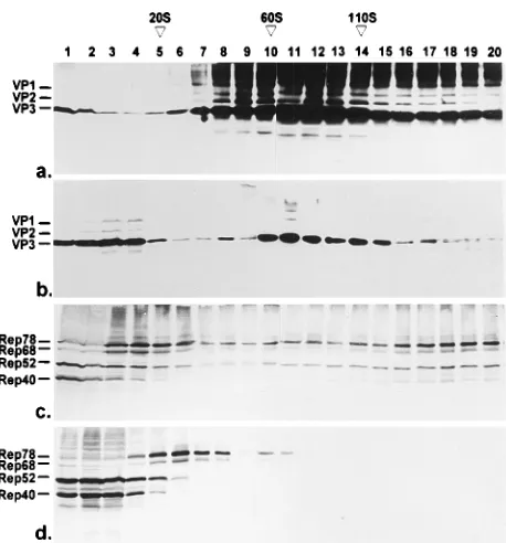
[image:3.612.319.548.67.313.2]Sedimentation profiles of soluble Rep and Cap proteins obtained during a productive AAV infection.The soluble nu-clear and cytoplasmic HeLa cell proteins harvested 24 h after infection with AAV-2 and Ad2 were fractionated by sucrose density gradient centrifugation designed to separate particles of up to 180S. Fractions were analyzed by Western blotting to detect the capsid and replication proteins. Figure 2 shows that most of the soluble nuclear capsid proteins sedimented be-tween 60 and 110S, the sedimentation values of empty and full capsids (24). These fractions probably contained AAV capsids and also showed the SDS-stable high-molecular-weight aggre-gates when precipitated with TCA (Fig. 2a; see above). Only a relatively small amount of capsid proteins sedimented below 20S. In the cytoplasm, we observed a larger fraction of all three capsid proteins sedimenting below 20S and a peak of VP3 between 60 and 110S similar to that in the nucleus. This peak was variable in intensity (see Fig. 4) and was not detected in the cytoplasmic fraction at earlier time points postinfection. We assume that it consists of capsid proteins released from the nucleus. The soluble nuclear Rep proteins were distributed over the whole gradient, with slightly increasing concentrations of the four Rep proteins toward the bottom of the gradient FIG. 1. Recovery of capsid and replication proteins from different
[image:3.612.99.257.72.206.2]subcellu-lar fractions of HeLa cells infected with AAV-2 and Ad2. Nuclei (N) and cytoplasms (C) of infected HeLa cells were prepared 24 h postinfection, and equivalent amounts of soluble (Sup) and insoluble (Sed) material were analyzed by SDS-PAGE and Western blotting with monoclonal VP antibody B1 for de-tection of capsid proteins (a) and monoclonal antibody 303/9 for dede-tection of Rep proteins (b). Immunoreaction was visualized by incubation with peroxidase-coupled secondary antibodies followed by enhanced chemiluminescence detec-tion. The effects of precipitation of soluble proteins with acetone [(A)] and TCA [(T)] on formation of high-molecular-weight protein bands are shown in separate slots.
FIG. 2. Sedimentation analysis of capsid and replication proteins by sucrose density gradient centrifugation in the range of 0 to 180S. At 24 h postinfection, the soluble proteins of nuclei (a and c) and cytoplasms (b and d) of HeLa cells coinfected with Ad2 and AAV-2 were prepared and analyzed by sedimentation through 5 to 30% sucrose gradients for 2 h at 160,0003gavand 48C. Fractions were analyzed by Western blotting with a VP antiserum mixed with monoclonal antibody A69, recognizing VP1 and VP2, to detect all capsid proteins with sufficient intensity (a and b), or with anti-Rep monoclonal antibody 303/9 (c and d). Immunocomplexes were visualized by incubation with peroxidase-coupled secondary antibodies followed by enhanced chemiluminescence (a and b) or with alkaline phosphatase-coupled secondary antibodies (c and d). S values were determined on parallel gradients by using purified virus (110S), empty VP2/VP3 particles (60S), and thyroglobulin (19S) as standards.
on November 9, 2019 by guest
http://jvi.asm.org/
(Fig. 2c). A peak of Rep78 and Rep68 was obtained between 10 and 20S, and Rep52 and Rep40 sedimented at higher con-centrations below 10S. Rep78 and Rep68 of the cytoplasmic fraction sedimented around 30S, whereas Rep52 and Rep40 predominantly sedimented below 10S.
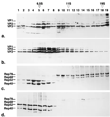
Higher-resolution analysis of the fractions in the sedimen-tation range below 20S showed that the capsid proteins in the nucleus and cytoplasm sedimented as monomers around 6.5S, with oligomers extending up to 19S (Fig. 3a and b). In the nucleus, we observed a distinct oligomer peak presumably of VP2 and VP3 sedimenting on average between 10 and 15S, whereas in the cytoplasm, a continuous pattern of oligomers of VP1, VP2, and VP3 without a defined peak was found. The nuclear 10 to 15S oligomers could theoretically represent VP2/ VP3 pentamers which become incorporated into capsids. In this experiment, the amount of VP1 and VP2 is slightly over-represented because of the enhancement of the VP1 and VP2 signal by addition of the VP1/VP2-specific monoclonal anti-body A69 to the VP antiserum. Nevertheless we observed nearly no VP1 in the 6.5 and 10 to 15S fractions of the nucleus (see also Fig. 4). Analysis of the Rep proteins in the sedimen-tation range of 0 to 20S showed that there was no significant amount of monomeric Rep78 and Rep68. Rep40 in the nu-cleus was found in two size fractions: a low-molecular-weight form sedimenting below 6.5S and a higher-molecular-weight form sedimenting above 6.5S in fractions 6 to 8 forming a separate peak with a retarded migration in SDS-PAGE (Fig. 3c). Rep52 never showed a similar distribution. Rep52 and
tergent. Sedimentation analysis of solubilized Rep proteins showed that Rep78 still existed in high-molecular-weight forms, although with a slightly different sedimentation pattern (data not shown), indicating that at least the high-molecular-weight Rep78 forms are not artifacts of binding to sheared cellular DNA. Since we included detergent in the lysis buffer, we have probably dissociated the other Rep proteins from the complexes described above. DNA digestion experiments sup-ported the notion that the Rep proteins are probably associ-ated with replicating AAV DNA (data not shown; see also Fig. 5).
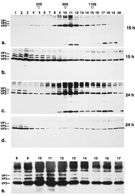
Changes of size of capsid protein pools with time.We com-pared the pools of cytoplasmic and nuclear capsid proteins obtained at an early (15 h) and a late (24 h) time point postin-fection (Fig. 4). The pool of nuclear capsid proteins sediment-ing above 60S increased with time, whereas the pool of cyto-plasmic capsid protein oligomers decreased. This result implicates a conversion of the undefined oligomers in the cy-toplasm into structures sedimenting in the range of empty and full capsids in the nucleus. As already mentioned, the low-molecular-weight fractions in the nucleus contained hardly de-tectable amounts of VP1, whereas VP1 was readily dede-tectable in cytoplasmic fractions. In this case, we used only monoclonal antibody B1, which recognizes all three capsid proteins (see Fig. 6c). Although the structures sedimenting around 60S clearly contained VP1, the stoichiometry of the three capsid proteins was different from the expected 1:1:10 ratio and showed a higher amount of VP2 (Fig. 4e). This changed stoi-chiometry of capsid proteins was accompanied by the appear-ance of a VP degradation product of approximately 10 kDa (Fig. 4a and c, arrowheads).
[image:4.612.58.290.68.316.2]Sedimentation patterns of insoluble nuclear Rep and Cap proteins after solubilization.To determine whether the insol-uble nuclear fractions of Rep and Cap proteins showed the same oligomers as the soluble fractions, we tried to solubilize them under different conditions (Fig. 5a and b). Treatment with low-salt buffers did not solubilize any Rep or Cap proteins compared with treatment with buffer A, whereas high-salt buff-ers solubilized a portion of both the structural and nonstruc-tural proteins of AAV. Also, treatment with detergents solu-bilized both of them to a certain amount. Treatment with DNase and RNase solubilized about 50% of the Cap proteins and only marginal amounts of the Rep proteins, as shown by Western blotting of the same sample with Rep and VP anti-bodies (Fig. 5a and b, lanes D). We compared the oligomer-ization states of Rep and Cap proteins solubilized by treatment with DNase and RNase and by high-salt buffer (Fig. 5c to f). The most striking result was that we never observed Rep oli-gomers after DNase-RNase treatment (Fig. 5c), whereas the Rep proteins solubilized under high-salt conditions still showed some high-molecular-weight oligomers of Rep78 and Rep68, suggesting that association with DNA accounts for the sedimentation pattern of Rep78 and Rep68 (Fig. 5e). The FIG. 3. Sedimentation analysis of capsid and replication proteins by sucrose
density gradient centrifugation in the range of 0 to 20S. The soluble fractions of HeLa cell nuclei (a and c) and cytoplasms (b and d) were prepared 24 h postinfection and analyzed by sedimentation through 5 to 30% sucrose gradients for 18 h at 160,0003gavand 48C. Fractions were analyzed by Western blotting with a VP antiserum mixed with monoclonal antibody A69, recognizing VP1 and VP2, to detect all three capsid proteins with sufficient intensity (a and b). Rep proteins were detected by monoclonal antibody 303/9, which recognizes all four Rep proteins (c and d). Immunoreaction was visualized by incubation with peroxidase-coupled secondary antibodies followed by enhanced chemilumines-cence detection (a and b) or with alkaline phosphatase-coupled secondary anti-bodies (c and d). S values were determined on separate gradients by using bovine serum albumin (6.5S), catalase (11S), and thyroglobulin (19S) as standards.
on November 9, 2019 by guest
http://jvi.asm.org/
majority of capsid proteins solubilized by DNase-RNase treat-ment still seditreat-mented between 60 and 110S (Fig. 5d). Solubi-lization under high-salt conditions probably led to some aggre-gation of the AAV capsids (Fig. 5f). Surprisingly, we recovered a proportionally larger amount of capsid proteins sedimenting below 20S from the insoluble nuclear fraction compared with the soluble fraction. In addition, this fraction also contained VP1 and showed the SDS-resistant cross-linking products.
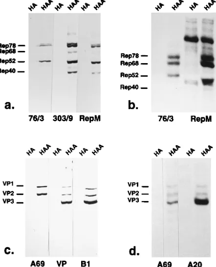
Immunoprecipitation of complexes containing Rep and Cap proteins.To test whether Rep and Cap proteins which cosedi-mented in overlapping fractions of the sucrose gradients are components of common complexes, we performed coimmuno-precipitation experiments and visualized the precipitated pro-teins with Rep- or Cap-specific antibodies by Western blotting. For these experiments, we first analyzed the specificities of various antibodies. For detection of Rep by Western blotting, we used either monoclonal antibody 303/9 or a polyclonal antiserum raised in guinea pigs (RepM) (Fig. 6a). The Rep proteins were precipitated with monoclonal antibody 76/3, which recognizes only the unspliced Rep proteins in Western blotting (Fig. 6a), but also coprecipitated Rep68 from lysates of metabolically labeled cells infected with Ad2 and AAV-2
[image:5.612.62.292.68.401.2](Fig. 6b). Alternatively, we used the polyclonal guinea pig Rep antiserum RepM, (Fig. 6a), which efficiently precipitated Rep78, Rep68, and Rep52 but also a cellular or adenovirus-encoded protein (Fig. 6b). Immunoprecipitation of lysates from infected [35S]methionine-labeled cells yielded only min-imal amounts of Rep40, although this Rep protein is recog-nized by the antiserum. For detection of VP proteins, we used either a rabbit VP antiserum (31) or, in more recent experi-ments, monoclonal antibody B1, which recognizes all three VP proteins (Fig. 6c). The capsid proteins were precipitated with monoclonal antibody A69 or A20 (Fig. 6d). A69 reacts with the VP proteins also in Western blotting and is specific for VP1 and VP2 (Fig. 6c), whereas A20 does not recognize the capsid proteins by Western blotting (data not shown).
[image:5.612.321.547.70.435.2]FIG. 4. Change of capsid protein pools harvested at different time points postinfection. HeLa cell nuclei (a, c, and e) and cytoplasms (b and d) were prepared 15 h (a and b) or 24 h (c, d, and e) postinfection, and the soluble fraction was analyzed by sucrose density gradient centrifugation (5 to 30% sucrose, 160,0003gav, 48C, 2 h). In panel e, fractions 8 to 17 of panel c are shown in an enlarged scale. Fractions were analyzed by Western blotting with mono-clonal VP antibody B1. Immunoreaction was visualized by incubation with per-oxidase-coupled secondary antibodies followed by enhanced chemiluminescence detection. S values were determined on parallel gradients by using purified virus (110S), empty VP2/3 particles (60S), and thyroglobulin (19S) as standards.
FIG. 5. Solubilization of insoluble nuclear fractions. Equal aliquots of insol-uble nuclear fractions harvested 20 h postinfection were treated with different buffers and analyzed for solubilization of Rep (a) or VP (b) proteins. They were treated with buffer A (A), with DNase and RNase in buffer A supplemented with 3 mM MgCl2and 5 mM MnCl2(D), with low-salt buffer (L), with high-salt buffer (H), and with Triton buffer (T) as specified in Materials and Methods. After sedimentation of insoluble material, equal amounts of pellets (P) and superna-tants (S) were analyzed by SDS-PAGE and Western blotting with monoclonal antibody 303/9 to detect the Rep proteins (a) and monoclonal VP antibody B1 to detect the capsid proteins (b). Fractions solubilized by treatment with DNase and RNase (c and d) and high-salt buffers (e and f) were analyzed by sucrose density gradient centrifugation as specified for Fig. 2 and 4. Gradients c and e show Rep proteins, and gradients d and f show the capsid proteins. Immunoreaction was developed by peroxidase-coupled secondary antibodies followed by enhanced chemiluminescence detection.
on November 9, 2019 by guest
http://jvi.asm.org/
To analyze common complexes of Rep and Cap, we per-formed immunoprecipitations of gradient fractions with anti-bodies against either Rep (Fig. 7a and c) or Cap (Fig. 7e and g). The immunoprecipitates were subsequently analyzed by Western blotting with antibodies against Rep (Fig. 7c and e) or Cap (Fig. 7a and g). As controls, gradient fractions were also precipitated with nonspecific antibodies and subjected to Western blotting (Fig. 7b, d, f, and h). We observed a strong coprecipitation of capsid proteins with the Rep antiserum (RepM) in fractions 4 to 8, ranging from 60 to 110S (Fig. 7a). The same pattern of coprecipitated VP proteins was observed with monoclonal antibody 76/3 (data not shown). Control pre-cipitations with an unrelated guinea pig antiserum or with unrelated monoclonal antibodies of the same subtype showed no significant precipitation of Rep or Cap proteins (Fig. 7b and d and data not shown). The distribution of Rep proteins pre-cipitated with the guinea pig antiserum or antibody 76/3 fol-lowed the pattern obtained by TCA precipitation and Western blotting, with a major peak of Rep78 and Rep68 between 10 and 20S (Fig. 7c; see also Fig. 2c). The Rep proteins in higher-molecular-weight fractions sedimenting above 110S, however, were not recovered in equivalent amounts. A different pattern of Rep proteins was obtained after coimmunoprecipitation with monoclonal capsid antibody A20 (or alternatively A69; data not shown) (Fig. 7e). Predominantly Rep78 and some Rep68 sedimenting between 10 and 60S were coprecipitated. Only few Rep proteins were detected in control precipitations
(Fig. 7f). In some experiments, we detected significant amounts of Rep52 and traces of Rep40 in fractions above 110S (fraction 9 and 10). Detection of precipitated VP proteins with antibody B1 revealed the capsid proteins in fractions overlap-ping the coprecipitated Rep proteins but with a different peak distribution (Fig. 7g). We did not observe coprecipitates of Rep with Cap proteins, or vice versa, from cytoplasmic frac-tions (data not shown).
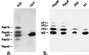
Prompted by the detection of Rep-Cap complexes in in-fected cell nuclei, we analyzed whether such complexes also exist in virus stocks or in purified AAV preparations. Figure 8 shows that VP proteins were efficiently coprecipitated from AAV stocks by two different guinea pig antisera directed against Rep proteins and that Rep was also coprecipitated with VP antibodies (A20). Since such virus stocks might contain immature virus particles due to cell lysis by freezing and thaw-ing without subsequent purification, we also analyzed CsCl-FIG. 6. Evaluation of the specificity of antibodies used for
immunoprecipi-tation experiments. The Rep and VP antibodies were tested by Western blotting (a and c) and immunoprecipitation (b and d) of radiolabeled proteins from extracts of HeLa cells infected with Ad2 (HA) or coinfected with AAV-2 and Ad2 (HAA). Western blots were developed by alkaline phosphatase-conjugated secondary antibodies (a and c). Immunoprecipitates were visualized by fluorog-raphy (b and d). 76/3 and 303/9 denote monoclonal Rep antibodies, RepM indicates a polyclonal guinea pig antiserum directed against Rep. A69, A20, and B1 indicate monoclonal VP antibodies, and VP indicates the rabbit VP
anti-serum. FIG. 7. Immunoprecipitation of capsid and replication proteins from sucrose
gradient fractions. Soluble nuclear proteins of HeLa cells harvested 24 h postin-fection with AAV-2 and Ad2 were fractionated on sucrose density gradients (5 to 30% sucrose, 160,0003gav, 48C, 2 h), and individual fractions were immu-noprecipitated with a polyclonal antibody against Rep proteins (RepM; a and c) or a monoclonal antibody against VP proteins (A20; e and g). Control precipi-tations were performed with a guinea pig antiserum directed against protea-somes (b and d) or with a monoclonal antibody against hepatitis B virus X protein (f and h). The immunoprecipitates were analyzed by SDS-PAGE and Western blotting with monoclonal VP antibody B1 (a, b, g, and h) or monoclonal Rep antibody 303/9 (c, d, e, and f). All Western blots were developed with a peroxidase-coupled secondary antibody and enhanced chemiluminescence de-tection. IgG, immunoglobulin G.
on November 9, 2019 by guest
http://jvi.asm.org/
[image:6.612.321.545.66.422.2] [image:6.612.67.288.74.347.2]purified virus of a density of 1.41. In such preparations, we could detect neither Rep when immunoprecipitated with VP monoclonal antibodies nor capsid proteins when precipitated with Rep antibodies (data not shown). These results indicate that the Rep-Cap complexes either were not present in mature AAV particles or became lost during CsCl density gradient purification of the virus.
DISCUSSION
In this study, we describe intermediates of AAV assembly at the protein level. We analyzed the sedimentation pattern of Rep and Cap proteins during a productive AAV-2 infection and also considered the nucleocytoplasmic distribution and solubility of the AAV gene products to obtain a more detailed picture of the AAV assembly process. In addition to the known pool of empty and full capsids, we observed steady-state pools of monomers and oligomers which had different compositions in the different cellular compartments. Typical 60 to 110S particles indicative of empty and full capsids were detectable only in the nucleus. The AAV Rep proteins sedimented in fractions overlapping with the capsid proteins, suggesting pos-sible interactions with the AAV capsid proteins. In coimmu-noprecipitation experiments, we detected two types of com-plexes containing both Rep and Cap proteins.
In their analysis of AAV assembly, Myers and Carter (24) described particles sedimenting at 66 and 110S which, on the basis of labeling experiments, they interpreted as empty and full AAV capsids. Pulse-chase experiments suggested a precur-sor-product relationship, leading to the model of an assembly pathway of AAV whereby AAV DNA is packaged into pre-formed empty capsids. Since they prefractionated by CsCl den-sity gradient centrifugation, they also preselected for particles of a certain density. Our analysis not only yielded 60 and 110S particles and intermediates in between but also showed several pools of capsid proteins sedimenting below 60S and above 110S. These pools were different in the nucleus and cytoplasm with respect to size, oligomerization state, and stoichiometry of capsid proteins. The interpretation of these pools within the dynamic process of virus assembly is based on changes of the pools with time and the cellular localization of the pools. We assume that the capsid proteins first accumulate in the cyto-plasm, where we observed the formation of a continuous array of oligomers. The capsid protein stoichiometry in these frac-tions was approximately 1:1:5 to 1:1:10. At early time points
postinfection (15 h), these cytoplasmic VP oligomers over-lapped the 60S region where empty capsids sediment, but they did not form a 60S peak, characteristic of empty particles. This oligomer pool decreased with time, and the nuclear pool show-ing a peak in the range of AAV capsids increased. Although one cannot exclude the possibility that the high-molecular-weight VP oligomers of the cytoplasm already contained empty capsids, the dramatic change in the sedimentation profile of the VP proteins after they had entered the nucleus suggests that capsid assembly occurs in the nucleus. Otherwise, one must assume that assembled capsid proteins preferentially en-ter the nucleus, which is rather unlikely. It can be assumed from transfection experiments with single capsid proteins that VP monomers and oligomers can enter the nucleus (30) and that assembly has to occur from a nuclear pool of VP mono-mers and oligomono-mers. This view is supported by the fact that we observed a significant proportion of VP proteins in the range of capsid protein monomers and oligomers in the nucleus, suggesting the existence of an equilibrium between capsids and free capsid proteins.
A relatively large proportion of the nuclear Rep and Cap proteins was associated with insoluble structures of the nu-cleus. The solubilization of part of these immobilized nuclear capsid proteins by DNase-RNase treatment suggests a direct or indirect association with DNA or RNA. The fact that only marginal amounts of Rep proteins were solubilized further suggests that the Rep and Cap proteins are not bound to the same sites of the nuclear structure. The most efficient solubi-lization of both proteins by high-salt treatment may indicate that they are immobilized by polar or ionic interactions. Com-parison of the sedimentation profiles of the Rep proteins sol-ubilized by DNase-RNase and high-salt treatment indicated that the Rep78 and Rep68 proteins sedimenting with high S values were bound to DNA, probably engaged in AAV DNA replication and/or packaging. Sedimentation of solubilized Cap proteins revealed that a relatively large portion of them was in an unassembled state and contained VP1, in contrast to the corresponding soluble fraction. Furthermore, the solubilized nuclear Cap proteins sedimenting below 20S showed an in-creased amount of TCA-induced cross-linking (similar to the 60 to 110S fractions) which was not observed in the soluble low-molecular-weight (,20S) fraction or in the soluble cyto-plasmic fraction. This finding probably indicates different mo-lecular properties of the capsid proteins in the immobilized state in the nucleus. Association of proteins of several viruses with an insoluble nuclear structure has been reported (6, 10, 12, 33), and the nuclear structure has been discussed as a site for viral capsid formation and/or viral DNA synthesis (6). Whether such functional implications also account for the in-soluble nuclear pools of AAV proteins is still a matter of speculation.
[image:7.612.97.252.73.170.2]The majority of soluble nuclear Rep proteins sedimented in complexes ranging from monomers to very high molecular weight forms. Only parts of the soluble Rep52 and Rep40 proteins of the nucleus sedimented in the range of monomers or dimers. A large portion of Rep52 and Rep40 is also distrib-uted over the whole gradient together with Rep78 and Rep68. Since Rep78 and Rep68 bind to the inverted terminal repeats (1, 19, 27), it seems plausible that the majority of Rep78 and Rep68 is engaged in DNA replication and possibly also in packaging. This view is supported by the result of DNase-RNase treatments of the soluble and insoluble nuclear frac-tions by which the high-molecular-weight Rep forms were de-stroyed. Rep52 and Rep40, which by themselves do not bind AAV DNA (20, 27), would have to be complexed with the large Rep proteins or with cellular factors.
FIG. 8. Coimmunoprecipitations of Rep and Cap proteins from virus stocks. Aliquots of a freeze-thaw supernatant of HeLa cells infected with AAV-2 and Ad2 and harvested 48 h postinfection were immunoprecipitated with the mono-clonal antibody against capsid proteins (A20) or, as a control, with an antibody against the X protein of hepatitis B virus and detected with antibody 303/9 against the Rep proteins (a). Alternatively, such supernatant aliquots were im-munoprecipitated with the guinea pig antiserum RepM or RepA and, as a control, with a guinea pig antiserum directed against proteasomes (20S) or protein N1 of Xenopus laevis oocytes (N1) and detected with monoclonal anti-body B1 against the capsid proteins (b). IgG, immunoglobulin G.
on November 9, 2019 by guest
http://jvi.asm.org/
nal antibodies against capsid proteins as well as coprecipitation of capsid proteins with monoclonal and polyclonal antibodies against Rep proteins strongly supported the existence of such complexes. Unexpectedly, the coprecipitates of Rep proteins with Cap antibodies and of Cap proteins with Rep antibodies from sucrose gradient fractions did not have the same peak distribution. We explain the complementary distribution of the coprecipitated Rep and Cap proteins along the sucrose gradi-ent by the relative proportions of these proteins in the com-plexes. We assume that in the range between 20 and 60S, where Rep78 and Rep68 were predominantly coprecipitated with anti-VP antibodies, only few capsid proteins might be associated with multiple Rep complexes, whereas between 60 and 110S, where capsids are coprecipitated with Rep antibod-ies, only few Rep molecules are associated with the capsids. In addition, possible differences in the sensitivities of Rep and Cap antibodies have to be considered. The coprecipitated VP complexes sedimenting between 60 and 110S probably repre-sent capsids in the process of DNA encapsidation, since their peak was generally shifted toward higher sedimentation values compared with the total capsid proteins precipitated with cap-sid antibodies (Fig. 7a and g). Furthermore, the precipitated capsid proteins of that sedimentation range contained AAV DNA protected against DNase digestion (data not shown). This interpretation would also explain the low Rep-to-VP pro-tein ratio. It is more difficult to assign a functional meaning to the Rep78-Rep68 complexes coprecipitated with the VP anti-bodies from fractions with a peak between 20 and 60S. The sedimentation range around 30S suggests that Rep complexes, possibly in association with DNA, are bound to capsid protein oligomers not yet assembled into capsids. Alternatively, single empty capsids could be bound to AAV genomes with multiple Rep replication complexes (to account for the stoichiometry) which form a very extended particle with an abnormally low sedimentation coefficient. In case of the first possibility, the 30S Rep-Cap complexes would implicate the involvement of Rep proteins and possibly of AAV DNA in the capsid assem-bly process. Recently the involvement of the simian virus 40 large T antigen in capsid assembly was demonstrated (35, 36), and similar results were obtained for the polyomavirus middle T antigen (13–15). Although in these cases no direct associa-tion of the large or middle T antigen with the capsid protein oligomers has been demonstrated, it is tempting to speculate that AAV Rep proteins play a role in AAV capsid assembly. If this analogy holds true, one has to reconsider whether the AAV assembly pathway involves an empty capsid which is filled with DNA, in contrast to the simian virus 40 and poly-omavirus pathways, in which capsids are formed around the viral minichromosomes.
It was previously shown by genetic experiments that the small Rep proteins Rep52 and/or Rep40 stimulate ssDNA accumulation (11). Since they also cosedimented with Rep78 and Rep68 over the whole sucrose gradient, we would have
a number of new questions. The composition and stoichiome-try of the Rep-Cap complexes, the involvement of DNA in the formation of these complexes, the role of the nuclear assembly precursors, and the role of the nuclear structure in the AAV assembly process remain to be elucidated. A major goal of this analysis is the determination of factors which stimulate viral assembly after accumulation of all components inside the nu-cleus.
ACKNOWLEDGMENTS
A. Wistuba is supported by a fellowship of the German-Israeli Co-operation in Cancer Research.
We are grateful to U. Bantl-Schaal and P. Klein-Bauernschmitt for providing purified AAV-2 virus. We thank M. Pawlita and H. G. Kra¨usslich for critical reading of the manuscript.
REFERENCES
1. Ashktorab, H., and A. Srivastava. 1989. Identification of nuclear proteins that specifically interact with adeno-associated virus type 2 inverted terminal repeat hairpin DNA. J. Virol. 63:3034–3039.
2. Atchinson, R. W., B. C. Castro, and W. M. Hammond. 1965. Adeno-associ-ated defective particles. Science 149:754–756.
3. Becerra, S. P., F. Koczot, P. Fabisch, and J. A. Rose. 1988. Synthesis of adeno-associated virus structural proteins requires both alternative mRNA splicing and alternative initiations from a single transcript. J. Virol. 62:2745– 2754.
4. Berns, K. I. 1990. Parvovirus replication. Microbiol. Rev. 54:316–329. 5. Berns, K. I., and R. A. Bohensky. 1987. Adeno-associated virus: an update.
Adv. Virus Res. 32:243–306.
6. Bibor-Hardy, V., M. Bernard, and R. Simard. 1985. Nuclear matrix modifi-cations at different stages of infection by herpes simplex virus type 1. J. Gen. Virol. 66:1095–1103.
7. Buller, R. M., J. E. Janik, E. D. Sebring, and J. A. Rose. 1981. Herpes simplex virus types 1 and 2 completely help adeno-associated virus replica-tion. J. Virol. 40:241–147.
8. Buller, R. M., and J. A. Rose. 1978. Characterization of adeno-associated virus-induced polypeptides in KB cells. J. Virol. 25:331–338.
9. Cassinotti, P., M. Weitz, and J. D. Tratschin. 1988. Organization of the adeno-associated virus (AAV) capsid gene: mapping of a minor spliced mRNA coding for virus capsid protein 1. Virology 167:176–184.
10. Chatterjee, P. K., and S. J. Flint. 1986. Partition of E1A proteins between soluble and structural fractions of adenovirus-infected and -transformed cells. J. Virol. 60:1018–1026.
11. Chejanovsky, N., and B. J. Carter. 1989. Mutagenesis of an AUG codon in the adeno-associated virus rep gene: effects on viral DNA replication. Vi-rology 173:120–128.
12. Fredman, J. N., and J. A. Engler. 1993. Adenovirus precursor to terminal protein interacts with the nuclear matrix in vivo and in vitro. J. Virol. 67:3384–3395.
13. Garcea, R. L., K. Ballmer-Hofer, and T. L. Benjamin. 1985. Virion assembly defect of polyomavirus hr-t mutants: underphosphorylation of major capsid protein VP1 before viral DNA encapsidation. J. Virol. 54:311–316. 14. Garcea, R. L., and T. L. Benjamin. 1983. Host range transforming gene of
polyoma virus plays a role in virus assembly. Proc. Natl. Acad. Sci. USA 80:3613–3617.
15. Garcea, R. L., D. A. Talmage, A. Harmatz, R. Freund, and T. L. Benjamin. 1989. Separation of host range from transformation functions of the hr-t gene of polyomavirus. Virology 168:312–319.
16. Harlow, E., and D. Lane. 1988. Antibodies, a laboratory manual. Cold Spring Harbor Laboratory, Cold Spring Harbor, N.Y.
17. Hermonat, P., M. A. Labow, R. Wright, K. I. Berns, and N. Muzyczka. 1984. Genetics of adeno-associated virus: isolation and preliminary
on November 9, 2019 by guest
http://jvi.asm.org/
tion of adeno-associated virus type 2 mutants. J. Virol. 51:329–339. 18. Im, D. S., and N. Muzyczka. 1989. Factors that bind to adeno-associated
virus terminal repeats. J. Virol. 63:3095–3104.
19. Im, D. S., and N. Muzyczka. 1990. The AAV origin binding protein rep68 is an ATP-dependent site-specific endonuclease with DNA helicase activity. Cell 61:447–457.
20. Im, D. S., and N. Muzyczka. 1992. Partial purification of adeno-associated virus Rep78, Rep52, and Rep40 and their biochemical characterization. J. Virol. 66:1119–1128.
21. Kotin, R. M., M. Siniscalco, R. J. Samulski, X. D. Zhu, L. Hunter, C. A. Laughlin, S. McLaughlin, N. Muzyczka, M. Rocchi, and K. I. Berns.1990. Site-specific integration by adeno-associated virus. Proc. Natl. Acad. Sci. USA 87:2211–2215.
22. Labow, M. A., P. L. Hermonat, and K. I. Berns. 1986. Positive and negative autoregulation of the adeno-associated virus type 2 genome. J. Virol. 60: 251–258.
23. Muralidhar, S., S. P. Becerra, and J. A. Rose. 1994. Site-directed mutagen-esis of adeno-associated virus type 2 structural protein initiation codons: effects on regulation of synthesis and biological activity. J. Virol. 68:170–176. 24. Myers, M. W., and B. J. Carter. 1980. Assembly of adeno-associated virus.
Virology 102:71–82.
25. Ni, T., X. Zhou, D. M. McCarty, I. Zolotukhin, and N. Muzyczka. 1994. In vitro replication of adeno-associated virus DNA. J. Virol. 68:1128–1138. 26. Owens, R. A., J. P. Trempe, N. Chejanovsky, and B. J. Carter. 1991.
Adeno-associated virus rep proteins produced in insect and mammalian expression systems: wild-type and dominant-negative mutant proteins bind to the viral replication origin. Virology 184:14–22.
27. Owens, R. A., M. D. Weitzman, S. R. Kyo¨stio¨, and B. J. Carter. 1993. Identification of a DNA-binding domain in the amino terminus of adeno-associated virus Rep proteins. J. Virol. 67:997–1005.
28. Redemann, B. E., E. Mendelson, and B. J. Carter. 1989. Adeno-associated virus Rep protein synthesis during productive infection. J. Virol. 63:873–882. 29. Rose, J. A., J. V. Maizel, J. K. Inman, and A. J. Shatkin. 1971. Structural
proteins of adeno-associated viruses. J. Virol. 8:766–770.
30. Ruffing, M., H. Heid, and J. A. Kleinschmidt. 1994. Mutations in the carboxy terminus of adeno-associated virus 2 capsid proteins affect viral infectivity: lack of an RGD integrin-binding motif. J. Gen. Virol. 75:3385–3392.
31. Ruffing, M., H. Zentgraf, and J. A. Kleinschmidt. 1992. Assembly of viruslike particles by recombinant structural proteins of adeno-associated virus type 2 in insect cells. J. Virol. 66:6922–6930.
32. Samulski, R. J., X. Zhu, X. Xiao, J. D. Brook, D. E. Housman, N. Epstein, and L. A. Hunter. 1991. Targeted integration of adeno-associated virus (AAV) into human chromosome 19. EMBO J. 10:3941–3950.
33. Schirmbeck, R., and W. Deppert. 1989. Nuclear subcompartmentalization of simian virus 40 large T antigen: evidence for in vivo regulation of biochem-ical activities. J. Virol. 63:2308–2316.
34. Smuda, J. W., and B. J. Carter. 1991. Adeno-associated virus having non-sense mutations in the capsid genes: growth in mammalian cells containing an inducible amber suppressor. Virology 184:310–318.
35. Spence, S. L., and J. M. Pipas. 1994. Simian virus 40 large T antigen host range domain functions in virion assembly. J. Virol. 68:4227–4240. 36. Spence, S. L., and J. M. Pipas. 1994. SV40 large T antigen functions as two
distinct steps in virion assembly. Virology 204:200–209.
37. Srivastava, A., E. W. Lusby, and K. I. Berns. 1983. Nucleotide sequence and organization of the adeno-associated virus 2 genome. J. Virol. 45:555–564. 38. Studier, W. F., A. H. Rosenberg, J. J. Dunn, and J. W. Dubendorff. 1990. Use of T7 RNA polymerase to direct expression of cloned genes. Methods En-zymol. 185:60–89.
39. Thomas, J. O., and R. D. Kornberg. 1975. An octamer of histones in chro-matin and free in solution. Proc. Natl. Acad. Sci. USA 76:2626–2630. 40. Tratschin, J. D., I. L. Miller, and B. J. Carter. 1984. Genetic analysis of
adeno-associated virus: properties of deletion mutants constructed in vitro and evidence for an adeno-associated virus replication function. J. Virol. 51:611–619.
41. Tratschin, J. D., J. Tal, and B. J. Carter. 1986. Negative and positive regulation in trans of gene expression from adeno-associated virus vectors in mammalian cells by a viral rep gene product. Mol. Cell. Biol. 6:2884–2894. 42. Trempe, J. P., and B. J. Carter. 1988. Alternate mRNA splicing is required for synthesis of adeno-associated virus VP1 capsid protein. J. Virol. 62:3356– 3363.
43. Trempe, J. P., E. Mendelson, and B. J. Carter. 1987. Characterization of adeno-associated virus rep proteins in human cells by antibodies raised against rep expressed in Escherichia coli. Virology 161:18–28.