JOURNAL OFVIROLOGY,
0022-538X/98/$04.0010Dec. 1998, p. 9514–9525 Vol. 72, No. 12
Copyright © 1998, American Society for Microbiology. All Rights Reserved.
Efficient, Repeated Adenovirus-Mediated Gene Transfer
in Mice Lacking both Tumor Necrosis Factor
Alpha and Lymphotoxin
a
KARIM BENIHOUD,1* ISABELLA SAGGIO,2PAULE OPOLON,1BARBARA SALONE,2
FRANCK AMIOT,3ELISABETH CONNAULT,1COLETTE CHIANALE,4
FRANC¸OIS DAUTRY,3PATRICE YEH,1
ANDMICHEL PERRICAUDET1
CNRS UMR1582/Rhoˆne Poulenc Gencell/IGR1and Service d’Expe´rimentation Animale,4
Institut Gustave Roussy, 94805 Villejuif Cedex, and CNRS UPR9044, Institut de Recherche sur le Cancer, 94807 Villejuif Cedex,3France, and Dipartimento
di Genetica, Universita di Roma “La Sapienza,” 00185 Rome, Italy2
Received 23 February 1998/Accepted 20 August 1998
The efficiency of adenovirus-mediated gene transfer is now well established. However, the cellular and the humoral immune responses triggered by vector injection lead to the rapid elimination of the transduced cells and preclude any efficient readministration. The present investigation focuses on the role of tumor necrosis factor alpha (TNF-a), a proinflammatory cytokine, and the related cytokine lymphotoxina(LTa), in mounting an immune reaction against recombinant adenovirus vectors. After gene transfer in the liver, mice genetically deficient for both cytokines (TNF-a/LTa2/2), in comparison with normal mice, presented a weak acute-phase
inflammatory reaction, a reduction in cellular infiltrates in the liver, and a severely impaired T-cell prolifer-ative response to both Adenoviral and transgene product antigens. Moreover, we observed a strong reduction in the humoral response to the vector and the transgene product, with a drastic reduction of anti-adenovirus immunoglobulin A and G antibody isotypes. In addition, the reduction in antibody response observed in TNF-a/LTa2/2and TNF-a/LTa1/2mice versus TNF-a/LTa1/1mice links antibody levels to TNF-a/LTagene
dosage. Due to the absence of neutralizing antibodies, the TNF-a/LTaknockout mice successfully express a second gene transduced by a second vector injection. The discovery of the pivotal role played by TNF-ain controlling the antibody response against adenovirus will allow more efficient adenovirus-based strategies for gene therapy to be proposed.
Adenovirus is a powerful vector for gene transfer to many tissues. Subsequent to infection, however, a strong two-phase immune response develops, impairing transgene expression: a polymorphonuclear leukocyte infiltration occurs within the first few days postinfection (p.i.) (24, 31, 60), followed by a specific immunoclearance of the infected cells. The immune effectors that come into play have been characterized in liver-and lung-directed gene transfer models. First, major histocom-patibility complex (MHC) class I-restricted cytotoxic T lym-phocytes (CTL) directed toward viral antigens and the trans-gene product target the transduced cells (11, 20, 34, 57, 61). Presentation of exogenous viral antigens by MHC class II mol-ecules has also been implicated to induce CD41T cells of the Th1 subset that strengthen the cytotoxic response, as well as CD41T cells of the Th2 subset involved in mounting an effi-cient humoral response (62). The B-cell response to an adeno-viral infection consists essentially of immunoglobulin G (IgG) serum antibodies, but IgA antibodies also appear within the lungs following airway administration (62). Since some of these antibodies are neutralizing, efficient adenovirus readministra-tion is prevented (9, 14, 19). Finally, serum antibodies have been implicated in reducing the levels of the transgene product
in cases for which the transgene encodes a secreted protein (31, 57).
Different strategies are being developed to counteract both arms of the host response to adenovirus infection. The first approach relies on modifying the vector backbone to limit its ability to induce a strong cellular response. E1-deleted vectors with a temperature-sensitive mutation introduced in the E2A gene were first shown to enhance transgene persistence by de-creasing the cellular response (16). Vectors defective for both E1 and E4 have also been shown to lead to long-term survival of transduced hepatocytes in C57BL/6 mice immunotolerant for the transgene product (11). Similar conclusions were reached by others who showed that systemic administration of an E1/E4-defective adenovirus correlated with fewer CTLs and a prolonged transgene expression (20, 59). Adenovirus vectors with larger deletions are now being engineered that may de-crease further the cellular arm of the immune response to the vector (29, 36). Although deletions of viral genes represent a potent approach for inhibiting the cellular response to the vector, it does not address the issues that stem from the hu-moral response directed against the capsid components.
A different means to control the host response aims at in-terfering directly with the many steps of this process, including inflammation. For example, a recombinant adenovirus encod-ing the interleukin-1 (IL-1) receptor antagonist was tested but failed to block virus-induced inflammation (40). In another study, tolerance induction following intrathymic or oral admin-istration of adenoviral antigens was shown to be effective in abrogating the recognition phase due to the deletion or anergy
* Corresponding author. Mailing address: Laboratoire de Vectorol-ogie et Transfert de Ge`nes, UMR1582 CNRS/Rhoˆne Poulenc Gencell/ IGR, Institut Gustave Roussy PR2, 39 rue Camille Desmoulins, 94805 Villejuif Cedex, France. Phone: (33) 1 42 11 50 82. Fax: (33) 1 42 11 52 45. E-mail: [email protected].
9514
on November 9, 2019 by guest
http://jvi.asm.org/
of the cognate lymphocytes, translating into long-term gene delivery and efficient readministration (10, 28, 58). Administra-tion of immunosuppressive drugs such as cyclophosphamide or cyclosporine has also been used to block the cellular and hu-moral arms of the immune response (50). Blocking of cell ad-hesion and costimulation molecules has also been tested. For example, neutralization of CD40 ligand by antibody adminis-tration has been reported to block CTL response and produc-tion of virus-specific neutralizing antibodies (63). IL-12 admin-istration aimed at increasing the Th1/Th2 ratio has been shown to inhibit production of IgA-neutralizing antibodies and to allow successful readministration in the lung (64). By contrast, inclusion of an IL-10-like cytokine of viral origin capable of decreasing the Th1/Th2 ratio has been reported to inhibit the cellular component of the response (47). Finally, overexpres-sion of adenovirus E3 gp19K protein has been shown to down-regulate both the levels of MHC class I molecules at the cell surface and CTL induction (6, 34). Although effective, most of these strategies, however, create a profound immunosuppres-sion. It is therefore important to assess the role of other ef-fectors in the host response against adenoviral vectors.
Tumor necrosis factor alpha (TNF-a) and lymphotoxin a (LTa) are known to be important players during the devel-opment of the immune response. TNF-a, which is mainly produced by activated macrophages and T cells, exists as a homotrimeric secreted molecule that binds either to TNF re-ceptor 1 (TNFRI or p55) or TNF rere-ceptor 2 (TNFRII or p75). TNF-a also exists as a transmembrane protein that binds to these same receptors for signal transduction (25). LTais anoth-er related homotrimanoth-eric secreted molecule, expressed by B and T lymphocytes, which also binds to TNFRI and TNFRII. LTa also associates with lymphotoxin b (LTb) to form different heterotrimeric complexes: the LTa2b1 trimer binds to both
TNF-areceptor types, the LTa1b2trimer binds exclusively to
a different receptor (LTbreceptor) (5, 8). Binding to TNFRI triggers various biological responses, such as cytolysis and in-flammation. For example, TNF-a-induced activation of phos-pholipase A2 is most likely involved in cytolysis and leads to the liberation of arachidonic acid and the synthesis of prosta-glandins and leukotrienes, which are very potent mediators of inflammation (32).
The importance of TNF-aas a major actor in the immune response against adenovirus is supported by several indepen-dent observations. For example, TNF-ahas been detected at a very early stage following adenovirus administration to the lungs (24). The pivotal role of TNF-ain controlling adenovirus infection is further emphasized by the existence of a set of adenovirus-encoded genes from the E3B region that counter-act its function: the 14.7-kDa protein and the 10.4-kDa–14.5-kDa heterodimer act by different mechanisms to inhibit TNF-a-induced cytolysis and phospholipase A2 activation (30). In particular, the 10.4-kDa–14.5-kDa complex blocks the TNF-a-induced translocation of cytosolic phospholipase A2 to the plasma membrane, impairing the release of inflammation me-diators (13). This complex also impairs FasL/Fas cytotoxicity by the release or degradation of cell surface Fas (49, 56). The E3 14.7-kDa protein interferes with TNF-a-induced apoptosis through its interaction with the caspase FLICE (7). The bio-logical significance of the E3B region in controlling the im-mune response is further supported by assessing the in vivo behavior of specific mutant adenoviruses. For example, admin-istration of mutants, lacking all or parts of the E3B region, into the lungs of both permissive (cotton rats) and nonpermissive (mice) animals has been correlated with an increased alveolar infiltration, compared with wild-type infection, an increase that was most likely made possible because the recruitment of
in-flammatory cells by TNF-abyproducts was no longer inhibited (23, 51). To investigate the contribution of TNF-aand its re-lated LTain the immune response against adenovirus, we have monitored the immune parameters affected by the systemic administration of vectors in mice genetically deficient for both cytokines (1).
MATERIALS AND METHODS
Mice.Generation of TNF-a/LTa2/2mice by homologous recombination and
the TNF-a/LTa1/2and TNF-a/LTa1/1mice have been described (1). The mice
were maintained on a mixed 129Sv3C57BL/6 genetic background in the animal facilities of the Institut Gustave Roussy (IGR) under specified pathogen-free conditions.
Adenovirus vectors.AdRSVbgal and Ada1hAT encoding a nuclearb -galac-tosidase and the humana1-antitrypsin (aAT), respectively, were as described previously (22, 54). Ad-dl324 was used as an antigen in the antibody assay (55). A recombinant adenovirus AdCO1 encoding no transgene was used in the proliferation assay (26). The viral stocks were prepared on 293 cells and purified twice on CsCl gradients. Desalting was performed by using Pharmacia G50 columns (Orsay, France), and viruses were frozen in phosphate-buffered saline (PBS)–10% glycerol at280°C. Titers were calculated as PFU on 911 cells (18). Animals.A volume of 100ml containing 53109PFU of recombinant
ade-novirus diluted in PBS was injected in the retroorbital plexus of 2-month-old mice. Blood samples were collected before the virus injection and at different intervals thereafter. The sera were prepared and analyzed for serum amyloid P component (SAP),aAT, and antibodies. When AdRSVbgal was injected, animals were sacrificed at different days, and livers were removed for histological analysis and transgene expression assay.
Measurement of SAP.The quantity of SAP present in mouse sera was quan-tified by enzyme-linked immunosorbent assay (ELISA). A 96-well microtiter plate (Nunc Maxisorp, Roskilde, Denmark) was coated with 50ml of sheep antimurine SAP antibody per well (Calbiochem, La Jolla, Calif.) diluted 1:1,000 in 50 mM NaHCO3; after overnight incubation at 4°C, the wells were washed five
times with TBSMT (Tris-buffered saline [TBS], 5% nonfat dry milk [Regilait; Nestle´, Saint-Martin-Belle-Roche, France], 0.02% Tween 20 [Sigma, Saint Quentin Fallovier, France]). Wells were blocked with TBSMT for 2 h at room temperature on a rocking platform shaker. After being washed as above, wells were incubated in TBSMT with serial dilutions of mice sera or standard murine SAP (Calbiochem). After a 2-h incubation with shaking and washing as above, wells were incubated with 50ml of rabbit anti-murine SAP antibody (Calbio-chem) diluted 1:2,000 in TBSMT. After incubation and washing as described above, wells were incubated with 50ml of anti-rabbit antibody conjugated with alkaline phosphatase (Promega, Madison, Wis.) diluted 1:5,000 in TBSMT. After incubation and a washing as described above, SAP was detected by a 20-min incubation with developing solution (alkaline phosphatase substrate kit; Bio-Rad Laboratories, Richmond, Calif.). The A405was determined by using a microplate
reader (Bio-Rad).
HumanaAT assay.The expression ofaAT was monitored by a sandwich ELISA as described earlier (42). Briefly, 96-well microtiter plates (Nunc) were coated with a rabbit anti-human aAT antibody (Cappel; Organon Teknika, Fresnes, France), blocked with 5% milk in TBS-Tween; dilutions of sera were then added. The linkedaAT was revealed by using a rabbit anti-humanaAT Ab (Boehringer Mannheim, Meylan, France) and an alkaline phosphatase-conju-gated goat anti-rabbit Ab (Promega).
Determination of adenovirus-specific antibodies.The presence of adenovirus-specific antibodies in the sera was determined by ELISA. Microplates with 96 wells (Nunc) were coated with 100 ng of inactivated adenovirus dl324 (55) and blocked with 5% milk in TBS-Tween; serial dilutions of the sera were then added. Bound antibody was detected with peroxidase-conjugated anti-mouse isotypes IgG1IgM (Jackson Immunoresearch Laboratories, West Grove, Pa.), IgG2a, IgG1, IgA, or IgM (Southern Biotechnology Associates, Birmingham, Ala.) goat antibodies. The peroxidase was revealed by incubation with the sub-strate o-phenylenediamine dihydrochloride (Sigma) for 30 min. The reaction was stopped by the addition of 3 N HCl, and a spectrophotometric reading was obtained at 490 nm.
Anti-adenoviral neutralizing antibody assay.293 cells cultured in complete minimal essential medium (GIBCO-BRL) in 96-well plates (50,000 cells/well) were incubated with recombinant adenovirus (multiplicity of infection510) and serum dilution. Typically, a suspension of AdRSVbgal (53105PFU in 100ml)
was mixed with serial dilutions of serum samples decomplemented for 30 min at 56°C. This mixture was incubated with the 293 cells for 1 h at 37°C. Then, 100ml of complete medium was added, and the cells were cultured for 19 h. Fluoro-metric methodology, which allows a more accurate estimation of transduction, was used. Briefly, cells were washed and incubated with 100ml of lysis buffer (6 mM Na2HPO4, 10 mM KCl, 0.1 mM MgSO4, 50 mM 2-mercaptoethanol, and
0.5% Triton X-100) containing 0.1 mg ofbgal substrate
(4-methyl-umbelliferyl-b-D-galactoside; Sigma) for 1 h at 37°C. After excitation at 360 nm, the resulting
fluorescence was measured at 460 nm with a Cytofluor 2300 (Millipore Corp., Bedford, Mass.). For each serum dilution, the percentage of transduction was calculated as follows: (experimental value2background value [without virus])/
on November 9, 2019 by guest
http://jvi.asm.org/
(positive control [100% transduction]2background value)3100. The titer of neutralizing antibody is defined as the highest dilution which gives a percentage of transduction lower than 50%.
Determination ofb-galactosidase-specific antibodies.Plates (96-well; Nunc) were coated with 100 ng ofb-galactosidase (Sigma) and treated as described above. Serial dilutions of the sera in 5% milk in TBS-Tween were added, and bound antibody was detected with peroxidase-conjugated anti-mouse isotypes IgG1IgM (Jackson Immunoresearch Laboratories). The peroxidase activity was measured as described above.
Histochemical analysis and histopathology.After sacrifice of the animals, livers were removed at different days p.i., immediately embedded in OCT com-pound (Tissue-Tek; Miles Laboratories Inc., Naperville, Ill.), and snap-frozen in liquid nitrogen. Forb-galactosidase activity, cryosections (10mm) were pro-cessed by postfixing in 0.37% formaldehyde–0.2% glutaraldehyde in PBS, stained with X-Gal (5-bromo-4-chloro-3-indolyl-b-D-galactosidase), and counterstained
with hematoxylin and eosin. For inflammation analysis, liver sections were coun-terstained with hematoxylin and eosin and then classified as described into four groups according to the level of cellular infiltrate as follows: group 1, absent or inconspicuous infiltration; group 2, slight infiltration; group 3, moderate infiltra-tion; and group 4, severe infiltration (11).
Immunostaining.Acetone-fixed frozen sections of liver (5mm) were incubat-ed with either a rat anti-CD4 antibody at a dilution of 1:3,000 or a rat anti-CD8 antibody at a dilution of 1:1,000 (PharMingen, Inc., San Diego, Calif.). For de-tection of macrophages, anti-Mac1 antibody was used at a dilution of 1:600 (Phar-Mingen). After being washed, an alkaline phosphatase-conjugated rabbit anti-rat antibody (DAKO, Trappes, France) was added at a dilution of 1:50. In order to amplify the signal, an APAAP complex specific for rat (rat antibodies to calf intestinal alkaline phosphatase plus calf intestinal alkaline phosphatase) was added at a dilution of 1:25 (DAKO). Endogenous phosphatases were inhibited with levamisole (DAKO). Alkaline phosphatase activity was revealed with Fast Red substrate (DAKO), and the liver sections were counterstained with hema-toxylin. As a negative control, the primary antibody was omitted in each immu-nostaining.
Lymphoproliferation assay.Mouse splenocytes were obtained at different times after AdRSVbgal administration. Quadruplicate cultures of lymphocytes (2.53105cells) were incubated in 96-well plates (Nunc) in 200ml of RPMI
supplemented with 0.6% mouse serum and 50mM 2-mercaptoethanol withb -galactosidase (0.6mg/ml; Sigma), AdRSVbgal (10 PFU/cell), or AdCO1 (10 PFU/ cell, heat inactivated or not) or without antigen. Proliferation was determined on day 5 in culture after an 18-h pulse with [3H]thymidine (specific activity, 5 Ci/
mmol; NEN, Le Blanc-Mesnil, France) with 1mCi/well. Results were expressed in counts per minute (Dcpm) and calculated asDcpm5mean cpm with anti-gen2mean cpm without antigen.
Cytokine release assay.Spleen cells (53106) from AdRSVbgal-infected
animals were cultured in 24-well plates (Nunc) in 2 ml of RPMI supplemented with 0.6% mouse serum and 50mM 2-mercaptoethanol. For detection of proin-flammatory cytokines, splenocytes were prepared at day 1 p.i. and cultured in the absence of any antigen, and cell-free supernatants were examined at 24 h in duplicate for the presence of IL-6 and TNF-a using commercial kits (R&D Systems, Abingdon, England). For analysis of the T-helper cytokine profile, spleen cells were removed at different times and cultured either with no antigen or with heat-inactivated AdCO1 (10 PFU/cell) or recombinantb-galactosidase (0.6mg/ml). After 48 h, cell-free supernatants were collected and tested in duplicate for the presence of IL-2, gamma interferon (IFN-g), and IL-4 by using commercial ELISA kits (R&D Systems).
RESULTS
A reduced inflammatory response was unaccompanied by a sustained expression of transgene in TNF-a/LTa2/2mice
fol-lowing recombinant adenovirus administration. In order to analyze the role played by TNF-a and LTa in the immune response directed against E1-deleted adenovirus, we adminis-tered 53109PFU of AdRSVbgal to TNF-a/LTa1/1, TNF-a/
[image:3.612.50.291.90.154.2]LTa1/2, and TNF-a/LTa2/2mice. We first monitored the level of SAP, an acute-phase protein produced at a very early step after the inflammatory stimulus with a pic at around 24 to 36 h (53). In each experimental group, an increase in the level of SAP is observed at day 1 p.i. after adenovirus administration and is maintained until day 2 p.i. (Table 1). However, the quantity of SAP remains less in TNF-a/LTa2/2than in TNF-a/LTa1/2and TNF-a/LTa1/1mice (80.75mg/ml versus 114.35 and 116.15, respectively, at day 1). These data lead to a differ-ence in SAP levels between day 1 before the virus injection and day 2 p.i. that is lower in TNF-a/LTa2/2 mice than in the control TNF-a/LTa1/2or TNF-a/LTa1/1mice (4.4mg/ml ver-sus 57 and 40.7mg/ml, respectively). We also analyzed two oth-er parametoth-ers of the inflammatory response: namely, the pro-duction of two proinflammatory cytokines TNF-a and IL-6. Because these cytokines were undetectable in mice sera, super-natants of splenocytes of animals infected with adenovirus for 1 day were tested for the presence of both cytokines. A strong
[image:3.612.316.535.275.675.2]FIG. 1. Correlation between TNF-a/LTa gene dosage and production of proinflammatory cytokines following adenovirus infection. The production of IL-6 (A) and TNF-a(B) in the supernatant of splenocytes of TNF-a/LTa2/2, TNF-a/LTa1/2, and TNF-a/LTa1/1mice infected for 1 day was monitored by ELISA. The bar graph depicts the mean of data (n53) from splenocyte cultures for each mouse genotype; each triangle represents individual mouse data.
TABLE 1. Measurement of SAP levels after administration of recombinant adenovirus
Mouse group SAP level (mg/ml)
aon day:
21 (preinfection) 1 p.i. 2 p.i.
TNF-a/LTa1/1 55.95625.6 116.15627.5* 96.65625.7** TNF-a/LTa1/2 61.95627.87 114.35633.68 118.9642.95* TNF-a/LTa2/2 54.45619.2 80.75635.1 58.8617.9
aThe values represent the means6the standard deviations for six mice.pand
pprepresent statistically significant differences (P,0.05 and P,0.01, respec-tively) compared to TNF-a/LTa2/2animals (Student t test).
9516 BENIHOUD ET AL. J. VIROL.
on November 9, 2019 by guest
http://jvi.asm.org/
reduction in IL-6 levels was observed in TNF-a/LTa2/2mice (10.467.4 pg/ml) versus TNF-a/LTa1/2(71.9644.7 pg/ml) and TNF-a/LTa1/1(489 6310.2 pg/ml) mice (Fig. 1A). In-terestingly, TNF-a/LTa1/2mice produce less TNF-a than TNF-a/LTa1/1mice (23.460 pg/ml versus 71.566.4 pg/ml [Fig. 1B]).
X-Gal staining performed on liver sections at days 4, 8, and 16 showed that in the TNF-a/LTa2/2 mice,b-galactosidase expression did not last any longer than it did in the controls; few hepatocytes were still expressingb-galactosidase at day 16 (data not shown).
Transgene-expressing cells are eliminated by the cellular im-mune response.Animals injected with AdRSVbgal were ana-lyzed for their liver cellular infiltrates (see Materials and Meth-ods). Figure 2 depicts the increased histopathological scoring observed for all three mouse groups between days 3 and 14. There is no significant difference in the quantity of cellular infiltrates between the different groups at days 3 and 7, but the infiltrate at day 14 is clearly less important in the TNF-a/ LTa2/2mice than in the TNF-a/LTa1/1mice. Indeed, the cel-lular infiltrate in TNF-a/LTa2/2mice never exhibits foci of in-flammatory cells (grades 3 and 4 of the histopathological scor-ing) in liver parenchyme as was observed in TNF-a/LTa1/1 mice.
We used immunostaining to characterize the immune cells involved in the cellular infiltrate and responsible for the elim-ination of transgene-expressing cells. Figure 3A and C show, respectively, CD41or CD81cells present at basal levels in the liver of noninfected TNF-a/LTa2/2and TNF-a/LTa1/1mice. While TNF-a/LTa1/1mice present a nearly complete absence of CD41and CD81cells, TNF-a/LTa2/2mice show clusters of cells in the periportal and perivascular regions containing CD41and CD81cells. A similar accumulation of lymphocytes was previously described for LTa2/2mice and was attributed to a homing of lymphocytes to liver because of a general defect in the organogenesis of secondary lymphoid tissues (3). At day 7 of the adenoviral infection, the levels of CD4 and CD8 cells are augmented in both TNF-a/LTa2/2 and TNF-a/LTa1/1 mice (Fig. 3B and D). The presence of CD81T cells in the liver parenchyme may explain the complete elimination of b-galactosidase-expressing cells at day 16 p.i. in both groups of mice.
Figure 3E shows the absence of Mac1-positive cells in noninfected TNF-a/LTa1/1 mice and the presence of few Mac1-positive cells among the lymphocytes clustered in the periportal and perivascular regions of TNF-a/LTa2/2mice. Interestingly, at day 7 p.i., foci of Mac1-positive cells are pres-ent in the liver parenchyme of TNF-a/LTa1/1mice, while TNF-a/LTa2/2mice exhibit an increase in Mac1 staining only in the periportal and perivascular regions (Fig. 3F).
Impaired antibody response in TNF-a/LTa2/2mice.We in-vestigated the humoral response against adenovirus in all three groups of mice. The antibody level against the adenoviral cap-sid was analyzed in the sera at day 29 after intravenous injec-tion of 5 3 109 PFU of Ada1hAT. We observed a marked
decrease in the level of total IgM and IgG antibodies in the sera of TNF-a/LTa2/2 mice compared with the other mice (Fig. 4A). A similar defect was noticed for antibodies of the IgA isotype (Fig. 4B). Quantification of IgG subisotypes IgG1 and IgG2a indicates that the levels of both of these are very much reduced (Fig. 4C and D, respectively); it should be pointed out, however, that while some TNF-a/LTa1/2and TNF-a/LTa1/1mice failed to produce significant levels of IgG2a, all of them produced IgG1 antibodies emphasizing the importance of this isotype in response to adenovirus systemic administration. On the contrary, levels of the IgM antibodies were significantly higher in the TNF-a/LTa2/2mice (Fig. 4E). To determine whether among the antibodies barely detect-able in the TNF-a/LTa2/2 mice sufficient neutralizing anti-bodies are present to neutralize a second adenovirus admin-istration, we specifically evaluated the level of neutralizing antibodies in the same sera at day 29. Table 2 shows that while the titers of neutralizing antibodies in the TNF-a/LTa1/1and TNF-a/LTa1/2mice range from 20 to 160 and from 40 to 160, respectively, the titers in the sera of the TNF-a/LTa2/2mice remain below 20 (the lowest dilution tested for technical rea-sons).
We also determined whether the TNF-a/LTa2/2mice are able to mount an immune response against the product of the transgene. Becauseb-galactosidase is known to be immuno-genic in a B6 background (10), mice were injected with 53109
PFU of AdRSVbgal. As illustrated in Fig. 5, sera (1:4,000 dilu-tion) from all of the TNF-a/LTa1/2and TNF-a/LTa1/1mice revealed a strong antibody response (IgG1IgM) against b-ga-lactosidase (optical densities [OD] of 2.3360.549 and 1.946 0.510, respectively). In sharp contrast, TNF-a/LTa2/2mice evi-denced a dramatically impaired response (OD50.00760.007).
The absence of neutralizing antibodies allows successful vi-rus readministration. Because of the absence of neutraliz-ing antibodies in the TNF-a/LTa2/2mice, we analyzed the ef-ficiency of a second administration. Mice were first injected with Ada1hAT (53109PFU), followed by AdRSVbgal (8.83
109PFU) 5 months later. As expected, measurement ofaAT
[image:4.612.50.288.68.247.2]levels in the serum of TNF-a/LTa1/1, TNF-a/LTa1/2, and TNF-a/LTa2/2mice revealed a long-term expression (data not shown). Indeed,aAT was previously reported as nonimmuno-genic in the C57BL/6 background (4), and this observation was valid for our animals established on a mixed C57BL/63129Sv background. Indeed, Figure 6 shows the level of b-galactosi-dase expression in the liver at day 3 after this second admin-istration. TNF-a/LTa1/1and TNF-a/LTa1/2mice failed to ex-pressb-galactosidase after readministration (Fig. 6A and B). In sharp contrast, a remarkable expression ofb-galactosidase was detected in TNF-a/LTa2/2mice (Fig. 6C). This expres-sion in TNF-a/LTa2/2mice is maintained at day 8 but is no longer detectable by day 15 (data not shown). The cellular infiltrate of mononuclear cells which develops just next to the b-galactosidase-positive hepatocytes in TNF-a/LTa2/2 mice FIG. 2. Cellular infiltrate in liver of TNF-a/LTa-deficient mice. The relative
cellular infiltrate in liver sections expressed as a histopathological score (see Materials and Methods) was evaluated at days 3, 7, and 14 p.i. Bars represent individual scores (n53) of TNF-a/LTa2/2(solid bars), TNF-a/LTa1/2
(stip-pled bars), or TNF-a/LTa1/1(open bars) mice.
on November 9, 2019 by guest
http://jvi.asm.org/
FIG. 3. Characterization of cellular infiltrate in the liver of adenovirus-infected mice. Immunostaining of cells positive for CD4 (A and B), CD8 (C and D), and Mac1 (E and F) was performed on liver sections of either noninfected (A, C, and E) or infected (B, D, and F) TNF-a/LTa2/2(left) and TNF-a/LTa1/1(right) mice.
Magnification,3200.
9518
on November 9, 2019 by guest
http://jvi.asm.org/
explains the loss of transgene expression. As was described for the first injection (Table 2), we observed that the titer of neutralizing antibodies at day 15 after the second injection in the TNF-a/LTa2/2mice still remains less than 20 (Table 3). In comparison, the antibody titer in TNF-a/LTa1/1 and TNF-a/LTa1/2mice rises and ranges from 1,280 to 10,240 and from 2,560 to 5,120, respectively.
To determine more accurately the efficiency of readminis-tration, we then sought to determine whether mice already primed with AdRSVbgal exhibit the same level ofaAT expres-sion after delivery of Ada1hAT as do naive mice. A similar level of aAT expression at day 3 p.i. was detected in both groups of mice (157.6673.7 and 20262 ng/ml for sensitized and naive mice, respectively), which demonstrates that
sensi-tized TNF-a/LTa2/2mice are as efficient as naive mice in their aAT expression.
The time course of antibody response correlates with the level of TNF-aand LTa.We analyzed the antibody response in the sera of TNF-a/LTa1/1, TNF-a/LTa1/2, and TNF-a/ LTa2/2mice at different time points from the first adminis-tration of adenovirus (53109PFU of Ada1hAT) till after the
[image:6.612.119.483.71.349.2]second injection (8.8 3 109 PFU of AdRSVbgal) 5 months
FIG. 4. Impaired humoral response against adenovirus in TNF-a/LTa-deficient mice. Sera of TNF-a/LTa1/1, TNF-a/LTa1/2, and TNF-a/LTa2/2mice were analyzed for adenovirus-specific antibodies of different isotypes: A, IgG1IgM antibodies; B, IgA antibodies; C, IgG1 antibodies; D, IgG2a antibodies; and E, IgM antibodies. The values expressed as OD490represent relative quantities of antibody at serum dilutions of 1:4,000 (IgG1IgM, IgG1, and IgG2a) and 1:125 (IgM and
IgA). The black triangles represent antibody levels at day 29 p.i. in the serum of individual mice, and the columns represent the mean of each group.
FIG. 5. Impaired antibody response against the product of the transgene in TNF-a/LTa-deficient mice. TNF-a/LTa1/1, TNF-a/LTa1/2, and TNF-a/LTa2/2 mice were injected with AdRSVbgal (53109PFU). The values expressed as OD
490
represent relative quantities of antibody at a serum dilution of 1:4,000 (IgG1
[image:6.612.353.499.514.674.2]IgM). The black triangles represent antibody levels at day 15 p.i. in the serum of individual mice, and the columns represent the mean of each group.
TABLE 2. Measurement of neutralizing antibody titer following administration of recombinant adenovirus
Mouse Antibody titer
afor group:
TNF-a/LTa2/2 TNF-a/LTa1/2 TNF-a/LTa1/1
1 ,20 160 80
2 ,20 40 160
3 ,20 20 40
4 ,20 40 80
5 ,20 40 80
6 ,20 80 160
7 ,20 80 160
8 ,20 80
aThe values represent antibody titers of individual mice in the sera at day
29 p.i. of 53109PFU of Ada1hAT.
on November 9, 2019 by guest
http://jvi.asm.org/
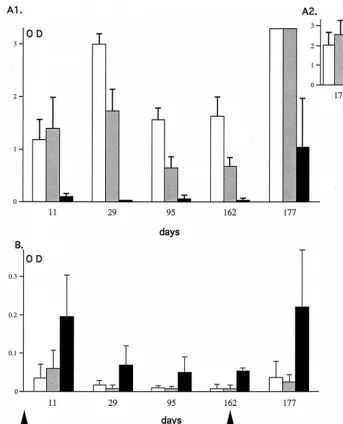
later. At all times tested, the IgG1IgM antibody response in TNF-a/LTa2/2mice was very low, whereas the antibody re-sponse in TNF-a/LTa1/2and TNF-a/LTa1/1mice rose to a peak at day 29 and then decreased and stabilized between days 95 and 162 (Fig. 7A1). Even after the second injection the antibody response was still lower in TNF-a/LTa2/2mice com-pared to the controls (day 177, Fig. 7A1). This is most obvious in Fig. 7A2, where a practically undetectable level of anti-adenovirus antibody in the sera of TNF-a/LTa2/2mice at a dilution of 1:32,000 can be seen compared to the controls. The antibody response of the IgM isotype was higher in TNF-a/LTa2/2, mice with a maximum level evident by day 11. It decreased and remained stable until it peaked again 15 days after adenovirus readministration (Fig. 7B). This higher level of IgM antibody observed in the TNF-a/LTa2/2mice com-pared to TNF-a/LTa1/2and TNF-a/LTa1/1mice is consis-tent with a block in the isotype switch of the humoral response.
Interestingly, Fig. 7 shows that at day 11, the IgG1IgM anti-body response was slightly higher in TNF-a/LTa1/2mice than in TNF-a/LTa1/1mice (Fig. 7A1), which correlates well with the difference in the IgM levels observed the same day (Fig. 7B). At the other time points analyzed (days 29, 95, and 162 p.i.), the IgG1IgM antibody response against adenovirus in TNF-a/ LTa1/2mice was lower than in TNF-a/LTa1/1mice. Experi-ments carried out on larger experimental groups demonstrat-ed that these differences between TNF-a/LTa1/1and TNF-a/ LTa1/2mice are statistically significant (P,0.05) at all of the times tested, suggesting a link between antibody response against adenovirus and TNF-a/LTagene dosage (data not shown).
Impaired proliferative response in TNF-a/LTa2/2mice.We analyzed the proliferative capacity of splenocytes of AdRSVbgal-infected mice at different times p.i. Data presented in Fig. 8 depicts the proliferative capacity of lymphocytes from mice of the three genotypes at day 14 p.i. with 53 109PFU of
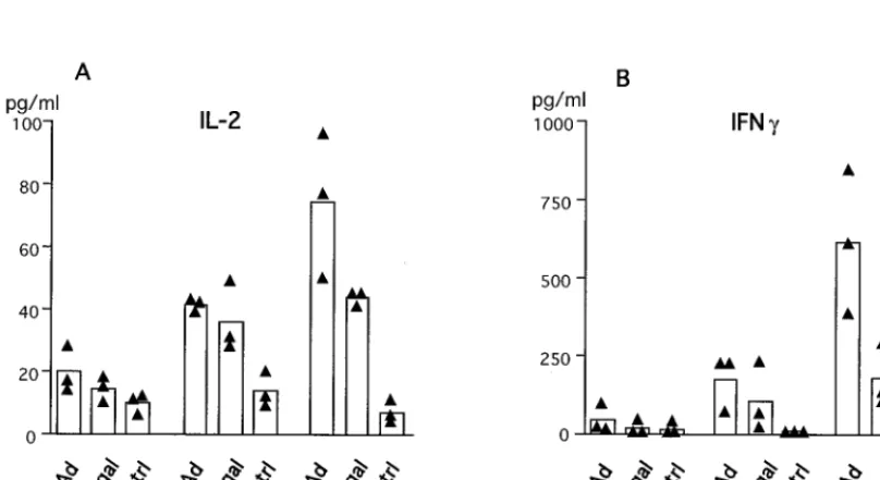
AdRSVbgal. TNF-a/LTa2/2mice were profoundly impaired in their proliferative response against both viral and b-galac-tosidase antigens, while TNF-a/LTa1/2mice present an inter-mediate profile compared to TNF-a/LTa1/1mice. Similar re-sults were consistently obtained with splenocytes at different times p.i. (data not shown). It should be noticed that the im-portant difference in the proliferative capacity between mice within each group may reflect the fact that the mice used in this study are not inbred but were maintained on a mixed 129Sv3 C57BL/6 background. Nevertheless, an intermediate pattern of proliferation was usually observed in TNF-a/LTa1/2 (Fig. 8 and data not shown), suggesting a link with TNF-a/LTagene dosage. Because the T-helper response to recombinant adeno-virus by intravenous delivery is known to implicate the Th1 sub-set in particular (61), we measured the level of IL-2 and IFN-g produced by splenocytes at day 22 p.i. after in vitro restimula-tion with antigen. Figure 9 indicates that viral proteins (heat-inactivated adenovirus) and b-galactosidase both induce the production of high levels of IL-2 (Fig. 9A) and IFN-g(Fig. 9B) by splenocytes of TNF-a/LTa1/1 mice (Fig. 9B) compared with unstimulated splenocytes. Upon restimulation, the levels of both cytokines were lower in TNF-a/LTa1/2mice and even lower in TNF-a/LTa2/2mice. These results are in agreement with the defect in proliferation observed for TNF-a/LTa1/2 and TNF-a/LTa2/2mice (Fig. 8). In all three groups of mice, we detected an inconspicuous level of IL-4 (data not shown), an indicator of Th2 T cells; this is most likely because of the FIG. 6. Efficient readministration of recombinant adenovirus in TNF-a/LTa
-deficient mice. Mice previously injected with Ada1hAT were secondarily in-jected with AdRSVbgal (8.83109PFU) and sacrificed at day 3 following the
second adenovirus administration.b-Galactosidase expression in liver sections after X-Gal staining is shown for TNF-a/LTa1/1(A), TNF-a/LTa1/2(B), and
[image:7.612.67.275.68.477.2]TNF-a/LTa2/2(C) mice. Magnification,3200.
TABLE 3. Neutralizing antibody titers following repeated injection of recombinant adenovirus
Expt group Mouse Antibody titer
aat
First injection Second injection
TNF-a/LTa2/2 1 ,20 ,20
2 ,20 ,20
3 ,20 ,20
TNF-a/LTa1/2 1 160 1,280
2 80 5,120
3 80 10,240
TNF-a/LTa1/1 1 160 2,560
2 80 5,120
3 160 5,120
aThe values represent neutralizing antibody titers after the first injection of
53109PFU of Ada1hAT (sera at day 29) and the second injection of 8.83109
PFU of AdRSVbgal (sera at day 15).
9520 BENIHOUD ET AL. J. VIROL.
on November 9, 2019 by guest
http://jvi.asm.org/
[image:7.612.308.548.565.700.2]paucity of this population, as described by others in another model of liver gene transfer (61).
DISCUSSION
The absence of TNF-aand LTain mice results in an im-paired immune response to recombinant adenovirus; this re-sponse is characterized by an attenuated acute-phase inflam-mation and the presence of a minor cellular infiltrate in the liver. These mice show a lack of proliferative response and an impaired humoral response against both the virus capsid and the transgene product. We report here for the first time that the absence of antiadenovirus neutralizing antibodies in this context allows an efficient secondary adenovirus-mediated gene transduction. An interesting observation lies in the correlation between TNF-a/LTagene dosage and the intensity of antibody response. A detailed analysis of antibody isotypes
[image:8.612.134.477.69.493.2]demon-strates the lack of serum antibodies not only of the IgG but also of the IgA isotype following adenovirus administration in the absence of both TNF-aand LTa. That an increase in the IgM/ IgG ratio is apparent in our study is consistent with the absence of isotype switching in TNF-a/LTa2/2mice, and this corre-lates with an abnormal splenic microarchitecture (33, 48). As shown in Fig. 6, these antibodies were, however, inefficient in preventing virus readministration. In another study, a disabled antibody response has been evidenced in mice after adminis-tration of an E1-deleted recombinant adenovirus expressing the E3 region from the cytomegalovirus promoter (27). In contrast to our study, a general impairment of virus-specific antibodies was documented, including IgM (27). Different im-munosuppression mechanisms are therefore involved upon ad-enovirus injection in TNF-a/LTa-deficient mice versus wild-type mice injected with such an E3-overexpressing virus. FIG. 7. Time course of antibody response in sera of mice deficient or not for TNF-a/LTa. Sera of TNF-a/LTa1/1(white bars), TNF-a/LTa1/2(gray bars), and
TNF-a/LTa2/2(black bars) mice were obtained at different days (11, 29, 95, and 162) after the first adenovirus administration (53109PFU of Ada1hAT) and at 15
days (day 177) after the second adenovirus administration (8.83109PFU of AdRSVbgal). The sera were tested for the presence of IgG1IgM antibodies (upper panel)
or IgM (lower panel). The values correspond to the mean (n53) of relative amounts (OD490) of antibodies in sera at dilutions of 1:4,000 (A1), 1:32,000 (A2), and
1:500 (B). Accurate estimation of antibody levels at day 177 was made on sera at a dilution of 1:32,000 in order to be within the linear range of the ELISA (A2). The standard deviations are shown.
on November 9, 2019 by guest
http://jvi.asm.org/
We observed a weak acute-phase inflammation process in TNF-a/LTa2/2and TNF-a/LTa1/2mice compared to wild-type animals, with the most obvious differences at day 2 p.i. One explanation for the weak acute-phase inflammatory pro-cess we observed may be due to the fact that TNF-a/LTa2/2 and TNF-a/LTa1/2express no, or only a low level of, TNF-a, a strong inducer of mouse acute-phase proteins (43). Because IL-6 is a promoter of acute-phase proteins (53), the low
lev-el of SAP observed in TNF-a/LTa2/2 and TNF-a/LTa1/2 splenocytes after adenovirus infection compared to TNF-a/ LTa1/1mice may also be explained by the limited ability of these mice to produce IL-6. Finally, it should be emphasized that the low levels of IL-6 in TNF-a/LTa2/2mice compared to TNF-a/LTa1/2and to TNF-a/LTa1/1mice are in agreement with the role of TNF-aas a positive regulator of IL-6 levels (52).
Binding of TNF-aon TNFRI is essential for upregulation of adhesion molecules such as E-selectin and VCAM-1 and for the production of chemoattractant cytokines that are required for leukocyte organ infiltration (17, 44, 45). We thus assessed the extent of liver infiltration in our TNF-a/LTa2/2model and found a significant reduction compared to TNF-a/LTa1/1 mice at day 16 p.i. but not at earlier time points. In contrast, an impaired intrahepatic recruitment of leukocytes has been re-ported as early as day 7 p.i. in TNF-a/LTa2/2mice (15). This difference most likely results from the absence of secondary lymphoid organs in our knockout mice due to the lack of LTb receptor signaling in the absence of LTa. We thus believe that leukocyte recruitment from the periportal and perivascular regions of the liver may be quicker in the absence of LTa, translating at early time points p.i. into a pattern of inflamma-tion in TNF-a/LTa2/2 mice that is similar to that in TNF-a/LTa1/1 mice. Indeed, immunostaining performed on liver sections demonstrated an increase of CD81and CD41cells in both groups of mice as early as day 7 p.i. These cells most likely illustrate an ongoing immune process by CTL and T-helper cells that might explain the absence of prolonged expression of transgene-expressing cells in TNF-a/LTa2/2mice. However, compared with TNF-a/LTa1/1mice, TNF-a/LTa2/2mice are impaired in their ability to recruit Mac1-positive cells since there is no evidence of clusters in the liver parenchyme.
After intravenous adenovirus administration in TNF-a/ LTa2/2mice, we observed a deficient ability of T-helper lym-phocytes to proliferate in response to either viral or transgene product antigen after in vitro restimulation. This deficiency is associated with a lack of production of Th1 cytokines. Such a FIG. 8. Deficient lymphoproliferative response in TNF-a/LTa-deficient
mice. Spleen cells from mice at day 14 p.i. with 53109PFU of AdRSVbgal were
[image:9.612.65.272.72.279.2]stimulated in vitro with AdRSVbgal (10 PFU/cell [Adbgal]), an adenovirus with no transgene (10 PFU/cell) that was either viable (AdCO1) or heat-inactivated (inact.), orb-galactosidase (bgal). The data presented on a logarithmic scale are expressed in counts per minute as indicated in Materials and Methods. The triangles represent the data (mean of quadruplicates) from the spleen of one animal, and the bars represent the mean (n53) of each experimental group.
FIG. 9. Defects in Th1 cytokine production in TNF-a/LTamice following adenovirus administration. Spleen cells from AdRSVbgal-infected mice at day 22 p.i. were cultured in the presence of AdCO1 (Ad) orb-galactosidase (bgal) or in medium alone (ctrl). Supernatants were collected after 2 days and were tested in duplicate for the presence of IL-2 (A) or IFN-g(B). Black triangles represent data from individual mice, and the bars represent the mean (n53) of each group.
9522 BENIHOUD ET AL. J. VIROL.
on November 9, 2019 by guest
http://jvi.asm.org/
[image:9.612.98.502.479.699.2]defect in Th1 response was reported for TNF-a/LTa2/2mice during infection with Candida albicans (41). The absence of a T-helper proliferative response may lead to the absence of cooperation with B cells for the production of antibodies di-rected against a T-dependent antigen such as the adenovirus. The absence of a T-helper response may also impair the co-operation with cytotoxic T cells and may explain why TNF-a/LTa2/2mice exhibit an attenuated CTL response directed against vaccinia virus or lymphocytic choriomeningitis virus (17). Because such a reduction of the cytotoxic or proliferative response was not observed in TNF-a2/2mice (15, 39), this defect most likely stems from the interruption of LTbreceptor signaling and is possibly correlated with the aberrant splenic microarchitecture.
Independent studies in different knockout models have helped to clarify the respective roles of LTa and TNF-ain the humoral immune response to various antigens. For ex-ample, mice lacking LTaexhibit a defect in secondary lym-phoid organ development and an unusual architecture of the spleen that translate into an impaired humoral immune response (3). LTa2/2mice also present a defective isotype switching that has been assigned to the breakdown of signal-ing through the LTb receptor, since normal switching oc-curs in both TNFRI2/2or TNFRII2/2mice (33). Both TNF-a2/2and TNFRI2/2mice exhibit a complete lack of splenic germinal centers, primary B-cell follicles, and follicular den-dritic cells, but they remain capable of mounting an IgM and IgG antibody response. These mice, however, fail to maintain a sustained IgG antibody response, apparently because follic-ular dendritic cells do not deliver a proper stimulation to activated B cells within the germinal centers. Because such a lack of IgG antibodies against adenovirus has been described in TNF-a knockout mice (15), signalization through TNF-a binding to TNFRI is likely to be required for a full IgG anti-body response (33, 46). Recently, the use of fusion proteins encoding soluble TNFR-p55 or LTb-R led to conclusions slightly different from those obtained with different TNF/LT knockout mice. In particular, this approach helped to discrim-inate between the role of TNF-a and LTa in development versus their physiological role in adults. Thus, in adult mice the inhibition of TNFR-p55 did not affect the splenic architecture or the humoral response against sheep erythrocytes, while in-hibition of LTa/bsignaling led to profound defects such as the absence of B-cell follicles in the spleen, the absence of germi-nal center formation, and impaired production of immuno-globulin to sheep erythrocytes (38). It remains to be clarified whether the apparent dominant influence of the LTa/bsystem relative to TNF in the humoral response in adults is true for adenoviral antigen. Indeed, while adenovirus encodes three proteins which antagonize the TNFR-p55 pathway (13, 30), no LTa/bpathway antagonist activity has yet been assigned to E3 region products; this discovery would support a crucial role of LTa/bin the antiadenovirus immune response.
The role of TNF-ain antibody response is further supported by the in vitro observation that soluble and membrane-bound TNF-a can rescue the Burkitt lymphoma B-cell line Ramos from B-cell receptor-induced cell death (35). Furthermore, cell-associated TNF-aon activated CD41T cells has been shown to deliver a costimulatory signal to human B cells (2, 12, 37). Taken together, these data clearly implicate TNF-a as a major player in B-cell activation and antibody response. Our comparison of the antibody response in TNF-a/LTa1/1 and TNF-a/LTa1/2mice correlates the magnitude of the an-tibody response with the TNF-a/LTagene dosage. Although we could not detect TNF-ain the serum of all virus-infected groups, levels of this cytokine in serum have been reported to
be 20- to 100-fold lower in samples from heterozygous TNF-a/ LTa1/2mice than in their wild-type counterparts when a very potent TNF-aactivator (e.g., lipopolysaccharide) was used (1). More interestingly, we demonstrated that the level of TNF-a produced by activated splenocytes is significantly lower in TNF-a/ LTa1/2mice than in TNF-a/LTa1/1mice. The overall data link antibody levels to TNF-aconcentrations. This correlation is further supported by an enhanced antibody response against T-cell-dependent antigen following administration of recombi-nant TNF-ato mice (21).
We have found no effect of TNF-aand LTaon the duration of transgene expression (aAT orb-galactosidase) in our mu-rine knockout model. In contrast, TNF-adeficiency has been recently reported to markedly attenuate the clearance of cir-culating chloramphenicol acetyltransferase enzyme encoded by a recombinant adenovirus (AdCAT) (15). This apparent con-tradiction may result from the use of different promoters (the adenovirus major late promoter for Ada1hAT, the LTR of Rous sarcoma virus [RSV] for AdRSVbgal, and the immedi-ate-early promoter of cytomegalovirus for AdCAT), as the weaker inflammatory setting in our knockout model might have impaired the activity of the RSV promoter. Indeed, an extinction of RSV-drivenb-galactosidase expression has been shown for an E1E4-defective adenovirus that correlated with a weaker inflammation in mice (11). The evaluation of transgene expression driven by a house-keeping gene promoter in our knockout model may help to answer this issue.
In summary, TNF-a plays roles at different levels of the immune response. First, as a proinflammatory cytokine it can trigger early events of inflammation. Second, as an effector of cytolysis of TNF receptor-bearing cells, it can eliminate the transduced cells while recruiting leukocytes and macrophages following the liberation of inflammation mediators. Third, TNF-acan provide a survival signal to B lymphocytes. Finally, mem-brane-bound TNF-aon T-helper cells can provide additional costimulatory signals to activated B cells. Strategies aimed at blocking this cytokine may thus prove useful for gene therapy applications requiring multiple adenovirus administrations.
ACKNOWLEDGMENTS
We are most grateful to S. Esselin for assistance. We are profoundly indebted to L. Franqueville for viral stock preparations. We also thank N. DiFalco and D. Grandjon, as well as all of the staff of the animal facilities of the IGR. We thank all of the staff of the Service Multime-dia of the IGR for photographic work. We express our gratitude to Gahe´ry-Se´gard for helpful advice in measurement ofb-galactosidase activity. Finally, we thank P. Bobe´, M. Robert-Le Meur, A. Maron, and K. Sollerbrant for helpful discussions.
This work was supported in part by the CNRS, the Association Franc¸aise de Lutte contre la Mucoviscidose (AFLM), and the Associ-ation Franc¸aise pour la Myopathie (AFM). K.B. was supported by the AFLM and Rhoˆne-Poulenc Gencell.
REFERENCES
1. Amiot, F., O. Boussadia, S. Cases, C. Fitting, M. Lebastard, J.-M. Cavaillon, G. Milon, and F. Dautry.1997. Mice heterozygous for a deletion of the tumor necrosis factor-alpha and lymphotoxin-alpha genes: biological impor-tance of a nonlinear response of tumor necrosis factor-alpha to gene dosage. Eur. J. Immunol. 27:1035–1042.
2. Aversa, G., J. Punnonen, and J. E. de Vries. 1993. The 26-kD transmembrane form of tumor necrosis factor alpha on activated CD41T cell clones
pro-vides a costimulatory signal for human B cell activation. J. Exp. Med. 177: 1575–1585.
3. Banks, T. A., B. T. Rouse, M. K. Kerley, P. J. Blair, V. L. Godfrey, N. A. Kuklin, D. M. Bouley, J. Thomas, S. Kanangat, and M. L. Mucenski.1995. Lymphotoxin-alpha-deficient mice. Effects on secondary lymphoid organ development and humoral immune responsiveness. J. Immunol. 155:1685– 1693.
4. Barr, D., J. Tubb, D. Ferguson, A. Scaria, A. Lieber, C. Wilson, J. Perkins, and M. A. Kay.1995. Strain-related variations in adenoviral mediated
on November 9, 2019 by guest
http://jvi.asm.org/
gene expression from mouse hepatocytes in vivo: comparisons between im-munocompetent and immunodeficient inbred strains. Gene Ther. 2:151–155. 5. Browning, J. L., A. Ngam-Ek, P. Lawton, J. DeMarinis, R. Tizard, E. P. How, C. Hession, B. O’Brine-Greco, S. F. Foley, and C. F. Ware.1993. Lympho-toxin-beta, a novel member of the TNF family forms a heteromeric complex with lymphotoxin on the cell surface. Cell 72:847–856.
6. Bruder, J. T., T. Jie, D. L. McVey, and I. Kovesdi. 1997. Expression of gp19k increases the persistence of transgene expression from an adenovirus vector in the mouse lung. J. Virol. 71:7623–7628.
7. Chen, P., J. Tian, I. Kovesdi, and J. T. Bruder. 1998. Interaction of the adenovirus 14.7-kDa protein with FLICE inhibits Fas Ligand-induced apo-ptosis. J. Biol. Chem. 273:5815–5820.
8. Crowe, P. D., T. L. VanArsdale, B. N. Walter, C. F. Ware, C. Hession, B. Ehrenfels, J. L. Browning, W. S. Din, R. G. Goodwin, and C. A. Smith.1994. A lymphotoxin-beta specific receptor. Science 264:707–710.
9. Dai, Y., E. M. Schwarz, D. Gu, W.-W. Zhang, N. Sarvetnick, and I. M. Verma.1995. Cellular and humoral responses to adenoviral vectors contain-ing factor IX gene: tolerization of factor IX and vector antigens allows for long term expression. Proc. Natl. Acad. Sci. USA 92:1401–1405. 10. De Matteo, R. P., G. Chu, M. Ahn, E. Chang, C. F. Barker, and J. F.
Markmann. 1997. Long-lasting adenovirus transgene expression in mice through neonatal intrathymic tolerance induction without the use of immu-nosuppression. J. Virol. 71:5330–5335.
11. Dedieu, J.-F., E. Vigne, C. Torrent, C. Jullien, I. Mahfouz, J.-M. Caillaud, N. Aubailly, C. Orsini, J.-M. Guillaume, P. Opolon, P. Delae`re, M. Perricaudet, and P. Yeh.1997. Long-term gene delivery into the livers of immunocom-petent mice with E1/E4-defective adenoviruses. J. Virol. 71:4626–4637. 12. Del Prete, G., M. De Carli, M. M. D’Elios, I. M. Fleckenstein, H.
Ficken-scher, B. Fleckenstein, F. Almerigogna, and S. Romagnani.1994. Polyclonal B cell activation induced by herpesvirus saimiri-transformed human CD41T
cell clones. J. Immunol. 152:4872–4879.
13. Dimitrov, T., P. Krajcsi, T. W. Hermiston, A. E. Tollefson, M. Hannink, and W. S. M. Wold.1997. Adenovirus E3-10.4K/14.5K protein complex inhibits tumor necrosis factor-induced translocation of cytosolic phospholipase A2 to membranes. J. Virol. 71:2830–2837.
14. Dong, J.-Y., D. Wang, F. W. Van Ginkel, D. W. Pascual, and R. A. Frizzell. 1996. Systematic analysis of repeated gene delivery into animal lungs with a recombinant adenovirus vector. Hum. Gene Ther. 7:319–331.
15. Elkon, K. B., C.-C. Liu, J. G. Gall, J. Trevejo, M. W. Marino, K. A. Abra-hamsen, X. Song, J.-L. Zhou, L. J. Old, R. G. Crystal, and E. Falck-Ped-ersen.1997. Tumor necrosis factor alpha plays a central role in immune-mediated clearance of adenoviral vectors. Proc. Natl. Acad. Sci. USA 94: 9814–9819.
16. Engelhardt, J. F., X. Ye, B. Doranz, and J. M. Wilson. 1994. Ablation of E2A in recombinant adenoviruses improves transgene persistence and decreases inflammatory response in mouse liver. Proc. Natl. Acad. Sci. USA 91:6196– 6200.
17. Eugster, H.-P., M. Mu¨ller, U. Karrer, B. D. Car, M. Schnyder, V. M. Eng, G. Woerly, M. Le Hir, F. di Padova, M. Aguet, R. Zinkernagel, H. Bluethmann, and B. Ryffel.1996. Multiple immune abnormalities in tumor necrosis factor and lymphotoxin-alpha double deficient-mice. Int. Immunol. 8:23–36. 18. Fallaux, F. J., O. Kranenburg, S. J. Cramer, A. Houweling, H. van Ormondt,
R. C. Hoeben, and A. J. van der Eb.1996. Characterization of 911: a new helper cell line for the titration and propagation of early-region-1-deleted adenoviral vectors. Hum. Gene Ther. 7:215–222.
19. Gahe´ry-Se´gard, H., V. Juillard, J. Gaston, R. Lengagne, A. Pavirani, P. Boulanger, and J.-G. Guillet.1997. Humoral immune response to the capsid components of recombinant adenoviruses: routes of immunization modulate virus-induced Ig subclass shifts. Eur. J. Immunol. 27:653–659.
20. Gao, G.-P., Y. Yang, and J. M. Wilson. 1996. Biology of adenovirus vectors with E1 and E4 deletions for liver-directed gene therapy. J. Virol. 70:8934– 8943.
21. Ghiara, P., D. Boraschi, L. Nencioni, P. Ghezzi, and A. Tagliabue. 1987. Enhancement of in vivo immune response by tumor necrosis factor. J. Im-munol. 139:3676–3679.
22. Gilardi, P., M. Courtney, A. Pavirani, and M. Perricaudet. 1990. Expression of human alpha-1 antitrypsin using a recombinant adenovirus vector. FEBS Lett. 267:60–62.
23. Ginsberg, H. S., U. Lundholm-Beauchamp, R. L. Horswood, B. Pernis, W. S. M. Wold, R. M. Chanock, and G. A. Prince.1989. Role of early region 3 (E3) in pathogenesis of adenovirus disease. Proc. Natl. Acad. Sci. USA 86: 3823–3827.
24. Ginsberg, H. S., L. L. Moldawer, P. Sehgal, M. Redington, P. L. Kilian, R. M. Chanock, and G. A. Prince.1991. A mouse model for investigating the molecular pathogenesis of adenovirus pneumonia. Proc. Natl. Acad. Sci. USA 88:1651–1655.
25. Grell, M., E. Douni, H. Wajant, M. Lo¨hden, M. Clauss, B. Maxeiner, S. Georgopoulos, W. Lesslauer, G. Kollias, K. Pfizenmaier, and P. Scheurich. 1995. The transmembrane form of tumor necrosis factor is the prime acti-vating ligand of the 80kDa tumor necrosis factor receptor. Cell 83:793–802. 26. Griscelli, F., H. Li, A. Bennaceur-Griscelli, J. Soria, P. Opolon, C. Soria, M. Perricaudet, P. Yeh, and H. Lu.1998. Angiostatin gene transfer: inhibition of
tumor growth in vivo by blockage of endothelial cell proliferation associated with a mitosis arrest. Proc. Natl. Acad. Sci. USA 95:6367–6372.
27. Ilan, Y., G. Droguett, N. Roy Chowdhury, Y. Li, K. Sengupta, N. R. Thum-mala, A. Davidson, J. Roy Chowdhury, and M. S. Horwitz.1997. Insertion of the adenoviral E3 region into a recombinant viral vector prevents antiviral humoral and cellular immune responses and permits long-term gene expres-sion. Proc. Natl. Acad. Sci. USA 94:2587–2592.
28. Ilan, Y., R. Prakash, A. Davidson, V. Jona, G. Droguett, S. M. Horwitz, N. R. Chowdhury, and J. R. Chowdhury. 1997. Oral tolerization to adenoviral antigens permits long-term gene expression using recombinant adenoviral vectors. J. Clin. Invest. 99:1098–1106.
29. Kochanek, S., P. R. Clemens, K. Mitani, H. H. Chen, S. Chan, and C. T. Caskey. 1996. A new adenoviral vector: replacement of all viral coding sequences with 28 kb of DNA independently expressing both full-length dystrophin and beta-galactosidase. Proc. Natl. Acad. Sci. USA 93:5731–5736. 30. Krajcsi, P., A. E. Tollefson, C. W. Anderson, and W. S. M. Wold. 1992. The adenovirus E3 14.5-kDa protein, which is required for down-regulation of the epidermal growth factor receptor and prevention of tumor necrosis factor cytolysis, is an integral membrane protein oriented with its C terminus in the cytoplasm. J. Virol. 66:1665–1673.
31. Kuzmin, A. I., M. J. Finegold, and R. C. Eisensmith. 1997. Macrophage depletion increases the safety, efficacy and persistence of adenovirus-medi-ated gene transfer in vivo. Gene Ther. 4:309–316.
32. Laster, S. M., W. S. M. Wold, and L. R. Gooding. 1994. Adenovirus proteins that regulate susceptibility to TNF also regulate the activity of PLA2. Semin. Virol. 5:431–442.
33. Le Hir, M., H. Bluethmann, M. H. Kosco-Villebois, M. Muller, F. di Padova, M. Moore, B. Ryffel, and H.-P. Eugster.1996. Differentiation of follicular dendritic cells and full antibody responses require tumor necrosis factor receptor-1 signaling. J. Exp. Med. 183:2367–2372.
34. Lee, M. G., M. A. Abina, H. Haddada, and M. Perricaudet. 1995. The constitutive expression of the immunomodulatory gp19k protein in E1-, E3-adenoviral vectors strongly reduces the host cytotoxic T cell response against the vector. Gene Ther. 2:1–7.
35. Lens, S. M. A., K. Tesselaar, B. F. A. Den Drijver, M. H. J. Van Oers, and R. A. W. Van Lier.1996. A dual role for both CD40-ligand and TNF alpha in controlling human B cell death. J. Immunol. 156:507–514.
36. Lieber, A., C.-Y. He, I. Kirillova, and M. A. Kay. 1996. Recombinant adeno-viruses with large deletions generated by Cre-mediated excision exhibit dif-ferent biological properties compared with first-generation vectors in vitro and in vivo. J. Virol. 70:8944–8960.
37. Macchia, D., F. Almerigogna, P. Parronchi, A. Ravina, E. Maggi, and S. Romagnani.1993. Membrane tumor necrosis factor alpha is involved in the polyclonal B-cell activation induced by HIV-infected human T cells. Nature 363:464–466.
38. Mackay, F., G. R. Majeau, P. Lawton, P. S. Hochman, and J. L. Browning. 1997. Lymphotoxin but not tumor necrosis factor functions to maintain splenic architecture and humoral responsiveness in adult mice. Eur. J. Im-munol. 27:2033–2042.
39. Marino, M. W., A. Dunn, D. Grail, M. Inglese, Y. Noguchi, E. Richards, A. Jungbluth, H. Wada, M. Moore, B. Williamson, S. Basu, and L. J. Old.1997. Characterization of tumor necrosis factor-deficient mice. Proc. Natl. Acad. Sci. USA 94:8093–8098.
40. McCoy, R. D., B. L. Davidson, B. J. Roessler, G. B. Huffnagle, S. L. Janich, T. J. Laing, and R. H. Simon.1995. Pulmonary inflammation induced by incomplete or inactivated adenoviral particles. Hum. Gene Ther. 6:1553– 1560.
41. Mencacci, A., E. Cenci, G. Del Sero, C. Fe d’Ostiani, P. Mosci, C. Montag-noli, A. Bacci, F. Bistoni, V. F. Quesniaux, B. Ryffel, and L. Romani.1998. Defective co-stimulation and impaired Th1 development in tumor necrosis factor/lymphotoxin-alpha double-deficient mice infected with Candida
albi-cans. Int. Immunol. 10:37–48.
42. Michalski, J. P., C. C. McCombs, S. Sheth, M. McCarthy, and R. deShazo. 1985. A modified double antibody sandwich enzyme-linked immunosorbent assay for measurement of alpha-1-antitrypsin in biologic fluids. J. Immunol. Methods 83:101–112.
43. Mortensen, R. F., J. Shapiro, B.-F. Lin, S. Douches, and R. Neta. 1988. Interaction of recombinant IL-1 and recombinant tumor necrosis factor in the induction of mouse acute phase proteins. J. Immunol. 140:2260–2266. 44. Neumann, B., T. Machleidt, A. Lifka, K. Pfeffer, D. Vestweber, T. W. Mak, B.
Holzmann, and M. Kro¨nke.1996. Crucial role of 55-kilodalton TNF receptor in TNF-induced adhesion molecule expression and leukocyte organ infiltra-tion. J. Immunol. 156:1587–1593.
45. Ohmori, Y., L. Wyner, S. Narumi, D. Armstrong, M. Stoler, and T. A. Hamilton.1993. Tumor necrosis factor-ainduces cell type and tissue-specific expression of chemoattractant cytokines in vivo. Am. J. Pathol. 142:861–870. 46. Pasparakis, M., L. Alexopoulou, V. Episkopou, and G. Kollias. 1996. Im-mune and inflammatory responses in TNF-alpha-deficient mice: a critical requirement for TNF-alpha in the formation of primary B cell follicles, follicular dendritic cell networks and germinal centers, and in the maturation of the humoral immune response. Proc. Natl. Acad. Sci. USA 184:1397– 1411.
9524 BENIHOUD ET AL. J. VIROL.
on November 9, 2019 by guest
http://jvi.asm.org/
47. Qin, L., Y. Ding, D. R. Pahud, N. D. Robson, A. Shaked, and J. S. Bromberg. 1997. Adenovirus-mediated gene transfer of viral interleukin-10 inhibits the immune response to both alloantigen and adenoviral antigen. Hum. Gene Ther. 8:1365–1374.
48. Ryffel, B., F. Di Padova, M. H. Schreier, M. Le Hir, H.-P. Eugster, and V. F. J. Quesniaux.1997. Lack of type 2 T cell-independent B cell responses and defect in isotype switching in TNF-lymphotoxin alpha-deficient mice. J. Immunol. 158:2126–2133.
49. Shisler, J., C. Yang, B. Walter, C. F. Ware, and L. R. Gooding. 1997. The adenovirus E3-10.4K/14.5K complex mediates loss of cell surface Fas (CD95) and resistance to Fas-induced apoptosis. J. Virol. 71:8299–8306. 50. Smith, T. A. G., B. D. White, J. M. Gardner, M. Kaleko, and A. McClelland.
1996. Transient immunosuppression permits successful repetitive intrave-nous administration of an adenovirus vector. Gene Ther. 3:496–502. 51. Sparer, T. E., R. A. Tripp, D. L. Dillehay, T. W. Hermiston, W. S. Wold, and
L. R. Gooding.1996. The role of human adenovirus early region 3 proteins (gp19k, 10.4K, 14.5K, and 14.7K) in a murine pneumonia model. J. Virol. 70: 2431–2439.
52. Starnes, H. F., M. K. Pearce, A. Tewari, J. H. Yim, J.-C. Zou, and J. S. Abrams.1990. Anti-IL-6 monoclonal antibodies protect against lethal
Esch-erichia coli infection and lethal tumor necrosis factor-achallenge in mice. J. Immunol. 145:4185–4191.
53. Steel, D. M., and A. S. Whitehead. 1994. The major acute phase reactants: C-reactive protein, serum amyloid P component and serum amyloid A pro-tein. Immunol. Today 15:81–88.
54. Stratford-Perricaudet, L. D., I. Makeh, M. Perricaudet, and P. Briand. 1992. Widespread long-term gene transfer to mouse skeletal muscles and heart. J. Clin. Invest. 90:626–630.
55. Thimmappaya, B., C. Weinberger, R. J. Schneider, and T. Shenk. 1982. Adenovirus VAI RNA is required for efficient translation of viral mRNAs at late times after infection. Cell 31:543–551.
56. Tollefson, A. E., T. W. Hermiston, D. L. Lichtenstein, C. F. Colle, R. A. Tripp, T. Dimitrov, K. Toth, C. E. Wells, P. C. Doherty, and W. S. M. Wold. 1998. Forced degradation of Fas inhibits apoptosis in adenovirus-infected cells. Nature 392:726–729.
57. Tripathy, S. K., H. B. Black, E. Goldwasser, and J. Leiden. 1996. Immune responses to transgene-encoded proteins limit the stability of gene expres-sion after injection of replication-defective adenovirus vectors. Nat. Med. 2: 545–550.
58. Walter, J., Q. You, J. N. Hagstrom, M. Sands, and K. A. High. 1996. Successful expression of human factor IX following repeat administration of an adenoviral vector in mice. Proc. Natl. Acad. Sci. USA 93:3056–3061. 59. Wang, Q., G. Greenburg, D. Bunch, D. Farson, and M. H. Finer. 1997.
Persistent transgene expression in mouse liver following in vivo gene transfer with a delta E1/delta E4 adenovirus vector. Gene Ther. 4:393–400. 60. Wolff, G., S. Worgall, N. van Rooijen, W.-R. Song, B.-G. Harvey, and R. G.
Crystal.1997. Enhancement of in vivo adenovirus-mediated gene transfer and expression by prior depletion of tissue macrophages in the target organ. J. Virol. 71:8798–8807.
61. Yang, Y., H. C. Ertl, and J. M. Wilson. 1994. MHC class I-restricted cytotoxic T lymphocytes to viral antigens destroy hepatocytes in mice infected with E1-deleted recombinant adenoviruses. Immunity 1:433–442.
62. Yang, Y., Q. Li, H. C. J. Ertl, and J. M. Wilson. 1995. Cellular and humoral immune responses to viral antigens create barriers to lung-directed gene therapy with recombinant adenoviruses. J. Virol. 69:2004–2015.
63. Yang, Y., Q. Su, I. S. Grewal, R. Schilz, R. A. Flavell, and J. M. Wilson. 1996. Transient subversion of CD40 ligand function diminishes immune responses to adenovirus vectors in mouse liver and lung tissues. J. Virol. 70:6370–6377. 64. Yang, Y., G. Trinchieri, and J. M. Wilson. 1995. Recombinant IL-12 prevents formation of blocking IgA antibodies to recombinant adenovirus and allows repeated gene therapy to mouse lung. Nat. Med. 1:890–893.