0022-538X/96/$04.0010
Copyrightq1996, American Society for Microbiology
Herpes Simplex Virus Type 1 Alkaline Nuclease Is Required for
Efficient Processing of Viral DNA Replication Intermediates
RIK MARTINEZ,1ROBERT T. SARISKY,2† PETER C. WEBER,3
ANDSANDRA K. WELLER1*
Department of Microbiology, University of Connecticut Health Center, Farmington, Connecticut 06030,1Department of Microbiology and Immunology, Milton S. Hershey Medical Center, Pennsylvania State University,
Hershey, Pennsylvania 17033,2and Infectious Diseases Section, Parke-Davis Pharmaceutical Research, a division of Warner-Lambert Company, Ann Arbor, Michigan 481053
Received 28 August 1995/Accepted 19 December 1995
Mutations in the alkaline nuclease gene of herpes simplex virus type 1 (HSV-1) (nucmutations) induce
almost wild-type levels of viral DNA; however, mutant viral yields are 0.1 to 1% of wild-type yields (L. Shao, L. Rapp, and S. Weller, Virology 195:146–162, 1993; R. Martinez, L. Shao, J. C. Bronstein, P. C. Weber, and
S. Weller, Virology 215:152–164, 1996).nucmutants are defective in one or more stages of genome maturation
and appear to package DNA into aberrant or defective capsids which fail to egress from the nucleus of infected
cells. In this study, we used pulsed-field gel electrophoresis to test the hypothesis that the defects in nuc
mutants are due to the failure of the newly replicated viral DNA to be processed properly during DNA replication and/or recombination. Replicative intermediates of HSV-1 DNA from both wild-type- and mutant-infected cells remain in the wells of pulsed-field gels, while free linear monomers are readily resolved. Digestion of this well DNA with restriction enzymes that cleave once in the viral genome releases discrete monomer DNA
from wild-type virus-infected cells but not fromnucmutant-infected cells. We conclude that both wild-type and
mutant DNAs exist in a complex, nonlinear form (possibly branched) during replication. The fact that discrete
monomer-length DNA cannot be released fromnucDNA by a single-cutting enzyme suggests that this DNA is
more branched than DNA which accumulates in cells infected with wild-type virus. The well DNA from cells
infected with wild-type and nuc mutants contains XbaI fragments which result from genomic inversions,
indicating that alkaline nuclease is not required for mediating recombination events within HSV DNA.
Furthermore,nucmutants are able to carry out DNA replication-mediated homologous recombination events
between inverted repeats on plasmids as evaluated by using a quantitative transient recombination assay. Well
DNA from both wild-type- and mutant-infected cells contains free ULtermini but not free UStermini. Various
models to explain the structure of replicating DNA are considered.
The molecular mechanisms of DNA replication vary widely from virus to virus. Circular duplex DNAs are generally thought to replicate in a theta structure (12) or through a rolling-circle mechanism (21). The small eukaryotic papovavi-ruses such as simian virus 40 replicate entirely by theta repli-cation. In contrast, bacteriophage lambda replicates initially as a theta form and switches to rolling-circle replication later in infection (17). The DNA bacteriophage T4 replicates as a linear DNA molecule, and initiation occurs by two markedly different strategies (36). The initial phase of T4 DNA replica-tion occurs at specific origins of replicareplica-tion (33); however, late DNA replication occurs by a recombination-dependent mech-anism in which DNA synthesis is primed from the ends of DNA strands that have invaded homologous segments of other DNA molecules (36, 44).
The 152-kb herpes simplex virus type 1 (HSV-1) genome is composed of two components, termed L and S. The L
compo-nent consists of unique sequences (UL) flanked by the
invert-ed-repeated sequences ab and b9a9, whereas, the S component
consists of unique sequences (US) flanked by
inverted-re-peated sequences ac and c9a9 (25, 54) (Fig. 1A). During the
course of viral DNA replication, the two unique regions invert
relative to one another (25). Genome isomerization most likely results from generalized recombination between inverted re-peats flanking the L and S components (50, 56, 57). DNA replication results in the formation of large head-to-tail con-catemers consisting of tandem repeats of the viral genome (6–8, 26, 29, 30) which can be chased into monomer-length viral genomes (30, 31, 47). Although rolling-circle DNA repli-cation may be involved in the generation of large concatemeric molecules, it is also possible that HSV DNA replication occurs by a mechanism which is inherently recombinogenic in a man-ner analogous to T4 bacteriophage DNA replication (36, 44). In T4, replication and/or recombination results in the genera-tion of branched forms of viral DNA which must be resolved prior to packaging into preformed phage heads, using the product of the T4 gene 49, endonuclease VII (32, 42). On the basis of the phenotype of HSV-1 mutants defective in the UL12 gene, we previously proposed that the viral alkaline nuclease may carry out an analogous function (53).
HSV alkaline nuclease (UL12) is a relatively abundant 85-kDa phosphoprotein in infected cells (2, 3). UL12 is encoded
by a 2.3-kb member of a family of unspliced 39 coterminal
mRNAs (13, 15). We have recently demonstrated that, as orig-inally proposed by Costa et al. (13), another member of this family of mRNAs (1.9 kb) encodes an N-terminally truncated protein (UL12.5) which shares its carboxy terminus with the alkaline nuclease protein (38). A viral frameshift mutant (AN-F1) that was constructed is predicted to abolish the gene prod-uct of the 2.3-kb mRNA (full-length alkaline nuclease) but leave intact the UL12.5 product of the 1.9-kb mRNA. Since no
* Corresponding author. Mailing address: Department of Microbi-ology, University of Connecticut Health Center, 263 Farmington Ave., Farmington, CT 06030. Phone: (860) 679-2310. Fax: (860) 679-1239. Electronic mail address: [email protected].
† Present address: Department of Pharmacology and Molecular Sci-ence, Johns Hopkins School of Medicine, Baltimore, MD 21205.
2075
on November 9, 2019 by guest
http://jvi.asm.org/
exonuclease activity was observed in cells infected with AN-F1, it appears that UL12.5 may have little or no enzymatic activity (38). We previously reported the construction of a null muta-tion (AN-1) in the HSV-1 alkaline nuclease gene (53, 62). Both
nuc mutants (AN-1 and AN-F1) form very small plaques on
Vero cells, and mutant virus yields on nonpermissive cells are approximately 0.1 to 1% of that seen for wild-type virus; how-ever, they synthesize wild-type levels of viral DNA and late proteins.
Although cleavage of viral DNA into monomeric units and packaging of viral DNA into capsids in the nucleus occurred efficiently in cells infected with nuc mutants, three lines of evidence suggest that the maturation of DNA-containing cap-sids into infectious virions is defective in nuc mutants (38, 53, 65). (i) In contrast to wild-type-infected cells, very small amounts of staphylococcal nuclease (SN)-protected DNA were detected in cytoplasmic extracts from nuc mutant-infected cells. (ii) Very few if any mature, DNA-containing capsids were observed in the cytoplasm upon analysis by electron micros-copy or sucrose gradient sedimentation. (iii) Analysis of mu-tant-infected cells by sucrose gradient sedimentation indicated an elevated ratio of A to B capsids. Type B capsids are believed to be precursors to the DNA-containing C capsids found in the
cytoplasm (27, 28); A capsids are believed to result from abor-tive, unsuccessful attempts to package viral DNA into B cap-sids (1, 34, 55). These data indicate that nuc mutants may be defective for the production of capsids competent to mature into the cytoplasm. We have proposed that the role of alkaline nuclease in the viral life cycle is to resolve or debranch recom-bination intermediates which are formed during the DNA rep-lication process (53, 65). The model posits that if reprep-lication/ recombination intermediates are not resolved properly, viral DNA can be cleaved and taken up into capsids, but these capsids are unstable and not competent to mature into the cytoplasm.
[image:2.612.141.462.69.410.2]We set out to analyze the DNA which accumulates in cells infected with wild-type and nuc mutant viruses. Previous stud-ies have shown that pulsed-field gel electrophoresis (PFGE) and field inversion gel electrophoresis are powerful tools in the analysis of large viral DNAs (4, 20, 41, 51, 70). Several intrigu-ing observations have been made regardintrigu-ing the structure of replicating viral DNA. First, replication intermediates in HSV-1-infected cells are present in complex nonlinear structures which cannot enter a pulsed-field gel. After digestion with a restriction enzyme which has a single recognition site within the HSV genome, some but not all of the viral DNA is released
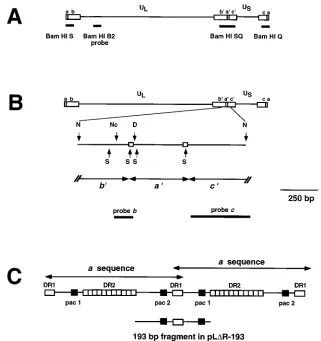
FIG. 1. Structure of the HSV-1 genome showing the locations of various probes and fragments used in this study. (A) Diagram of the HSV-1 genome showing the ULand USregions of the genome as well as the flanking repeated sequences ab, b9a9c9, and ca. The location of the BamHI B2 probe used in this study is shown (described in Materials and Methods). The locations of the BamHI SQ, S, and Q fragments are also shown. (B) Diagram of the HSV-1 genome showing an expanded view of the NarI fragment in pNar. Restriction sites are shown for NarI (N), SmaI (S), NcoI (Nc), and DraI (D). Boxes on the second line denote DR1 sequences which flank the a sequence. The third line shows the locations of the a, b, and c sequences within pNar. On the fourth line, the positions of probes b and c are shown. (C) A tandem HSV-1 a-sequence junction with locations of sequences DR1, DR2, and the pac1 and pac2 elements. The portion of the a sequence present in pLDR-193 is shown on the bottom line. This sequence represents the minimal signal sufficient for mediating cleavage and packaging of viral DNA (46).
on November 9, 2019 by guest
http://jvi.asm.org/
into a unit-length molecule (51). Second, analysis of newly replicated DNA by PFGE indicates that inversion has occurred at the earliest times that replicated DNA can be detected (4, 40, 51, 70). In this report, PFGE was used to analyze the replication intermediates which accumulate in cells infected with wild-type or nuc mutant HSV-1. We find that complex nonlinear structures arise during replication of both wild-type and nuc mutant viruses and that inversion events have oc-curred early in the process in both viruses. After digestion with a single-cutting enzyme, no discrete fragments are released from replicating DNA from cells infected with nuc mutants, whereas discrete fragments are released in DNA from cells infected with wild-type virus. This finding indicates that the DNA which accumulates in these cells is even more complex in structure than that in cells infected with wild-type virus.
MATERIALS AND METHODS
Cells and viruses.African green monkey kidney cells (Vero; American Type Culture Collection) were grown and maintained as described previously (64). Cell line 6-5, which is permissive for nuc mutants, was generated and maintained as described previously (53). The KOS strain of HSV-1 was used as the wild-type virus. AN-1 is a null mutant in the alkaline nuclease gene in which 917 bp from the N-terminal half of the gene (corresponding to residues 70 to 375) were deleted and replaced by an insertional mutagen consisting of the lacZ gene of
Escherichia coli under the control of the regulatory elements of the large subunit
of ribonucleotide reductase (ICP6) (65). AN-F1 and AN-F3 are independent isolates containing a 2-bp insertion which results in the premature termination of translation of the 2.3-kb alkaline nuclease mRNA (38). The growth phenotypes of the null and frameshift mutants are identical (38). AN-1-2a is a recombinant virus in which the deletion-insertion of AN-1 has been replaced by the wild-type UL12 gene by marker rescue; the growth properties of this virus resemble those of wild-type KOS (65).
Plasmids and bacteria.Plasmids pLR and pLDR-193, carrying HSV-1 oriSand different sets of inverted-repeat sequences, were previously described (50). pLR is a construct containing two copies of the IS50 element of Tn5. pLDR-193 contains a 193-bp (Fig. 1C) fragment from the a sequence inserted into a deleted version of the IS50 element (all but 236 bp of the IS50Relement deleted). The 193-bp fragment contains the pac2-DR1-pac1 region from two tandemly re-peated a sequences and lacks the DR2 sequences. This fragment is similar to one which has been shown to function as a minimal cleavage and packaging signal (46). Plasmid pSG124 containing the EcoRI A fragment of HSV-1 strain KOS (map coordinates 0.495 to 0.635) was previously described (22). Plasmids pSG10, containing the EcoRI D fragment of HSV-1 strain KOS (map coordinates 0.086 to 0.194), and pSG10-B2, containing an internal BamHI fragment (map coordi-nates 0.145 to 0.165) from pSG10, were described previously (63). Plasmid pNN9, containing two copies of the 5.9-kb BamHI K fragment from strain KOS cloned into pUC19, was generously provided by Mark Challberg (National In-stitutes of Health, Bethesda, Md.). pNar contains a 1,209-bp NarI fragment (sequence coordinates 125585 to 126793 [39]) from pNN9 cloned into the NarI site of pSL301 (Invitrogen); this NarI fragment contains the entire a sequence (400 bp) and parts of the flanking b (389 bp) and c (420 bp) sequences from HSV-1 strain KOS (Fig. 1B). The published sequence for HSV-1 strain 17 (39) lists an additional NarI site in this region at position 125789 which is not present in strain KOS (38a). Recombinant plasmids were propagated in E. coli UT481 or DH5aby standard procedures (37).
DNA isolation and Southern blotting.Virion DNA was isolated from purified virions following equilibrium centrifugation in CsCl as described previously (66). Cellular DNA was isolated as previously described (66). For analysis of DNA by Southern blotting, virion or infected-cell DNA was digested with a restriction endonuclease(s) as directed by the manufacturer, subjected to agarose gel electrophoresis, and transferred to a GeneScreen Plus nylon membrane (New England Nuclear Corp., Boston, Mass.) as recommended by the supplier. Blots to be rehybridized with separate probes were stripped by boiling in TE (10 mM Tris [pH 8.0], 1 mM EDTA [pH 8.0]) with 1% sodium dodecyl sulfate for 30 min.
DNA fragments to be used as probes for hybridization were labeled as de-scribed previously (18), using the random hexamer-primed method (Boehringer Mannheim, Indianapolis, Ind.). Virion DNA was digested with BamHI and
EcoRI prior to labeling. In several experiments, subfragments of recombinant
plasmids were isolated following restriction enzyme digestion, agarose gel elec-trophoresis, and electroelution of the appropriate fragment (37). For fragments smaller than 150 bp in length,b-agarase (New England Biolabs [NEB], Beverly, Mass.) was used to dissolve the agarose as directed by the manufacturer. For the transient recombination assay, a radiolabeled 3.4-kb HindIII fragment of Tn5 was used as a probe as described previously (50). Other HSV genomic fragments used as probes were a probe from UL, the BamHI fragment B2 from pSG10-B2 (coordinates 0.145 to 0.165; Fig. 1A), the 8-kb BamHI H fragment (coordinates
0.522 to 0.572) isolated from pSG124, and the 5.0-kb HindIII N fragment from the middle of US(see Fig. 7). For the detection of viral termini, two probes were used: a 132-bp NcoI-to-Dra fragment from pNar, a fragment from within the repeated b sequences which detects the Bam S terminal fragment flanking UL (Fig. 1B, probe b); and a 429-bp SmaI-NarI fragment from pNar, a fragment within the repeated c sequences which detects the Bam Q terminal fragment flanking US(Fig. 1B, probe c).
PFGE.PFGE was performed on Bio-Rad (Melville, N.Y.) DR-II and DR-III apparatuses. Vero or 6-5 cells were infected at a multiplicity of infection (MOI) of 5 PFU per cell in 60-mm-diameter tissue culture plates and harvested at various times after infection. Cells were scraped off the plates, centrifuged in medium at 2,000 rpm for 5 min in a Beckman model TJ-6 tabletop centrifuge, rinsed in phosphate-buffered saline (PBS), resuspended in approximately 600ml of 558C 1% low-melting-point agarose (SeaKem LMP) in PBS, and cast into two blocks of approximately 270ml each in a casting mold provided by the manu-facturer (Bio-Rad). Liquid stocks of HSV virions were cast into blocks by using 2% SeaKem LMP agarose and contained approximately 108
PFU per block. Blocks were stored at 48C in 50 mM EDTA (pH 8.0). Lysis of the cells or virions in blocks was performed by incubation in 1% laurylsarcosine–0.4 M EDTA (pH 9.0) with proteinase K at 1 mg/ml for 24 h at 378C. Blocks were then rinsed in 13 TE at 508C, five times for 15 min each, to remove the detergent and the proteinase K and stored at 48C in TE.
Enzyme digestions were carried out by incubating one-quarter of a lysed and rinsed block in enzyme digestion buffer and enzyme (30 to 60 U) overnight at 48C and then for 4 to 6 h at 378C. Restriction enzymes BamHI and XbaI were obtained from NEB, and restriction enzyme SpeI was obtained from U.S. Bio-chemical (Cleveland, Ohio). When double digestions were performed, the rinses and equilibration steps were repeated for the second enzyme and buffer condi-tion. The digested blocks were sealed into the well of a 1.3 or 1.5% SeaKem GTG agarose gel made in 0.53TBE (13TBE is 0.089 M Tris [pH 8.0], 0.089 M boric acid, and 0.002 M EDTA [pH 8.0]). Run conditions were 6 V/cm (approximately 200 V) for 18 to 22 h at 148C, with a linear pulse increase from 2 to 70 s over the course of the run. Lysed and rinsed HSV virions, lambda ladder (NEB), and lambda DNA digested with HindIII were used as molecular weight markers. In most cases, gels were stained in ethidium bromide at 0.5mg/ml for 1 h at room temperature, photographed, and then prepared for Southern blotting (37) by incubation in 0.25 M HCl for 45 min, 0.6 M NaCl–0.4 M NaOH for 30 min, and 1.5 M NaCl–0.5 M Tris (pH 7.5) for 30 min. DNA was transferred to GeneScreen Plus membranes in 103SSC (1.5 M NaCl, 0.15 M sodium citrate [pH 7.0]).
Well DNA was obtained by subjecting lysed, rinsed blocks to PFGE, using parameters described above. This procedure results in the separation of viral DNA into two bands, one which does not leave the well and one which migrates as a unit-length linear genome. Viral DNA from the well was isolated by excising the block from the well of an unstained gel. For some experiments, this process was repeated to rule out possible contamination of unit-length DNA. Well DNA was then subjected to restriction enzyme digestion and electrophoresis as de-scribed above.
SN digestion.Vero cells were infected with KOS, nuc mutant AN-1 or AN-F1, or the recombinant AN-1-2a at an MOI of 5 PFU per cell for 18 h at 378C. Wild-type- or nuc mutant-infected cells were harvested and resuspended in cold lysis buffer (150 mM NaCl, 10 mM Tris [pH 7.5], 1.5 mM MgCl2) with 0.2% Nonidet P-40 and incubated on ice for 3 min. The nuclei were spun (2,000 rpm for 10 min) and embedded in agarose, and each agarose block was divided in half. One half of each block was incubated overnight at 48C and then for 2 h at 378C in SN at 6 U/106
cells in SN digestion buffer (53). Treated and untreat-ed samples still in blocks were lysuntreat-ed for 24 h at 378C in 0.4 M EDTA (pH 9.5)–1% lauryl sarcosine–1 mg of proteinase K per ml. Blocks were rinsed in TE five times for 15 min each, to remove the detergent and the proteinase K, and then stored at 48C in TE. These samples were then subjected to PFGE as described above.
Analysis of inversion events in a transfection-superinfection assay.Vero cells were transfected in triplicate with 6mg of each plasmid by calcium phosphate precipitation as described previously (50). The medium was changed after 18 h, and the cells were superinfected with HSV-1 (KOS) or AN-1 at an MOI of 10 to 20. After 24 h, the infected-cell DNA was isolated as described previously (61). The DNA was resuspended and digested with the appropriate restriction endo-nucleases, reprecipitated, and subjected to agarose gel electrophoresis and blot-ting onto GeneScreen Plus as described previously (50). Filters were hybridized to Tn5-specific probes as described above. Autoradiograms were quantified with a Betagen imager.
RESULTS
Characterization of replicating DNA from wild-type- and
nuc mutant virus-infected cells. To analyze the structure of
viral DNA which accumulates in wild-type- and nuclease mu-tant-infected cells, PFGE was used. PFGE has been used pre-viously to separate large viral DNA genomes and to investigate the structures of nonlinear DNAs such as circles, concatenates, or branched molecules which are unable to enter these gels (9,
on November 9, 2019 by guest
http://jvi.asm.org/
10, 49). To avoid shearing of large viral DNA molecules, in-fected cells were embedded in blocks of agarose and manipu-lated in situ. DNA from cells infected with KOS, 1, AN-1-2a, or AN-F1 were harvested at 18 h postinfection and subjected to PFGE as described in Materials and Methods.
The gel was blotted and hybridized to a32P-labeled BamHI B2
probe (Fig. 1A). Figure 2 shows that total HSV-1 DNA from wild-type KOS and a wild-type recombinant AN-1-2a consists of two major bands (lanes 2 and 6, respectively). The upper band has been termed well DNA and does not enter the gel, even if excised and rerun on a new gel (data not shown). The lower band, which comigrates with the 152-kb band of the virion DNA (lane 1), represents the cleaved linear monomer form that is packaged into capsids. In lanes 3 and 7, total DNA from KOS- or AN-1-2a-infected cells was treated with SN as described in Materials and Methods. Only the 152-kb mono-mer band is protected, indicating that it has most likely become encapsidated, whereas the well DNA is sensitive to SN diges-tion. Other groups have also reported the accumulation in infected cells of HSV-1 DNA which fails to enter a pulsed-field or field inversion gel (4, 40, 51, 70). Severini et al. showed that well DNA represents replicating HSV-1 DNA, as determined
by pulse-labeling with [3H]thymidine (51). At 3 h postinfection,
at an MOI of 5 PFU per cell, the viral DNA signal is barely detectable as either monomer or well DNA; between 5 and 8 h, faint signals consisting primarily of well DNA are observed (data not shown). These results are consistent with the previ-ous report of Zhang et al. showing that even at an MOI of 20 PFU per cell, input DNA is barely detectable at early time points (3 to 5 h postinfection) (70). Therefore, the well and monomeric DNA seen at 18 h postinfection (Fig. 2) is believed to represent newly replicated and not input DNA.
The profile of DNA which accumulates in nuc mutant-in-fected cells is identical to that seen in wild-type virus-inmutant-in-fected cells (Fig. 2). Specifically, the SN-treated DNAs from AN-1-and AN-F1-infected cells (Fig. 2, lanes 5 AN-1-and 9, respectively) were similar to DNA from wild-type virus-infected cells, indi-cating that the encapsidated DNA from nuc mutant-infected cells did not differ in size or structure from that in wild-type virus-infected cells. Furthermore, the relative amounts of pro-tected DNA were similar in all four types of infected cells when
the signals were quantified and normalized to amount of viral DNA synthesized (data not shown). These results confirm our previous observations that efficient encapsidation occurs in the nuclei of infected cells (53).
Analysis of well DNA from wild-type- andnuc
mutant-in-fected cells.The structure of the replicative well DNA was also
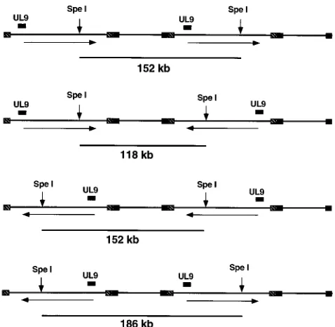
examined by PFGE following digestion with restriction en-zymes. DNA samples from cells infected with KOS, AN-1, AN-1-2a, or AN-F1 at various times postinfection were sub-jected to PFGE, and the band at the well was excised and digested to completion with SpeI, which cuts the HSV-1 ge-nome once. Digested well DNA was subjected to PFGE. This treatment would be expected to produce linear monomers from either circular or concatemeric viral DNA. If genomic inversion has occurred within the concatemeric DNA, three bands, 152, 118, and 186 kb, would be expected (Fig. 3). In the experiment shown in Fig. 4, only two of the three fragments, 152 and 186 kb, would be expected to react with the HSV-1
BamHI B2 probe. At 8 h postinfection, the 186- and 152-kb
bands are visible in KOS- and AN-1-2a-infected cells (Fig. 4, lanes 1 and 3, respectively). An additional band of 118 kb is visible on ethidium bromide-stained gels (not shown). The fact that both the 186- and 152-kb bands are seen indicates that genomic inversions have occurred by 8 h postinfection. These results confirm the recent report of Zhang et al. that inverted concatemers were present in wild-type virus-infected cells at early times after infection (70). Furthermore, the presence of the 186- and 118-kb bands indicates that these linear molecules arise from replicating concatemeric DNA and could not pos-sibly be the product of circularized input DNA.
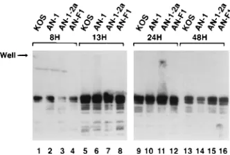
[image:4.612.316.555.426.659.2]At later time points (13, 24, and 48 h postinfection), the 186-and 152-kb b186-ands are very prominent in well DNA from KOS-and AN-1-2a-infected cells (Fig. 4, lanes 5, 7, 9, 11, 13, KOS-and 15),
FIG. 2. Southern blot analysis of a pulsed-field gel of total and encapsidated DNA from KOS-, AN-1-, AN-1-2a-, and AN-F1-infected Vero cells. Vero cells were infected with the indicated virus at an MOI of 5 PFU per cell for 18 h. Samples for PFGE were prepared by suspending infected cells in a block con-taining melted low-melting-point agarose as described previously (51). Blocks were either used as is (total [T]) or digested with SN as described in Materials and Methods. Virion DNA was prepared by lysis of purified KOS virions in agarose blocks. PFGE was performed with a CHEF DR-II apparatus (Bio-Rad). Linear fragments up to 864 kb could be resolved under these conditions. The Southern blot was probed with a32P-labeled BamHI B2 probe. The positions of well and 152-kb monomer DNAs are indicated. Lambda ladder PFGE markers consisting of concatemers of lambda DNA (ranging in size from 48.5 to 1,018.5 kb) (NEB) were used as molecular weight markers.
FIG. 3. Schematic diagram of the HSV-1 genome showing expected frag-ments of SpeI digestion. The positions of SpeI internal fragfrag-ments expected from replicating DNA are shown, as are four different concatemers representing various inversion events. Concatemers in which the orientations of both long arms remain constant generate a 152-kb band; concatemers in which the long arms have inverted with respect to each other generate a 118- or 186-kb band. The BamHI B2 probe (corresponding to the position marked UL9) will detect the 152- and 186-kb bands but not the 118-kb band.
on November 9, 2019 by guest
http://jvi.asm.org/
although a considerable amount of viral DNA remains in the well. The fact that most of the DNA remained in the well even after digestion with a single-cutting enzyme strongly suggests that the replicating DNA is composed of complex nonlinear DNA. The DNAs remaining in the wells following SpeI diges-tion are not the products of incomplete digesdiges-tion; this was determined by treating SpeI-digested DNA with BamHI and analyzing the products by conventional agarose gel electro-phoresis and Southern blotting. The blot was probed with a DNA fragment (BamHI-H) which spans the SpeI site. In each of the DNA samples shown in Fig. 4, digestion with BamHI yielded the predicted digestion products, indicating that SpeI digestion was nearly complete; in this experiment, very little DNA corresponding to partial BamHI digestion products were seen, indicating that BamHI digestion was also complete (data not shown).
When the well DNA from Vero cells infected with AN-1 or AN-F1 was digested with SpeI, no discrete fragments were released from the well even at late times after infection (Fig. 4, lanes 2, 4, 6, 8, 10, 12, 14, and 16). SpeI digestion was shown to be complete as described above (data not shown). Some addi-tional smearing of DNA was seen at positions corresponding to between 50 and 375 kb. These results indicate that in cells infected with nuc mutants, the viral DNA is structurally differ-ent from that in cells infected with wild-type virus. Taken together, these results indicate that complex nonlinear DNA accumulates in cells infected with both wild-type HSV-1 and
nuc mutants of HSV-1. Severini et al. have proposed that
replicating DNA is composed of large branched structures (51), and our results are consistent with this hypothesis. Fur-thermore, our results suggest that the DNA which accumulates in cells infected with nuc mutants is even more complex or branched than that found in cells infected with wild-type virus. We suggest that the processing or debranching of viral DNA is aberrant in cells infected with nuc mutants.
To further characterize the well DNA, we have also digested it with enzymes which cleave viral DNA more frequently. For instance, XbaI cleaves HSV DNA four times, generating frag-ments of between 24 and 64 kb. In Fig. 5, well DNA from cells infected with KOS, AN-1, AN-1-2a, or AN-F1 was isolated as described above. After digestion with XbaI, DNA was sub-jected to PFGE, blotted, and hybridized to a probe consisting of whole HSV genomic DNA as described in Materials and
Methods. Figure 5 shows that linear fragments can be released from well DNA from both mutant- and wild-type-infected cells following XbaI digestion. The predominant band of released linear DNA represents several different fragments ranging from 24 to approximately 62 kb which are not resolved on this gel. A significant amount of replicating DNA remains in the well, indicating that some DNA is still in a complex configu-ration, possibly containing branches which prevent the DNA from migrating into the gel. In Fig. 6, well DNA was digested with BamHI, which cleaves the HSV-1 genome at least 41 times and generates fragments smaller than 12 kb. In this case, almost all viral DNA can be released from well DNA from mutant- and wild-type-infected cells (Fig. 6). The fact that most of the DNA can be released by BamHI digestions indi-cates either that small fragments with branches migrate nor-mally on gels and are not retained in the well or that in small fragments, branch migration may occur readily (48). Since
BamHI is a frequent cutter, putative branch points would likely
be found close to an end and be able to migrate, generating linear fragments which can enter a gel.
[image:5.612.344.522.74.167.2]Taken together, these results indicate that replicating DNA assumes a complex, possibly branched structure. If the DNA is in fact in a branched configuration, we estimate that the fre-quency of branches must be at least once per genome in nuc mutant-infected cells. Although the frequency is less in cells infected with wild-type virus, it is impossible to accurately estimate the number of branches per genome. This is due in part to the fact that although some DNA was released, a
FIG. 4. Southern blot analysis of well DNA from wild-type- and nuc mutant-infected cells digested with SpeI. Cells were mutant-infected with KOS, AN-1, AN-1-2a, and AN-F1 at an MOI of 5 PFU per cell for various times after infection, and well DNA was isolated as described in Materials and Methods. Well DNA was digested in an agarose block with SpeI, which cleaves HSV-1 DNA once per monomer unit, and subjected once again to PFGE as described in the legend to Fig. 2. If linear monomers are released from well DNA, three fragments, 186, 152, and 118 kb, are expected as a result of genomic inversions. The blots were probed with the BamHI B2 probe, which will detect only the 186- and 152-kb genome isomers (see Fig. 3). Lambda ladder PFGE markers consisting of con-catemers of lambda DNA (ranging in size from 48.5 to 1,018.5 kb) (NEB) were used as molecular weight markers.
FIG. 5. Southern blot analysis of well DNA from KOS-, AN-1-, AN-1-2a-, and AN-F1-infected cells digested with XbaI. Cells were infected and well DNA was isolated as described in the legend to Fig. 2. Agarose blocks were digested with XbaI, which cleaves HSV-1 DNA four times per monomer unit.32
[image:5.612.351.517.556.668.2]P-labeled genomic HSV DNA digested with BamHI and EcoRI was used as a probe. Lambda ladder PFGE markers and lambda DNA digested with HindIII were used as molecular weight markers.
FIG. 6. Southern blot analysis of well DNA from wild-type- and nuc mutant-infected cells digested with BamHI. Cells were mutant-infected and well DNA was isolated as described in the legend to Fig. 2. Agarose blocks were digested with
BamHI, which cleaves HSV-1 DNA 41 times per monomer unit.32 P-labeled genomic HSV DNA digested with BamHI and EcoRI was used as a probe. Lambda ladder PFGE markers and lambda DNA digested with HindIII were used as molecular weight markers.
on November 9, 2019 by guest
http://jvi.asm.org/
significant amount of DNA from cells infected with wild-type virus remained in the well. The possibility of branch migration when the branch point is near an end liberated by digestion with a restriction enzyme also makes it difficult to precisely estimate the frequency of these structures. Furthermore, it is not clear how a molecule with a single branch would migrate in PFGE.
Inversion in replicating DNA from wild-type- andnuc
mu-tant-infected cells.In the experiment described above (Fig. 4),
well DNA from KOS-infected cells was digested with SpeI. The appearance of both the 186- and 152-kb bands indicates that genomic inversions have occurred by 8 h postinfection. Since
SpeI failed to release discrete bands from AN-1- and
AN-F1-infected cells, it was impossible to determine whether inver-sions had occurred in these cells. Because XbaI digestion was more successful in liberating well DNA from nuc mutant-infected cells, we used XbaI-digested well DNA from KOS-, AN-1-2a-, AN-F1-, or AN-F3-infected cells to determine wheth-er nuc mutants wwheth-ere able to carry out genomic invwheth-ersions. In the experiment shown in Fig. 8, the 5-kb HindIII N fragment
from the middle of USwas used as a hybridization probe. This
probe will detect a 64.5-kb band if excised from concatemeric
DNA in which the two ULsegments are in the same
orienta-tion with respect to one another (either parental or inverted orientation) (Fig. 7, lines 1 and 2); on the other hand, if two
adjacent ULsegments are inverted with respect to one another,
an 84- or a 47-kb band will be detected (Fig. 7, lines 3 and 4). In Fig. 8, lane 2, well DNA harvested at 8 h postinfection with
KOS was digested with XbaI; the three expected bands at 84, 64.5, and 47 kb (bands 1, 2, and 3, respectively) are seen, although the 47-kb band (band 3) is somewhat underrepre-sented. An identical pattern was seen in cells infected with the wild-type recombinant virus, AN-1-2a (Fig. 8, lane 3). In cells infected with AN-F1 or AN-F3, the same three bands can be seen; however, in this case the 47-kb band (band 3) is even more prominent than it was in well DNA from wild-type virus-infected cells (Fig. 8, lanes 4 and 5). Both nuc mutant DNAs exhibit smearing in the region of the gel surrounding the ob-served bands. The smearing is reminiscent of the smearing seen after SpeI digestion (Fig. 4) and may represent increased fragility in the DNA as a result of unusual structures in the nuc mutant DNA. The appearance of XbaI fragments 1, 2, and 3 indicates that inversions occur in nuc mutant DNA at a fre-quency similar to that seen in the wild-type DNA.
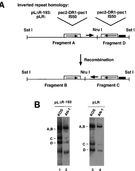
To confirm that nuc mutants do not exhibit a defect in inversion events, we used a quantitative recombination assay originally developed to assess the role of the a sequences in HSV-1 genome isomerization (50). Two of the constructs used in that study have been used here to analyze the ability of nuc mutants to carry out DNA inversion-type events. pLR, a con-struct containing an HSV origin of replication and Tn5 se-quences, was shown to undergo inversion as a result of
recom-bination between its reiterated IS50 sequences. pLDR-193
contains inverted copies of a small subfragment from within the a sequence; these reiterations are much smaller than the IS50 elements of pLR and are able to promote high-frequency recombination only because they can be recognized and cleaved by the cleavage and packaging machinery (50). Both plasmids had previously been shown to undergo high-fre-quency recombination in cells infected with wild-type virus;
however, high-frequency inversion did not occur within pLD
R-193 when the superinfecting virus was an HSV-1 mutant which is defective for the cleavage and packaging process. We previ-ously showed that AN-1 is capable of carrying out genome cleavage and packaging (53). In this study, we were interested in whether AN-1 was capable of directing high-frequency re-combination and also in whether the inversion frequency of
pLR and pLDR-193 would differ in HSV-1 (KOS) and AN-1.
Vero cells were transfected with pLR and pLDR-193 and
[image:6.612.66.295.72.357.2]FIG. 7. Schematic diagram of the HSV-1 genome showing several possible concatemers after digestion with XbaI. Concatemers in which the orientations of both long arms remain constant generate a 64.5-kb band (lines 1 and 2); con-catemers in which the long arms have inverted with respect to each other generate an 84- or 47-kb band (lines 3 and 4). Horizontal arrows represent the orientations of the long-arm segments. The lengths of the segments contributing to the observed XbaI fragments are shown above each line. The position of the hybridization probe, the HindIII N fragment from the middle of US, is shown.
FIG. 8. Southern blot analysis of well DNA from wild-type- and nuc mutant-infected cells digested with XbaI to analyze genomic inversions. Cells were infected with KOS, AN-1-2a, AN-F1, and AN-F3 at an MOI of 5 PFU per cell for various times after infection. Well DNA was purified twice by PFGE to rule out contamination with free monomeric DNA and subjected to XbaI digestion, PFGE, and Southern blotting. Lane 1, virion DNA digested with SpeI to serve as size markers; lane 2, well DNA from KOS-infected cells; lane 3, well DNA from AN-1-2a-infected cells; lane 4, well DNA from AN-F1-infected cells; lane 6, well DNA from AN-F3-infected cells. Bands 1, 2, and 3 correspond to 84-, 64.5-, and 47-kb fragments, respectively.
on November 9, 2019 by guest
http://jvi.asm.org/
superinfected the following day with either wild-type HSV-1 strain KOS or AN-1. DNA was isolated and examined in Southern blots to determine if plasmid DNA had undergone sequence inversion as a result of DNA replication-directed recombination. The plasmids typically yield two SstI-NruI ments (A and D in Fig. 9A) but will generate two novel frag-ments (B and C) if a sequence inversion event has occurred. Representative blots are shown in Fig. 9B. The signal in each of the bands was quantified as described in Materials and Methods, and the results from duplicate experiments are shown in Table 1. Inversion frequency represents the percent-age of the C-fragment signal relative to the total signal of the C and D fragments for a given lane. The frequency of inversion of AN-1 relative to the KOS inversion frequency for a given plasmid is shown. In addition, the total signal of all bands within each lane was determined, and the efficiency of DNA replication of plasmid in AN-1-infected cells relative to that for KOS-infected cells for a given plasmid is shown. These results
indicate that both pLR and pLDR-193 are capable of
under-going high levels of recombination in both wild-type- and AN-1-infected cells. Both plasmids undergo inversion at maximal levels in KOS-infected cells, and this recombination is reduced
by only 13 to 19% in AN-1-infected cells. The latter effect is probably due to the fact that both plasmids are significantly underreplicated in AN-1-infected cells. We previously re-ported that DNA synthesis of viral genomes in AN-1-infected cells varies somewhat from experiment to experiment between 50 and 90% of wild-type levels. The present experiment indi-cates that the efficiency of replication of HSV-1 origin-contain-ing plasmids may be lower than the efficiency of genome rep-lication. We do not have an explanation for this observation. Nevertheless, the high levels of recombination seen in AN-1-infected cells clearly indicate that alkaline nuclease is not re-quired for HSV-1 genome isomerization. This finding is in agreement with the previous finding that the UL12 gene is not a member of the minimum complement of genes required for mediated HSV-1 recombination events in a transient expres-sion assay (61). Thus, the results of the transfection assays confirm the results obtained by directly examining replicating DNA, which indicate that inversion is not compromised in AN-1-infected cells.
Presence of ULtermini in replicating DNA from wild-type
andnucmutant virus-infected cells.Previous reports showed
that after electrophoretic removal of linear viral genomes, ter-minal fragments in well DNA were barely detectable (51, 70). Upon further study, however, Zhang et al. were able to detect
genomic termini corresponding to the UL segment of the
HSV-1 genome (70). We have confirmed this observation by using SpeI-digested well DNA from cells infected with wild-type virus (data not shown). We wanted to extend this analysis to well DNA from cells infected with nuc mutants; however, because SpeI digestion does not release fragments efficiently from replicating DNA from these cells, we decided to use a more frequent cutter, BamHI. Wild-type and nuc mutant DNAs were analyzed following BamHI digestion and Southern blot hybridization using defined probes which are expected to
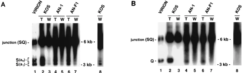
[image:7.612.62.298.70.368.2]hybridize specifically to ULor US. In the experiment shown in
Fig. 10, virion DNA, total DNA, or twice-purified well DNA from KOS-, AN-1-, and AN-F1-infected cells taken at 24 h postinfection was digested with BamHI and hybridized sequen-tially with probes b and c. Probe b was from the reiterated b fragment and would be expected to hybridize to the SQ
junc-tion and to the BamHI S fragment from ULbut not to the
BamHI Q fragment from US(Fig. 1B). Probe c was from the
reiterated c sequence and would be expected to hybridize to
the SQ junction and to the BamHI Q fragment from USbut
not to the BamHI S fragment from UL(Fig. 1B). In this region
of the genome, considerable heterogeneity has been reported. One source of heterogeneity results from multiple reiterations
FIG. 9. Ability of AN-1 and KOS to induce inversion events in plasmids. (A) Schematic diagram of plasmids pLDR-193 and pLR, which were used to assess inversion events. pLDR-193 contains a 193-bp region from the a sequences including pac2, DR1, and pac1 (illustrated in Fig. 1C). pLR contains the IS50 repeat elements from Tn5. The repeat homology in each case was cloned in an inverted orientation into pUC19, which also contains oriS, an HSV-1 origin of replication. The third line shows the predicted fragments (A and D) expected if the parental plasmid is digested with NruI and SstI. The bottom line shows the predicted fragments (B and C) if the plasmid had undergone an inversion event. (B) Cells were transfected with either pLDR-193 (lanes 1 and 2) or pLR (lanes 3 and 4). Transfected cells were superinfected with KOS (lanes 1 and 3) or AN-1 (lanes 2 and 4). Total DNA was digested with NruI and SstI, and the DNA was subjected to electrophoresis and Southern blot transfer as described in Materials and Methods.
TABLE 1. Inversion frequencies of plasmids pLDR-193 and pLR in KOS- and AN-1-infected cells
Transfected
plasmid Strain
Inversion frequencya
(SD)
% of wt recombinationb
(SD)
% of wt replicationc
(SD)
pLDR-193 KOS 51.3 (0.8) 100 100
AN-1 41.8 (4.4) 81.5 (8.6) 18.3 (4.0)
pLR KOS 50.3 (3.4) 100 100
AN-1 43.9 (2.9) 87.3 (5.8) 29.8 (16.1)
a
Percentage of the C-fragment signal relative to the total signal of the C and D fragments for a given lane.
b
Percentage of the AN-1 recombination frequency relative to the frequency for KOS (wild type [wt]). The KOS values were set at 100% for comparison purposes.
c
Efficiency of DNA replication for each plasmid in AN-1-infected cells com-pared with KOS-infected cells. The KOS values were set at 100% for comparison purposes.
on November 9, 2019 by guest
http://jvi.asm.org/
[image:7.612.315.554.587.662.2]of the a sequence itself. The L-S junction (BamHI SQ) and the
ULterminus (BamHI S) contain multiple copies of the a
se-quence, while the USterminus (BamHI Q) possesses only one
copy (35, 60). Virion and total DNAs from all infected cell samples contain several bands detected with probe b (Fig. 10A,
lanes 1, 2, 4, and 6): the band marked S(a1) likely corresponds
to an S band with a single copy of the a sequence, and the
group of bands marked S(an) each differ from the bottom band
and from each other by approximately 250 bp. These bands
likely correspond to reiterations of the a sequence (an). On the
other hand, when probe c was used (Fig. 10B, lanes 1, 2, 4, and 6), a single band corresponding to the Q fragment was ob-served. The only difference between wild-type and nuc mutant total DNAs is an increased amount of heterogeneously sized DNA in the nuc mutants, migrating as a smear.
Well DNA from KOS-infected cells shows a strong band corresponding to the SQ internal junction fragment and con-tains very little DNA corresponding to terminal fragments (Fig. 10A and B, lanes 3 and darker exposures of the same lanes shown in lanes 8). By comparing the well DNAs which hybridize to probes b and c (Fig. 10A and B, lanes 3 and 8), it
is clear that small amounts of DNA from the ULterminus but
no DNA from the USterminus are present in replicating DNA.
The multiple bands seen in Fig. 10A, marked S(a1) and S(an),
likely correspond to S fragments with single or multiple copies of the a sequence. Well DNA from AN-1- or AN-F1-infected cells (Fig. 10A and B, lanes 5 and 7) is also similar to well DNA from cells infected with KOS. Thus, bands corresponding to
the ULterminus, marked S(an) and S(a1), are seen (Fig. 10A,
lanes 5 and 7). Although considerable smearing is observed in
nuc mutant well DNA with both probes, the absence of a
discrete Q band in Fig. 10B, lanes 5 and 7, confirms that no US
terminus is present. Thus, it appears that well DNA from
KOS-and nuc mutant-infected cells contains a ULterminus but not
a US terminus. The major difference between wild-type and
mutant well DNAs is in the considerable size heterogeneity observed in DNA from the nuc mutants. As mentioned above, the heterogeneity may reflect fragile sites in the mutant DNA as a result of extensive branching or other alterations in
struc-ture. Thus, we have demonstrated that ULand not US
seg-ments are found at the termini of replicating well DNA in nuc mutants as well as wild-type well DNA. Although other expla-nations are possible, we consider it most likely that the ob-served ends are generated by an asymmetry in the cleavage and
packaging process, resulting in ULtermini being left behind.
The presence of ULtermini in nuc mutant-infected cells
pro-vides another piece of evidence that the cleavage machinery is functional in these mutants.
DISCUSSION
The precise role of the viral alkaline nuclease in the life cycle is unknown; however, the fact that well-conserved homologs of the alkaline nuclease are found in all herpesviruses suggests that it may carry out an important function in the virus life cycle. The major conclusions drawn from this study are as follows: (i) both wild-type and mutant HSV-1 DNAs exist in a complex nonlinear form (possibly branched) during replica-tion; (ii) digestion with a single-cutting enzyme cannot release discrete monomer-length DNA from replicating DNA isolated from nuc mutant-infected cells whereas it can do so from wild-type virus-infected cells; (iii) replication intermediates from cells infected with the wild type and nuc mutants contain genomic inversions indicating that alkaline nuclease is not es-sential for genomic inversion; (iv) plasmids containing inverted repeats undergo high-frequency recombination in cells in-fected with the wild type and nuc mutants, indicating that alkaline nuclease is not required for replication driven recom-bination in plasmids; (v) DNA encapsidated by nuc mutants is similar in size and structure to DNA encapsidated by wild-type virus; and (vi) replicating DNA in cells infected with the wild
type and nuc mutants contains ULtermini and is devoid of US
termini. Our previous data indicated that although DNA syn-thesis, cleavage of viral DNA into monomeric units, and en-capsidation of monomeric units in the nucleus occur in nuc mutants, the capsids are not competent to migrate into the cytoplasm (53, 65). These results and those of others suggest that DNA with a complex structure accumulates during HSV DNA replication (51). In nuc mutants, the structure is even more complex than that seen in wild-type virus-infected cells; this leads to the production of aberrant capsids which are not competent for egress.
Models for the generation of replication intermediates.
[image:8.612.117.509.72.190.2]Sev-eral models for the formation of concatemers during DNA replication can be considered. (i) It has been suggested that HSV DNA replicates through a rolling-circle mechanism (5, 29); however, a simple rolling-circle model does not account for recent reports describing the structure of viral DNA which arises during DNA replication in HSV-infected cells (de-scribed below). In addition, the rolling-circle model does not account for the rapid amplification of viral DNA seen in the initial hours after infection (23, 24). (ii) It is possible that the
FIG. 10. Southern blot analysis of virion, total, and well DNAs from wild-type- and nuc mutant-infected cells. Cells were infected with KOS, AN-1, or AN-F1 at an MOI of 5 PFU per cell for 24 h. Virion, total, and twice-purified well DNAs were subjected to BamHI digestion, conventional gel electrophoresis, and Southern blotting. Two different probes were used in this experiment. In panel A, a 132-bp NcoI-to-DraI fragment from pNar, within the repeated b sequences, detects the Bam S terminal fragment flanking UL(Fig. 1B, probe b). Bands marked S(a1) and S(an) represent S fragments with single and multiple a sequences (see text). In panel B, a 429-bp SmaI-NarI fragment from pNar, within the repeated c sequences, detects the Bam Q terminal fragment flanking US(Fig. 1B, probe c). Molecular weight markers were 1-kb markers (ranging in size from 12 to 0.075 kb) (GIBCO-BRL, Grand Island, N.Y.).
on November 9, 2019 by guest
http://jvi.asm.org/
initial rounds of HSV DNA replication occur by theta replica-tion structures initiated at one or more of the three origins of replication. At a later stage, a rolling-circle structure could be formed which would generate the head-to-tail concatemers seen late in infection. Another version of this model (70) posits that theta-type amplification of a circular template followed by a recombination event similar to those which occur between
two copies of a repeated sequence in the yeast 2mm circle may
occur (19, 45, 58). The inversion occurs between a replicated copy of an inverted repeat unit and an unreplicated copy, resulting in a situation in which two forks chase each other, generating concatemeric DNA. (iii) Concatemers could be formed by a recombination mechanism similar to that used during late T4 bacteriophage DNA replication (36, 44). Strand
invasion provides 39hydroxyl primers for initiation of further
DNA replication, and these recombination intermediates can be converted into replication forks (44). Branch migration and subsequent resolution of these recombination intermediates are required for efficient late viral DNA synthesis and packag-ing in T4. In models ii and iii, on the basis of DNA replication
strategies in either the 2mm circle and bacteriophage T4,
re-spectively, recombination is an obligatory event in the gener-ation of the head-to-tail concatemers of DNA. The notion that replication and recombination in HSV are intimately inter-twined and possibly dependent on one another is attractive for several reasons. First, replication and recombination seem to be temporally linked (16, 70). Second, replication and recom-bination appear to be functionally linked in that Sarisky and Weber showed that inversion events are mediated by the viral DNA replication machinery (50). Third, a precedent exists for a linkage between replication and recombination in other DNA viruses such as T4 (36) and adenovirus (69).
The demonstration that genomic inversions can be detected at 3 to 4 h postinfection (70) has several implications for the models of DNA replication. First, this result is not consistent with simple rolling-circle replication (model i, above), since this model makes no provision for inversion. Second, the rep-lication and/or recombination machinery must include a nick-ing activity responsible for initiatnick-ing the inversion event in a circular genome. Although it is possible that the cleavage and packaging system of HSV can generate a free DNA end capa-ble of strand invasion, Zhang et al. have shown that inversion occurs before the viral enzymes responsible for cleavage and packaging are functional (70). Furthermore, the demonstra-tion that the seven HSV replicademonstra-tion funcdemonstra-tions are sufficient for recombination (61) further argues that the inversion events do not require the cleavage and packaging machinery. Another possible candidate for an activity which could initiate recom-bination through the formation of double-stranded breaks is a cellular endonuclease described by Wohlrab et al. which cleaves DNA in the DR2 array of the a sequence of HSV-1 (67, 68). However, inversions occur even in the absence of the DR2 array (50, 56). Furthermore, Martin and Weber (37a) have recently constructed a virus whose L-S junctions lack a se-quences. This virus still undergoes normal isomerization, indi-cating that neither cleavage and packaging at the a sequence nor nicking of its DR2 repeats is required for genomic inver-sion events. Thus, while generation of a single- or double-stranded end of DNA by either the cleavage and packaging enzymes or by a DR2-specific endonuclease has the potential to promote recombination, these cleavage events cannot ex-plain the early inversion events which occur independently of these two mechanisms. A third possibility is that the process of HSV DNA replication per se is recombinogenic. According to this model, breaks which occur normally at an HSV replication
fork, either at the 39end of the leading strand or at the 39end
of an Okazaki fragment on the lagging strand, may provide the required break to initiate a strand invasion leading to a recom-bination event. We propose that DNA replication which ini-tiates at an HSV origin is more likely to be involved in such an event than in other systems such as simian virus 40 DNA replication.
Resolution of replication intermediates.We propose that as
a result of one or more of the replication and/or recombination mechanisms outlined above, branched structures arise. These structures may initially resemble three-way or Y junctions. Direct evidence for the presence of Y junctions in replicating HSV DNA has recently been provided by Severini et al., using agarose two-dimensional gel electrophoresis and electron mi-croscopy (52). Three-way junctions can be converted to four-way junctions (X or Holliday junctions) by branch migration (43). Severini et al. also have evidence for X junctions in replicating HSV DNA by two-dimensional gel electrophoresis, although Y junctions were more prevalent (52). Regardless of how they are formed, and whether Y or X junctions predom-inate, these branched structures must be resolved prior to packaging genomic DNA into viral capsids. Bacteriophage T4 encodes a phage protein endonuclease VII (product of gene 49) which can process or resolve Holliday junctions to produce unconnected linear DNA molecules (33, 42). A similar en-zyme, called endonuclease I, is encoded by T7 gene 3 (14). Severini et al. have recently demonstrated that branched struc-tures which accumulate in HSV-infected cells can be resolved by T7 phage endonuclease I, which is known to specifically linearize Y and X junctions (52). Therefore, although it is not clear exactly how they are generated, considerable evidence indicates that replicating HSV DNA contains branches which must be resolved before infectious virus can be produced.
We have previously suggested that the alkaline nuclease functions to resolve recombination intermediates which arise during DNA replication (53, 65). However, it is possible that structures which result from recombination in infected cells require host enzymes as well. The high frequency of recombi-nation observed between infecting viruses (11) and in plasmid recombination systems suggests that numerous Holliday junc-tions likely exist within the replicating DNA. Brown et al. have estimated that the average frequency of recombination be-tween infecting genomes is 0.43/kbp, or almost 70 per genome length (11). In the quantitative plasmid recombination assay, inversion frequencies are very high (50). Branched DNA ac-cumulates in cells infected with wild-type virus (4, 40, 51, 70), and we have now shown that even more complex structures accumulate in cells infected with nuc mutants. Although it has not been possible to accurately estimate the number of branches per genome, the frequencies of branches even in cells infected with nuc mutants do not seem to be as high as would be suggested by the overall recombination frequencies. It is possible that host cell resolvases function to eliminate at least some of the Holliday junctions in viral DNA. This explanation is consistent with the observation that plasmid recombination occurs with very high efficiency in cells infected with AN-1. The possible involvement of a cellular resolvase is also consistent with the observation that transfected DNA replication genes are capable of mediating inversion in the absence of alkaline nuclease.
The observation that the DNA accumulating in cells infected with nuc mutants has an aberrant structure indicates that the cellular enzymes are incapable of resolving all unusual struc-tures which occur in replicating DNA. This may indicate that replicating HSV DNA exists in a complex structure unique to replicating herpesvirus DNA which is different from standard Holliday junctions and not recognized by host resolvases. A
on November 9, 2019 by guest
http://jvi.asm.org/
comprehensive biochemical analysis of the activities of the alkaline nuclease and continued investigation of the structure of replicating viral DNA will be required to fully understand the role that the alkaline nuclease plays in the maturation of herpesvirus DNA.
DNA encapsidated bynuc mutants is similar in size and
structure to DNA encapsidated by wild-type virus.Our
previ-ous results indicated that nuc mutants have a severe defect in the egress of DNA-containing capsids from the nucleus and are consistent with the observation that no C capsids were found in the cytoplasm of AN-1-infected cells on sucrose gra-dients (53). These findings are reminiscent of a report in 1982 by Vlazny et al. describing the formation of defective genomes (59); these investigators demonstrated that viral DNA mole-cules substantially smaller than unit length can be packaged in the nucleus but do not appear in the cytoplasm. Apparently, a selection mechanism exists such that only capsids containing a full complement of viral DNA actually mature into the cyto-plasm; this mechanism may involve a conformational change or ‘‘locking step’’ involving the capsid. We previously demon-strated that a significant amount of AN-1 DNA in the nucleus of infected cells does become protected in a nuclease sensitiv-ity assay (53). In this study, we used PFGE to determine the size and structure of viral DNA protected in the nuclei of AN-1-infected cells. One hypothesis was that this encapsidated DNA was abnormal in some way, for instance, perhaps shorter than unit length as was seen by Vlazny et al. (59). Our PFGE analysis indicates that this DNA is indistinguishable from wild-type monomeric DNA. Thus, the DNA encapsidated by the mutant is certainly not smaller than unit length; however, we cannot rule out alterations such as small branches which may not be detected by PFGE. It is possible that the full-length monomeric DNA which is taken up into the capsids in nuc mutant-infected cells contains branches that are capable of branch migration and that the branches simply migrated off each linear molecule during extraction. Spontaneous branch migration is known to occur readily in molecules containing a branch point flanked by DNA sequence homology (48). Fur-ther experiments designed to freeze putative branch points will be required in order to more fully understand the nature of the viral DNA which becomes nuclease resistant in nuc mutant-infected cells.
The two major defects in nuc mutants are (i) the inability to correctly process viral DNA genomes (discussed above) and (ii) the failure of DNA-containing capsids to migrate into the cytoplasm. It is possible that the alkaline nuclease plays a role in the conformational or maturational changes required for egress of DNA-containing capsids which is completely separate from its role in resolution of recombination intermediates. However, we favor the notion that the defect in processing of viral DNA is responsible for the failure of DNA-containing capsids to mature into the cytoplasm.
ACKNOWLEDGMENTS
We are grateful to members of the laboratory for critical discussions and reading of the manuscript. We thank Alberto Severini for making results available prior to publication.
This work was supported by ACS grant VM9 and by grant AI37549. S.K.W. was an American Heart Association/Genentech Established Investigator during the initial stages of this work.
REFERENCES
1. Addison, C., F. J. Rixon, and V. G. Preston. 1990. Herpes simplex virus type 1 UL28 gene product is important for the formation of mature capsids. J. Gen. Virol. 71:2377–2384.
2. Banks, L., D. J. Purifoy, P. F. Hurst, R. A. Killington, and K. L. Powell. 1983. Herpes simplex virus non-structural proteins. IV. Purification of the
virus-induced deoxyribonuclease and characterization of the enzyme using mono-clonal antibodies. J. Gen. Virol. 64:2249–2260.
3. Banks, L. M., I. W. Halliburton, D. J. M. Purifoy, R. A. Killington, and K. L.
Powell.1985. Studies on the herpes simplex virus alkaline nuclease: detection of type-common and type-specific epitopes on the enzyme. J. Gen. Virol. 66: 1–14.
4. Bataille, D., and A. Epstein. 1994. Herpes simplex virus replicative concate-mers contain L components in inverted orientation. Virology 203:384–388. 5. Becker, Y., Y. Asher, E. Weinberg-Zahlering, S. Rabkin, A. Friedmann, and
E. Kessler.1978. Defective herpes simplex virus DNA: circular and circular-linear molecules resembling rolling circles. J. Gen. Virol. 40:319–335. 6. Ben-Porat, T., and F. J. Rixon. 1979. Replication of herpesvirus DNA. IV:
analysis of concatemers. Virology 94:61–70.
7. Ben-Porat, T., F. J. Rixon, and M. L. Blankenship. 1979. Analysis of the structure of the genome of pseudorabies virus. Virology 95:285–294. 8. Ben-Porat, T., and S. A. Tokazewski. 1977. Replication of herpesvirus DNA.
II. Sedimentation characteristics of newly synthesized DNA. Virology 79: 292–301.
9. Beverley, S. M. 1988. Characterization of the ‘unusual’ mobility of large circular DNAs in pulsed field-gradient electrophoresis. Nucleic Acids Res.
16:925–939.
10. Beverley, S. M. 1989. Estimation of circular DNA size using gamma-irradi-ation and pulsed-field gel electrophoresis. Anal. Biochem. 177:110–114. 11. Brown, S. M., J. H. Subak-Sharpe, J. Harland, and A. R. MacLean. 1992.
Analysis of intrastrain recombination in herpes simplex virus type 1 strain 17 and herpes simplex virus type 2 strain HG52 using restriction endonuclease sites as unselected markers and temperature-sensitive lesions as selected markers. J. Gen. Virol. 73:293–301.
12. Cairns, J. 1963. The chromosome of Escherichia coli. Cold Spring Harbor Symp. Quant. Biol. 28:43–46.
13. Costa, R. H., K. G. Draper, L. Banks, K. L. Powell, G. Cohen, R. Eisenberg,
and E. K. Wagner.1983. High-resolution characterization of herpes simplex virus type 1 transcripts encoding alkaline exonuclease and a 50,000-dalton protein tentatively identified as a capsid protein. J. Virol. 48:591–603. 14. deMassy, B., F. W. Studier, L. Dorgai, E. Appelbaum, and R. A. Weisberg.
1984. Enzymes and sites of genetic recombination: studies with gene-3 en-donuclease of phage T7 and with site-affinity mutants of phage lambda. Cold Spring Harbor Symp. Quant. Biol. 49:715–726.
15. Draper, K. G., G. Devi-Rao, R. H. Costa, E. D. Blair, R. L. Thompson, and
E. K. Wagner.1986. Characterization of the genes encoding herpes simplex virus type 1 and type 2 alkaline exonucleases and overlapping proteins. J. Virol. 57:1023–1036.
16. Dutch, R. E., R. C. Bruckner, E. S. Mocarski, and I. R. Lehman. 1992. Herpes simplex virus type 1 recombination: role of DNA replication and viral a sequences. J. Virol. 66:277–285.
17. Enquist, L. W., and A. M. Skalka. 1978. Replication of bacteriophage lambda DNA. Trends Biochem. Sci. 3:279–283.
18. Feinberg, A. P., and B. Vogelstein. 1983. A technique for radiolabeling DNA restriction fragments to high specific activity. Anal. Biochem. 132:6–13. 19. Futcher, A. B. 1986. Copy number amplification of the 2 micron circle
plasmid of Saccharomyces cerevisiae. J. Theor. Biol. 119:197–204. 20. Garber, D. A., S. M. Beverley, and D. M. Coen. 1993. Demonstration of
circularization of herpes simplex virus DNA following infection using pulsed field gel electrophoresis. Virology 197:459–462.
21. Gilbert, W., and D. Dressler. 1968. DNA replication: the rolling circle model. Cold Spring Harbor Symp. Quant. Biol. 33:473–484.
22. Goldin, A. L., R. M. Sandri-Goldin, M. Levine, and J. C. Glorioso. 1981. Cloning of herpes simplex virus type 1 sequences representing the whole genome. J. Virol. 38:50–58.
23. Hammerschmidt, W., and J. Mankertz. 1991. Herpes viral DNA replication: between the known and unknown. Semin. Virol. 2:257–269.
24. Hammerschmidt, W., and B. Sugden. 1990. DNA replication of herpesvi-ruses during the lytic phase of their life-cycles. Mol. Biol. Med. 7:45–57. 25. Hayward, G. S., R. J. Jacob, S. C. Wadsworth, and B. Roizman. 1975.
Anatomy of herpes simplex virus DNA: evidence for four populations of molecules that differ in the relative orientations of their long and short components. Proc. Natl. Acad. Sci. USA 72:4243–4247.
26. Hirsch, I., G. Cabral, M. Patterson, and N. Biswal. 1977. Studies on intra-cellular replicating DNA of herpes simplex virus type 1. Virology 81:48– 61.
27. Irmiere, A., and W. Gibson. 1983. Isolation and characterization of a non-infectious virion-like particle released from cells infected with human strains of cytomegalovirus. Virology 130:118–133.
28. Irmiere, A., and W. Gibson. 1985. Isolation of human cytomegalovirus in-tranuclear capsids, characterization of their protein constituents, and dem-onstration that the B-capsid assembly protein is also abundant in noninfec-tious enveloped particles. J. Virol. 56:277–283.
29. Jacob, R. J., L. S. Morse, and B. Roizman. 1979. Anatomy of herpes simplex virus DNA. XII. Accumulation of head to tail concatemers in the nuclei of infected cells and their role in the generation of four isomeric arrangements of viral DNA. J. Virol. 29:448–457.
30. Jacob, R. J., and B. Roizman. 1977. Anatomy of herpes simplex virus DNA.