FLEXIBLE INTRAMEDULLARY NAILING IN THE
MANAGEMENT OF SIMPLE BONE CYST
CERTIFICATE
This is to certify that this dissertation titled “FLEXIBLE INTRAMEDULLARY NAILING IN THE MANAGEMENT OF SIMPLE BONE CYST ” is a bonafide work done by Dr KAUSHIK BHOWMICK, in the Department of Orthopaedic Surgery, Christian Medical College and Hospital, Vellore, in partial fulfillment of the rules and regulations of the Tamil Nadu Dr M.G.R. Medical University for the award of M.S. Degree (Branch-II) Orthopaedic Surgery under the supervision and guidance of Prof. VRISHA MADHURI during the period of his post-graduate study from March 2008 to February 2011.
This consolidated report presented herein is based on bonafide cases, studied by the candidate himself
Prof. Vrisha Madhuri
D.Ortho, M.S.Ortho., M.Ch.Ortho.(L’Pool) Professor of Orthopaedics
CERTIFICATE
This is to certify that this dissertation titled “FLEXIBLE INTRAMEDULLARY NAILING IN THE MANAGEMENT OF SIMPLE BONE CYST ” is a bonafide work done by Dr KAUSHIK BHOWMICK, in the Department of Orthopaedic Surgery, Christian Medical College and Hospital, Vellore, in partial fulfillment of the rules and regulations of the Tamil Nadu Dr M.G.R. Medical University for the award of M.S. Degree (Branch-II) Orthopaedic Surgery under the supervision and guidance of Prof. VRISHA MADHURI during the period of his post-graduate study from March 2008 to February 2011.
This consolidated report presented herein is based on bonafide cases, studied by the candidate himself.
Prof. Vernon N. Lee D.Ortho, M.S.Ortho., M.Ch.Ortho.(L’Pool) Professor and Head
ACKNOWLEDGEMENT
First and foremost, I thank God who is able to do immeasurably more than we can ask or imagine. This thesis would never have been accomplished without the blessings of my God Almighty.
I owe my deepest gratitude to my teacher Prof. Vrisha Madhuri, Professor and Head of the Paediatric Orthopaedics unit for her supervision, advice and guidance from the initial to the final level of this study. Her constant oasis of ideas and constructive comments has exceptionally inspired and enriched my growth as a student.
I gratefully acknowledge my teachers Prof. G.D. Sunderaraj, Prof. Ravi Jacob Korula, Prof. Samuel Chittaranjan, Prof. Vernon N. Lee, Prof. Alfred Job Daniel, Prof. Issac Jebaraj, Prof. Vinoo Mathew Cherian, Prof. V.T.K. Titus, Prof. Tilak Jepegnanam and Prof. Kenny S. David for their unflinching encouragement and support throughout the preparation of this thesis.
INDEX
S.NO. CONTENTS PAGE NO.
1. AIMS AND OBJECTIVES 1
2. INTRODUCTION 2
3. HISTORICAL REVIEW 3
4. REVIEW OF LITERATURE 7
5. MATERIALS AND METHODS 32
6. RESULTS 37
7. DISCUSSION 47
8. CONCLUSION 51
9. SCOPE OF THE STUDY 52
10. BIBLIOGRAPHY 53
AIMS
The aim of this study is to evaluate the efficacy and safety of flexible intramedullary
nails in the treatment of simple bone cyst supplemented with bone substitutes in
children.
OBJECTIVES
1. To define the clinical profile of children with simple bone cyst.
2. To evaluate the outcome of surgery with respect to cyst healing.
INTRODUCTION
Simple bone cysts constitute 2-3% of benign lytic bone lesions in children with
preponderance in the first and second decade of life with 70% of patients presenting
with pathological fractures. (4, 32, 35) Since it’s description by Virchow in 1876, the
etiopathogenesis and treatment had confounded surgeons till now. Till the 1950’s
simple bone cyst was managed by masterly inactivity, irradiation, curettage and bone
grafting with or without chemical cauterisation. (12, 32, 34) All these techniques were
associated with complications such as high rates of recurrence, pathological fracture
premature physeal closure and infection. (1, 2, 12, 13). In the late 1970’s,steroids
and bone marrow was introduced in the treatment of simple bone cyst but the results
were found to be unpredictable. (46) In the 2000’s bone ceramics such as medical
grade calcium sulphate and high porosity hydroxyapatite was introduced in the
treatment. (22) But all these modes of treatment did not address the problem of
postoperative stability because patients continued to be treated with cast
immobilisation which continued the morbidity in these patients.
Flexible intramedullary nail was introduced in the 1980’s in the treatment of simple
bone cyst but it became popular as an option of treatment in the 2000’s. The stated
advantage of intramedullary nail was that it provided continuous decompression as
well as internal splinting in cases of pathological fracture. (26, 27, 28) We have
added ceramic scaffolds to the flexible intramedullary nail to hasten the healing time.
HISTORICAL REVIEW
Virchow in 1876 is credited with the first radiological description of a simple bone
cyst by many authors. (22,30,43) Bloodgood in 1910 described the pathology of
simple bone cyst as a distinct entity from other cystic lesions.(2) They are not true
neoplasms but they create structural defect in the long bones like the humerus and
femur causing fractures. Patients usually present when they have pain due to
pathological fracture or whenincidentalradiographsshows a defect in the bone.
Jaffe and Lichtenstein traced the natural history of simple bone cyst and suggested
that the disease is most active in children below the age of 10 years and becomes
inactive after that. They found that a cyst does not progress after closure of the
growth plates. About 15% of simple bone cysts undergo spontaneous resolution.(32)
Many theories exist regarding its etiopathogenesis,butthe etiology remains
elusive.(43)
Treatment has varied since the original description, however, simple bone cyst are
known for their fortenacity and high rates of recurrence.(26,27)
Alldredge et al in 1942 reported treatment in 152 cases of cystic lesions of the bone
with various modalities like irradiation alone, irradiation before or after simple
curettage, simple curettage, bone grafting or chemical cauterization, complete
resection and amputation. There were many complications associated with
irradiation such as stiffness,high rates of fractures of the cystic lesion , physeal injury
evidence that preoperative or postoperative radiation was of any benefit in these
patients. It was also stated that resection of bone is a curative option for
nonessential or non weight bearing bones like the fibula.(34)
Neer et al in 1966 showed that currettage and bone graftingwith or without
cauterisation were associated with recurrence rates of 40%.They made an opening
in the cortex, curetting the membranous lining and filling the defect cavity with chips
or strips of autogenous or homogenous bone graft. They proved that surgical
treatment showed better results than masterly inactivity.(12,32,34)An analysis was
done for the conservatively treated cyst with respect to the proximity to the epiphysis.
Cysts which were 0.5.cm from the epiphysis were treated conservatively. In their
study, 92% of the conservatively treated cases were eventually operated.
Spence et al in 1969 used cancellous freeze dried allograft to pack the cyst
cavityafter curettage showing a healing rate of 64% .In 1976, Spence et al used
crushed cortical bone allograft showing a healing rate of 88%.They also showed that
cortical bone is better than cancellousbone for packing the cystic cavity because
they are retained in the cyst for a longer period of time providingosteogenic stimulus
for new bone formation and healing. (33,35)
Fahey et al in 1973described subtotal resection of the cystic lesion with bone
grafting with recurrence rate of 5%.They described the procedure as resection of
part of the cyst wall, curretting the lesion and filling the defect with strut graft from the
iliac crest or tibia in the major long bones.(9)McKay et al in 1977 described subtotal
resection without bone grafting with recurrence of 9%.(16)
rate. Dreesmann in 1892 was the first one to use plaster of Paris pellets to fill
cavities in the bone. In their study, they listed many advantages of using POP pellets
as they are cheap and readily available ,could be easily fabricated into pellets or
rods, They are radiopaque, easily absorbed ,have a long shelf life ,easily sterilised
and does not give local inflammatory reactions in the body.(21)
Scaglietti et al in 1979 reported 90% favourable results using intracystic injections of
methylprednisolone acetate, with no growth inhibition or secondary deformity of the
involved bone. When these results were carefully reviewed, a number of treated
cysts designated as successes never completely healed radiographically, but were
clinically stable and thus were reported as a favourable result.(17)
Campanacci et al produced similar results in another series of patients with 15%
recurrence but had incomplete healing in 50% of the reported cures.(14,19)
Schreuder et al in 1997 described currettage and bone grafting with cryosurgery
using liquid nitrogen with recurrence rates of 12%. (40)
Drilling alone of these cysts has also been reported as well as trepanationwith
drainage of the cyst fluid and saline lavage of the cavity. The technique consisted of
introducing a trocar to aspirate the cyst fluid and making multiple holes through the
cyst wall in all directions. The excretion of cyst fluid is an important step in the
mode of treatment.(20)
Lockiec et al in 1996 used autologous bone marrow injections for the treatment of
simple bone cyst showing comparable results to steroid injections.(19,30).Rougraff
et al used demineralised bone matrix in combination with bone marrow showing 22%
injection.(8)Dissatisfaction with steroid injections in some patients, especially those
under 10 years of age, has regenerated interest in use of bone graft substitutes.
Such substitutes include bioactive ceramics liketricalcium phosphate, calcium
sulphate pellets and high porosity hydroxyapatite.(22,24)
Catier et al in 1981 described the use of flexible intramedullary nail in the fixation of
pathological fractures in simple bone cyst. Before this, patients with pathological
fractures were either treated with a sling or plaster immobilisation and plate screw
fixation. He had reported 100% rate of healing in all the patients in his study. Since
then, there have been many studies that have shown excellent results with flexible
LITERATURE REVIEW
Simple bone cyst is a benign,single chambered fluid filled cystic lesion that occurs in
the metaphysis of long bones in children recognised as distinctive entity since 1910.
DEMOGRAPHIC CHARACTERISTICS
It is the most common benign lytic lesion in childhood and represents 2-3% of all
bone lesions in childhood.(4,26,36)
Incidence is 1 in 10000 cases per year.(4)
Male preponderance exists in the ratio of 2:1 to 3:1(4,32,35)
The lesion is relatively common and usually manifests during the first two decades of
life.(32,38).
70% of patients present with pathological fracture.(32)
ANATOMICAL LOCATION
The common location of the cyst tends to be in the proximal end ofhumerus(75-80%)
and the proximal femur(47%).(12,32,35)
Less common locations are pelvis,calcaneum,talus, distal radius, distal femur and
PATHOGENESIS
There are many theories for formation of the lesion:-
-Virchow’s theory-According to Virchow, simple bone cyst is a chondromatous lesion
which has undergone cystic softening. This theory was not accepted as there were
no facts supporting this theory. (3)
-Monckeberg’s theory- it is the healing form of a pre-existing giant cell tumor or
osteitisfibrosa. This theory was negated by the fact that giant cell tumorsappear at a
later age and usually start in the epiphysis whereas simple bonecysts starts in the
metaphysis. The histological architecture also does not correspond to simple bone
cyst. Strands of giant cells if present are not embedded in the stromal tissue. (1,3)
-Phemister and Gordon’s theory - it is secondary to a form of osteomyelitis.
Phemister recovered Streptococcus viridans from four cases of simple bone cyst but
did not yield anything in rest of the cases. The argument against this theory was that
the cysts were of long standing duration and may have sterilised themselves.
Furthermore, histological studies showed that the lining of these cysts resembled
lining of cyst found in the end of long bones in arthritis deformans.(1,13)
-Pommer’s theory - there is an encapsulation of a metaphysealhemorrhage.After the
encapsulation,it is kept distended by the transudation of fluid into the cyst and the
pressure from the cyst causes stagnation of the lymph and blood vessels resulting in
pressure erosion and expansion of the cortex. Objections were raised as patients
changes. Hence it was postulated that cysts developed in mild trauma without
fractures but intramedullary haemorrhage.(1,3,32)
-Mikulicz’s theory-he postulated that simple bone cyst represent a local
post-traumatic dystrophy. He suggested that the cyst represented some local disturbance
in bone growth and development as it had a predilection towards young people and
for regions of active growth of long bones.There is microtrauma at the epiphyseal
line leading to defect in the enchondral bone formation with subsequent cyst
formation.(3)
-Developmental anomaly occurs in the veins of the affected bone with resultant
accumulation of interstitial fluid and subsequent equilibration of this fluid with that in
unblocked vessels. It was very difficult to prove developmental anomaly in small
veins through intraosseous venography. (5,7)
Out of these, the main theories which have gained acceptanceare:-
1. Mechanical theory (Cohen’s hypothesis)
In the bones of children, the metaphysesare the sites of most rapidremodelling of
bone. This is a consistent property of bone with a process of deposition and
resorption. When the process of resorption is very rapid, it is accompanied by
formation of foci of loose fibrous tissue which resembles that seen in the wall of the
cyst. One or more of these foci of fibrous tissue represents the first step in the
formation of these cysts.
The vessels present in this area are extremely thin walled sinusoids with extremely
into the vascular system and the interchange of plasma proteins between the
interstitial fluid and plasma, which depends on the lymphatic system, the formation of
a bone cyst may be predicted on the blockage in the drainage of interstitial fluid
within the metaphysis.
The site of accumulation of this fluid might be well in an area of interstitial tissue
where the sinusoidal vessels were occluded by trauma or thrombosis. Another
possibility is that one of the branches of the sinusoidal vessels proliferating at the
face of epiphyseal cartilage or the rapidly remodelling metaphyseal zone may be
partially or completely blocked. Indirect evidence which support this hypothesis is
complete interruption of the central medullary vein by intraosseousvenography
anddilation of the nutrient artery by angiography. On histological examination,
congested capillaries and veins are seen in the newly formed cortex and between
the spongiformtrabeculae near the cyst.
Since the hemodynamic pressure in this area is very low, only a small increase in
pressure of the blocked area would be necessary for continuing expansion of the
cyst.
The lack of bone trabeculaein the cyst is due to its resorption due to the elevated
hydrodynamic pressure in the cyst or altered composition of the fluid stagnating
inside the cyst.
The lining of the cysts are flattened fibroblastic cells which had lead to the earlier
speculation of the fluid being formed by either secretory or diffusion mechanism
which has been negated now. There is no characteristic resemblance of the cyst
2. Biological theory (Komiya’s hypothesis)
Studies by Komiya et al have shown bone resorptive factors in the cyst fluid such as
IL-1, PG and gelatinase. Values of Interleukin- 1β of cyst fluid were examined with
an enzyme-linked immunosorbent assay (ELISA) kit. Gelatinase activity in cyst fluid
was detected by sodium dodecyl sulfate polyacrylamide gel electrophoresis
(SDS-PAGE)
PG E2 is produced by osteoblastic cells in the presence of IL-1β which induce bone
resorptionthrough stimulation of osteoclasticactivity.Interleukin-I produced by
monocytes or polynuclear cells present in the exudation fluid, stimulates osteoblasts,
fibroblasts, or other connective tissue cells to generate PGE 2. IL1 also acts directly
on osteoclasts by direct osteoclastic precursor proliferation or maturation.
Lymphotoxin, TNF, PDGF and EGF also stimulate bone resorption.
The degradation of the extracellular matrix components of collagens and
proteoglycans are attributed to proteolyticenzymes such as collagenase,
gelatinaseandstromeolysin. In the process of bone resorption in a simple bone cyst,
gelatinase may play a role in collagenolysis by splitting collagen into two fragments
Komiya et al Oxygen scavengers in simple bone cyst CORR no 287 Feb 1993
Studies have shown high levels of oxygen free radicals in the cyst fluid which
contribute to bone destruction. Venous obstruction causes localised ischemia in its
drainage field generating oxygen free radicals like superoxide anion(O2-), hydrogen
peroxide(H2O2), hypochlorous acid(HOCl) and hydroxyl radical(OH) all of which
cause cell membrane damage and breakdown of matrix by activating latent
collagenase and metalloproteinases .High levels of oxygen scavengers are present
in the cyst fluid reflecting the presence of oxygen free radicals as these radicals have
a very short half life. These oxygen scavengers are superoxide dismutase and
Superoxide dismutase levels were determined by electron spin resonance
spin-trapping technique and the spectra was recorded at 100KHz magnetic field
modulation.The spin resonance technique is based on a method for determining
superoxide radical (O2-). The cyst fluid to be assayed is added in a reaction medium
containing hypoxanthine-xanthine oxidase system. Catalase activity is determined by
a spectrophotometer by the decomposition of H2O2.(20)
Blockage in the drainage of interstitial fluid leads to ischemia and the generation of
oxygen species and IL-1 in the cyst fluid. Oxygen species damage bone cells and
degrade bone matrix by activating collagenase and metalloproteinases of latent type.
IL-1 also activates metalloproteinases causing matrix breakdown. Oxygen
scavengers play a protective role against oxygen species.
Summarising, Cohen’s hypothesis is the currently accepted theory of pathogenesis
of simple bone cyst.(4,30)Komiya et al have studied the cyst fluid characteristics
hypothesis by showing indirect evidence of elevated cyst pressure, dilation of the
nutrient artery and interruption of the central medullary vein by intra-osseous
venography.(20)
GROSS ANATOMY
Simple bone cysts are found in tubular bones in 90-95% of patients. It usually
reaches maximum size before skeletal maturity. Within the long bones, most simple
bone cysts are situated in the proximal metaphysis. The involved bone is not wider
than the adjacent physis. It is usually a unilocular or rarely multilocular cavity filled
lesion with serous or serosanguinous fluid which is yellow or greenish incolour with
low viscocity. Inner surface has ridges separating the depressed zone, sometimes
the wall is covered by a layer of fleshy tissue of 1cm or more. Occasionally, partial or
complete septa are seen,the latter making it multicameral. Periosteal bone formation
is slight or usually absent.(7,9,12,38)
CYST FLUID
The serous fluid has been analysed and has been found to have prostaglandins E,
HISTOLOGICAL FEATURES
The cyst is lined by a thin connective tissue membrane beneath a thin distended
cortex. The cyst is filled with coarse red –brown granulations loosely attached to the
lining.Thicker areas when formed are composed of fibrogenic connective tissue that
contain numerous benign giant cells, hemosiderin pigment, few chronic inflammatory
cells and lipophages.
The granulations are composed of immature fibrous tissue in which are scattered
areas of recent and old hemorrhage. The granulation tissue and the lining are
extensively infiltrated with lymphocytes plasma cells,polymorphonuclear leucocytes
and giant cells of foreign body type.Proliferating fibroblast tissue and callus may be
prominent outside and within the cyst.
The bone surrounding the cyst cavity exhibits lacunar resorption alternating with
areas of osteoid tissue which may exhibit calcification and new bone formation.
Rarely,there are small islets of cartilage also.(12,13,38)
RADIOLOGICAL FEATURES
A.Radiographicfeatures- The typical radiographic appearance is that of a lesion
concentrically located in the medullary cavity of the metaphysis of a long bone with
expansion in all directions, creating an expanded and thinned but unpenetrated
cortex. Cysts located in flat bones such as the pelvis are centred between the inner
and outer tables of the ilium.
Cortex is thin and eroded but intact unless pathological fracture has occurred. Fine
it. The long axis of the lesion in the bone almost always exceeds its width, giving the
appearance of a truncated cone. The distal extent of the long axis of the cyst,
especially in the humerus, is frequently hard to see on plain films and appears to
blend into the metadiaphyseal bone outline. If periosteal reaction is present along the
thinned cortex of the cyst wall, one should search carefully for a pathologic fracture
in the area. Serial radiographs show epiphysis growing away from the region of the
cyst so that it lies in the centre of the shaft.(32,38)
The "fallen leaf" sign on plain films is virtually pathognomic of a multiloculated bone
cyst. This results when a pathologic fracture in the thinned cortical wall of the bone
dislodges a fragment of the cortex. The fragment "drops" into the fluid-containing
(but often not filled) cavity of the cyst. This fragment can also shift in the fluid,
depending on positioning of the patient's involved bone during serial imaging. The
cyst is infrequently distributed from one end of the bone to its opposite end. This
appearance is almost always found in the skeletally mature or close to skeletally
mature patient.(14, 15)
Pathogenesis of the fallen fragment sign:-
A. Solitary bone cyst in the proximal end of the humerus. Thinned cortex shows
"'eggshell" cracks following minimal trauma. Periosteum remains intact around the
comminutedundisplaced fragments.
B.A large cortical fragment attached to the underlying periosteum only at the bone
C. Cortical fragment has now become completely displaced and lies at the bottom of
the cyst cavity creating the "fallen fragment sign".
D. Lying free in the cyst cavity, the fragment is able to gravitate toward the top of the
cavity on change in position of the arm. (15)
Radiographs are also used for finding various aspects of simple bone cyst:-
1. Activity
A cyst is considered active if it is
-abutting the physis
-symptomatic with activities of daily living
-has fractured once or more
-has increased in size within an observation period of 3 to 6 months(3,8)
Latent cyst- lesser chance of recurrence. They are further classified as:-
-Latent primary cyst is one in which the distance between the nearest
epiphyseal plate and the cyst does not exceed one- third the length of the
shaft.
-Latent secondary cyst is one in which the distance betweenthe nearest
epiphyseal plate and the cyst exceeds one third the length of the shaft.(9)
2. Size of the cyst- The size of cysts was determined with regard to the length of the
affected bone. Cysts involving up to one tenth of the bone length were defined as
small, those upto one fifth as medium, and those exceeding one fifth of the bone
length as large.(10)
Cyst index of Kaelin and MacEwen- One or more trapezoids were drawn round
the cyst to measure the cyst area. The diameter of the diaphysis was measured in its
tubular part. The cyst index is the cyst area divided by diameter squared. The lower
limits of the cyst index for pathologic fracture were 4 for the proximal humerus and
3.5 for the proximal femur. Above these limits, a cyst was prone to fracture.
Mechanically, a cyst was considered healed when the index was less than 3, with a
cortical width more than 2 mm.
Cyst diameter method of Ahn and Park - A cyst occupying more than 85% of the
bone diameter is at high risk of fracture.(11)
4. Joint incongruity
5. AVN
6. Pathological fracture
Radiographic differences between cystic lesions
SBC ABC FIBROUS DYSPLASIA
Lucencycentral eccentric central
Locationabuts physismetaphysealmetadiaphyseal
Appearanceuniloculatedmultiloculatedground glass
Cortexthinned unless fracdestroyed thinned
Calcificationrare present present
Cartilagenil nilproduced
maturity moves away no change no change
fromphysis
B. CT –It is most helpful in evaluating the extent of pelvic cysts but can be used for
evaluation of cysts in any location. When the cyst is found in the midshaft of a bone
or in an unusual location, CT may be useful in determining the extent of the defect. If
the CT scan is utilized to evaluate these cysts, Houndsfield units should always be
included on the films to confirm the existence of fluid, since lipomas of bone also
occur especially in the calcaneum, vertebral bodies and the proximal femur (the
second most common location for cysts) have a similar plain film and CT scan
appearance. Fluid within cystic defects is typically less than 20 Houndsfield units
(2-18 U). Fat is less than 0 Houndsfield unit (0 to -200), and is helpful for differentiation.
Rising bubble sign-This sign indicates presence of a hollow lesion such as simple
bone cyst in which a tiny gas bubble is formed at the time of pathological fracture
and rises to the most non-dependent margin of the bone lesion. The bubble of gas
can arise by one of two mechanisms: rapid tissue compression and decompression
with the atmosphere (as seen in open fractures or instrumentation).It is seen in acute
setting of a pathological fracture in simple bone cyst.(39)
C. MRI -Typical bone cyst will show a low [T1] and high [T2] signal on MRI. It is more
easier to detect intracystic fluid or a single gas bubble than on CT. MRI is also used
to exclude the presence of intralesional soft tissue or solid matrix. However, these
findings do not clearly differentiate the aneurysmal bone cyst from the simple cyst in
the child under age of eight years.(39)
D.Cystogram- Injecting a radio-opaque dye in the cystic defect as an additional
diagnostic aid, stating that if this material fills the cavity, the diagnosis of a cyst can
be safely made. A cyst that does not fill with dye implies a solid tumor. Limited recent
reports also show that injection of fluid into the closed medullary cavity of the bone
increases the intraluminal pressure fourfold. There has been one case of fatal
embolism during injection techniques into the humeralcanal for the treatment of a
cyst. Inactive cysts occasionally have well-formed septations that prevent complete
filling of the defect (especially if a fracture has previously occurred), thereby giving
DIFFERENTIAL DIAGNOSIS
Some lesions which should be considered in differential diagnosis are :- (32,38)
1.Fibrousdysplasia-Lesion in the metadiaphyseal region of the bone with well defined
zone of rarefaction which is surrounded by a narrow rim of sclerotic bone.
Sometimes, the tumor produces a large expansile mass that bulges in the soft
tissue ex. Base of skull and maxilla .The tumor may have large amounts of cartilage
which appears as dot or ring like calcification on radiographs.Sometimes, they have
superimposed aneuyrsmal bone cyst.
2.Nonosteogenic fibroma-These are metaphyseal defects in the first and second
decade of life, mostly in the distal femur. On radiographs, it usually present as
lesions located eccentrically in long tubular bones with bulging of the cortical outline
which appear to migrate towards the centre of the boneas the physis grows away
from it. On histopathogenesis, they show a characteristic spindle cell proliferation
with a loose storiform arrangement of cells. Usually no treatment is needed.
3.Giant cell tumor-These are epiphyseal tumors that usually arise in the second and
third decade of life. Radiographs show expanding zone of radiolucency in the end of
long bone of an adult. It usually extends to the articular cartilage,but there may be
thin zone of normal bone intervening between the two. It frequently destroys the
cortex extending to the soft tissues. On histopathogenesis, the characteristic cells
are spindle shaped cells .Giant cells containing 40-60 nuclei are scattered through
the lesion. Treatment usually consist of extended curettage and providing structural
4.Osteitisfibrosacystica- It is usually produced due to hyperparathyroidism. The
lesion is filled with rich fibroblastic connective tissue rich in osteoclastic like giant
cells resulting in resorption of bones resulting in weakening of the bones as their
calcified supporting structures are replaced with fibrous tissue (peritrabecular
fibrosis) and the formation of cyst-like brown tumors in and around the bone.
Radiographs show thin bones which are bowed or fractured. Blood tests will show
increased serum calcium and parathyroid hormone. Treatment is usually directed
towards parathyroidectomy.
5.Enchondroma-It is a benign tumor of mature hyaline cartilage, usually located
centrally in the metaphyseal bone. The incidence is evenly distributed in all the
decades of life. Majority are located in the phalanges of the hand and feet. On
radiographs, they produce a localised area of rarefaction. Long bone enchondromas
are usually associated with mineralisation which is described as ring like or popcorn
like. There is scalloping of the endosteal part of the cortex.Histopathogenesis shows
presence of cartilage nodules with intervening bone trabeculae which lies in the
marrow and does not involve the medullary bone. Usually no treatment is required or
simple curettage is sufficient.
6.Neurofibroma-Neurofibromatosis is usually associated with various skeletal
changes caused by contiguous neurogenic tumors like scoliosis, defects in the
posterior orbital wall, congenital bowing and pseudoarthrosis.
7.Lipoma-Intraosseouslipomas are very rare and can involve any part of the
skeleton with proximal femur the most common location. On radiographs,it presents
the fatty nature of the lesion becomes apparent. It may undergo malignant
transformation.
MANAGEMENT
Since the first description of simple bone cyst in 1910, many methods of treatment
have been tried with varying degrees of success.(2)
Initially Brunschwig et al in 1930had described treatment of simple bone cyst with
expectant management or in cases where there was pathological fracture,patients
were usually treated with curettage of the lesion and swabbing out the lesion with
antiseptic solution such as iodine.(13).
Alldredge et al in 1942 and Garceau et al in 1954 described treatment of simple
bone cyst with various methods such as irradiation, curettage, curettage and bone
grafting and various combinations.They found 100% recurrence with irradiation only.
They found 33% recurrence rate using curettage alone. Their best results were with
curettage and using bone chips. Alldredge et al used chemical cauterisation such as
Zinc chloride and carbolic acid. On important finding in their study was that better
results were obtained when cysts were treated in the latent phase.(32,34)
Neer et al in 1966 showed that operative treatment showed better rates of healing
than non-operative treatment. They found immobilisation and restriction of activities
did not help in treatment and 90% of the patients came back for surgery. In cases of
curettage,cauterization using zinc chloride, phenol and bone grafting. In their
series,curettage and bone grafting without cauterisation showed 39% recurrence
compared to curettage and bone grafting with cauterisation with a recurrence rate of
30%. They showed 31% recurrence using allograft compared to 23% when
autogenous graft is used. (12)
Fahey et al in 1968 described subtotal resection of the cyst with bone grafting in
which the involved area of the bone is exposed along with the cyst and transverse
cuts are made on the bone adjacent to the proximal and distal end of the cyst upto
3\4th of the diameter of the width of the bone,curretting the cyst and filling it with bone
graft. Similar study was done without bone grafts showing comparable healing
rates.(9,16).
There were many complications associated with currettage and bone grafting such
-high recurrence rates (30-40%)
-pathological fracture
-limb length discrepancy (14%)
-premature epiphyseal closure(2-4%)
-infection(1%)(4,14)
This resulted in advancement of procedures with minimal surgical interventions like
steroid injections, injection of demineralised bone matrix and autologous bone
Scaglietti et al in 1979 described the use of methylprednisolone in the treatment of
simple bone cyst showing are recurrence rate of 10%.In another series,the injection
was repeated in 76% of the patients with an average of three or four times with a
maximum of nine injections. In their procedure,they injected 40-200mg of
methylprednisolone in the cystic cavity. In the course of treatment, 2% of patients
developed pathological fractures.(17,18).
Komiya et al in 1991 described a procedure drilling multiple puncture drill holes in the
cyst (trepanation) after aspiration of the cyst fluid through a trocar. This method is
effective because first, by removing cyst fluid, the internal pressure of the cyst
becomes depressed, and the venous flow around the cyst wall is improved.
Removing cyst fluid is also useful for removal of bone resorptive factors and,
therefore, for inhibiting a progressive osteolysis. Second, making multiple drill holes
through the cortical bone of the cyst wall stimulates the periosteum to induce bone
formation and makes cyst fluid escape through the drilled holes. Third, making
multiple drill holes through the medullary bone of the cyst wall improves the
intramedullary venous flow. When a pathologic fracture occurs, cyst fluid escapes
from the cyst and the periosteum is stimulated to accelerate bone formation. There
was a recurrence rate of 27% using this procedure.(20)
Lokiec et al in 1996 reported 100% results with autologous bone marrow injection in
the treatment of simple bone cyst using a trocar to disrupt the lining
membrane,aspirating the cyst fluid and injecting bone marrow aspirate through the
same trocar. A maximum of 25ml of aspirate was introduced in the cyst cavity.(19)
Docquier et al in 2003 did a similar study using autologous bone marrow with 12%
a part of the trial. All the patients were a maximum of three injections. They showed
that the healing rate was better in the steroid group compared to the bone marrow
injection group and the risk of fracture was lower in the steroid group.(41)
As both steroids and bone marrow procedures showed comparable rates of
recurrence of 10-15%, demineralised bone matrix was used more and more alone or
in combination with bone marrow injections. Killian et al in 1998 used DBM for the
treatment of simple bone cyst in which they obtained 22% recurrence.(42) Rourgraff
et al in 2002 and Kanellopoulos et al in 2005 used DBM with bone marrow and
showed a recurrence rate of 20-25%.Demineralized bone matrix is commercially
available as an injectable substrate. It is prepared from freeze-dried allograft with a
method that may preserve some of its osteoinductive and osteoconductive
properties, andthen it is mixed into a glycerine carrier, giving the material agel-like
consistency. This substance (Grafton; MusculoskeletalTransplant Foundation,
Holmdel, New Jersey) is commerciallyavailable pre-packaged in syringes and can be
injected throughlarge-gauge needles. (6,8)
Peltier et al have operated on simple bone cyst with curettage and packing the cyst
area with plaster of Paris pellets showing 90% success rates.(21)
The predictors of failure of treatment were described by Sung et al in 2008, which
were:-
1. Younger age
2. Treatment with steroids
Newer techniques such as bone scaffolds are increasingly being used over the last
10years ex.Tricalciumphosphate, calcium sulphate and high porosity calcium
hydroxyapatite. Dormans et al in 2005 used the technique of percutaneous
intramedullary decompression,curettage and grafting using MGCS pelletsshowed a
healing rate of 91.7%. Inoue et al in 1993 have described the use of high porosity
hydroxyapatite in filling the cavity of the cyst after curettage. It is composed of
calcium hydroxyapatite [Ca10(PO4)6(OH)2].The HA is composed of 70% of highly
porous connecting air cells of 90um in diameter and sintered at 900◦.The authors
have found a healing of 78% using high porosity HA.They are used for their
osteoinductive and osteoconductive properties accelerating the rate of healing after
decompression of the cyst by various methods. The advantage over traditional bone
grafting methods are decreasing the donor site morbidity in children, decreased
chance of infection and more quantity obtained to fill the cyst.(22,23,24)
The recurrence rates with the various techniques were:-
-Expectant management- 80-100% (13)
-Irradiation - 90-100%(13, 32)
-Currettage alone- 50-60% (12, 32, 34)
-Currettage with bone grafting- 30-40% (12, 27)
-Currettage ,bone grafting and chemical cauterisation- 30-35% (12)
-Subtotal resection with or without bone grafting- 5-10% (35)
-Trepanation- 20-25% (5)
-Steroids \ autogenous bone marrow - 10-15% (18, 41)
-Demineralised bone matrix – 20-25% (6, 8 ,30)
-POP pellets- 12% (21)
-Bone ceramics such as calcium sulphate or hydroxyapatite- 10% (22, 24)
In patients presenting with pathological fracture, they were usually treated with
immobilisation casts till bone healing was visualised on radiographs. Bumci et al
have used Kuntscher nail in displaced fractures with or without cerclage wires
achieving comparable results.(10)
As the recurrence rates were high and unpredictable in the above mentioned
techniques,management has evolved towards flexible intramedullary nail as the
method of treatment of simple bone cyst. One of the main advantages of using an
intramedullary nail was the absence of postop immobilisation. A review of the
literature showed that approximately 50 % of patients with a bone cyst have a
complete pathological fracture and another 25 % have infraction of a thinned cortex
at the time of presentation. Although local methods such as steroid injections,
bone-marrow injections, and decompression produce consolidation in most patients, they
offer no immediate mechanical stability to the weakened bone. Nailing has the
early mobilization, thus obviating the need for a plaster cast and decreasing the
prevalence of the most common complicationie; a pathological fracture. (27,43,46)
Catier et al in 981 first described the use of Ender nails in fixation of pathological
fractures due to simple bone cyst in the femur.(28) Santori et al used flexible IM
nailing in 1986 for pathological fracture in the proximal humerus and femur
achieving 100% healing rates.Roposch et al in 2000 operated in 32 children with
flexible intramedullary nailing, out of which 30 presented with pathological fracture.
Healing in these patients were assessed with Capanna‘s grading and 30 of the 32
patients had shown grade 1 and 2 healing. They had a recurrence rate of 6%. They
had another6% recurrence after removal of the nail. The other complications they
encountered were change of nails in 28% of patients because of shortening in the
growing bone and varus deformity of the proximal femur in 15% of the
patients.Sanctis et al in 2006 operated on 47 patients with simple bone cyst with no
recurrences. All the lesions had healed,60% completely and 40% with residual
radiolucency according to Capanna’s grading. In two patients where the cyst was
close to the physis, there was evidence of limb length discrepancy of 1cm.
Pogorelićet al did not find any recurrence in their 18 patients in a followup of 1.5-5
years. Their only complication was irritation at the nail entry site. Kanellopoulos et al
in 2007 used flexible intramedullary nailing in simple bone cysts with DBM and bone
marrow to fill the cystic cavity in 9 patients. They introduced the DBM into the cyst
cavity after calculating the cyst ratio. In their study, they reported no recurrence in a
5 year followup with the only complication, being,irritation at the nail entry site.The
As shown in the above studies, flexible intramedullary nailing in simple bone cyst are
associated rarely with complications. The recurrence reported in one study was
6%,rest of the studies did not show any recurrence. The major complication reported
in the above studies was varus deformity at the fracture site, change of nail due to
MATERIALS AND METHODS
All cases of children admitted with a diagnosis of simple bone cyst between January
2004 to December 2009 were identified from the operation register, biopsy reports
and in-patient records. Discharge summaries were searched from the clinical
workstation, a hospital information archival and retrieval software. Radiographs of
these patients were obtained from PACS.
38children, in the Paediatric Orthopaedic unit, who were under the age of 18 years,
were identified to have a diagnosis of simple bone cyst of femur,humerus and radius.
28had undergone flexible intramedullary nailing, 26 of these had cyst filled with
commercially available bone substitutes (hydroxyapatite or calcium sulphate). The
other 10 had undergone treatment with alternative techniques and do not form part
of this study.
Postoperative follow up was available on 27 patients and one was lost to follow up
after the surgery. The mean duration of follow up was 16 months and ranged from 3
months to three and a half years. The patient who has no follow up was excluded
from this study.
The pre-operative data collected from these patients included site, age, gender,
location of the cyst,activity of the cyst, presence or absence of pathological fracture,
use of bone substitutes and any prior surgeries. The postoperative data collected
from these patients were duration of followup, radiographic signs of healing and
presence or absence of postop complications.
The choice of treatment was made by the paediatric orthopaedic surgeon. The size
internal diameter of the bone in both the antero-posterior and lateral radiographs
and this was divided by 2 and 1 mm subtracted to arrive at the nail diameter to be
used.(37)
OPERATIVE TECHNIQUE
Proximal femur
The patient was placed in supine position on the standard operating table. After the
extremity has been surgically prepped and draped, the proximal femur cyst was
located under image intensifier and aspirated A clear fluid aspiration confirmed the
diagnosis of simple bone cyst. In case of a pathological fracture or a blood filled cyst
the cyst was opened for grafting. In other cases an opening was made and biopsy
obtained to rule out other cystic lesions such as aneurysmal bone cyst and fibrous
dysplasia.
When opening the cyst the incision was made on the lateral aspect of the thigh,
dividing the fascia reaching the anterolateral cortical wall of the cyst. Now, a cortical
window is made on the most prominent part of the cyst wall draining the cyst fluid
and a thorough curetting the membranous lining of the cyst wall is carried out.
Distally symmetrical skin incisions are made on the medial and lateral side. The
distal skin mark was the upper pole of the patella and progressed 2-3cm proximally.
The skin and the fascia were incised together. Blunt dissection was continued
through the muscle to the bone. The entrance points for nail should be outside the
joint capsule and away from the edge of the physis. The entry sites were perforated
awl was initially placed at 90 degrees to the cortex to keep it from slipping off. Once
the awl was firmly seated on the cortex, it was reduced to an angle of 45 degrees to
the shaft axis and the perforation of the bone was continued at an upward angle.
Carefully the pre-bent nail with a curve at the tip was inserted into the medullary
canal by hand or using the T – handle inserter. Following its insertion, the position of
the nail was confirmed with the image intensifier. The curve of the tip was
accentuated to facilitate its bouncing off the opposite cortex. Carefully the first nail
was advanced up to the region of pathological fracture zone. Following this the
second nail was inserted from the medial side and advanced to the cyst.At the cyst
the nail was rotated three hundred sixty degrees and advanced in increments to
provide an opening of all septae usually present in children with previous fractures.
Both nails were advanced into the proximal fragment crossing the proximal cyst wall.
Once the nailscross the cavity, it is packed manually with bone substitutes. The
medial nail was directed to the femoral neck and the lateral nail toward the greater
trochanter andan attempt is made to have a purchase in the physis. This prevents
backing out of the nail. Just prior to advancing to their final position, the nails were
cut and terminal part bent to lie against the bone, leaving enough length to
manipulate and advance them to their final position. Once both the nails have
entered the proximal part they were then tapped to their final position. Once the nail
tips were in their final position, the end of each nail was cut, leaving 1-2cm
protruding from the cortex. The cyst size and adequacy of curettage is measured by
a cystogram and then the cyst is packed with bone substitute. The wounds are
closed with drains.
Postoperatively, the child is ambulated on the second post operative day partial /non
clinic at six weeks to three months. If there are sufficient signs of healing, full weight
bearing is started. During follow up we recorded the range of motion of hip and knee,
radiographic healing, limb length discrepancy, the condition of wound and skin, other
symptoms and other complications.
Radiographic healing was measured according to Capanna’s grading.
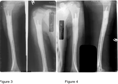
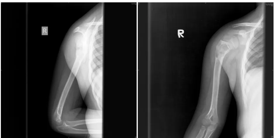
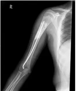
2. Proximal humerus - Patient lies supine on the radiolucent table. After
localising the cyst under image intensifier, the cyst isopened through a mini
incision (2.5to 5 cm) in the deltopectoral groove .The most affected cortex is
opened,the cyst fluid is drained and the cyst lining is aggressively curetted
and cyst volume and quality of curetting assessed by cystogram.A single
humeral nail is used in cysts with no fracture or sufficient structural integrity.
Distally, one incision is made on the lateral aspect of the distal humerus. The
entrance point for nail should be outside the joint capsule and away from the
edge of the physis. The entry site were perforated by an awl in the most
proximal end of the incision. The awl was initially placed 90 degrees to the
cortex to keep it from slipping off. Once the awl was firmly seated on the
cortex, it was reduced to an angle of 45 degrees to the shaft axis and the
perforation of the bone was continued at an upward angle. Carefully the nail
was inserted into the medullary canal by hand. Following its insertion, the
position of the nail was confirmed with the image intensifier. The curve of the
tip was accentuated to facilitate its bouncing off the opposite cortex. Carefully
the nail was advanced up to the region of pathological zone. The nail is
advanced into the proximal fragment crossing the proximal cyst wall as
described under the femur technique. Once the nail crosses the cavity, it is
Postoperatively, the patient was kept in a broad arm sling for a period of 2-3 weeks.
Xrays were taken serially at 6 weeks, 3 months,1 year for followupand periodically
after that for unresolved cysts to assess the healing according to Capanna’s grading.
ASSESSMENT OF OUTCOME
Radiological assessment
Capanna’sgrading(46):-
Grade 1-healed when cyst is completely filled with bone and cortical margins are
thickened.
Grade2- healed with residual radiolucency when most of the cyst is filled with bone
and cortical margins are thickened, but there were small residual areas of
radiolucency.
Grade 3- recurrence,when cyst had healed initially and had filled with bone but large
areas of radiolucency and cortical thinning subsequently developed.
Grade 4 –no response when there is no evidence of any effect of treatment.
Radiographic assessment of healing was taken at the minimum duration when the
cyst showed the best grade of healing and the minimum period for evaluation was at
RESULTS
Flexible intramedullary nailing was done in (21children in the proximal humerus, 6 in
the proximal femur) for a confirmed diagnosis of simple bone cyst in children in the
paediatric unit in Christian Medical College and Hospital,Vellore.
27 patients with followup were included in the study. There were nineteen males and
eight females.The mean age of the patients is 10.3 years ranging from 3 to18 years.
The mean duration of postoperative stay in the hospital was 4.9 days.
All were symptomatic with symptoms ranging from ache, swelling, decreased range
of motion and inability to walk (for femur) or use upper limb.
1. Sex ratio
Male=19
2. The location of cysts
Proximalhumerus=21
Proximal femur=6
Active = 18
Inactive =9
Present =20
Absent = 7
Nil 22
Currettage\BG\substitutes 4
Nail\rods 0
Others 1
Flexible nailing with bone substitutes=25
7. Duration of followup
3months =4
6months =4
12months =8
18months =2
24months = 6
36months= 1
>36months= 2
Healing by Capanna’s grading
grade 1 =19
grade 2 =7
grade 3 =1
grade 4 =0
There were no complications in 23 children Four children had 5 complications
requiring repeat surgery.
Nil= 23
Recurrence = 3
Angular deformities =0
Physeal damage =0
Growth arrest =0
Infection =1
Re-fracture =0
Implant loosening =1
One had infection of the nail which was treated with implant removal. The cyst
recurred after one year and a second nailing procedure was done for the recurrence.
Another child had a recurrence of the cyst in the femur and this was treated with rush
rod and bone substitute filling of the cyst. The third child developed a second cyst
occurred at a geographically different level distal to the previous site and this was
treated with a bigger size nail and grafting. The fourth patient had implant loosening
DISCUSSION
Many treatment methods exist in the literature showing that as of now there is no
widely accepted effective treatment for simple bone cyst .(30) Flexible intramedullary
nailing to drain and heal the cyst has been described three decades ago but has
gained acceptance only recently. However the time to heal for the cyst by this
technique is still averages about 5 months. Since these lesions limit the patient
activity on account of a fear of pathological fractures we have added bone
substitutes as they do not increase the morbidity or risk but increase the strength of
the affected area during the phase of healing. This study looks at the outcome of this
previously undescribed combination of interventions in simple bone cyst in children.
Postop range of motion was almost fully obtained during the time of follow up of
these patients. In our study we have found the sex ratioshowing preponderance of
malesto be similar to the sex ratio in literature.(4) The mean age of the children in
our study was 10.3 years which was similar to that described in the literature.(27,43)
The site of the lesion is predominantly proximal humerusin most studies which is
similar to the findings of our study as 78% of our children had a cyst in the proximal
humerus.(12,32)
2\3rd (66.6%) of the cysts in our study were present in the active phase which were
more than studies done by Roposch et al and Sanctis et al.(27,43) This is probably
because this study only involves children. As many as 74% had an event of fracture
before presentinghere which is again comparable to the findings in the literature.
Recurrence is the hall mark of simple bone cyst and out of 27 patients in our study,
4(15%) had a prior surgical intervention such as curettage and bone grafting\bone
pathological fracture. In studies done by Roposch et al and Sanctis et al no patients
had previous surgery. Those who presented with pathological fracture were
managed conservatively with cast immobilization before being included in their
study.
In our study 25 patients(93%) had undergone flexible intramedullary nailing and
bone substitutes to fill the cystic cavity. In literature, the only similar study is
byKanellopoulos et al. They however used demineralised bone matrix with added
bone marrow in addition toputting the flexible nail. Roposch et al and Sanctis et al
did not use any bone substitutes to fill the cystic cavity.
In our study 19 patients (70%) showed Capanna’sgrade 1 healing,7 patients (26%)
showed grade 2 healing and 1 patient (1%) showed a grade 3 outcome on
radiographs. The grade 3 child was characterized as a recurrence and underwent
second surgery. These results are comparable to those published in the literature for
flexible intramedullary nail in literature. Roposch et al in a series of 32 patients have
shown 6% recurrence rates. We had 11% recurrence,4% infection and 4% implant
loosening.
Many treatment strategies have been tried for simple bone cyst till now which
includes currettage and bone grafting, subtotal resection, trepanation and
microdrilling , use of autogenous bone marrow demineralised bone matrix and use of
artificial bone substitutes like calcium phosphate and hydroxyapatite and use of
steroids with overall recurrence rates on 15-20% .Each of these modes of treatment
One important shortcoming in the various methods used to treat simple bone cyst in
literature was the inability to describe methods to provide internal splinting as a part
of the treatment. This is very important as it limits the activities of daily living in
patients if the cyst occurs in the weight bearing bones. This is the distinct advantage
of using flexible intramedullary nail in the treatment as it acts an internal splint for
weight bearing as well as providing continuous decompression.
One of the presently popular techniques at present of using repeated steroid
injections is difficult in the Indian population as patients are unable to travel long
distances for repeated followup, they need support to avoid pathological fractures or
limit their activities and multiple anesthesia make the procedure more expensive.
Steroid injections are also associated with various complications such as avascular
necrosis,pathological fracture during the course of treatment and LLD. In literature, a
recurrence rate of 12-15% is documented and the healing is unpredictable. (46)
In cases of pathologicalfractures, the affected patients are either treated with cast
orimmobilisation. K nails have been described for treatment of pathological fractures
of the femur.The use of flexible intramedullary nail for the treatment of simple bone
cyst provides fixation by internal splinting and has been associated with excellent
results.In 2006 Sanctis et al in 47 and Kanellopoulos et al in 9 patients obtained
100% results with the only complication being irritation at the nail entry site. Roposch
et al have shown a small recurrence rate.
In this study, the recurrence rate is 11% with a low complication rate. This makes it a
patients also received some form of bone substitutes it is difficult to compare and say
if it was really necessary.
On the basis of these results, we posit flexible Intramedullary nailing with and
without the addition of bone substitutes an efficient and safe procedure for the
CONCLUSION
This study validates the result of 27 patients treated with flexible intramedullary nail
with or without bone substitutes. We conclude this treatment regime is a safe and
SCOPE OF THE STUDY
This is a retrospective review, thus it has its limitations. We were unable to
completely assess the healing time because of presence of the scaffolds. A
randomized controlled trial comparing this technique with other techniques of
treatment in our country and looking at the cost effectiveness of various techniques
might give us a better insight into which is the best method of treatment in simple
BIBLIOGRAPHY
1. Broder, H Possible Precursor of Unicameral bone Cysts JBJS A 1968;50:503-507
2.Bloodgood, JC Benign bone cysts, osteitisfibrosa, giant cell sarcoma and bone
aneurysm of long pipe bone. Annsur 1910;52:145-89
3. Jaffe, HL and Lichtenstein, L Solitary unicameral bone cyst with emphasis on
the roentgen picture: the pathological appearance and pathogenesis. Arch Sur
1942;44:1004-25.
4.Docquier,P and Delloye,CTreatment of Simple Bone Cysts With Aspiration and a
Single Bone Marrow Injection JPO A 2003;23:766–773
5. Komiya, S ; Inamitan, K ; Asaguri, Y; Ashimoto, S ; Orimatsu, M and Inoue,
A Simple Bone Cyst Treatment by Trepanation and Studies on Bone Resorptive
Factors in Cyst Fluid With a Theory of Its Pathogenesis CORR February. 1993
204-211
6. Kanellopoulos,A ; Yiannakopoulos, CKand Soucacos,P Percutaneous Reaming of Simple Bone Cysts in Children Followed by Injection of Demineralized
Bone Matrix and Autologous Bone Marrow JPO A 2005;25:671–765
7. Cohen, J Simple Bone Cysts: Studies of Cyst Fluid in Six Cases with a Theory of
Pathogenesis JBJS A 1960;42:609-616
8. Rougraff, B and Kling,TTreatment of Active Unicameral Bone Cysts with Percutaneous Injection of Demineralized Bone Matrix and Autogenous Bone Marrow
9. Fahey, JandO'Brien, E Subtotal Resection and Grafting in Selected Cases of
Solitary Unicameral Bone Cyst JBJS A 1973;55:59-68.
10. Igor, B and Tomislav, VSignificance of Opening the Medullar Canal in Surgical
Treatment of Simple Bone Cyst JPO A 2002
11. Vasconcellos, D and Yandow, S Cyst IndexA Nonpredictor of Simple Bone
Cyst Fracture JPO A 2007;27:307-310
12. Neer,C ;Francis,K ; Marcove,R ; Terz, J and Peter, NTreatment of unicameral
bone cysts.A follow-up study of one hundred and seventy-five cases.JBJS A.
1966;48:731-45
13. Brunschwig, A Histology of solitary bone cyst of long duration JBJS A 1930;12:141-149
14Campanacci,M ;Campanna, R and Picci, P Radiology xray of Unicameral and
aneurysmal bone cysts. CORR 1986;204:25-36.
15. Struhl,A ;Pritzker, H and Seimon, LP Solitary (unicameral) bone cyst. The
fallen fragment sign revisited. Skelet Rad 1989;18:261-265
16. McKay, DW and Nason, SS Treatment of unicameral bone cysts by subtotal
resection without grafts JBJS A 1977;59:515-519
17. Scaglietti,O; Marchetti,PG andBartolozzi P. The effects of methylprednisolone
acetate in the treatment of bone cysts. Results of three years follow-up JBJS B
18. Scaglietti, O ; Marchetti, PG and Bartolozzi, P Final results obtained in the treatment of bone cysts with methylprednisolone acetate (depo-medrol) and a
discussion of results achieved in other bone lesions. CORR. 1982;165:33-42.
19. Lokiec, F ; Ezra, E ; Khermosh, O and Weintroub, S. Simple bone cysts
treated by percutaneous autologous marrow grafting. JBJS B 1996;78:934-7
20. Komiya, S ;Tsusuki, K and Mangham, D Oxygen scavengers in simple
bone cysts. CORR. 1994;308:199–206
21. Peltier LF and Jones RH Treatment of unicameral bone cysts by curettage and
packing with plaster-of-Paris pellets. JBJS A 1978;60:820-822
22. Dormans,JP ;Sankar, W ; Moroz,L and Erol,B Percutaneous Intramedullary Decompression, Curettage, and Grafting With Medical-Grade Calcium SulfatePellets
for Unicameral Bone Cysts in Children A New Minimally Invasive Technique JPO A
2005;25:804–811
23. Wilkins, RM. Unicameral bone cysts. J Am AcadOrthopSurg 2000;8:217-224
24. Inoue, O ; Ibaraki, K and Shimabukuro, H Packing with high-porosity
hydroxyapatite cubes alone for the treatment of simple bone cyst. CORR
1993;293:287-292
25. Santori,FS ;Ghera, S and Castelli,VPossibilita` di guarigione di estesecisti
Osseegiovanilitrattatemedianteinfibuloendomidollare: interpretazione
26. Pogorelić, Z ; Furlan, D ; Biočić, M ; Meštrović, J; Jurić, I and Todorić, D
Titanium Intramedullary Nailing for Treatment of Simple Bone Cysts of the Long
Bones in Children SCM August 2010
27. Roposch, A ;Saraph, V and Linhart, WE Flexible intramedullary nailing forthe
treatment of unicameral bone cyst in long bones. JBJS A2000;82-A:1447-1453
28. Catier, P ;Bracq, H and Canciani, JP Indication inhabituelle de
l’enclouage de Ender: le kysteosseuxfe´moral de l’enfant. Rev Chir
Orthop. 1981;67:147-149
29. Givon,U ;Sher-Lurie, N ; Schindler , A and Ganel, ATitanium Elastic Nail—a
Useful Instrument for the Treatmentof Simple Bone Cyst JPO A 2004;24:317–318
30Sung, A ; Anderson, M ; ZurakowskiD; Hornice , F and Gebhardt, M
Unicameral Bone CystA Retrospective Study of Three Surgical Treatments CORR
(2008) 466:2519–2526
31. Cohen, J Etiology of Simple Bone Cyst JBJS A 1970;52:1493-1497
32. Garceau, GJ and Gregory, CF. Solitary unicameral bone cyst.JBJS A
. 1954;36:267–280
33.Spence, K ; Bright, C and Fitzgerald. P: Solitary unicameral bone cyst:
Treatment with freeze-dried crushed cortical-bone allograft. JBJS A 58A:636. 1976
34. Alldredge, RLOCALIZED FIBROCYSTIC DISEASE OF BONE: Results of
35 Spence, K ; Sell, K and Brown,R Solitary Bone Cyst: Treatment with Freeze-Dried Cancellous Bone Allograft: A STUDY OF ONE HUNDRED SEVENTY-SEVEN
CASES JBJS A. 1969;51:87-96
36.Dormans, JPPediatricorthopaedics Core knowledge in orthopaedics Elvesier Mosby 2005
37.Kasser, J and Beaty, J Femoral shaft fractures in children. eds. Rockwood and Wilkin’s . Lippincot Williams and Wilkins. Fractures in children . 6th edition 2006
38. Unni,K and Inwards, CDahlins bone tumor Williams and Wilkins. 6th edition
39.Jordanov, M The “rising bubble” sign: a new aid in the diagnosis of unicameral
bone cysts Skelet Rad (2009) 38:597– 600
40. Schreuder, H; Conrad, E; Bruckner, J ; Howlett, A and Sorensen,
LTreatment of Simple Bone Cysts in Children with Curettage and Cryosurgery JPO
A November/December 1997, pp 814-820
41. Wright, J ;Yandow, S ; Donaldson, S and Marley, L A Randomized Clinical Trial Comparing Intralesional Bone Marrow and Steroid Injections for Simple Bone
Cysts JBJS A 2008;90:722-730.
42. Killian, J; Wilkinson, L; White, S and Brassa,M Treatment of Unicameral Bone
Cyst with Demineralized Bone Matrix JPO ASeptember/October 1998, pp 621-624
43. Sanctis, NandAndreacchio,A Elastic Stable Intramedullary Nailing Is the Best Treatment of Unicameral Bone Cysts of the Long Bones in Children? Prospective
44. Kanellopoulos, A ;Mavrogenis, A ; Papagelopoulos, P and Soucacos, P
Elastic intramedullary nailing and DBM-Bone marrow injection for the treatment of
simple bone cysts WJSO 2007
45Barry, M and Paterson, J. M. H Flexible intramedullary nails for fractures in
children JBJS B SEPTEMBER 2004
46. Capanna, R; Dal Monte, A; Gitelis, S and Campanacci, M :The natural history
of unicameral bone cyst after steroid injection. CORR 166: 204-211, 1986