Copyright © 1999, American Society for Microbiology. All Rights Reserved.
Protection against a Lethal Avian Influenza A Virus in a
Mammalian System
JANICE M. RIBERDY,1KIRSTEN J. FLYNN,1JUERGEN STECH,2ROBERT G. WEBSTER,2 JOHN D. ALTMAN,3ANDPETER C. DOHERTY1*
Department of Immunology1and Department of Virology,2St. Jude Children’s Hospital, Memphis, Tennessee 38101, and Emory Vaccine Center, Emory University, Atlanta, Georgia 303223
Received 21 September 1998/Accepted 2 November 1998
The question of how best to protect the human population against a potential influenza pandemic has been raised by the recent outbreak caused by an avian H5N1 virus in Hong Kong. The likely strategy would be to vaccinate with a less virulent, laboratory-adapted H5N1 strain isolated previously from birds. Little attention has been given, however, to dissecting the consequences of sequential exposure to serologically related influ-enza A viruses using contemporary immunology techniques. Such experiments with the H5N1 viruses are limited by the potential risk to humans. An extremely virulent H3N8 avian influenza A virus has been used to
infect both immunoglobulin-expressing (Ig1/1) and Ig2/2mice primed previously with a laboratory-adapted
H3N2 virus. The cross-reactive antibody response was very protective, while the recall of CD81T-cell memory
in the Ig2/2mice provided some small measure of resistance to a low-dose H3N8 challenge. The H3N8 virus
also replicated in the respiratory tracts of the H3N2-primed Ig1/1mice, generating secondary CD81and CD41
T-cell responses that may contribute to recovery. The results indicate that the various components of immune memory operate together to provide optimal protection, and they support the idea that related viruses of nonhuman origin can be used as vaccines.
Any doubt that avian influenza A viruses can cross naturally into mammals and cause severe disease was removed by the recent outbreak in Hong Kong. A highly pathogenic H5N1 virus that circulates in domesticated birds infected at least 18 people and caused six deaths. Though the situation was con-trolled rapidly by the concerted efforts of viral epidemiologists and regulatory authorities, the experience served as a stark reminder that a human pandemic caused by a novel influenza A virus constitutes a very real danger. Experiments with a human isolate in laboratory mice have shown evidence of ex-treme virulence. Furthermore, this particular H5N1 strain kills chicken embryos so rapidly that there is little production of progeny virus. Since influenza virus vaccines are generally made from the infected allantoic fluid of hen eggs, developing appropriate strategies for dealing with such pathogens is a matter of some urgency (3, 4, 6, 14, 29, 33).
Both public health safety requirements and the lack of any preexisting human herd immunity impose major limitations on animal experiments with the H5N1 viruses. However, there is evidence that the human H3N2 viruses, which have been re-sponsible for the recurring influenza epidemics over the past 30 years, also came originally from birds (27, 28). Furthermore, avian viruses carrying the H3 hemagglutinin (HA) molecule that provides the major determinants for the neutralizing an-tibody response (11, 30) are available for laboratory use (5). We have thus chosen to analyze the nature of protective im-munity to an extremely pathogenic avian H3N8 influenza A virus (A/Duck/Hokkaido/8/80) that is conferred by prior prim-ing with a mouse-adapted human H3N2 virus. This mimics the situation that would occur if a less virulent avian H5N1 virus were to be used to develop a vaccine intended for humans, a strategy that is currently under development (29).
MATERIALS AND METHODS
Viruses.The analysis concentrated on the avian H3N8 virus A/Duck/Hok-kaido/8/80. The isolate provided to us by Yoshihiro Kawaoka (St. Jude Chil-dren’s Research Hospital) had been passaged eight times through BALB/c mouse lungs and once in embryonated hen eggs. It was then passaged an addi-tional four times in C57BL/6 (B6) mouse lungs, and graded doses of the final, frozen lung homogenate (MP12) were used to infect B6 mice. The MP12 virus was also passaged a further three times in chicken embryos (MP12EP3) to give a high-titer stock for infecting stimulators and target cells for the in vitro immu-nology analysis. The MP12EP3 virus was no less virulent for mice (data not shown) than the MP12 virus (Fig. 1). The studies described here used the H3N2 influenza A virus HKx31. HKx31, hereafter referred to as H3N2 virus, is a laboratory-generated reassortant between A/Aichi/68 (H3N2) and A/PR/8/34 (A/PR8; H1N1) which contains the surface HA and neuraminidase molecules of Aichi and the internal components of A/PR8 (17). Comparative sequence anal-ysis of the H3N8 and H3N2 virus stocks used in the in vivo experiments showed differences in both the nucleoprotein (NP) and HA genes. The immunodominant epitope derived from the NP molecule (NP366–374and presented in association with H-2Dbwas completely conserved between the H3N8 and H3N2 viruses, but residues flanking this epitope differed between the two (Table 1; GenBank entry nucleoprotein AF079571). Each of the five antibody domains of the HA1 com-ponent of the H3 glycoprotein contained amino acid changes (Table 1; GenBank entry HA1 chain AF079570). The unrelated B/Hong Kong/8/73 (B/HK) virus was used as a control. All viruses were titrated by allantoic inoculation into chicken embryos, and titers are expressed throughout as log1050% egg infectious doses (EID50) (1). The H3N8 virus did not cause rapid death of the chicken embryos. Mice, infection, and sampling.The B6 mice were purchased from The Jackson Laboratory, Bar Harbor, Maine. A homozygous colony was established from
mMT mice (18) backcrossed onto the B6 background that were obtained origi-nally from The Jackson Laboratory. Apart from the exposure to influenza virus, the mice were maintained under specific-pathogen-free conditions throughout. Female mice aged 8 to 12 weeks were anesthetized with avertin (2,2,2-tribromo-ethanol) and then infected intranasally (i.n.) with 106.8EID
50of the H3N2 virus, graded i.n. doses of the H3N8 virus, 105.6EID
50of B/HK, or 103.0EID50of A/PR8. Some of the immunoglobulin-deficient (Ig2/2)mMT mice were also primed intraperitoneally (i.p.) with 105.2EID
50of H3N8, as they succumbed to even a minimal i.n. challenge. Those mice used in survival studies were moni-tored daily and euthanized when severely clinically affected. Secondary challenge experiments were done with mice that had been primed for at least 4 weeks.
Inflammatory cells were obtained from anesthetized, infected mice by bron-choalveolar lavage (BAL). The BAL cells were first allowed to adhere on plastic petri dishes (Falcon, Lincoln Park, N.J.) for 1 h at 37°C to remove macrophages. Single-cell suspensions were made from the cervical lymph nodes (CLN), medi-astinal lymph nodes (MLN), and spleens. The lungs, brain, liver, spleen, and blood (0.5 ml) were frozen (270°C). Thawed samples were used for virus titra-tion, with the solid tissues first being homogenized in 1 ml of Dulbecco’s
phos-* Corresponding author. Mailing address: Department of Immunol-ogy, St. Jude Children’s Hospital, 332 N. Lauderdale St., Memphis, TN 38101-2794. Phone: (901) 495-3470. Fax: (901) 495-3107. E-mail: peter .doherty@stjude.org.
1453
on November 9, 2019 by guest
http://jvi.asm.org/
phate-buffered saline (PBS; GibcoBRL, Grand Island, N.Y.). BAL was also done to sample mucosal Ig, and cells were removed by centrifugation.
Titration of H3N2-specific IgG in the lung.The 96-well enzyme-linked immu-nosorbent assay (ELISA) plates (Nunc, Roskilde, Denmark) were coated with detergent-disrupted H3N2 virus at a concentration of 0.5mg/well, washed with PBS–0.5% Tween 20 (Sigma, St. Louis, Mo.), and blocked with PBS–3% bovine serum albumin (Sigma). The plates were then incubated with threefold serial dilutions of the 1 ml of PBS used for BAL, followed by washing and incubation with anti-mouse IgG conjugated to alkaline phosphatase (Southern Biotechnol-ogy Associates, Birmingham, Ala.). The ELISAs were then developed with the sub-stratep-nitrophenyl phosphate, and optical density readings at 405 nm were done on a Bio-Rad Microplate Reader (model 3550; Bio-Rad, Richmond, Calif.).
Staining virus-specific CD81T cells.Tetramers of major histocompatibility complex (MHC) class I glycoprotein plus viral peptide (2) were made from H-2Dbcomplexed with influenza NP
366–374(ASNENMETM; NPP) or Sendai virus NP324–332(FAPGNYPAL; SEV9) and avidin conjugated to phycoerythrin (PE) (10). The BAL cells were adhered, while the MHC class II1and CD41 populations were removed from the MLN and spleen cells by using Dynabeads (Dynal, Oslo, Norway) and a magnet (10). The Fc receptors were then blocked with purified anti-mouse CD16/CD32 (Fc-gRIII/II receptor; Pharmingen, San Diego, Calif.), and the lymphocytes were stained with either the NPP or SEV9 tetramer for 1 h at room temperature and then with fluorescein isothiocyanate-conjugated anti-CD8 (53-6.7; Pharmingen) for 30 min on ice. They were then washed and analyzed (2) on a FACScan using Cell Quest software (Becton Dickinson, Mountain View, Calif.).
Assaying functional CD81T cells.Spleen or MLN cells were incubated under bulk culture conditions for 5 days in 12-well tissue culture plates (Costar, Cam-bridge, Mass.) at a responder-to-stimulator ratio of 2:1 in SMEM (GibcoBRL), 10% fetal calf serum (Atlanta Biologicals, Atlanta, Ga.), antibiotics, and 53
1025M 2-mercaptoethanol (Sigma) in a humidified 10% CO
2incubator (17). The split-well limiting-dilution analysis (LDA) used a 7-day culture period in round-bottomed 96-well tissue culture plates (Costar) in the presence of inter-leukin-2. Percent specific lysis was determined as [(experimental release2 spon-taneous release)/(maximum release2spontaneous release)]3100. Levels of specific51Cr release.3 times the standard deviation of the mean for the value in medium alone were considered to be positive for the LDA microcultures (21). Assaying CD41T cells.The CD41T-cell population was enriched by incu-bating MLN or spleen populations with anti-CD8 (53-6.72; American Type Culture Collection) and anti-MHC class II (TIB 120; American Type Culture Collection), followed by anti-rat Ig- and anti-mouse Ig-coated Dynabeads and depletion with a magnet (25). The final population contained.85% CD41 T cells. The gamma interferon (IFN-g) ELISPOT assay used 96-well filtration plates (Millipore, Bedford, Mass.) that were coated with purified rat anti-mouse IFN-g(Pharmingen) at 4mg/ml, washed with PBS, and blocked with SMEM (GibcoBRL) containing 10% fetal calf serum (Atlanta Biologicals) for 1 h at room temperature. The CD41T cells were plated at a maximum concentration of 43105per well and serially diluted twofold. These responders were then cultured for 68 h with either uninfected or H3N2-infected irradiated (2,500 rads)
splenocytes dispensed at a final concentration of 53105per well. The plates were washed four times with PBS–0.05% Tween 20 (Sigma), stained overnight at 4°C with 2mg of biotin anti-mouse IFN-g(Pharmingen) per ml, then washed again, and incubated with peroxidase-labeled goat antibiotin (Vector Laborato-ries, Burlingame, Calif.) at 5mg/ml for 1 h at room temperature. After a further washing, the plates were incubated with the developing substrate (3-amino-9-ethylcarbazole; Sigma) for 15 min at room temperature and then washed with distilled H2O to stop the reaction. The peroxidase-positive spots were then counted microscopically, and the data were used to determine the virus-specific CD41T-cell frequency.
In vivo T-cell depletion.Mice were injected with ascites fluid containing the CD4-specific monoclonal antibody (MAb) GK1.5, the CD8-specific MAb 2.43, or a control rat Ig, commencing 5 days before infection and continuing at 2- to 3-day intervals thereafter (24). The efficacy of the protocol was checked at time of sampling, with flow cytometric analysis (CD4-PE antibody RM4-4 and
anti-TABLE 1. Differences in amino acid residues for NP and HA molecules of H3N2 and H3N8 virusesa
Viral component
(reference) Residue
Amino acid
H3N2 H3N8
Flanking region of NP366–374b 351 K R
353 L V
357 K Q
375 E D
HA1 antibody domains (9)
a 144 G A
226 L Q
b 193 S N
c 92 K N
278 I V
d 248 K Ec
e 62 I R
81 N D
aThe NP molecules of the H3N2 and H3N8 viruses are 94% homologous by
identity, while the HA1 subunits are 96% homologous. There are a total of 12 amino acid differences in the HA1 subunits.
bThe Db-restricted CTL epitope ASNENMETM is completely conserved between the H3N2 and H3N8 viruses.
cK in published sequence.
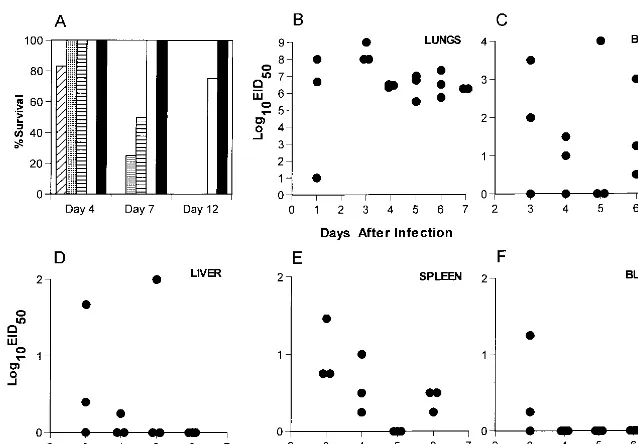
FIG. 1. (A) Naive B6 mice were infected i.n. with 10-fold dilutions of the H3N8 virus and monitored daily for survival. Four mice per group were infected as follows: diagonal stripes, 105EID
50; dotted, 104EID50; horizontal stripes, 103EID50; open, 102EID50; filled, 101EID50. (B to F) Naive B6 mice were infected i.n. with 104 EID50of H3N8 virus, and virus titers were determined for the lungs, brain, spleen, blood, and liver in embryonated chicken eggs.
on November 9, 2019 by guest
http://jvi.asm.org/
[image:2.612.145.464.68.290.2]CD8-PE antibody 53-5.8; Pharmingen) always showing,1% of the respective population remaining.
RESULTS
Infection in immunologically naive mice.The first step was
to analyze the nature of the infectious process caused by the H3N8 virus. The mean survival time following respiratory chal-lenge of naive, adult B6 mice with a uniformly lethal dose (104.0EID
50) of the mouse-passaged H3N8 influenza A virus was 5.861.7 days (Fig. 1A). Those remaining alive on day 7 had lung titers of.106.0EID
50(Fig. 1B). Evidence of signif-icant systemic spread, a characteristic observed only with ex-tremely virulent influenza A viruses (15), was detected in sam-ples of liver, spleen, and brain (Fig. 1C to E). The titers were variable in all sites other than the lung (Fig. 1B to E) and could not be attributed to the concurrent presence of infected blood (Fig. 1F), though there was early evidence of minimal viremia. This H3N8 virus is clearly very pathogenic for laboratory mice.
Protection conferred by H3N2 priming.The next question
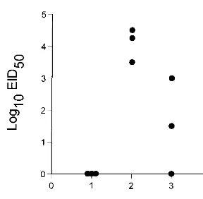
was whether mice can be protected against this fatal influenza virus infection by prior exposure to a related but much less virulent virus. The HA molecule of the HKx31 influenza A virus, which has been analyzed extensively in mouse model systems, is 96% homologous to that of the H3N8 virus, with sequence differences in each of the five antibody-binding do-mains (Table 1; references 13a and 30). Mice that had recov-ered from respiratory exposure to the HKx31 virus, hereafter referred to as H3N2 virus, were challenged i.n. with 104.0 EID50of the H3N8 virus. No virus was recovered from the lung at 24 h after infection, but titers as high as 104.5EID
50were detected in mice sampled at 48 h and were obviously declining by the next day (Fig. 2). None of the H3N2-primed mice showed any obvious signs of clinical impairment, and all were clearly protected from a dose of the H3N8 virus that achieved titers of .108.0 EID
50 in the lungs of naive mice and was uniformly lethal (Fig. 1A and B).
Role of antibody.The protective effect of prior H3N2
prim-ing (Fig. 2) could be mediated by humoral immunity, by the recall of T-cell memory, or by both sets of mechanisms. In terms of the humoral response, specific antibodies to the H3N2 and H3N8 viruses are cross-reactive in the standard hemag-glutination inhibition test; preincubating the H3N8 virus in 10% mouse serum from H3N2-primed mice prior to i.n. chal-lenge completely prevented the development of symptoms, whereas preincubation with 10% normal mouse serum resulted in 100% mortality (data not shown). The next step was to
determine the susceptibility profile of immune mice that lack one or more components of the specific host response. Chal-lenge with a lethal dose of the H3N8 virus (104EID
50) was thus repeated in B6 mice and congenic, Ig2/2mMT mice which had been primed 2 to 4 months previously with either the H3N2 or the H3N8 virus. The immune B6 (Ig1/1) mice were protected and showed 100% survival, while the immunemMT (Ig2/2) mice were completely susceptible to the secondary challenge (Table 2). Eliminating both the CD41and the CD81T cells by treating the Ig1/1 B6 mice with lymphocyte subset-specific MAbs commencing prior to challenge with the H3N8 virus still resulted in 100% survival (Table 2). Levels of H3N2-specific IgG in lung washes of mock-treated and T-cell-depleted mice were determined on days 0, 7, and 10 after secondary infection with the H3N8 virus. Significant virus-specific antibody titers were found when both mock-treated and T-cell-depleted (Fig. 3) mice were compared to naive mice. It seems that although the levels of H3N2-specific antibody at the mucosal surface were not sufficient to prevent the H3N8 virus from infecting at least some respiratory epithelial cells, the Ig response gener-ated by H3N2 priming protected against the development of lethal pneumonia (compare Fig. 1 and 2).
Secondary CD41 and CD81 T-cell responses in primed
Ig1/1 mice. The H3N2-specific antibody levels in the lung
[image:3.612.103.247.66.207.2]increased between days 0 and 7 after challenge with the H3N8 virus in control mice but remained constant in mice depleted of both CD41and CD81T cells (Fig. 3). Furthermore, viral lung titers of these same animals showed that the control group had completely cleared virus by day 7 after infection with the H3N8 virus (data not shown). In contrast, virus was still present in lung samples from two of three T-cell-depleted mice (101.5 EID50and 102.5EID50, respectively) at day 10 after secondary challenge (data not shown). Despite the apparent delay in clearance, all of nine T-cell-depleted animals survived for.45 days (data not shown). Taken together, these data suggest that although antibody-mediated protective mechanisms are suffi-cient to ensure survival following a potentially lethal secondary challenge, the CD41and CD81T-cell subsets also play a role in the secondary responses of Ig1/1 mice. Thus, secondary CD41and CD81T-cell responses were examined as follows. Mice that had been given the H3N2 virus 8 months previously were infected i.n. with (i) the lethal H3N8 virus, which shares HA-specific CD41T-cell epitopes with the H3N2 virus, (ii) the serologically different A/PR8 (H1N1) virus, which shares the NP366–374epitope recognized by the majority of the responding FIG. 2. Naive B6 mice were primed i.n. with 106.8EID
50of the H3N2 virus and rested for 1 month prior to challenge i.n. with 104EID
50of the H3N8 virus. Virus titers in the lung were determined on days 1 to 3 after infection.
TABLE 2. Susceptibility of primed mice to challenge with the H3N8 virusa
Mouse
strain depletionT-cell b
Mean survival time (days)6SD Naive mice Immune mice
B6 Nil 5.660.6 .21
CD4 4.360.5 .21
CD8 4.860.5 .21
CD41CD8 5.361.3 .21
mMT Nil 7.562.9 6.161.0, 4.762.1c
aB6 andmMT mice were infected i.n. with 106.8EID
50of the H3N2 virus and rested for 2 to 4 months. Groups of four mice were challenged i.n. with 104.0 EID50of the H3N8 virus. The data are cumulated from several experiments and are for a total of 8 to 12 mice for each treatment.
bThe mice were treated with MAb GK1.5 to CD4, MAb 2.43 to CD8, or rat Ig as a control (Nil), commencing 5 days prior to challenge and continuing every 2 or 3 days thereafter (20).
cThis group ofmMT mice was primed i.p. with 105.2EID
50of the H3N8 virus and rested for 7 weeks prior to i.n. challenge with 104.0EID
50of the H3N8 virus.
on November 9, 2019 by guest
http://jvi.asm.org/
[image:3.612.311.549.562.652.2]CD81T cells, and (iii) an influenza B virus (B/HK) that is not known to cross-react in any way with the influenza A viruses but causes a similar inflammatory pathology in the murine lung (10).
We have previously shown that staining of lymph node, spleen, and BAL populations with the NPP tetramer (tetra-meric complex of Dbbound to NP peptide) allows enumera-tion of virus-specific CD81T cells during an influenza virus infection (10). The recruitment of tetramer-positive (CD81 NPP1) T cells to the lung was comparable for the mice chal-lenged with the H3N8 and H1N1 viruses (Table 3), which is intriguing since the extent of replication (and thus antigen load) for the H1N1 virus would be expected to be much greater (reference 16 and Fig. 2). The value for the B/HK challenge presumably reflects the background associated with the non-specific recruitment of memory cytotoxic T lymphocytes (mCTL) to the pneumonic lung (26). Both of the influenza A viruses, but not the B/HK virus, stimulated the development of effector CTL (eCTL) in the lymph nodes. The greatest increase in the prevalence of the virus-specific CD41 T-helper precursor (Thp) population was seen for the mice challenged with the H3N8 virus (Table 3). This is not surprising, as many of the epitopes recognized by CD41T cells inH-2bmice are derived
from the HA molecule (21a). Clearly, even in the presence of neutralizing antibody, the antigen load is sufficient to restim-ulate both the CD41and CD81T-cell responses. This, in turn, may account for a more vigorous humoral response and accel-erated viral clearance in animals not depleted of T cells.
Limited protection of the Ig2/2mice by CD81
T-cell-medi-ated immunity.The Ig2/2mMT mice were primed i.n. with the
H3N2 virus or i.p. with the H3N8 virus and then challenged i.n. with the H3N8 virus to analyze the protective efficacy of the
[image:4.612.70.279.71.322.2]secondary CD81T-cell response (Fig. 4). A similar protocol involving the challenge of H3N2-immunemMT mice with the homologous H3N2 virus resulted in a rapid clearance of virus from the lung. This protective effect was greatly diminished by
FIG. 3. Naive B6 mice were infected i.n. with 106.8EID
50of the H3N2 virus and rested for 4 months. Rat Ig-treated control mice (A) or mice depleted of both CD41and CD81T cells (B) were infected i.n. with 104EID
[image:4.612.309.554.90.160.2]50of the H3N8 virus. BAL was done on days 0, 7, and 10 after secondary infection. The BAL cells were removed by centrifugation, and H3N2-specific IgG titers were deter-mined by ELISA. Each curve represents a single animal, and three animals are shown per time point. O.D. 405, optical density at 405 nm.
TABLE 3. CD41and CD81T-cell responses in
H3N8-challenged, H3N2-primed B6 micea
Challenge virus
% CD81NPP1
T cellsb % Specificlysisc Reciprocal Thpfrequencyd BAL MLN Spleen MLN Spleen MLN Spleen
B/HK 3.8 0.1 0.4 0 0 20,000 33,333
H3N8 9.6 2.3 0.8 23 0 523 1,785
H1N1 7.3 1.8 1.4 32 9 1,754 8,333
aB6 mice were infected i.n. with 106.8EID
50of H3N2 virus and rested for 7 to 8 months; they were then challenged i.n. with 105.6EID
50of the B/HK virus, 103 EID50of the A/PR8 (H1N1) virus, or 104EID50of H3N8 virus and sampled 5 days later.
bThe BAL cells were processed, stained with the tetramer and anti-CD8, and analyzed as described in Materials and Methods to determine the prevalence of virus-specific (CD81NPP1) T cells. The MLN and spleen cells were enriched for
CD81T cells and stained with the tetramer and anti-CD8 prior to analysis. The
level of background staining with the SEV9 tetramer was,0.1%.
cFreshly isolated MLN and spleen cells were used directly as effectors in a 6-h
51Cr release assay with H3N2-infected MC57G targets at an effector-to-target ratio of 100:1. Lysis of uninfected targets at this ratio was,4.0%.
dEnriched CD41T cells from the MLN and spleen were incubated for 68 h
with either uninfected or H3N2-infected stimulators and assayed for IFN-g
production by ELISPOT as described in Materials and Methods. The results are expressed as reciprocal virus-specific CD41Thp frequencies.
FIG. 4. (A and B)mMT mice were primed i.n. with 106.8EID
50of the H3N2 virus (A) or i.p. with 105.2EID
50of the H3N8 virus (B) and rested for 6 weeks. They were then either mock (rat Ig)-treated (F) or depleted of CD41T cells
(h), CD81T cells (Œ), or both (‚) as described in Materials and Methods and
then challenged i.n. with 104.0EID
50of the H3N8 virus. Virus titers in the lungs were determined. (C)mMT mice were infected i.n. with 106.8EID
50of H3N2 virus, rested for 10 weeks, and then challenged i.n. with 103EID
50of the H3N8 virus. Virus titers in the lungs were determined.
on November 9, 2019 by guest
http://jvi.asm.org/
[image:4.612.315.550.382.655.2]the elimination of the CD81T-cell subset (24). The immuno-dominant NP366–374peptide (5) recognized in association with H-2Dbis present and completely conserved in both the H3N2 and the H3N8 viruses. However, there is variation in the flank-ing regions (Table 1) which might potentially influence the
processing and presentation of the epitope (7) and thus the magnitude of the CD81T-cell response. Restimulating the immune T-cell populations with virus under conditions of bulk culture followed by testing in a standard 51Cr release assay showed that cross-reactive eCTL were generated in both the H3N2- and H3N8-primed mMT mice (Table 4). Similarly, determining mCTL frequencies by LDA established that the spectra of T-cell priming were comparable following i.n. (H3N2) or i.p. (H3N8) exposure to these two influenza viruses (Table 4).
What are the characteristics of the secondary CD81T-cell response in these primed mice? Recruitment of substantial numbers of CD81NPP1T cells to the pneumonic lung was apparent at 6 days after i.n. challenge of the H3N2- and H3N8-immune, but not naive, mMT mice with 104.0 EID
50 of the H3N8 virus (Fig. 5A to C). However, the lung titers were still high (Fig. 4A and B), and this dose of virus is uniformly lethal in the absence of antibody (Table 2). Furthermore, any control mediated by the CD81 T cells was more apparent for the H3N2-primed mice than for the H3N8-primed mice (see below and Table 5), reflecting the greater eCTL numbers recovered from the virus-infected lung (Fig. 5B and C). The extreme vir-ulence of the H3N8 virus (Fig. 1) is clearly the key determinant of susceptibility, as a secondary response of similar magnitude was previously shown to accelerate the rapid CD81 T-cell-TABLE 4. Cross-reactivity of the CTL response for
the H3N2 and H3N8 virusesa
Immunizing
virus restimulationIn vitro
% Specific51Cr
releaseb Reciprocal CTLpfrequencyc
Nil H3N2 H3N8 H3N2 H3N8
H3N2 H3N2 1 58 59 1,722 1,615
H3N8 16 50 53 2,684 2,091
H3N8 H3N2 7 63 56 2,013 2,065
H3N8 4 49 45 3,914 2,781
aIg2/2mMT mice were uninfected (Nil) or infected i.n. with 106.8EID 50of the H3N2 virus or i.p. with 105.2EID
50of the H3N8 virus and rested for 4 weeks prior to harvesting of CLN, MLN, and spleen cells for bulk culture or LDA assays.
bCells were restimulated in vitro with either H3N2- or H3N8-infected
stimu-lators as indicated; values for bulk cultures are shown at an effector-to-target ratio of 25:1 in a 6-h assay using MC57G target cells.
cLDA cultures were assayed for levels of specific51Cr release.3 times the standard deviation to calculate reciprocal precursor CTL (CTLp) frequencies (21).
FIG. 5. (A to C) Staining profiles for CD81NPP1lymphocytes obtained for BAL cells harvested from naive (A), H3N2-immune (B), or H3N8-immune (C)mMT
mice on day 6 after i.n. challenge with 104.0EID
50of the H3N8 virus. (D to F) Prevalence of virus-specific CD81T cells for BAL (D), MLN (E), and spleen (F) cells harvested frommMT mice on day 12 after i.n. challenge with 250 EID50of the H3N8 virus. Staining with the control SEV9 tetramer was always,0.1%. Percentages in parentheses denote levels of specific51Cr release (see footnotes to Table 4) after direct assay of freshly isolated lymphocyte populations; numbers in brackets are reciprocal Thp frequencies for IFN-g-producing CD41T cells (see footnotes to Table 3).
on November 9, 2019 by guest
http://jvi.asm.org/
[image:5.612.53.292.88.178.2] [image:5.612.76.530.349.681.2]mediated clearance of the less pathogenic A/PR8 (H1N1) in-fluenza virus from H3N2-primed B6 mice (10).
Decreasing the magnitude of the H3N8 challenge to 103.0 EID50resulted in the long-term survival of 10% of the primed
mMT mice, with evidence that the infection was being con-trolled with time (Fig. 4C). The early stage of the infectious process was much more variable following exposure to this lower dose, though lung titers were uniformly high by day 8 after infection (Fig. 4C). Reducing the virus challenge a fur-ther fourfold (250 EID50) increased both the interval to the development of severe symptoms and the numbers of mice that recovered (Table 5). Depleting the CD81subset in mice sec-ondarily challenged with the 250 EID50dose of H3N8 virus showed that this protective effect was mediated largely by the secondary eCTL response (Table 5). The eCTL were present at high frequency in the BAL (Fig. 5D) of mice challenged with the 250 EID50dose and could also be detected in the MLN and spleen by staining with the NPP tetramer (Fig. 5E and F). Furthermore, the lymphoid tissue contained both eCTL and virus-specific CD41T cells that could be stimulated by in vitro culture (Fig. 5E and F). Taken together, the results in Fig. 4, Fig. 5, and Table 5 indicate that the CD81T-cell response is capable of handling a very low dose of the H3N8 influenza virus in the absence of antibody, but not in a way that is uniformly protective (Table 5).
DISCUSSION
The extreme susceptibility of laboratory mice to respiratory challenge with the H3N8 influenza A virus demonstrates very clearly that clinical outcome depends on a race between the growth characteristics of the pathogen and the development of the specific host response. The extent of virus-induced damage to lung epithelium in immunologically naive mice is simply too great by the time that virus-specific antibody and eCTL pop-ulations become available at the site of pathology. Previous analysis utilizing an H3N2 challenge in H1N1-primed B6 mice has shown that the secondary CTL response develops in the MLN and takes at least 4 to 5 days before the effectors are available in the infected respiratory tract (10). Our studies (24, 25) and others (8, 12) using Ig2/2 mMT mice indicate that primed CD81 T cells can provide at least some protection against novel influenza A viruses, which may be the reason that not all those infected during the recent H5N1 outbreak in Hong Kong succumbed (29). However, although priming the CD81 T-cell compartment can protect Ig2/2 mMT mice against homologous challenge with high titers of the HKx31 virus (24), the recall of the mCTL to eCTL function is still
too slow to protect from all but a very low dose of the H3N8 virus.
Though the H3N8 virus has a variable capacity for systemic spread, naive mMT mice suffered no obvious consequences following i.p. challenge with a dose 1,000 times higher than that causing uniformly lethal disease following i.n. exposure. Thus, the requirement for enzymatic cleavage of the viral HA molecule that substantially limits the production of infectious influenza A viruses to the murine lung mucosa still determines the pathogenesis of the disease (13). The secondary localiza-tion of the H3N8 virus to the brain detected following i.n. infection of B6 mice is probably a consequence of the much greater viral load resulting from the continued replication in the respiratory tract. The present experiments show that this can result in measurable viremia, although of very limited duration. The analysis also makes the point that it may be possible to protect mammals against a novel influenza virus infection by injecting fully virulent virus subcutaneously or intramuscularly, though such a strategy would obviously be too risky to consider using in humans. However, an attenuated, live virus vaccine might be given safely via this route to people that lack cross-reactive neutralizing antibody, with much less risk than for administration via a respiratory route. Such an ap-proach could be considered for limiting a rapidly spreading pandemic caused by an extremely virulent avian influenza A virus, provided that an attenuated variant is available.
The H3-specific antibody response in the B6 mice clearly prevents the development of lethal pneumonia following re-spiratory challenge with the H3N8 virus, though there is still some replication in the lung. In general, preexisting antibody has been shown to be the major mechanism of protection against secondary influenza virus infection (11). This may re-flect direct neutralization of the inoculum (reviewed in refer-ence 11) by virus-specific IgG or IgA already present at the surface of the lung mucosa. Also, the virus-specific Ig could act to prevent further spread in the respiratory tract by the neu-tralization of free virions or by opsonizing the virus for uptake by macrophages. Ig may also enable macrophages to mediate antibody-dependent cellular cytotoxicity (11).
The rapid control of the H3N8 infection in the H3N2-im-mune B6 mice by neutralizing antibody in no way inhibited the development of the secondary CD41 and CD81 T-cell re-sponses. Previous studies have shown that virus-specific CD81 T-cell responses are generated in the presence of neutralizing antibody (23). The present analysis extends this observation to show that both CD41and CD81T-cell responses develop in the presence of neutralizing antibody. Furthermore, though the recall of CD41T-cell memory has been shown to have relatively little protective effect in Ig2/2mMT mice depleted of the CD81T-cell subset (24), such primed T-cell help could obviously be a significant factor in the Ig1/1group. Overall, the results indicate that cross-reactive CD81eCTL (20) and the neutralizing antibody response may both play a part in recovery from a virulent influenza virus infection, with the latter mechanism being much more important.
[image:6.612.53.291.80.182.2]The laboratory-adapted viruses used in these experiments express H3 molecules derived from viruses circulating in hu-mans (H3N2) and birds (H3N8) in 1968 and 1980, respectively. Sequencing the HA1 subunits showed at least one amino acid difference for each of the five antibody-binding domains. The 96% sequence homology for these two HA1 regions is fairly representative of that seen for many of the “drifted” H3N2 viruses that cause sequential pandemics (16, 19, 22, 31, 32). Such antibody-mediated selection pressure was shown to result in a 9.2% change in the HA1 region of human H3N2 viruses isolated over a 10-year period. The avian viruses do not show TABLE 5. CD81T-cell-mediated protection in Ig2/2micea
Immunizing
virus depletionT-cell b Groupsize Mean survival time(days)6SD % Survival.21 days
None Nil 5 10.462.2 0
CD8 5 5.861.5 0
H3N2 Nil 9 1468.4 78
CD8 10 12.961.9 20
H3N8 Nil 10 9.762.6 30
CD8 6 7.761.8 0
amMT animals were primed either i.n. with 106.8EID
50of H3N2 or i.p. with 105.2EID
50of H3N8 and rested for 14 to 16 weeks prior to i.n. challenge with 250 EID50of the H3N8 virus.
bThe mice were given rat Ig (Nil) or were CD8 depleted as described for
Table 2.
on November 9, 2019 by guest
http://jvi.asm.org/
much evidence of such drift in their natural, maintaining host (16). The present results indicate that any recent incursion of an avian virus into the human population could be dealt with by using a virus that is likely to be already available as a nonpathogenic laboratory strain adapted for growth in embry-onated hen eggs, the basic starting point for the currently prevalent vaccine technology. The same is true for the H7N7 viruses, which also loom as possible human pathogens (28). The use of contemporary molecular approaches to develop attenuated vaccine candidates for the influenza A viruses that seem most likely to cross from domestic animals into humans is worth considering.
ACKNOWLEDGMENTS
We thank Vicki Henderson for help in preparing the manuscript, and we thank Kristin Branum for expert technical assistance.
This work was supported by Public Health Service grants AI08831, AI29579, AI38359, and CA21765 and by the American Lebanese-Syrian Associated Charities.
REFERENCES
1.Allan, W., Z. Tabi, A. Clearly, and P. C. Doherty.1990. Cellular events in the lymph node and lung of mice with influenza: consequences of depleting CD41T cells. J. Immunol.144:3980–3986.
2.Altman, J. D., P. A. H. Moss, P. J. R. Goulder, D. H. Barouch, M. G. McHeyzer-Williams, J. I. Bell, A. J. McMichael, and M. M. Davis.1996. Phenotypic analysis of antigen-specific T lymphocytes. Science274:94–96. 3.Claas, E. C. J., A. D. M. E. Osterhaus, R. van Beek, J. C. De Jong, G. F.
Rimmelzwaan, D. A. Senne, S. Karauss, K. F. Shortridge, and R. G. Webster. 1998. Human influenza A H5N1 virus related to a highly pathogenic avian influenza virus. Lancet351:472–477.
4.Cohen, J.1997. The flu pandemic that might have been. Science277:1600– 1601.
5.Deckhut, A. M., W. Allan, A. McMickle, M. Eichelberger, M. A. Blackman, P. C. Doherty, and D. L. Woodland.1993. Prominent use of Vb8.3 T cells in H-2Dbnucleoprotein epitope. J. Immunol.151:2658–2666.
6.De Jong, J. C., A. D. M. E. Osterhaus, R. G. Webster, and W. L. Lim.1997. A pandemic warning? Nature389:554.
7.Eisenlohr, L. C., J. W. Yewdell, and J. R. Bennink.1992. Flanking sequences influence the presentation of an endogenously synthesized peptide to cyto-toxic T lymphocytes. J. Exp. Med.175:481–487.
8.Epstein, S. L., C. Lo, J. A. Misplon, and J. R. Bennink.1998. Mechanisms of protective immunity against influenza virus infection in mice without anti-bodies. J. Immunol.160:322–327.
9.Flynn, K., A. Mu¨llbacher.1997. The generation of memory antigen-specific cytotoxic T cell responses by CD28/CD80 interactions in the absence of antigen. Eur. J. Immunol.27:456–462.
10. Flynn, K. J., G. T. Belz, J. D. Altman, R. Ahmed, D. L. Woodland, and P. C. Doherty.1998. Virus-specific CD81T cells in primary and secondary influ-enza pneumonia. Immunity8:683–691.
11.Gerhard, W., K. Mozdzanowska, M. Furchner, G. Washko, and K. Maiese. 1997. Role of the B-cell response in recovery of mice from primary influenza virus infection. Immunol. Rev.159:95–103.
12.Graham, M. B., and T. J. Braciale.1997. Resistance to and recovery from lethal influenza virus infection in B lymphocyte-deficient mice. J. Exp. Med. 186:2063–2068.
13. Horimoto, T., and Y. Kawaoka.1995. The hemagglutinin cleavability of a virulent avian influenza virus by subtilisin-like endoproteases is influenced by the amino acid immediately downstream of the cleavage site. Virology210: 466–470.
13a.Stech, J.Unpublished data.
14. Subbarao, K., A. Klimov, J. Katz, H. Regnery, W. Lim, H. Hall, M. Perdue, D. Swayne, C. Bender, J. Huang, M. Hemphill, T. Rowe, M. Shaw, X. Xu, K. Fukuda, and N. Cox.1998. Characterization of an avian influenza A (H5N1) virus isolated from a child with a fatal respiratory illness. Science279:393– 396.
15. Kawaoka, Y.1991. Equine H7N7 influenza A viruses are highly pathogenic in mice without adaptation: potential use as an animal model. J. Virol.65: 3891–3894.
16. Kida, H., Y. Kawaoka, C. W. Naeve, and R. G. Webster.1987. Antigenic and genetic conservation of H3 influenza virus in wild ducks. Virology159:109– 119.
17. Kilbourne, E. D.1969. Future influenza vaccines and the use of genetic recombinants. Bull. W.H.O.41:643–606.
18. Kitamura, D., J. Roes, R. Kuhn, and K. Rajewsky.1991. A B cell-deficient mouse by targeted disruption of the membrane exon of the immunoglobulin
mchain gene. Nature350:423–426.
19. Krystal, M., J. F. Young, P. Palese, I. A. Wilson, J. J. Skehel, and D. C. Wiley. 1983. Sequential mutations in hemagglutinins of influenza B virus isolates: definition of antigenic domains. Proc. Natl. Acad. Sci. USA80:4527–4531. 20. Lalvani, A., R. Brooks, S. Hambleton, W. J. McMichael.1997. Rapid effector
function in CD81memory T cells. J. Exp. Med.186:859–865.
21. Mu¨llbacher, A., A. B. Hill, R. V. Blanden, W. B. Cowden, N. J. King, and R. T. Hla.1991. Alloreactive cytotoxic T cells recognize MHC class I antigen without peptide specificity. J. Immunol.147:1765–1772.
21a.Riberdy, J.Unpublished data.
22. Rogers, G. N., J. C. Paulson, R. S. Daniels, J. J. Skehel, I. A. Wilson, and D. C. Wiley.1983. Single amino acid substitutions in influenza haemagglu-tinin change receptor binding specificity. Nature304:76–78.
23. Seiler, P., M. A. Bru¨ndler, C. Zimmermann, D. Weibel, M. Bruns, H. Hen-gartner, and R. M. Zinkernagel.1998. Induction of protective cytotoxic T cell responses in the presence of high titers of virus-neutralizing antibodies: implications for passive and active immunization. J. Exp. Med.187:649–654. 24. Topham, D. J., and P. C. Doherty.1998. Clearance of an influenza A virus by CD41T cells is inefficient in the absence of B cells. J. Virol.76:882–885. 25.Topham, D. J., R. A. Tripp, A. M. Hamilton-Easton, S. R. Sarawar, and P. C.
Doherty.1996. Quantitative analysis of the influenza virus-specific CD41T cell memory in the absence of B cells and Ig. J. Immunol.157:2949–2952. 26.Tripp, R. A., S. Hou, A. McMickle, J. Houston, and P. C. Doherty.1995.
Recruitment and proliferation of CD81T cells in respiratory virus infec-tions. J. Immunol.154:6013–6021.
27. Webster, R. G. 1997. Influenza virus: transmission between species and relevance to emergence of the next human pandemic. Arch. Virol.13:105– 113.
28. Webster, R. G.1997. Predictions for future human influenza pandemics. J. Infect. Dis.176:514–519.
29. Webster, R. G., and A. J. Hay.1998.InK. G. Nicholson, R. G. Webster, and A. J. Hay (ed.), Textbook of influenza, p. 561–565. Blackwell Science Ltd., Oxford, England.
30. Wiley, D., and J. J. Skehel.1987. The structure and function of the hemag-glutinin membrane glycoprotein of influenza virus. Annu. Rev. Biochem.56: 365–394.
31. Wiley, D. C., I. A. Wilson, and J. J. Skehel.1981. Structural identification of the antibody-binding sites of Hong Kong influenza haemagglutinin and their involvement in antigenic variation. Nature289:373–378.
32. Wilson, I. A., J. J. Skehel, and D. C. Wiley.1981. Structure of the haemag-glutinin membrane glycoprotein of influenza virus at 3 Å resolution. Nature 289:366–373.
33. Yuen, K. Y., P. K. S. Chan, M. Peiris, D. N. C. Tsang, T. L. Que, K. F. Shortridge, P. Y. Cheung, W. K. To, E. T. F. Ho, R. Sung, A. F. B. Cheng, and members of the H5N1 study group.1998. Clinical features and rapid viral diagnosis of human disease associated with avian influenza A H5N1 virus. Lancet351:467–471.