Copyright © 2000, American Society for Microbiology. All Rights Reserved.
NOTES
Cloning the Antibody Response in Humans with Chronic Inflammatory
Disease: Immunopanning of Subacute Sclerosing Panencephalitis
(SSPE) Brain Sections with Antibody Phage Libraries Prepared
from SSPE Brain Enriches for Antibody Recognizing
Measles Virus Antigens In Situ
GREGORY P. OWENS,1R. ANTHONY WILLIAMSON,2MARK P. BURGOON,1OMAR GHAUSI,2
DENNIS R. BURTON,2ANDDONALD H. GILDEN1,3*
Departments of Neurology1and Microbiology,3University of Colorado Health Sciences Center, Denver, Colorado
80262, and Department of Immunology, Scripps Research Institute, La Jolla, California 920372
Received 23 July 1999/Accepted 20 October 1999
In central nervous system (CNS) infectious and inflammatory diseases of known cause, oligoclonal bands represent antibody directed against the causative agent. To determine whether disease-relevant antibodies can be cloned from diseased brain, we prepared an antibody phage display library from the brain of a human with subacute sclerosing panencephalitis (SSPE), a chronic encephalitis caused by measles virus, and selected the library against SSPE brain sections. Antibodies that were retrieved reacted strongly with measles virus cell extracts by enzyme-linked immunosorbent assay and were specific for the measles virus nucleocapsid protein. These antibodies immunostained cells in different SSPE brains but not in control brain. Our data provide the first demonstration that diseased brain can be used to select in situ for antibodies directed against the causative agent of disease and point to the potential usefulness of this approach in identifying relevant antibodies in chronic CNS or systemic inflammatory diseases of unknown cause.
A common feature of chronic infectious and inflammatory diseases of the central nervous system (CNS) is the intrathecal synthesis of oligoclonal immunoglobulin G (IgG). In known CNS infectious diseases, oligoclonal bands (OGBs) are di-rected against the etiologic agent (reviewed in reference 10). We hypothesize that in CNS inflammatory disease of unknown cause, such as multiple sclerosis (MS), the specificities of OGBs may similarly yield important clues for identifying a putative pathogen and contribute to our understanding of dis-ease pathogenesis.
Standard immunological techniques have not revealed a vi-ral or cellular antigen that consistently reacts with MS OGBs. Recombinant antibody technology combined with phage dis-play offers a novel strategy to identify the disease-relevant IgG in MS cerebrospinal fluid and brain plaques (1). Combinatorial antibody library technology, in which Fab fragments are ex-pressed on the surface of phage, has been used to describe human antibody responses to both infectious agents (2, 5, 8, 9, 15, 19) and autoantigens (4, 11, 12, 16–18). We tested the feasibility of using this technology to clone and characterize intrathecal IgG found in the prototype chronic CNS infectious disease, subacute sclerosing panencephalitis (SSPE), caused by persistent measles virus (MV) infection in brain. Large com-binatorial antibody libraries were constructed from heavy and light chain sequences expressed in a single SSPE brain and
panned against MV-infected cell extracts. Fabs specific for the MV phosphoprotein and nucleocapsid protein were selected, proving that IgG mRNA expressed in brain during a chronic CNS infection can be used to generate high-affinity antibodies specific for the disease-causing pathogen (6). However, use of this strategy in a CNS inflammatory disease of unknown cause requires that panning be performed in situ with diseased tissue. In this study, we panned for MV-specific Fabs by using SSPE brain tissue sections as a source of antigen.
Construction of the recombinant antibody phage display li-brary used for panning has been described previously (6). This library contains heavy (␥1) and kappa light chain sequences PCR amplified from a cDNA library generated from patho-logically verified SSPE brain (SSPE 83) of a 14-year-old boy. IgG extracted from this brain was shown to be oligoclonal by isoelectric focusing and reactive with multiple MV proteins (6, 14). Phage library panning was performed as described previ-ously (8) except for the use of SSPE brain sections (SSPE 81) instead of antigen-coated microtiter wells. Sections of fresh-frozen brain were prepared for panning as follows. Slides were dehydrated under vacuum for 2 h at room temperature (RT), fixed for 10 min in⫺20°C acetone, hydrated in cold phosphate-buffered saline (PBS) (10 mM sodium phosphate [pH 7.4], 150 mM NaCl), treated with 1% H2O2in 100% methanol for 10
min at 4°C, washed twice for 1 min at RT in 100% methanol, and then air dried and stored at⫺20°C. Tissue sections on the slide were shaved to approximate the area of a microtiter well and surrounded by a glue wall to provide a reservoir. Two sets of slides were used: one set was pretreated for 10 min with 0.1 M HCl, pH 2.2 (low-pH elution), and neutralized in PBS, and the other set was untreated (no elution). In a humid chamber,
* Corresponding author. Mailing address: Department of Neurol-ogy, Mailstop B182, University of Colorado Health Sciences Center, 4200 E. 9th Ave., Denver, CO 80262. Phone: (303) 315-8281. Fax: (303) 315-8720. E-mail: [email protected].
1533
on November 9, 2019 by guest
http://jvi.asm.org/
sections were blocked with 75l of 3% bovine serum albumin in water for 60 min at 25°C.
After removal of blocking solution, 50l of freshly prepared phage suspension was added and sections were incubated for 2 h at RT. Unbound phage were removed by immersing the slides into a petri dish containing PBS–0.5% Tween 20 and agitated for 5 min on a 120-rpm rotary shaker. Washing was repeated four additional times with fresh buffer for each wash. After the final wash, excess buffer was removed, and 50l of elution buffer (0.1 M HCl, adjusted to pH 2.2 by the addition of glycine) was added onto the tissue section. After disruption of the tissue with a pipette tip and a 10-min incubation, the eluate was transferred to an Eppendorf tube and adjusted to neutral pH by addition of 6l of 1 M Tris base. The tissue was rinsed with 50l of PBS, and this material was pooled with the
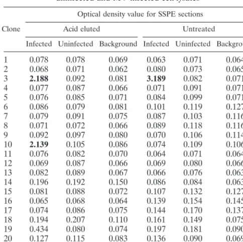
clones taken from the unpanned SSPE library. No immunore-activity was found in any of the unpanned Fabs. However, a single Fab (Fab 1) from the panning against untreated SSPE sections and two Fabs (Fabs 2 and 3) from the panning against acid-treated SSPE sections demonstrated strong immunoreac-tivity against MV-infected cell extracts, but not uninfected cell extracts (Table 1).
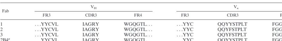
We next determined the heavy chain amino acid sequences of the three MV-specific Fabs. Each contained the same se-quence within complementarity-determining region 3 (CDR3) (Table 2). This sequence was also identical to that found in Fab 2B4, a recombinant antibody specific for the MV nucleocapsid protein that was selected by panning against MV-infected cell extracts (6). Importantly, we did not find this heavy chain sequence in the heavy chain sequences of 38 Fab clones ran-domly selected from the unpanned library (data not shown). A comparison of kappa light chain variable region sequences revealed that Fab 1 had the same light chain as 2B4, whereas Fabs 2 and 3 had a light chain that differed only by a single amino acid substitution in the CDR3. Alignment of these two light chain variable regions showed extensive homology but also revealed several distinct amino acid differences character-istic of clonal variants (data not shown).
[image:2.612.52.294.90.332.2]We next sought to test the reactivity of the selected antibod-ies against SSPE brain sections compared to normal human brain white matter, acute MS plaques, and a brain infarct. However, the presence of large amounts of endogenous IgG in SSPE and MS brain precluded the use of an antihuman sec-ondary antibody in evaluating recombinant Fab reactivity. To circumvent this problem, we expressed the recombinant anti-bodies in pFLAG, a modified form of the pComb3H vector (3) in which an 8-amino-acid flag epitope tag (13) is fused to the carboxyl terminus of the heavy chain (Fd region) moiety of the Fab. The flag peptide sequence was introduced into the back-bone of the pComb3H phagemid vector between the more downstream of twoSfiI sites and the uniqueNheI site, replac-ing sequence encodreplac-ing cpIII with a 38-bp insert created by annealing the oligonucleotide primers HCFLAG(⫹) (5⬘-CGG CCC AGA CTA CAA GGA CGA TGA CGA TAA GGG CTA AG-3⬘) and HCFLAG(⫺) (5⬘-CTA GCT TAG CCC TTA TCG TCA TCG TCC TTG TAG TCT GGG CCG GCC-3⬘). Phagemid DNA recovered following library panning was digested withSfiI, and the excised fragment, containing antibody sequences, was directionally religated into the SfiI sites of pFLAG. Flag-tagged soluble Fab was expressed and purified as described previously (8). The flagged 2B4 antibody (identical in heavy and light chain composition to Fab 1) re-acted specifically with MV-infected cells in culture, intensely staining the cytoplasm of multinucleated cells (Fig. 1A). Bound
TABLE 1. Binding of soluble Fabs from panned SSPE library to uninfected and MV-infected cell lysatesa
Clone
Optical density value for SSPE sections Acid eluted Untreated
Infected Uninfected Background Infected Uninfected Background
1 0.078 0.078 0.069 0.063 0.071 0.064 2 0.068 0.071 0.062 0.080 0.073 0.065
3 2.188 0.092 0.081 3.189 0.082 0.071
4 0.077 0.087 0.066 0.071 0.091 0.071 5 0.076 0.085 0.071 0.084 0.099 0.071 6 0.086 0.079 0.081 0.101 0.119 0.127 7 0.079 0.091 0.075 0.087 0.103 0.116 8 0.071 0.072 0.066 0.089 0.118 0.116 9 0.092 0.097 0.080 0.070 0.106 0.114
10 2.139 0.105 0.086 0.074 0.109 0.106
11 0.076 0.082 0.070 0.064 0.071 0.064 12 0.069 0.087 0.066 0.069 0.080 0.066 13 0.082 0.089 0.067 0.066 0.076 0.063 14 0.196 0.192 0.150 0.086 0.084 0.063 15 0.081 0.088 0.072 0.107 0.132 0.127 16 0.065 0.068 0.064 0.139 0.154 0.145 17 0.074 0.086 0.075 0.144 0.170 0.137 18 0.194 0.207 0.110 0.161 0.149 0.075 19 0.434 0.080 0.074 0.197 0.181 0.090 20 0.127 0.115 0.083 0.136 0.090 0.069
IF9b 1.301 0.076 0.062 1.381 0.085 0.062
aELISA measures binding of soluble Fabs to detergent extracts of
MV-infected and unMV-infected Vero cells. Background binding measures immunoreac-tivity of secondary antibody to MV-infected cell lysates in the absence of soluble Fab. Optical density was measured at 405 nm after a 60-min incubation with
p-nitrophenol phosphate as substrate.
bIF9 is an MV phosphoprotein-specific Fab obtained by panning on
MV-infected cell lysates (6).
on November 9, 2019 by guest
http://jvi.asm.org/
on November 9, 2019 by guest
http://jvi.asm.org/
(1:200 dilution in 10% NGS-PBS) per ml. After incubation overnight at 4°C and an additional 2 h at RT, sections were washed five times with PBS. Sections bound with 2B4 antibod-ies were incubated for 3 h at RT with mouse monoclonal IgG specific for the flag peptide sequence (1:200 dilution in 10% NGS-PBS). After washing in PBS, sections were overlaid with mouse anti-IgG conjugated to alkaline phosphatase (a 1:200 dilution in 2% NGS-PBS) for 1 h at RT and again washed extensively with PBS. Staining was developed with New Fuch-sin (DAKO, Carpinteria, Calif.) as substrate, and sections were counterstained with Gill no. 2 hematoxylin (Sigma Chemical Co.) and mounted with aqueous Immunomount (Fisher Scien-tific, Pittsburgh, Pa.) for light microscopy. Bound rabbit an-ti-MV IgG was detected with a 1:200 dilution of a goat anti-rabbit IgG conjugated to alkaline phosphatase.
The flagged 2B4 Fab reacted strongly with infected cells in the fresh-frozen acetone-fixed SSPE brain used for panning (Fig. 1C). This staining pattern was also seen when the same SSPE brain was incubated with a polyclonal anti-MV antibody (Fig. 1D). Immunoreactivity was not observed when adjacent sections from the same SSPE brain were stained with un-flagged 2B4 (Fig. 1E). A second fresh-frozen paraffin-embed-ded SSPE brain (SSPE 2D) was also stained by flagged 2B4 (Fig. 1F). In this tissue, cytoplasmic staining of individual cells was accompanied by punctate staining that appeared to be extracellular. No 2B4-flag immunoreactivity was detected in sections from a formalin-fixed paraffin-embedded brain infarct (Fig. 1G) or fresh-frozen acetone-fixed normal human brain white matter (Fig. 1H) or acute MS plaques (data not shown). Our findings demonstrate that a complex mixture of brain antigens in tissue sections can be used to specifically enrich for immunoreactive Fabs. After five rounds of panning, approxi-mately 10% (3 of 36) of the eluted phage represented a single MV-specific Fab. The heavy chain of this Fab was not detected in 38 random clones sequenced from the unpanned library. Thus, the presence of this Fab after extensive panning does not simply reflect carryover of an abundant heavy chain sequence.
capsid protein from infected cell extracts and recognizes the denatured form of the protein by immunoblotting (6) but also recognizes the nucleocapsid antigen in different SSPE brains. Staining with both 2B4 and anti-MV IgG revealed that only a small fraction of cells (⬍1%) in the panned sections harbored MV proteins, and most immunoreactivity was confined to small areas (Fig. 1C). Thus, despite the apparent paucity of MV antigens, modest numbers of MV-specific phage were still recovered.
Phage display of combinatorial antibody libraries is a pow-erful tool to rapidly generate specific high-affinity antibodies from immune human donors (7). We have extended this tech-nology to infectious inflammatory diseases of the CNS. The selection of specific Fabs with a complex antigen source such as brain sections suggests that this technology may be valuable to demonstrate antibody specificity not only in CNS diseases of unknown cause such as MS but also in other chronic inflam-matory systemic disorders of unknown cause such as sarcoid-osis, Wegener’s granulomatsarcoid-osis, polyarteritis nodosa, and sys-temic lupus erythematosus.
This work was supported in part by Public Health Service grants NS 32623 to D.H.G. and AI 39162 to D.R.B. from the National Institutes of Health.
SSPE brain was kindly provided by the National Neurological Re-search Specimen Bank, VAMC, Los Angeles, Calif. SSPE-2D and brain infarct were kindly provided by John W. Prineas, Veterans Ad-ministration Hospital, East Orange, N.J. We also thank Paul Rota from the Centers for Disease Control and Prevention for giving us the Edmonston strain of MV and Thomas Moench for providing rabbit serum against purified MV. We thank Marina Hoffman for editorial review and Cathy Allen for preparing the manuscript.
REFERENCES
1.Barbas, C. F., III, A. S. Kang, R. A. Lerner, and S. J. Benkovic.1991. Assembly of combinatorial antibody libraries on phage surfaces: the gene III site. Proc. Natl. Acad. Sci. USA88:7978–7982.
[image:4.612.55.551.92.165.2]2.Barbas, C. F., III, T. A. Collett, W. Amberg, P. Roben, J. M. Binley, D. TABLE 2. CDR3 amino acid sequence of heavy chain and associated kappa light sequences of Fabs selected by panning on SSPE
brain sections
Fab VH V
FR3 CDR3 FR4 FR3 CDR3 FR4
1 . . .YYCVL IAGRY WGQGTL. . . .YYC QQYYSTPLT FGGGT. . .
2 . . .YYCVL IAGRY WGQGTL. . . .YYC QQYFSTPLT FGGGT. . .
3 . . .YYCVL IAGRY WGQGTL. . . .YYC QQYFSTPLT FGGGT. . .
2B4a . . .YYCVL IAGRY WGQGTL. . . . . .YYC QQYYSTPLT FGGGT. . .
aSee reference 6.
on November 9, 2019 by guest
http://jvi.asm.org/
natorial libraries. Adv. Immunol.57:191–280.
8.Burton, D. R., C. F. Barbas III, M. A. A. Persson, S. Koenig, R. M. Chanock, and R. A. Lerner.1991. A large array of human monoclonal antibodies to type 1 human immunodeficiency virus from combinatorial libraries of asymp-tomatic seropositive individuals. Proc. Natl. Acad. Sci. USA88:10134–10137. 9.Ditzel, H. J., S. M. Barbas, C. F. Barbas III, and D. R. Burton.1994. The nature of the autoimmune antibody repertoire in human immunodeficiency virus type 1 infection. Proc. Natl. Acad. Sci. USA91:3710–3714. 10. Gilden, D. H., M. E. Devlin, M. P. Burgoon, and G. P. Owens.1996. The
search for virus in multiple sclerosis brain. Mult. Scler.2:179–183. 11. Graus, Y. F., M. H. de Baets, P. W. H. I. Parren, S. Berrih-Aknin, J. Wokke,
P. J. van Breda Vriesman, and D. R. Burton.1997. Human anti-nicotinic acetylcholine receptor recombinant Fab fragments isolated from thymus-derived phage display libraries from myasthenia gravis patients reflect pre-dominant specificities in serum and block the action of pathogenic serum antibodies. J. Immunol.158:1919–1929.
12. Hexham, J. M., J. Furmaniak, C. Pegg, D. R. Burton, and B. R. Smith.1992. Cloning of a human autoimmune response: preparation and sequencing of a human anti-thyroglobulin autoantibody using a combinatorial approach. Au-toimmunity12:135–141.
13. Hopp, T. P., K. S. Prickett, V. L. Price, R. T. Libby, C. J. March, D. P.
Cervetti, D. L. Urdal, and P. J. Corbon.1988. A short polypeptide marker sequence useful for recombinant protein identification and purification. Bio/ Technology6:1204–1210.
14. Owens, G. P., M. P. Burgoon, M. E. Devlin, and D. H. Gilden.1997. Extrac-tion and purificaExtrac-tion of active IgG from SSPE and MS brain. J. Virol. Methods68:119–125.
15. Parren, P. W. H. I., and D. R. Burton.1997. Antibodies against HIV-1 from phage display libraries: mapping of an immune response and progress to-wards antiviral immunotherapy, p. 18–56.InJ. D. Capra (ed.), Antibody engineering. S. Karger, Basel, Switzerland.
16. Portolano, S., and S. M. R. B. McLachlan.1993. High affinity, thyroid-specific human autoantibodies displayed on the surface of filamentous phage use V genes similar to other autoantibodies. J. Immunol.151:2839–2851. 17. Siegel, D. L.1995. Isolation of human anti-red blood cell antibodies by
repertoire cloning. Ann. N. Y. Acad. Sci.764:547–548.
18. Siegel, D. L.1998. The human immune response to red blood cell antigens as revealed by repertoire cloning. Immunol. Res.17:239–251.
19. Williamson, R. A., R. Burioni, P. P. Sanna, L. J. Partridge, C. F. Barbas III, and D. R. Burton.1993. Human monoclonal antibodies against a plethora of viral pathogens from single combinatorial libraries. Proc. Natl. Acad. Sci. USA90:4145.