TRANSFUSION SUPPORT IN LEUKEMIC
CHILDREN
Dissertation submitted to the
THE TAMILNADU Dr. M.G.R MEDICAL UNIVERSITY IN PARTIAL FULFILMENT OF REQUIREMENTS
FOR THE AWARD OF DEGREE OF M.D (BRANCH XXI)
IMMUNOHEMATOLOGY AND BLOOD TRANSFUSION
DEPARTMENT OF TRANSFUSION MEDICINE,
THE TAMILNADU DR. M.G.R. MEDICAL UNIVERSITY,
CHENNAI – 600 032.
CONTENTS
PAGE NO.
1. INTRODUCTION
2. AIMS AND OBJECTIVES
3. REVIEW OF LITERATURE
4. MATERIALS AND METHODS
5. RESULTS
6. DISCUSSION
7. SUMMARY
8. CONCLUSION
9. BIBLIOGRAPHY
1. INTRODUCTION
The incidence of different types of leukemia varies with age through out the world.1 In India, Parkienstal et al reported that leukemias are the most common cancer affecting the children accounting for 25 – 35% of malignancies.2 The majority of them were ALL and most were in age groups 0 – 3 yrs, 4 – 6 yrs and 7 – 9 yrs. ALL is most frequent in South India3. In Chennai, leukemia was reported as the highest among the first five childhood cancers.1
Acute leukemia is a malignancy arising due to the clonal proliferation of abnormal hematopoietic cells leading to disruption of normal marrow function resulting in increased number of blast cells > 20% (Normally blast cells <5%).4
Valleri et al suggested that transfusion of RBC to anemic thrombocytopenic patients would be effective before transfusing viable platelets. Maintenance of hemoglobin concentration of a patient with thrombocytopenia at higher levels may contribute to improve hemostasis.6
In tertiary care centres, platelet transfusions are widely used for the management of bleeding in thrombocytopenic leukemic patients.7 Clinicians faced with an abnormal laboratory value may use transfusion therapy to correct the value rather than to achieve a clinical result.8 Prophylactic transfusion may or may not improve platelet survival when compared to transfusion in response to active bleeding.5
A reliable platelet count and appropriate clinical evaluation of the leukemic patients showed that a significantly lower threshold is needed more for therapeutic transfusion than for prophylactic transfusion.9
aggressive chemotherapies producing more acute and prolonged thrombocytopenia.10 So the present study was undertaken to find the
(i) need for transfusion in leukemic children and
2. AIMS AND OBJECTIVES
(i) To study the need for transfusion in leukemic children.
(ii) To identify the blood component most commonly used for transfusion.
(iii) To find the pre and post Hemoglobin estimation.
3. REVIEW OF LITERATURE
3.1 Methods of component preparation
Platelet rich plasma (PRP) can be separated from whole blood by a soft centrifugation. The processing of blood and PRP are done at room temperature (22°C) and holding the sedimented platelets stationary for 30 – 60 minutes before resuspending them as platelet concentrates (PC).11 Platelet rich plasma was the first method used to prepare PC12. Platelet concentrates from Whole blood are often referred to as “random donor platelets”. Platelet concentrates are prepared by centrifugation of standard amounts of whole blood.13 Preparation of platelet concentrates are done from anti coagulated whole blood by the “platelet rich plasma” and or “buffy coat techniques”.11
There are two methods for doing platelet preparation
(i) Platelet rich plasma (PRP) method. (ii) Buffy coat (BC) Method.
0.75 x10¹¹ platelets / unit or approximately 60to 75% of the platelets from the original unit of blood.13 One draw back is that this method results in 108 WBC or approximately 50% or more Leukocyte contamination occurs during seperation from the original unit of Whole Blood.13
In the second method i.e. Buffy coat method, anticoagulated whole blood is subjected to hard spin, resulting in separation of platelet poor plasma (supernatant), buffy coat (intermediate) and PRBC at the bottom. Buffy coat is separated and subjected to soft spin leading to separation of PRP and WBC with residuals.
Platelet Rich Plasma Preparation
Anti coagulated whole blood
PRBC Platelet Rich Plasma
Soft Centrifugation
Hard Centrifugation
Platelet poor plasma Platelet
Buffy coat preparation
3.2 Storage
Platelets are stored at 20˚C to 24˚C using continuous gentle horizontal agitation in storage bags specifically designed to permit Oxygen and Carbon dioxide exchange to optimize platelet quality.13 This combination of storage bag, constant agitation preservative solution, temperature and the use of approximately 50 ml of plasma permit satisfactory preservation of platelets up to 5 days.
3.3 Transfusion
Platelet storage also has an impact on platelet transfusion efficacy. Transfusion with platelets stored from day 0 to day 5 were
Anti coagulated whole blood
Plasma Buffy PRBC Coat
Soft Centrifugation
Platelet Concentrate
used14. All the platelets used were ABO group identical between Donor and the recipient. A single unit transfusion was considered as one transfusion episode and is defined as either RBC unit or unit of “whole blood derived platelet concentrate”.14
3.4 Hematology Analyzer
In blood banks hematology analyzers, are used to count cells in blood products. These blood products are derived from centrifuged whole blood.15 Platelet counting in platelet concentrates should be validated separately for platelet collected from whole blood because most of the analyzers are designed to count platelets in the presence of RBC. Platelet counting in the absence of RBC might result in deviating from correct platelet counts. The samples were taken in a EDTA tube. Significantly higher platelet count in the EDTA tube was observed by the cell analyzer than in the dry tube.15
3.5 Role of RBCs in bleeding
surgical blood loss refers to generalized, systemic bleeding that is not corrected by surgical interventions. A correlation exists between anemia and prolonged bleeding time. When the anemia is corrected, the bleeding time is reduced. Platelet function is known to be a major determinant of bleeding time and anemia produces a reversible platelet dysfunction.6
A Hematocrit level of 35 volume percent at the site of bleeding minimizes shear stress and reduces nitric oxide produced by endothelial cells.16 Therefore trigger for prophylactic platelet transfusion should consider both the Hematocrit and the platelet count. If WBC depleted platelets are used, it will modify the development of allo immunisation in leukemia.17
and enhanced hemostasis.6 Haemoglobin level increase is always associated with the transfusion of red cells in anemic cancer patients.
3.6 Clinical features
In leukemia, the blast cells replace normal hematopoietic cells in the marrow resulting in Anemia, thrombocytopenia and neutropenia. Most of the clinical symptoms are due to anemia, neutropenia and thrombocytopenia.21
The most common presenting clinical features of leukemia are :
1. Fatigue and pallor
2. Respiratory infection – Brochopnemonia 3. Bone pain – sternal tenderness
4. Lymphadenopathy 5. Hepatosplenomegaly 6. CNS involvement and
7. Petechial hemorrhages and mucosal bleeds
3.7 Hematological findings in Leukemia
Peripheral smear reveals blast cells which vary from as few as 1% to as high as 98 – 99% .Blast cells are either lymphoblasts in ALL or myeloblasts in AML. Lymphoblast may be of L1 L2 or L3 morphology and demonstrate block positivity with Periodic Acid Schiff stain. Myeloblasts are positive for Sudan Black and Myeloperoxidase B stains.
3.8 Bleeding in Leukemic Children
In leukemic children, skin bleeding (Petechiae) is a sufficient clinical sign to transfuse platelets.22 The other common clinical indicators for therapeutic platelet transfusion are gastrointestinal, genitourinary (Hematuria) and retinal hemorrhages. Significant bleeding included all bleeding except petechiae formation in dependant areas, ecchymoses not larger than 1 cm in diameter and/or more than 5 in number or oozing of blood from the periodontal groove.23
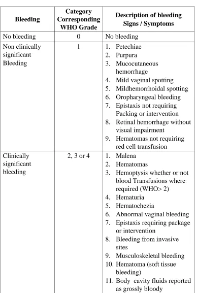
3.9 Categorization of bleeding events
Table 1 Categorization of bleeding events
Bleeding
Category Corresponding
WHO Grade
Description of bleeding Signs / Symptoms
No bleeding 0 No bleeding
Non clinically significant
Bleeding
1 1. Petechiae 2. Purpura
3. Mucocutaneous hemorrhage
4. Mild vaginal spotting 5. Mildhemorrhoidal spotting 6. Oropharyngeal bleeding 7. Epistaxis not requiring
Packing or intervention 8. Retinal hemorrhage without
visual impairment
9. Hematomas not requiring red cell transfusion
Clinically significant bleeding
2, 3 or 4 1. Malena 2. Hematomas
3. Hemoptysis whether or not blood Transfusions where required (WHO> 2) 4. Hematuria
5. Hematochezia
6. Abnormal vaginal bleeding 7. Epistaxis requiring package
or intervention
8. Bleeding from invasive sites
9. Musculoskeletal bleeding 10. Hematoma (soft tissue
bleeding)
3.10 Role of platelets in bleeding
3.11 Prophylactic platelet transfusion
Prophylactic platelet transfusion is an important part of supportive care measures in modern treatment. The aim of prophylactic transfusion is the prevention of major as well as minor bleedings during the time of disease or therapy induced thrombocytopenia.18 Hospitals have tried to reduce platelet use and the cost of platelet transfusion by transfusing platelets at lower platelet counts more frequently. But it is not optimal for cancer patients receiving intensive chemotherapy who require multiple platelet transfusion.10 There are studies determining the optimal transfusion trigger for prophylactic platelet transfusions in patients who have chemotherapy induced thrombocytopenia. Most of the centers now use a trigger of 10x109/L to transfuse platelet products.25
Among 20% of institutions surveyed reported that, among clinicians, the most common reason for platelet transfusions was for hemostasis in response to active bleeding.20
Recent studies suggest that the threshold for prophylactic platelet transfusion may be safely lowered to 10x109 / L from previous standards of 20x109/ L Earlier studies of spontaneous bleeding demonstrated that bleeding risk increased dramatically only at platelet counts below 5x10⁹ litre.26 In current practice, most platelet transfusion is given for prophylaxis. one random unit/10 kg for pediatric patients is the usual dose. Platelet survival varies with platelet count because of consumption of a fixed number of platelets each day to maintain vascular integrity. If prophylactic transfusion for severe thrombocytopenia are intended to maintain vascular integrity, then the total platelet usage may be determined by that fixed consumption, rather than by senescence of excess transfused platelets.26
is often cited as the rule of thumb for the expected increase in uncomplicated patients.5
Clinical trials have shown that one hour post transfusion count is a good measure of response to transfusion whereas the 24 hour count can be used to monitor platelets survival. Astler and Jandl showed that the recovery of platelets when labeled and re-infused into an autologous normal donor is not 100 percent but rather 52 percent. The decreased recovery is attributed to pooling of normal platelets in the spleen. This pooling is not reversible as platelets go back and forth between the spleen and the general circulation. On an average, one platelet is in the spleen for two in the circulation.27 A fixed number of platelets is required for hemostasis each day. As thrombocytopenia worsens, the percentage of platelets used each day increases. So the choice of 50 percent for mean cell life (MCL) was based on physiological and medical considerations.27
and that the number of platelet transfusion administered to many patients can be decreased with proper clinical evaluation.20
3.12 Group matched platelets
It has been an accepted practice for platelets to be transfused out of ABO group as a second line therapy when ABO matched platelets are unavailable. The mismatch is tolerated in most cases owing to the assumption that the antibodies and volume of incompatible plasma transfused will be diluted in the total blood volume of the recipient. This dilution prevents passively acquired ABO antibodies from causing a clinically significant problems.27 Intravascular hemolysis secondary to minor ABO mismatch is defined as donor plasma containing isohemagglutunin to antigens on recipient RBC causing severe morbidity and mortality. This is especially true for those transfusions of group O plasma or platelets to group A, B or AB recipients.
routinely measure isohemagglutunin titres (IgM or IgG) before transfusing ABO mismatched plasma, although some recommend this. A policy was instituted to test anti A, anti B, anti AB titres of all group O Single Donor Platelets before transfusion to group A patients. If IgM> 1: 64 and or IgG is > 1:256, the platelet product is not transfused out of group.27
3.13 Leukemia and Alloimmunisation
3.14 Corrected count increment (CCI)
The corrected count increment has been used as a surrogate measure for bleeding in platelet dose studies and also for platelet transfusion triggers studies.
CCI may not be a valid and reproducible surrogate marker as it depends upon (a) BSA – Calculation of BSA is variable as per the formula used. (b) Total No. of Platelets in the Platelet product transfused.25
Hematology Laboratory using electronic counter were not designed to accurately count cells in platelets rich plasma. The potential sources of variation in results includes
1. Method of taking the sample
2. Type of test tube that the sample is placed (plastic or glass) 3. Duration of time that the sample sits before being counted 4. The accuracy of making the dilution
5. The instrument on which the counting is being performed Platelet count increment (Post count – Pre count) X BSA CCI =
There are many studies which showed that there is significant variability in counting depending on the instrument that is being used. BEST study demonstrated the variability in platelet counts performed on samples of platelet rich plasma in eight different laboratories in five different countries. 20 – 60% of patients requiring regular platelet transfusions failed to achieve adequate post transfusion increments and are considered to be refractory to platelet transfusion.29 The factors influencing CCI are 1. Splenectomy, 2. Bone marrow transplantation, 3. Concurrent IV administration of Amphotericin B., 4. Palpable Spleen, 5. Temperature.28 Amphotericin B also binds to the sterol moiety of membrane to produce increased permeability and lysis of red cells.28
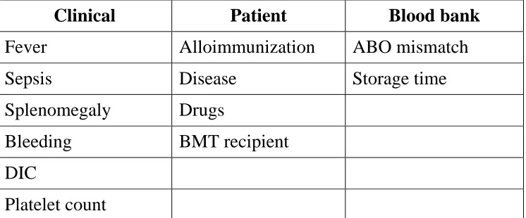
3.15 Guidelines for platelet transfusion in infants
3.16 Factors that affect the response to platelet transfusion10
Table 2 Factors that affect the response to platelet transfusion10
Clinical Patient Blood bank
Fever Alloimmunization ABO mismatch
Sepsis Disease Storage time
4. MATERIALS AND METHODS
4.1 Study Design and Place
• An Observational Study conducted in the Department of hematology, Institute of Child Health during the period 2007 – 2008. The study population included children diagnosed as acute leukemia in the age group 1 – 12 years.
4.2 Inclusion Criteria
• All acute leukemic Children, 1 – 12, years both male and female.
• Children diagnosed as acute lymphoblastic leukemia and acute myeloid luekemia.
• All acute leukemic Children requiring transfusion support.
• Both treated and follow up children.
4.3 Exclusion Criteria
• Leukemic children below 1 year and above 12 years.
• Lymphoma evolving to leukemic phase.
• Juvenile myelomonocytic leukemia.
4.4 Variables Studied
• Age, Sex, Weight
• Hemoglobin Level
• Platelet Count
• Hematocrit
4.5 Sample Size
Sample Size is calculated by the formula N = Z2 P (1-P) / d2
Where N = Sample Size
Z = Z statistic for a level of confidence. P = expected prevalence or proportion d = Precision
Z statistic (z): CI-95%, Z value is 1.96
N = (1.96)2 x 0.1 (1-0.1) / (0.05)2 = 135 (This is for 10% Prevalence & for 20 - 50% Prevalence it is 245)
So the total sample taken for the study is 250 and in this the transfusion of each unit is taken as one sampling unit.
4.6 Components prepared
The components used in this study were platelets and packed red cells and were prepared as follows:
The red cells were stored at 2-6°C (shelf life 35 days) and the platelets were stored at 20-24°C (shelf life 5 days) under constant gentle agitation.
The platelets were neither leukoreduced nor irradiated. No apheresis platelets were used.
The transfusion was given depending upon the clinical sign of bleeding and the hemoglobin estimation. The following data have been documented in this study for analysis.
Anti coagulated whole blood
PRBC Platelet Rich Plasma
Soft Centrifugation
Hard Centrifugation
Plasma Platelet
The hematology analyser used in this study was Sysmex4000 working on the principle of Impedance .The pre and post transfusion hemoglobin and platelet count were estimated 24 hours after the transfusion.
Hemoglobin content of the RBC and platelets were estimated as a quality measure once in a month randomly. The hematology analyser used was calibrated once in three months using known samples.
4.7 Variables
Before transfusion the following data were documented and the variables used in the study are
• Pre Hemoglobin count
• Pre platelet count
• No of units of RBC and platelets transfused
• Post hemoglobin count after 24 hrs of transfusion
• Post platelet count after 24 hrs of transfusion
5. RESULTS
Two hundred and fifty episodes of transfusion given to 30 leukuemic children were analysed . The data is analysed as follows:
5.1 Demographic details
5.2 Clinical features
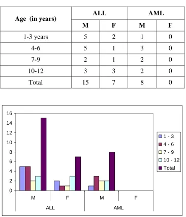
5.1 (a) Demographic details
Table 3 Age & Sex distribution
ALL AML Age (in years)
M F M F
1-3 years 5 2 1 0
4-6 5 1 3 0
7-9 2 1 2 0
10-12 3 3 2 0
Total 15 7 8 0
0 2 4 6 8 10 12 14 16
M F M F
ALL AML
1 - 3 4 - 6 7 - 9
10 - 12 Total
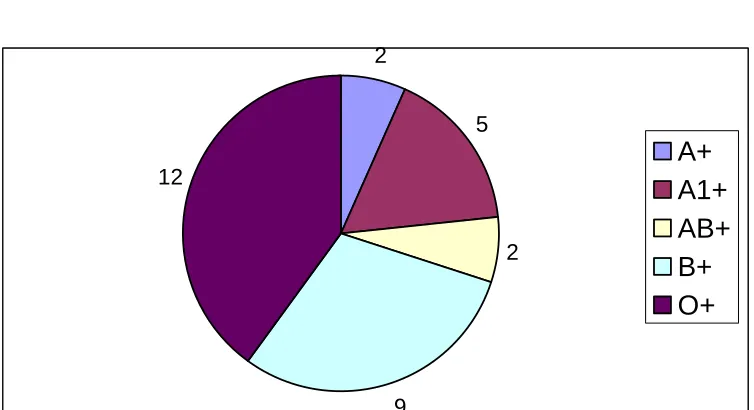
5.1 (b) Blood group distributions
Table 4 Blood group distribution
Blood Group Number of children
A+ 2 A1+ 5 AB+ 2 B+ 9 O+ 12 Total 30
2
5
2
9 12
A+ A1+ AB+ B+ O+
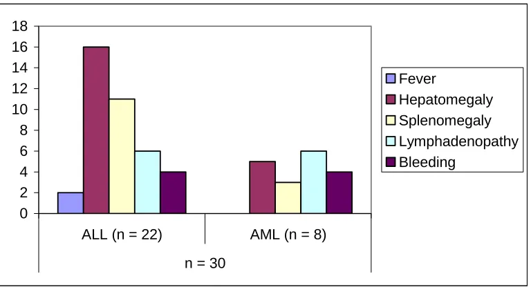
5.2 Clinical Features
The Leukemic children presented with the following major clinical features
1. Fever
2. Hepatomegaly 3. Splenomegaly 4. Lymphadenopathy 5. Anemia
Table 5 Clinical features
n=30
Clinical Features ALL
n=22
AML
n=8
Fever 2(9.1%) 0
Hepatomegaly 16(72.7%) 5(62.5%)
Splenomegaly 11(50%) 3(37.5%)
Lymphadenopathy 6(27.8%) 6(75%)
Bleeding 4(18.2%) 4(50%)
0 2 4 6 8 10 12 14 16 18
ALL (n = 22) AML (n = 8) n = 30
Fever
Hepatomegaly Splenomegaly Lymphadenopathy Bleeding
Table 6 Presence of Anemia
Hb% in grams No. Of Children
< 7 15
7.1 to 10 10
10 to 12 5
15
10 5
< 7 7.1 to 10 10 to 12
5.3 Analysis of Transfusions
Algorithm of Transfusion episodes Components Transfused
Total no. of transfusion episodes : 250 units
30 patients (23 males, 7 females)
Packed red blood cells
Random donor platelets
No of transfusion episodes 52
No of transfusion episodes 198
Single unit 47
Double unit 47
More than 2 units 15
26 patients
Mean post transfusion increment 1.12 grams %
(SD +/- 0.48)
Mean post transfusion increment Mean post transfusion increment Mean post transfusion increment 4900/µl (SD+/-1943) 9700/µl (SD+/-3596) 11200/µl (SD+/-8925)
5.3 (a) Clinical Factors Influencing Platelet Count
Table 7 Bleeding Vs Platelet count
Bleeding Platelet Count
< 5000 / µl
Platelet Count
> 5000 / µl
Present 4 4 Absent 1 21
Odds ratio = 21(p < 0.05)* Bleeding is definitely associated with thrombocytopenia.
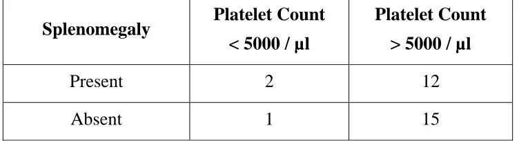
Table 8 Splenomegaly Vs Platelet Count
Splenomegaly Platelet Count
< 5000 / µl
Platelet Count
> 5000 / µl
Present 2 12 Absent 1 15
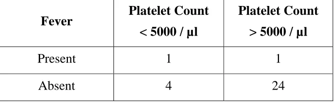
[image:37.612.126.502.442.544.2]Table 9 Fever Vs Platelet Count
Fever Platelet Count
< 5000 / µl
Platelet Count
> 5000 / µl
Present 1 1
Absent 4 24
Odds ratio = 6 (p<0.05)* Presence of fever also contributes to thrombocytopenia.
Table 10 Distribution of platelet count in children with bleeding
Platelet Count No. of children with bleeding
< 5000/µl 4
> 5000/µl 4
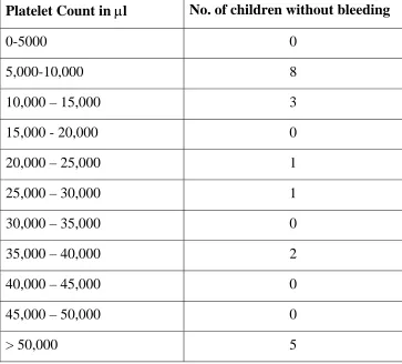
[image:38.612.128.493.389.475.2]Table 11 Distribution of platelet count in children without bleeding
Platelet Count in µl No. of children without bleeding
0-5000 0 5,000-10,000 8
10,000 – 15,000 3
15,000 - 20,000 0
20,000 – 25,000 1
25,000 – 30,000 1
30,000 – 35,000 0
35,000 – 40,000 2
40,000 – 45,000 0
45,000 – 50,000 0
> 50,000 5
0 1 2 3 4 5 6 7 8 9 0-5000 5,000 -10 ,000 10,0 00 –
15 ,000
15,0 00 - 2
0,000
20,00 0 – 2
5,000
25,00 0 – 3
0,000
30,0 00 –
35 ,000
35,00 0 – 4
0,000
40,00 0 – 4
5,000
45,00 0 –
50, 000
> 50, 000
No. of children without bleeding
No. of children with bleeding
Statistically there is no correlation with the platelet count between the number of children with and without bleeding
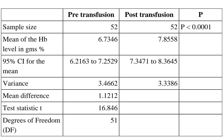
5.3 (b) Analysis of RBC transfusions
Table 12 Analysis of RBC transfusions
Paired samples t-test n = 52
Pre transfusion Post transfusion P
Sample size 52 52 P < 0.0001
Mean of the Hb level in gms %
6.7346 7.8558
95% CI for the mean
6.2163 to 7.2529 7.3471 to 8.3645
Variance 3.4662 3.3386
Mean difference 1.1212
Test statistic t 16.846
Degrees of Freedom (DF)
51
In the above analysis it is found that there is a mean difference of 1.1212 grams % and for 95% CI for the mean p < 0.0001, the post transfusion rise in Hb is significant.
Histogram of differences
0.0 5.0 10.0 15.0 20.0
0.0 0.8 1.5 2.3 3.0
Differences
5.3 (c)Analysis of Platelet Transfusions
Out of 198 platelet transfusion analysis about the Pre & Post platelet count were done for single unit, double unit & for 3 & 4 units separately as follows:
Single unit platelets transfusion = 47 Double unit platelets transfusion = 51 More than 2 units platelets transfusion = 15
5.3 (d) Single Unit Platelet Transfusion
Table 13 Analysis of single unit platelet transfusion
Paired samples t-test n = 47
Pre transfusion Post transfusion P
Sample size 47 47 P < 0.0001
Mean of the platelet count in 103 µl/l
39.3745 44.3553
95% CI for the mean
30.4899 to 48.2590 35.3566 to 53.3541
Variance 915.6493 939.3299
Mean difference 4.9809
Test statistic t 17.572
Degrees of Freedom (DF)
46
In the above analysis it is found that there is a mean difference of 4.9809X10³ µl / l from the pre and post platelet single unit transfusion. For 95% CI for the mean, the P < 0.0001 and so the rise in post transfusion platelet count is significant.
Histogram Of Difference
0.0 3.0 6.0 9.0 12.0
2.0 4.0 6.0 8.0 10.0
Differences
5.3 (e) Double units platelets transfusion
[image:44.612.125.501.297.506.2]51 double units were given and the following analysis done to find out the post platelet count increment
Table 14 Analysis of Double unit’s platelets transfusion
Paired sample t-test n = 51
Pre transfusion Post transfusion P
Sample size 51 51 P < 0.0001
Mean of the platelet count in 103 µl/l
35.5843 45.3784
95% CI for the mean 27.4889 to 43.6798 36.9598 to 53.7971
Variance 828.4809 895.9477
Mean difference 9.7941
Test statistic t 19.448
Degrees of Freedom (DF)
50
Histogram Of Differences
0.0 5.0 10.0 15.0 20.0
0.0 6.3 12.5 18.8 25.0
Differences
Count
5.3 (f) Three & Four unit platelet Transfusion
Table 15 Analysis of Three & Four unit platelet Transfusion
Paired sample t-tests n=15
Pre transfusion Post transfusion P
Sample size 15 15 P = 0.0002
Mean of the platelet count in 103 µl/l
27.7133 38.9600
95% CI for the mean
16.9953 to 38.4314 27.0850 to 50.8350
Variance 374.5898 459.8240
Mean difference 11.2467
Test statistic t 4.880
Degrees of Freedom (DF)
14
In the above analysis it is found that there was a mean difference 11.2467X10³ µl/l Platelets between the pre and post platelets transfusion from those who received more than 2 units. For 95% CI for the mean, P < 0.00001 and the rise in platelet count is significant.
Histogram Of Differences
0.0 3.0 6.0 9.0 12.0
-20.0 -7.5 5.0 17.5 30.0
Histogram of Differences
Differences
6. DISCUSSION
Hemato-oncology services require many transfusions for a prolonged period. Profound anemia can alter hemostasis and thus should be avoided in patients with thrombocytopenia.10 Kathryn E. Webert et al hypothesized that maintaining the hemoglobin concentration of a patient with thrombocytopenia at a higher level may contribute to improved homeostasis. In this study, they found that once the patients’ hemoglobin level reached the targeted threshold, there were no differences between groups with respect to number of RBC or platelet transfusion.16 Normal platelet survival is approximately nine days. Hansor SR et al suggested that patients under going induction chemotherapy for leukemia often require platelet transfusion at least every three days. E. Pattern and Visania et al published that acute leukemic patients receive on average 80 – 110 units of platelets and 20 – 40 units of red cells.20
In this study, 50 leukemic children were admitted and among them only 30 (60%) children needed transfusion support.
198 were random donor platelets (mean 2.3). Of 52 packed red cell transfusion episodes given to 22 patients, five of them received more than four units of red cells during the study period. The mean haemoglobin increment for single unit of packed red cell transfusion was 1.12 gms%. 15 children had haemoglobin <7 gms%, 10 of them had haemoglobin level between 7.1 to 10 gms% and 5 of them presented >10 gms%. Of this 15 children, only two presented with bleeding, but among 10 children who had haemoglobin level between 7.1 to 10 gms%, four of them presented with bleeding. Cause and effect of anemia with bleeding could not be ascertain from their presentation. There was no optimal RBC transfusion trigger followed. Out of 198 random donor platelets given to 28 leukemic children, 17 of them received less than 5 units, 8 of them received 5 to 10 units and 3 of them received more than 20 units. The most common component used in the study was platelets and the number of platelets provided per transfusion was not ascertained.
approach can prevent bleeding, as opposed to therapeutic approach, in which platelet transfusion is given after a certain degree of hemorrhage has occurred. High et al in 21 patients with acute leukemia found that fever preceded hemorrhage in 10 of the 13 patients who experienced bleeding23. In the present study, 2 out of 30 patients (6.3%) had fever and among these two, one had bleeding episode with a platelet count of <5000/µl.
Table -16 Summary of Platelet Transfusion Trigger Trials
Study Year N-type Patients
Bayer wl et al 1992 31 Malignancy Gumur et al 1992 105 Acute Leukemia Heckman et al 1997 78 Acute Leukemia Rebulla et al 1997 255 Acute Leukemia
Wandt et al 1998 105 Acute Myeloid Leukemia
[image:50.612.117.509.133.300.2]the risk of bleeding. In the present study no transfusion trigger was followed. Platelets were transfused to 28 Leukemic children based on their platelet count and bleeding episodes. Out of 198 transfusion episodes given, 8 patients (28.5%) presented with bleeding. In this study 4 of them presented with a platelet count <5000/µl. In this study the clinical signs presented by the leukemic children – bleeding, splenomegaly and fever were analyzed with the platelet count below and above 5000 per µl and the odds ratio calculated and were found to be significant as shown in table 7,8 & 9. The distribution of platelet count children with bleeding and without bleeding were analysed as shown in table 10 & 11. Bleeding was significantly present with platelet count <5000/µl also.
The problems that might have been faced by the physician to decide on an optimal prophylactic threshold could be
(1) Serious hemorrhage was rare at lower platelet numbers.
(2) Presence of minor clinical bleeding.
practice it is difficult to evaluate the efficiency of platelet transfusion due to the fact that
(a) Severe bleeding due to thrombocytopenia alone is rare.
(b) Mortality due to hemorrhage in thrombocytopenia is not common. The bleeding time is not helpful in determining the effectiveness of platelet transfusion. Some times platelet count does not increase to the expected level after platelet transfusion.
there was no refractoriness. The mean increment for single unit transfusion (2.6) is 4900/µl and for double unit transfusion (2.6) is 9700/µl and for more than 2 units (1.36) is 11200/µl and the standard deviation is +/- 1943, 3596 and 8925 respectively.
Table 16 Studies addressing the issue of optimal platelet dose25
Study Year Patients N Dosage of platelets
Noral et al 1998 AML 69 Medium Dose = 4- 6 x10¹¹
High Dose = 6 – 8x10¹¹ Very High Dose = >8x10¹¹
Goodnough et al
2001 Chemotherapy induced
thrombocytopenia
120 Medium Dose = 3.4- x1011 High Dose = 5.7 x10¹¹
Very High Dose = 11x1011
Klumpp et al
1993 Human progenitor cell transplant
46 High Dose = 5 x10¹¹ Low Dose = 3x10¹¹
TinMouth et al
2005 ALL / AML 111 High Dose = 5 WB
derived platelets poole Low Dose = 3 WB derived
platelets pooled
Sensebe’ et al
2005 Acute Leukemia 96 Single Dose = .5x1011
Pl / 10kg
Double Dose = Twice this amount
7. SUMMARY
¾ All leukemic children included in this study were given transfusion and all of them who were transfused showed significant clinical improvement showing that there is a need for transfusion support in this group.
¾ Most common blood component used in this study was platelets (79%) and the remaining were red cells (21%).
¾ There was a mean hemoglobin rise of 1.12g% after transfusion of one unit of red cells.
¾ The platelet increment were as follows:
For single unit – 4.98 x 10³ / µl
For double unit – 9.74 x 10³ / µl
For more than 2 units – 11.2 x 10³ / µl
Platelet count increment was present in all transfusion episodes and there was no refractoriness.
All children were negative for transfusion transmissible infections at the end of the study.
¾ 50% of the children who presented with bleeding had platelet count < 5000 per microlitre.
8. CONCLUSION
9. BIBLIOGRAPHY
1. Swaminathan R, Rama R, Shantha V. Childhood cancers in Chennai. Int J cancer 2008; 122(11):2607-11.
2. Parkeins et al. Childhood cancer incidence in Delhi 1996-2000. Indian journal of medical and paediatric oncology 2006; 27:4
3. D’ costa GG Siddiqui HN,Pradhan RM. Pattern of leukemias: a ten year incidence study of 242 cases. Journal of postgraduate Medicine 1989; 35; 4:191-5.
4. Richard E. Behrman, Robert M. Kliegman, Hal B. Jenson : Nelson Text Book Of Pediatrics. ed 17, W.B. Saunders 2006.
5. Bruce Cameron, Gail Rock, Bernard Olberg, and Doris Neurath Evaluation of platelet transfusion triggers in a tertiary – care hospital. Transfusion 2007; 47:206-2011.
7. Joan aid and Miguel Lozano. Lower or higher doses for prophylactic transfusions : results of a meta analysis of randomized controlled trials. Transfusion 2008; 47: 464-470.
8. Anthony D. Slonim, Jill G. Joseph, Wendy M. Turenne, Aditi Sharangapani, and Naomi L.C. Luban. Blood Transfusions in children : A multi institutional analysis of practices and complications. Transfusion 2008; 48:73-80.
9. Jurg Gmur Johanna, Burger Urs Schanz, Jorgfehr Andreas, Schaffner. Safety of stringent prophylactic platelet transfusion policy for patients with acute Leukemia. The Lancet 1991; 338: 1223-26.
10. Jeffrey Mc Cullough. Current issues with platelet transfusion in patients with cancer. Seimars in Hematology, 2000; 37, 2, 4:3-10.
11. S.Murphy, W.A. Heaton and P.Rebulla. Platelets production in the old world and the new. Transfusion 1996; 36:751-754.
13. Cherles A. Schiffer, Kenneth C. Anderson, Charles L.Bennelt, Steven Berstein, Linda S. Elting, Miriam Gold Smith, et al. Platelet transfusion for patients with cancer : Clinical practice guidelines of the American Society of clinical oncology. Journal of clinical Oncology, 2001; 19: 5 :1519-1538.
14. France Gauvin, Martin A. Champagne, pierre Robillard, Jean – Pierre Le Cruguel, Helene Lapointe ,and Heather Hume. Long-Term survival rate of pediatric patients after blood transfusion. Transfusion 2008; 48:801-808.
15. Margriet J. Dijkstra – Tiekstra, Willeke Kuipers, Airies C. Setrokromo, and Janny de Wildt – Eggen. Platelet counting in platelet concentrates with various automated hematology analyzers. Transfusion 2007; 47:1651-1657.
17. CA Shiffer, JP Dutcher and Jaisner. A randomized trial of leucocyte depleted platelet transfusion to modify alloimmunisation in patients with leukemia. Blood 1983; 62: 815-820.
18. Dominik Heim, Jakob Passweg, Michael Gregor, Andreas Buser, Alexander Theocharides and Caroline Arber et al. Patient and product factors affecting platelet transfusion results. Transfusion 2008; 48:681-687.
19. Patrick Davies, Simon Robertson, Shilpa Hedge, Rosemary Greenwood, Edwin Massey and Peter Davis. Calculating the required transfusion volume in children .Transfusion 2007; 47: 212-216.
20. E. Patten Controversies in transfusion medicine. Prophylactic platelet transfusion revisited after 25 years. Transfusion 1992; 32; 4:381-385.
22. P.T. Pisciotto, K. Benson, H. Hume, A.B. Glassman, H.Oberman and M.Popovsky et al. Prophylactic versus therapeutic platelet transfusion practices in hematology and / or Oncology patients. Transfusion 1995; 35; 6:498-502.
23. D.J. Higby, E. Cohen, J. F.Holland and L Sinks. The Prophylatic treatment of thrombocytopenic Leukemic patients with platelets : a double blind study. Transfusion 1974; 14:440-446.
24. F.norol, M. Kuentz, C. Cordoiiner, F. Beaujean, C.Haioun, J.P. Vernant and N.Deudari. Influence of clinical status on the efficiency of stored platelet transfusion .British Journal of Haematology, 1994; 86:125-129.
25. N.M.Hoddle. Controversy concerning platelet dose .ISBT science series 2007; 2:220-25.
27. Scott Murphy. The case for a new approach for documentary platelet viability - Transfusion 2006; 46:495-525.
28. James F. Bishop Katherine Mc Grath, Max M . Wolf, Jane P. Mathews, Trudy De Luise and Kally Yuen et al. Clinical Factors Influencing the Efficacy of pooled platelet Transfusions. Blood 1998; 71 : 2:383-387.
29. Inaki Alcorta Arturo Pereira And Antonio Ordinas. Clinical and Laboratory Factors associated with platelet transfusion refractoriness: A case control study. British journal of Hematology. 1996; 93:220-224.
30. Platelet transfusion to infants. ISBTI 2006 : 37-39. ISBT Science series 2006; 1:37-39.
67 S.NO PRE HB NO OF UNITS POST
HB S.NO PRE UNIT POST
1 GANI 1806/08 3 M 13 ALL AB + 2 5 7 1 1 2 2 2 1. ALL 1 3.1 1 3.9 3 80x103 1 89X103
2 4.1 1 5.4 4 88x103
2 100X103
5 120x103
2 131X103
2 HARI ROSHAN 436/08 2 M 11 ALL A1 + 2 4 6 1 1 2 2 2 2. ALL 1 4.8 1 6.1 3 14x103 3 29x103
2 5.2 1 7.8 4 29x103 4 36x103
5 36x103
5 42X103
3 PRASANTH 331/08 12 M 30 AML A + 4 29 33 2 2 2 2 2 3. AML 1 5 1 6.8 5 4x10³
2 9X10³
2 6.8 1 7.2 6 8x10³ 2 19X103
3 7.1 1 8 7 19x103 4 32X103
4 7.2 1 8.2 8 32x103
3 17X103
9 17x103
1 20X103
10 20x103 3 28X103
11 28x103 1 31X103
12 31x103
1 36X103
13 36x103
1 39X103
14 39x103 2 48X103
15 48x103 2 59X103
16 59x103
1 63X103
17 63x103
1 65X103
18 65x103 2 71X103
19 71x103 3 84x103
4 SURYA 1737/07 2 M 12 AML O + - 2 2 1 1 2 1 2 4.AML 1 40x103 2 19X103
5 PRIYADARSHINI 359/08 3.5 F 13 ALL B + 4 5 9 2 2 2 2 2 5.ALL 1 5.2 1 7.1 1 40x103
2 57X103
2 7.1 1 7.9 2 57x103 2 67X103
3 6.1 1 7.2 3 67x103 1 75X103
4 7.2 1 8.8
6 NIROSHA 514/08 10 F 25 ALL B + 1 3 4 2 2 1 2 2 6. ALL 1 5.2 1 7.1 1 26x103
3 56X103
7 SURYA 1026/08 11 M 28 ALL B + 1 - 1 1 1 2 1 2 7.ALL 1 6.8 1 7.4
8 SHARUKA 1552/07 7 M 22 ALL B + 2 2 4 2 2 2 1 2 8. ALL 1 7 1 8.2 1 120x103 1 125X103
2 7.8 1 8 2 80x103 1 85X103
9 JOSHUA 846/03 11 M 28 ALL O + - 3 3 1 1 2 1 2 9. ALL 1 10x103
3 25X103
10 VIJAY 220/08 5 M 18 ALL O + 1 2 3 1 2 2 2 2 10. ALL 1 6.8 1 7.2 1 10x103 2 18X103
11 LATHA 1623/07 2 F 12 ALL B + 1 1 2 1 1 2 2 2 11. ALL 1 5.2 1 6.4 1 11x103 1 15X103
12 KARTHIKA 711/08 12 F 31 ALL B + 1 - 1 2 2 2 2 2 12. ALL 1 6.2 1 7.8
13 VENKATESH 273/08 3.5 M 12 ALL A1 + 4 7 11 1 1 1 2 2 13. ALL 1 7.2 1 8 1 9x10³
3 20X103
2 8 1 9.9 2 20x103
1 24X103
3 9.7 1 10.8 3 4x10³ 1 10X103
4 8.9 1 10.1 4 10x103 1 16X103
14 ABINASH 286/08 8 M 22 AML O + 4 20 24 1 1 1 1 2 14. AML 1 8.6 1 10.8 1 2x10³
1 7.2X10³
2 10.8 1 11.9 2 7.2x10³
1 13.3X10³
3 4.9 1 5.4 3 13.3x10³ 1 19.8X10³
4 5.4 1 7 4 19.8x10³ 1 22.4X10³
5 22.4x10³
2 31.3X10³
6 31.3x10³
2 39X103
7 39x103 4 52X103
8 52x103 1 59.3X10³
9 59.3x10³
1 62X103
10 62x103
1 68.7X10³
11 68.7x10³ 3 82.4X10³
12 70x103 1 74X103
13 72x103
1 76X103
15 PRADEEP 873/08 4 M 15 AML O + 1 6 7 1 1 1 2 2 15. AML 1 2.1 1 3.4 1 75x103
2 87X103
2 87x103 2 93X103
3 93x103 2 100X103
16 ELUMALAI 609131 6 M 18 AML B + 3 7 10 1 2 1 1 2 16. AML 1 7.8 1 9.2 1 5x10³
1 8X10³
2 9 1 9.8 2 8x10³
1 10X103
3 9.8 1 10.5 3 10x103 1 16X103
4 16x103 1 20X103
5 20x103
2 32X103
6 32x103
1 38X103
7 38x103 1 46x103
17 RAHUL 605432 4 M 16 AML B + - 2 2 2 2 1 2 2 17. AML 1 90x103 1 100X103
18 MONICA 627/08 12 F 30 ALL O + - 1 1 1 2 1 2 2 18. ALL 1 37x103
1 41X103
19 SAI SIDDARTH 1540/07 9 M 22 AML O + 1 28 29 2 2 1 1 2 19. AML 1 7 1 7.8 1 10x103
2 18X103
2 18x103 2 26X103
3 26x103 2 31X103
4 31x103
2 39X103
5 39x103
2 47X103
6 10x103 2 20X103
7 20x103 1 24X103
8 24x103
4 37X103
9 37x103
2 44X103
10 44x103 2 54X103
11 54x103 2 61X103
12 61x103 1 65X103
13 65x103
2 76X103
14 76x103 1 81X103
15 81x103 1 86X103
20 NANDA KUMAR 764/08 4.5 M 16 ALL O + 2 6 8 1 2 1 2 2 20. ALL 1 7.6 1 8.8 1 10x103
2 18X103
2 8 1 9.2 2 18x103
2 29X103
3 10x103 1 16X103
4 16x103 1 24X103
21 FARHUL RAHUMAN 733/08 3.5 M 15 ALL A1 + 1 8 9 1 1 2 2 2 21. ALL 1 3 1 4.1 1 4x10³ 1 11X103
SUBJECT NO & DIAGNOSIS
RBC TRANSFUSION PLATELET TRANSFUSION SUB NAME IP NO AGE SEX WeightDIAGBLOOD SPLE LYMPH BLEEDING FEVER